neuroscience: myths, metaphors and marketing
TRANSCRIPT

Neuroscience
Myths, Metaphors and Marketing.
!
James Lawley
NLPtCA Conference
2012
www.cleanlanguange.co.uk
1

Policy Statement
I admire the scope, scale and achievement of neuroscience.
This talk is not intended to diminish the work of any neuroscientist or psychotherapist.
My aim is to help us distinguish between:
– quality neuroscience and inferences drawn therefrom and
– what Raymond Tallis calls “Neuromania”.
2
James Lawley

EXERCISE !
1.Name a piece of neurological research that has influenced the way you do therapy.
2.What do you do differently with your clients as a result of the research?

Watch first two minutes of video : http://www.schoolsworld.tv/node/2887

A connectome is
the totality of connections between the neurons in a
nervous system.

A connectome of C. elegans roundworm
1 mm in length 302 neurons.

What do you make of this quote from a leading psychotherapist?: !
“The role of the psychotherapist is brought into a new light through the implications of working with psychoneuroimmunology.
... Validating the client’s model of the world and negative feelings, the therapist enables neurotransmitters associated with the negative state to be released from the subcortex and the existing neural networks activated.”
7

And this one?:
“As soon as the client accesses the future-oriented state, the neurological potential is then created for change to happen.
Solution-oriented therapy ... ensures that the client fires the neurological pathway a number of times in therapy, making it easier to re-access once the session has ended. ...
This process reinforces the ‘not problem’ state, again reinforcing positive neurological patterning.
At this next session, we both noted that he could more easily move towards future-oriented thinking ... This was evidence that the neurological re-patterning that we had done in the previous week had started to work.”
psycho
Does changing one prefix change the meaning?:
psycho
psycho
psycho

“Your prefrontal cortex is the biological seat of your conscious interactions with the world. It’s the part of your brain central to thinking things through. ... Getting everything ‘just right’ for the prefrontal cortex is what Emily needs to learns to do, to get on top of the extra information she is juggling in her new job.” !David Rock, Your Brain at Work p. 6 !!“A question I ask my clients all time is: What does your brain need right now to move forward?” !David Rock, A Brain-Based Approach to Coaching, International Journal of Coaching in Organizations 2006 4(2)
What do you think Emily and other clients make of this question?

“[Neuro-talk] is often accompanied by a picture of a brain scan, that fast-acting solvent of critical faculties.”
Matthew Crawford ‘The Limits of Neuro-Talk’

CAT Computed axial tomography DOT Diffuse optical tomography EEG Electroencephalography EROS Event-related optical signal
MEG Magnetoencephalography MRI Magnetic resonance imaging (structural)
fMRI Functional MRI
dMRI Diffusion MRI
NIRS Near infrared spectroscopy
PET Positron emission tomography
SPECT Single-photon emission computed tomography
BRAIN IMAGINING TECHNIQUES

Can you guess what experience this brain scan is showing ?
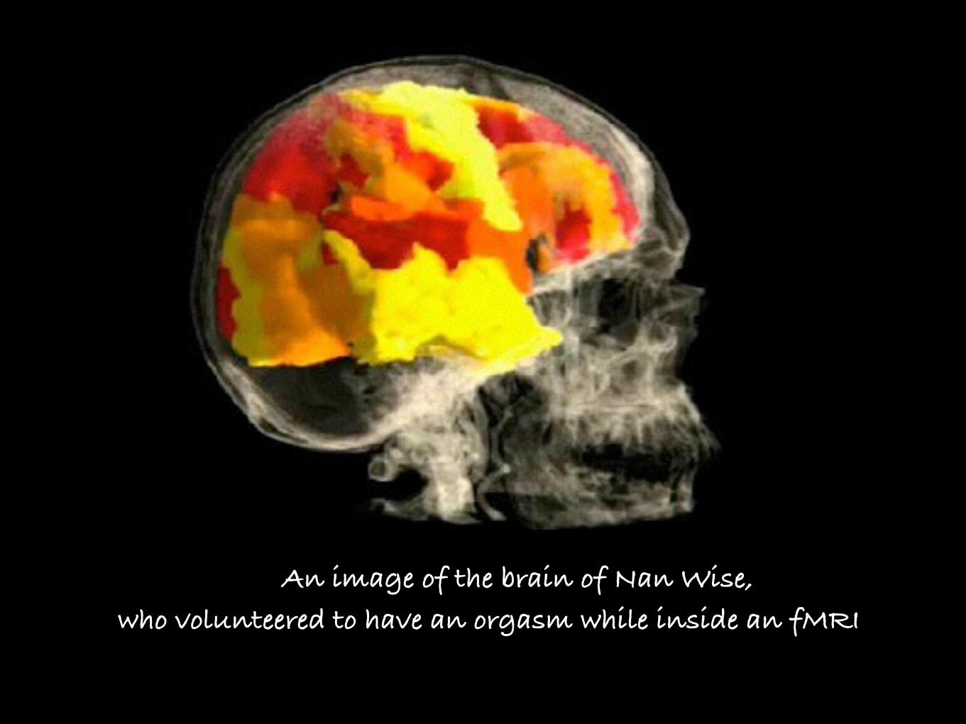
An image of the brain of Nan Wise, who volunteered to have an orgasm while inside an fMRI

• Technical Problems with fMRI ‣ Measures oxygen in the blood as a proxy for brain activity. Millions of
neurons have to be activated for a change in blood flow to be detected. ‣ Neuronal activity lasts milliseconds while detected changes in blood
flow lag by 2-10 seconds. ‣ Brain is changing all the time – somewhere is always ‘lit up’. ‣ Images made up of voxels, each representing at best 10,000+ neurons. ‣ Each scan has 50,000 data points; thousands of scans in a study means
many millions of comparisons. Massively complex analysis required – 7 million lines of code.
‣ A big problem is false positives – thousands of published studies conducted without corrections for false positives.
‣ Some researchers pick out the ‘best’ results. ‣ Spurious ‘brain activity’ related to non-existent tasks found with
standard settings on the most popular fMRI analysis software.

Functional MRI scans of six people who took the same spatial memory test
26 SCIENTIFIC AMERICAN MIND
tal functions to particular brain regions. Critics feel that fMRI overlooks the networked or dis-tributed nature of the brain’s workings, empha-sizing localized activity when it is the communi-cation among regions that is most critical to men-tal function.
“This is a very gross technique,” says critic Steven Faux , who heads the psychology depart-ment at Drake University. “It’s like a blurry pho-to—better than no photo but still blurry, with real limitations that are too often overlooked. It’s very easy to overextend [the value of] this technology.”
Many fMRI practitioners seem bewildered that this powerful new tool has created contro-versy. “It is a huge surprise to me how big this issue has become,” says Marcus E. Raichle, a Washington University neurologist who has re-
searched brain scanning for more than two decades.
Vague PrecisionBrain imaging began with an early 20th-cen-
tury method called pneumoencephalography, a dangerous procedure in which the skull’s cere-brospinal fl uid was replaced with air to show the brain more clearly on x-ray. The angiograph, de-veloped in the 1920s, produced improved results by capturing images of dyes injected into the bloodstream. (Angiography is still used to help diagnose and track blood vessel defects and some tumors.) These early methods showed only static structure rather than function. Computed axial tomography (CAT or CT) scans, developed in the 1970s, exploited x-ray technology and took static pictures, too, but with far greater detail.
The 1970s also brought the fi rst functional imaging technology—scans designed to show not just how the brain is structured but how it functions. Positron emission tomography (PET) measures increases in blood fl ow associated with neuronal activity, giving a sense of which neu-rons may be processing information. A subject is injected with radioactive elements that tag molecules such as glucose that are delivered to the brain by blood. The tags emit positrons and reveal the relative rates at which cells consume the glucose, a marker of which cells are active during mental processes. The scans are captivat-ing, but there are a number of drawbacks. Sub-jects worry about taking in radioactive material; the process requires the better part of an hour for a scan; and the images provide a rather broad temporal resolution of 60 seconds (meaning it
takes that long to measure the blood fl ow to an area) and a spatial resolution of six to nine cubic millimeters—large for a nuanced understanding of what is happening.
In contrast, fMRI can scan a brain cross sec-tion in less than two seconds, enabling it to mod-el most of the brain in one to two minutes. It can work at spatial resolutions as fi ne as two to three cubic millimeters, although in practice it usually collects information in voxels (a term that merg-es “volume” with “pixel”) about two millimeters square and four to fi ve millimeters long, about the size of a grain of rice. FMRI requires no injec-tions, allowing more extensive scanning. In a typical study, a subject lies in a doughnut-shaped machine and is fi rst scanned at rest with his eyes closed to provide a baseline reading. He is then scanned again while performing some mental task: identifying faces, threading a computerized
Functional MRI scans of six people who took the same spatial memory test show how varied brain activation pat-terns can be. Scientists must design fMRI ex-periments care-fully to avoid misleading conclusions.
DA
VID
C.
OS
MO
N U
niv
ers
ity
of
Wis
con
sin
–M
ilw
au
ke
e
COPYRIGHT 2005 SCIENTIFIC AMERICAN, INC.
www.sc iammind.com 27
maze, engaging in a role-playing game. In the most common technique, called BOLD (for blood oxygen level–dependent) fMRI, the ma-chine measures increases in blood fl ow by spot-ting a change in magnetism that occurs when a blood surge raises the ratio of fresh, oxygenated hemoglobin to “used,” deoxygenated hemoglo-bin, which has a signifi cantly different charge. The regions creating surges appear as brighter colors on the images, red changing to yellow as fl ow rises. Doubts about whether these increases correspond to actual neuronal activity have been answered by several studies tying blood fl ow di-
rectly to neuron signaling, including recent ani-mal models that used probes to match the fi ring of individual neurons to the heightened fl ow seen in fMRI scans.
Yet the link is decidedly rough. Abigail A. Baird, a Dartmouth College psychologist who uses fMRI to study brain changes during adoles-cence, puts it succinctly: “Hemodynamic re-sponse is a sloppy thing.” For starters, neuronal action takes milliseconds, whereas the blood surge follows by two to six seconds; a detected increase in blood fl ow therefore might be “feed-ing” more than one operation. In addition, be-cause each voxel encompasses thousands of neu-rons, thousands or even millions may have to fi re to signifi cantly light up a region; it is as if an entire section of a stadium had to shout to be heard.
Meanwhile it is possible that in some cases a
small group of neurons drawing little blood, or a thin network of neurons connecting large re-gions, may perform functions as crucial as a larg-er group elsewhere but either go undetected or show up as minor activity. Likewise, some neu-rons might operate more effi ciently than others, consuming less blood. All these factors could mean that an fMRI image misrepresents actual neurodynamics.
Processing the scan’s gigabytes of raw data so that they become images introduces other cave-ats. Researchers must choose among and adjust many different algorithms to extract an accurate
image, compensating along the way for varia-tions in skull and brain confi guration, movement of subjects in the scanner, noise in the data, and so on. This “chain of inferences,” as a recent Na-ture Neuroscience article called it, offers much opportunity for error.
Finally, most fMRI studies use univariate processing, which critics say shortchanges the distributed nature of neurodynamics. The charg-es rise because univariate (literally “one vari-able”) algorithms consider the data coming in from each voxel during a scan as one sum, which makes it impossible to know how the activity in a particular voxel accrued (all at once, for in-stance, or in several pulses) or how it related se-quentially with activity in other voxels. Univar-iate processing does see all the parts working—thus the multiple areas lit up in most images—but not in a way that shows how one area follows or
The beautiful graphics fMRI produces imply much more precision than there actually is. )(
COPYRIGHT 2005 SCIENTIFIC AMERICAN, INC.
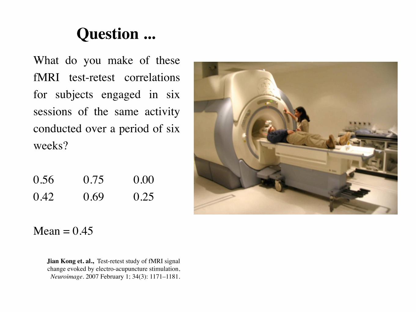
• Question ... What do you make of these fMRI test-retest correlations for subjects engaged in six sessions of the same activity conducted over a period of six weeks? !0.56 0.75 0.00 0.42 0.69 0.25 !Mean = 0.45 !!
Jian Kong et. al., Test-retest study of fMRI signal change evoked by electro-acupuncture stimulation, Neuroimage. 2007 February 1; 34(3): 1171–1181.

These subjects engaged in a simple finger-tapping task and yet the correlations ranged between 0 and 0.76 – imagine the subjects were doing something useful! !
A review of papers published in top-ranking journals, including Science, concluded:
“A disturbingly large and quite prominent segment of fMRI scan research on emotion, personality and social cognition is using seriously defective research methods and producing a profusion of numbers that should not be believed.”
!Edward Vul1, Christine Harris, Piotr Winkielman and Harold Pashler, ‘Puzzlingly High Correlations in fMRI Studies of Emotion, Personality, and Social Cognition’ Perspectives on Psychological Science, May 2009 vol. 4 no. 3 274-290.

Example of a neuro-imaging research methodology
‣ The subject was placed in a fMRI scanner. ‣ Subject was shown a series of photographs depicting human
individuals in social situations with a specified emotional valence.
‣ The subject was asked to determine what emotion one of the individuals in the photo must have been experiencing.
‣ Each photo was presented for 10 seconds followed by 12 seconds of rest. A total of 15 photos were displayed.
‣ Total scan time was 5.5 minutes.

This is the brain scan of the subject

“By complete, random chance, we found some voxels that were significant ... [even though] the salmon was not alive at the time of scanning.”
Craig Bennett, neuroscientist, University of California

“An fMRI study has shown that
men’s amygdalas light up when they view Ferraris” !!
What is wrong with this statement?

“An fMRI study demonstrated heightened activity in the
amygdala’s of Democrats and Republicans watching videos
of John Kerry and George W. Bush, concluding
the volunteers were actively trying to dislike the opposition”.
!!
What is wrong with this statement?

• Design flaws in fMRI studies ‣ Less activity in frontal lobes and more in the amygdala of
adolescents than adults looking at black-and-white photographs of faces of frightened middle-aged people.
But in a much less widely reported follow-up study using colour photographs, adolescent subjects scored much like adults.
‣ Over 30 studies found physiological markers of ADHD in children but failed to control for the effects of their subjects’ Ritalin use.

• And guess what ... University students told of fictitious studies such as “watching television improves maths ability” judged results to be more scientific and believable when presented in the form of brain scans rather than in charts or words.

• Conceptual Problems with MRI ‣ Parts of the brain appear again and again, serving different functions.
‣ The same cognitive functions show up in different regions of the brain.
‣ MRI are blind to the connectional anatomy of the human brain.
‣ Activities subjects do are necessarily isolated and simple compared to the everyday actions of humans.
‣ Conclusions subject to a long ‘chain of inferences’.
‣ Often a confusion between correlation, causation and identity.
‣ Results are extended way beyond their remit, e.g.
Neuroarthistory by John Onians. Professor Emeritus of World Art at the University of East Anglia.
Time Magazine ran a “Guide to the Neuroscience of Shopping”

!=>
!!=>
!!!=>

<= !
<= !!<= !!!
<= !!!!

fMRI sequence shows how grey matter is gradually replaced or overgrown with white matter between ages 5 and 21.
NOTE: This statement refers to physical changes rather then mental acts.
30 SCIENTIFIC AMERICAN MIND
culprit, whereas its regions may just be lighting up because executive function underlies so many brain activities that it may pretty much always be “on.”
In part, critics such as Faux and Uttal are pro-testing the arbitrary nature of terms that are nec-essarily abstract; they are questioning judgment calls about the reality of an unseen thing. A scan is only a representation of activity. But fMRI pro-ponents counter that everyone seems to accept when physicists and astronomers describe dis-tant cosmological objects that are not seen at all but that are inferred from data. The same goes for the ultimate building blocks of matter. “You can’t see or measure subatomic particles direct-ly,” notes John Darrell Van Horn, who directs operations at the fMRI Data Center at Dart-mouth. “But they’re useful, well-supported mod-
els we can refi ne based on experiment. I think many of these functions are quite similar.” Yet as Van Horn points out, the central executive con-cept pushes the limit for many, including him; he considers it more metaphor than model. Further evidence will be needed to resolve these fuzzy nomenclature issues.
A Wider ViewIt is not happenstance that fMRI controver-
sies concern matters both conceptual and tan-gible. This duality is inherent in scientists’ at-tempts to connect the ephemeral mind to the corporeal brain. One basic concern is that fMRI is a new wrinkle on the old temptation to tie specifi c mental processes to particular brain regions.
Few researchers seriously believe that brain
Gray Areas
Functional MRI can map the brain’s composition with exquisite clarity. This sequence shows how gray matter is gradually replaced or overgrown with white matter between ages 5 and 21. A defense attorney could ostensibly use such information to ask that a teenager convicted of a violent crime not be sentenced as an adult since his cognitive capacity is not as fully developed.
Age 5Age 8 Age 12
Age 16 Age 20
------------------------------------
>0.5
0.4
0.3
0.2
0.1
0.0
Graymattervolume
IMA
GE
S C
OU
RT
ES
Y O
F P
AU
L T
HO
MP
SO
N,
KIR
AL
EE
HA
YA
SH
I A
ND
AR
TH
UR
TO
GA
Un
ive
rsit
y o
f C
ali
forn
ia,
Los
An
ge
les
AN
D N
ITIN
GO
GTA
Y,
JAY
GIE
DD
AN
D J
UD
ITH
RA
PO
PO
RT
Na
tio
na
l In
stit
ute
of
Me
nta
l H
ea
lth
COPYRIGHT 2005 SCIENTIFIC AMERICAN, INC.

EXERCISE
- Look around the room
!
[Lead group through exercise in changing attention.]
!!
There is nothing in neuroscience that can remotely explain what you all just did so easily.

“The majority of neuroimaging studies
I come across are so flawed,
either due to design or statistical errors,
they add virtually nothing to my knowledge.”
!
Daniel Bor PhD in cognitive neuroscience, Medical Research Council Cognition and Brain Sciences Unit, Cambridge University.
Now at Sackler Centre For Consciousness Science, University of Sussex.
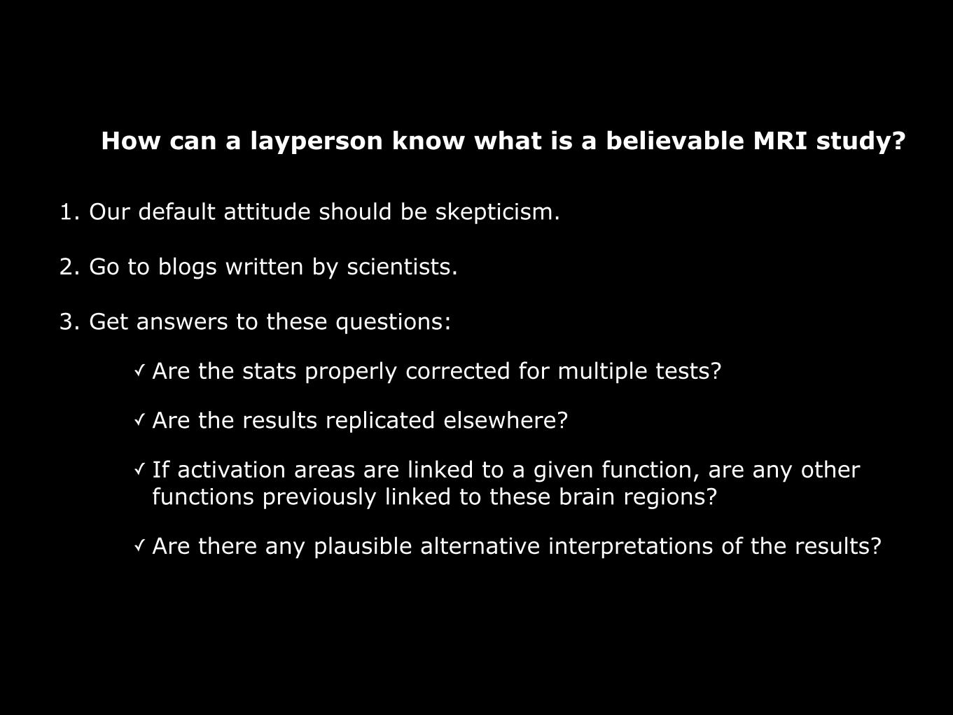
How can a layperson know what is a believable MRI study?
1. Our default attitude should be skepticism.
2. Go to blogs written by scientists.
3. Get answers to these questions:
✓ Are the stats properly corrected for multiple tests?
✓ Are the results replicated elsewhere?
✓ If activation areas are linked to a given function, are any other functions previously linked to these brain regions?
✓ Are there any plausible alternative interpretations of the results?
Neuroscience
Myths

1891 - 1976 !
!!
What percentage of Wilder Penfield’s patients
experienced spontaneous memories when he
inserted an electrode into their brain?
Watch one minute video: https://www.youtube.com/watch?v=kNdM9JhTPJw

1 in 20 and contemporary surgeons have found it
difficult to replicate some of Penfield’s results. !
Raymond Tallis, Aping Mankind (2011, p. 93)

“... allow us to grasp the minds of others not through conceptual reasoning but through direct simulation.”
Giacomo Rizzolatti, co-discover of mirror neurons in macaque monkeys (New York Times, 10 Jan 2006, ‘Cells That Read Minds’)
!!!
"... the driving force behind the great leap forward in human evolution."
V.S. Ramachandran (2000)

Except that, most evidence for mirror neurons in humans is indirect.
“Mirror neurons have not been demonstrated unequivocally in humans”
Raymond Tallis, Aping Mankind (2011, p. 190)
!“fMRI’s resolution is not fine enough to distinguish whether
the neurons firing are mirror neurons or just motor cortex neurons, which fire both when we think about an action and
when we actually perform an action.” !
Marco Iacoboni, a mirror neuron expert at the University of California, quoted in Monkey See, Monkey Don't by Nikhil Swaminathan February 3, 2011 Scientific American Mind
!!
37
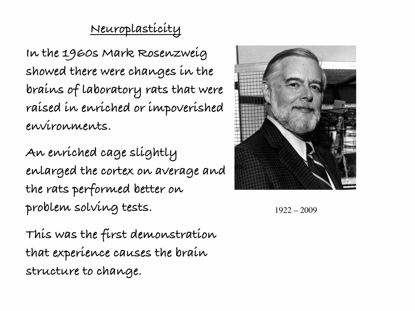
Neuroplasticity !In the 1960s Mark Rosenzweig showed there were changes in the brains of laboratory rats that were raised in enriched or impoverished environments. !An enriched cage slightly enlarged the cortex on average and the rats performed better on problem solving tests.
This was the first demonstration that experience causes the brain structure to change. !
1922 – 2009
38

London taxi drivers have an
enlarged right posterior
hippocampus, which is the region
of the cortex thought to be involved
in navigation.
In musicians, the cerebellum is
larger and certain cortical regions
are thicker.
Bilinguals have a thicker cortex in
the lower part of the left parietal
lobe.

However ... !
“[Rosenzweig] did not prove that it was the thickening that
caused the improvement in the intelligence. We can only say
that cortical thickening with learning are correlated.
Furthermore, the correlation is weak, revealed only by averages
over groups. Cortical thickening is not a reliable predictor of
learning in individuals.” !
Sebastian Seung Connectome: How the Brain’s Wiring Makes Us Who We Are. (2012, p. 25)
Professor of Computational Neuroscience at MIT.

Minds differ because neural networks differ. Personality, IQ and memories are encoded in neural networks.
!
“Although this theory has been around for a long time, neuroscientists still don’t know if it’s true. These ideas may sound powerful, but there’s a catch: they have never been subjected to conclusive experimental tests ... because neuroscientists have lacked good techniques for mapping the connections between neurons”
Sebastian Seung (2012) Connectome: How the Brain’s Wiring Makes Us Who We Are. p. xiv-xx Professor of Computational Neuroscience at MIT.

Yes, but ... !
“Although this theory has been around for a long time, neuroscientists still don’t know if it’s true. These ideas may sound powerful, but there’s a catch: they have never been subjected to conclusive experimental tests ... because neuroscientists have lacked good techniques for mapping the connections between neurons”
Sebastian Seung Connectome: How the Brain’s Wiring Makes Us Who We Are. (2012, p. xiv-xx)

Thinking leading to Neuromania Mixing Logical Levels
“There is only one sort of stuff, namely matter – the physical stuff of physics, chemistry and physiology – and the mind is somehow nothing but a physical phenomenon. !In short, the mind is the brain ... We can (in principle!) account for every mental phenomenon using physical principles, laws and raw materials.”
Daniel Dennett, Consciousness Explained, p. 33MATTER
CELLS
BRAIN
MIND

Thinking leading to Neuromania Mixing metaphors
“Nervous systems are information-processing machines.” Patricia Churchland, Neurophilosophy.
“The brain can now be described as an incredibly powerful microprocessor, the mother of all motherboards.”
Dr Vinoth Ramachandra
“Artificial intelligence is the science and engineering of making intelligent machines.” John McCarthy
!!!!!!!
Metaphor
Metaphor
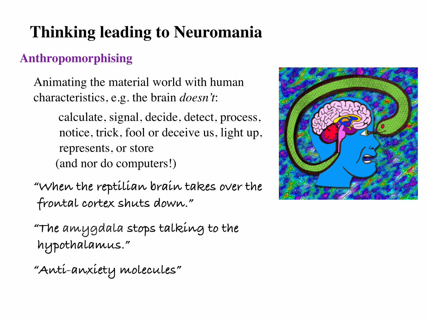
Thinking leading to Neuromania Anthropomorphising
Animating the material world with human characteristics, e.g. the brain doesn’t:
calculate, signal, decide, detect, process, notice, trick, fool or deceive us, light up, represents, or store
(and nor do computers!)
“When the reptilian brain takes over the frontal cortex shuts down.”
“The amygdala stops talking to the hypothalamus.”
“Anti-anxiety molecules”

Thinking leading to Neuromania Unwitting metonymy ‣ Mistaking a part for the whole. e.g.
Nobel prize winner Eric Kandel claimed he could capture “memory in a dish”.
Localising a distributed, massively interconnected, small-world network (<3 degrees of separation).
!!!!!!!!!!
“The brain - that's my second most favourite organ!” Woody Allen

The truth is ... !
“As a neuroscientist myself, I have come to know firsthand
[the] feeling of dread [when] I speak to the public about the
state of our field. My audience [is] curious about brains that
malfunction or excel, but even the humdrum lacks
explanation.
Every day we recall the past, perceive the present, and imagine
the future. How do our brains accomplish these feats?
It’s safe to say that nobody really knows.”
!Sebastian Seung

"To map the human brain at the cellular level, we're talking about 1m petabytes of information. Most people think that is more than the digital content of the world right now.
I'd settle for a mouse brain, but we're not even ready to do that. We're still working on how to do one cubic millimetre."
!"Sooner or later humans are going to have to confront the fact that we don't know how the brain works." !!Jeff Lichtman, Professor of Molecular and Cellular Biology, Harvard University quoted in The Guardian. 7 May 2012.

!“Fifty years of research shows that
we don’t understand what neural networks are doing.” !!
Dr. Michael Harré, Principle Investigator at Large, Centre for the Mind, Faculty of Science, University of Sydney
!“There is no science of the individual”
!!
Aristotle

References Bateson, G., 1972, Steps to an Ecology of Mind, Ballantine. Bateson, G., 1988, Mind and Nature: A Necessary Unity, Bantam. Bateson, G. & Bateson, M.C., 1988, Angels Fear: Towards an Epistemology of the Sacred, Bantum. Bateson, G., 1991, A Sacred Unity: Further Steps to an Ecology of Mind, Harper Collins. Begley, S., 2009, The Plastic Mind: New Science Reveals Our Extraordinary Potential to Transform Ourselves, Constable. Blackmore, S., 2000, The Meme Machine, Oxford University Press. Bloom, P. 2004 Descartes' Baby: How the Science of Child Development Explains What Makes Us Human, Arrow. Calvin, W.H., 1996, The Cerebral Code: Thinking a Thought in the Mosaics of the Mind, MIT Press. Capra, F., 1996, The Web of Life: A New Synthesis of Mind and Matter, Harper Collins. Chalmers, D.J, 1996 The Conscious Mind: In Search of a Fundamental Theory, Oxford University Press. Claxton, G., 1997, Hare Brain Tortoise Mind: Why Intelligence Increases When You Think Less, Fourth Estate. Claxton, G., 2006. The Wayward Mind: An Intimate History of the Unconscious, Abacus. Cytowic, R.E., 1993/2003, The Man Who Tasted Shapes, MIT Press. Damasio, A., 2006. Descartes’ Error: Emotion, Reason and the Human Brain, Vintage. Damasio, A., 2003, Looking for Spinoza: Joy, Sorrow and the Feeling Brain, Harcourt. Damasio, A., 2000. The Feeling Of What Happens: Body, Emotion and the Making of Consciousness New ed., Vintage. Dennett, D.C., 1993, Consciousness Explained, Penguin. Dennett, D.C., 2004, Freedom Evolves, Penguin. Doidge, N., 2008, The Brain That Changes Itself: Stories of Personal Triumph from the Frontiers of Brain Science, Penguin. Donald, M., 2001, A Mind So Rare: The Evolution of Human Consciousness, Norton. Dreyfus, H.L. & Dreyfus, S.E., 1988, Mind over Machine: The Power of Human Intuition and Expertise in the Era of the Computer, Free Press. Edelman, G.M., 1992, Bright Air, Brilliant Fire: On the Matter of the Mind, Basic Books, Basic Books. Edelman, G.M. & Tononi, G., 2000, The Universe of Consciousness: How Matter Becomes Imagination, Basic Books. Freeman, W., 2000 How Brains Make Up Their Minds, Phoenix Hauser, M. D., 2007 Moral Minds: The Nature of Right and Wrong, Harper Perennial. Hofstadter, D., 2008, I Am A Strange Loop, Basic Books. Huppert, F.A., Baylis, N. & Keverne, B., (eds) 2005, The Science of Well-Being, Oxford University Press. Jaynes, J., 1990, The Origin of Consciousness in the Breakdown of the Bicameral Mind, Hougton Mifflin. Kandel, E.R., 2007. In Search of Memory: The Emergence of a New Science of Mind, Norton LeDoux, J., 1999. The Emotional Brain: The Mysterious Underpinnings of Emotional Life Maturana, H.R. & Varela, F.J., 1992, The Tree of Knowledge: The Biological Roots of Human Understanding, Shambala. McGilchrist, I., The Master and his Emissary: The Divided Brain and the Making of the Western World, Yale University Press. Minsky, M., 2006, The Emotion Machine: Common Sense Thinking, Artificial Intelligence, and the Future of the Human Mind, Simon & Schuster Montague, R., 2006. Why Choose this Book?: How We Make Decisions, Dutton. Penrose, R., 1996. Shadows of the Mind: A Search for the Missing Science of Consciousness, Oxford University Press. Pinker, S., 1998. How The Mind Works, The Softback Preview. Pinker, S., 2007, The Stuff of Thought: Language as a Window into Human Nature, Allen Lane. Plotkin, H., 2003, The Imagined World Made Real: Towards A Natural Science of Culture, Penguin. Ramachandran, V.S. & Blakeslee, S., 1999. Phantoms in the Brain: Human Nature and the Architecture of the Mind, Fourth Estate. Robertson, I., 2000, Mind Sculpture: Your Brain’s Untapped Potential, Bantum. Rose, S., (ed) 1999, From Brains to Consciousness?: Essays on the New Sciences of the Mind, Penguin Rose, S., 2006, The 21st Century Brain: Explaining, Mending and Manipulating the Mind, Vintage. Searle, J.R., 1997, The Mystery of Consciousness, Granta. Seung, S., 2012, Connectome: How the Brain’s Wiring Makes Us Who We Are, Allen Lane. Sacks, O., 1986. The Man Who Mistook his Wife for a Hat, Picador. Sacks, O, 1995. An Anthropologist on Mars, Picador. Solso, R.L., & Massaro, D.W., 1995, The Science of the Mind: 2001 and Beyond, Oxford University Press. Tallis, R, 2011. Aping Mankind: Neuromania, Darwinitis and the Misrepresentation of Humanity, Acumen. Varela, F.J. & Thompson, E. & Rosch, E., 1993, The Embodied Mind: Cognitive Science and Human Experience, MIT Press. Zeman, A., 2002. Consciousness: a User's Guide, Yale University.