neuropsychological findings of extrapontine myelinolysis
TRANSCRIPT
Behavioural Neurology 18 (2007) 131–134 131IOS Press
Neuropsychological findings of extrapontinemyelinolysis without central pontinemyelinolysis
Jung Im Seok∗, Dong Kuck Lee, Min Gu Kang and Jae Han ParkDepartment of neurology, School of Medicine, Catholic University of Daegu, Korea
Abstract. Central pontine myelinolysis (CPM) and extrapontine myelinolysis (EPM) are well recognized syndromes related tothe rapid correction of hyponatremia, which are reported to show brain stem signs and various movement disorders. Cognitivedysfunction and neuropsychological findings, however, have seldom been reported. Cognitive manifestations in osmotic myeli-nolysis may have been underestimated due to the prominent brain stem symptoms and movement disorders. We report a case ofEPM without CPM and describe the neuropsychological findings of EPM. The absence of CPM in this case made it possible totest neuropsychological function in the acute stage.Neuropsychological testing showed severe impairment of attention, verbal and visual memory, visuospatial function, andfrontal/executive function. Language and language-related functions were normal except naming.
Keywords: Extrapontine myelinolysis, neuropsychological, central pontine myelinolysis
1. Introduction
Central pontine myelinolysis (CPM) and extrapon-tine myelinolysis (EPM) are distinctive clinical syn-dromes with characteristic magnetic resonance fea-tures, and demyelination is frequently related to a rapidcorrection of an electrolyte imbalance [1,2].
Two studies have examined the cognitive aspect ofCPM [3,4], and only one reported the findings of neu-ropsychological testing in CPM with EPM [5]. Wedescribe a case of EPM without CPM in which neu-ropsychological testing showed cognitive impairmentin multiple domains.
2. Case report
A 69-year-old man was admitted to the hospital withcomplaints of dizziness over a 1-week period. The
∗Corresponding author: Jung Im Seok, M.D., Department of Neu-rology, School of Medicine, Catholic University of Daegu, 3056-6,Daemyung-Dong, Nam-Gu, Daegu, 705-718, Korea. Tel.: +82 53650 3043; Fax: +82 53 654 9786; E-mail: [email protected].
sensation of imbalance was aggravated by walking, butnausea or vomiting did not accompany the dizziness.Three days prior to admission, the patient lost his bal-ance and fell down. He did not lose consciousness. Hismedical history was unremarkable except for a 15-yearhistory of hypertension that was currently being treatedwith diuretics (started four weeks earlier). He smoked20 cigarettes a day for 30 years and had no past historyof chronic alcohol abuse. Prior to admission, he wasa manager of his own company and had 16 years ofeducation.
On examination, he was alert and well oriented,and an ataxic gait comprised the only abnormal find-ing. Laboratory evaluation revealed severe hypona-tremia (100 mEq/L) and a low potassium concentra-tion (3.0 mEq/L). Other routine biochemical test re-sults, including a white blood cell count with differ-ential, hemoglobin, liver enzyme levels, renal func-tion, and glucose, were all within normal limits. AnECG showed no abnormalities. We diagnosed diuretic-induced hyponatremia, and the patient’s hyponatremiawas corrected to 126 mEq/L over 2 days.
ISSN 0953-4180/07/$17.00 2007 – IOS Press and the authors. All rights reserved

132 J.I. Seok et al. / Neuropsychological findings of extrapontine myelinolysis
Table 1Results of neuropsychological tests in the patient
Cognitive domain Results Cognitive domain Results
Attension Memorydigit span: forward 3 (<1%ile) Rey CFT: immediate recall 3 (<1%ile)
backward 2 (1.5%ile) 20-min delayed recall 0 (<1%ile)Language and related functions recognition 7 − 9 = −2
fluency NL Frontal/Executive functionauditory comprehension NL motor impersistance NLrepetition NL contrasting program NLnaming (K-BNT) 34/60 (<1%ile) go-no-go test NLreading NL fist-edge-palm NLwriting NL alternating hand movement NLcalculation NL alternating square and triangle Deformedfinger naming AB Luria loop Deformedright-left orientation NL semantic word fluency: animal; supermarket items 2;0 (AB)body part identification NL phonemic word fluency: sum of three consonants 0 (AB)limb praxis NL K-CWST: word reading: correct/incorrect 33/4 (AB)
Visuospatial functions color reading: correct/incorrect 30/8 (AB)K-MMSE: interlocking pentagon NL General cognitive indexRey-Osterrieth Complex Figure Test (Rey CFT) 5.5/36 (<1%ile) MMSE 19/30
Memory CDR 1K-MMSE: registration 3 GDS 4
recall 2 Neuropsychiatric symptomsHVLT: free recall (1st; 2nd; 3rd) 4/12;4/12;4/12 (5.5%ile) Geriatirc Depression Scale 12/30
20-min delayed recall 0 (<1%ile) K-NPI 2/144recognition (true positive-false positive) 10 − 7 = 3
K-BNT: The Korena version of the Boston Naming Test, HVLT: Hopkins Verbal Learning Test (Korean version),K-CWST: Korean Color Word Stroop Test, K-MMSE: Mini-Mental State Examination(Korean version),CDR:Clinical Dementia Rating Scale, GDS:Global Deterioration Scale, K-NPI: Neuropsychiatric Inventory (Korean version),NL: within normal limit, AB: abnormal.
On the seventh day after correcting the sodium lev-el, the patient developed progressive dysarthria, dys-phagia, and a gait disturbance. On examination, hewas alert, but would stare at the speaker’s eyes with afixed gaze and reduced blinking. Muscle strength wassymmetrical and nearly normal in all four extremities,and reflexes were normal in all four extremities. Nopathologic plantar responses were observed. Lead piperigidity was present, and the patient could not sit orstand without support.
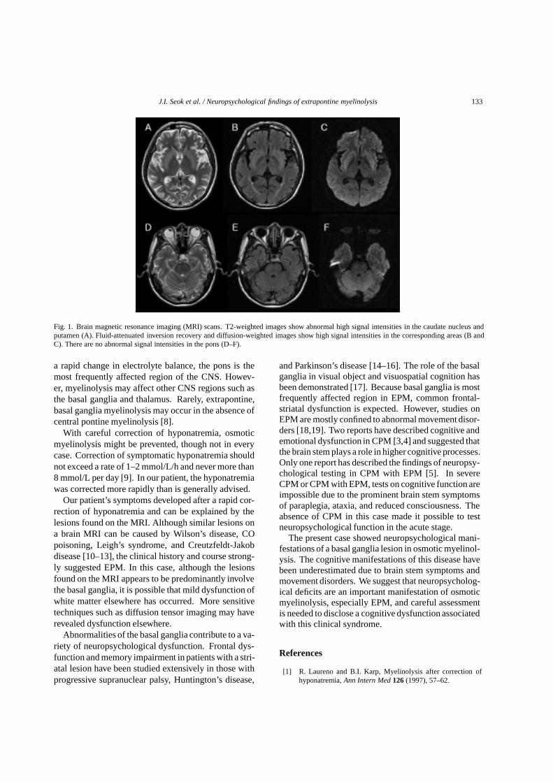
We performed a brain MRI to confirm osmotic myeli-nolysis because parkinsonian features had occurred af-ter the correction of hyponatremia. A T2-weighted andfluid-attenuated inversion recovery brain MRI showedsymmetric high signal intensities in the bilateral cau-date nucleus and putamen (Fig. 1A, B). No abnormalsignal intensity occurred in the pons (Fig. 1D-F). Thediffusion-weighted imaging (DWI) showed high signalintensities in the corresponding areas (Fig. 1C). TheApparent Diffusion Coefficient map showed no definiteabnormality. A clinical and radiological diagnosis ofEPM was made.
Since steroid administration or symptomatic treat-ment with dopaminergiccompoundshas provedbenefi-cial in several cases [6,7], we started steroid pulse ther-
apy that was continued for 3 days and was followed bymedication with a dopaminergic agent. After the initi-ation of treatment, the patient’s symptoms slowly im-proved. On the fifth day of treatment, the patient’s mus-cle strength was normal; the dysarthria was also muchimproved. However, the rigidity persisted, and reflexeswere increased in all four extremities. He could standwith support and could walk with substantial difficultydue to a short stride and postural instability.
Neuropsychological examination was performed onthe tenth day of treatment. The results of neuropsy-chological testing are presented in Table 1. In summa-ry, the patient was severely impaired in regard to at-tention, naming, verbal and visual memory, visuospa-tial function, and frontal/executive function. The pa-tient demonstrated impaired performance on tests ofrecognition memory as well as free recall memory.Language and language-related functions were normalexcept naming. No prominent psychiatric symptomswere observed.
3. Discussion
Osmotic myelinolysis may develop after a rapid cor-rection of hyponatremia. Due to its vulnerability to
J.I. Seok et al. / Neuropsychological findings of extrapontine myelinolysis 133
Fig. 1. Brain magnetic resonance imaging (MRI) scans. T2-weighted images show abnormal high signal intensities in the caudate nucleus andputamen (A). Fluid-attenuated inversion recovery and diffusion-weighted images show high signal intensities in the corresponding areas (B andC). There are no abnormal signal intensities in the pons (D–F).
a rapid change in electrolyte balance, the pons is themost frequently affected region of the CNS. Howev-er, myelinolysis may affect other CNS regions such asthe basal ganglia and thalamus. Rarely, extrapontine,basal ganglia myelinolysis may occur in the absence ofcentral pontine myelinolysis [8].
With careful correction of hyponatremia, osmoticmyelinolysis might be prevented, though not in everycase. Correction of symptomatic hyponatremia shouldnot exceed a rate of 1–2 mmol/L/h and never more than8 mmol/L per day [9]. In our patient, the hyponatremiawas corrected more rapidly than is generally advised.
Our patient’s symptoms developed after a rapid cor-rection of hyponatremia and can be explained by thelesions found on the MRI. Although similar lesions ona brain MRI can be caused by Wilson’s disease, COpoisoning, Leigh’s syndrome, and Creutzfeldt-Jakobdisease [10–13], the clinical history and course strong-ly suggested EPM. In this case, although the lesionsfound on the MRI appears to be predominantly involvethe basal ganglia, it is possible that mild dysfunction ofwhite matter elsewhere has occurred. More sensitivetechniques such as diffusion tensor imaging may haverevealed dysfunction elsewhere.
Abnormalities of the basal ganglia contribute to a va-riety of neuropsychological dysfunction. Frontal dys-function and memory impairment in patients with a stri-atal lesion have been studied extensively in those withprogressive supranuclear palsy, Huntington’s disease,
and Parkinson’s disease [14–16]. The role of the basalganglia in visual object and visuospatial cognition hasbeen demonstrated [17]. Because basal ganglia is mostfrequently affected region in EPM, common frontal-striatal dysfunction is expected. However, studies onEPM are mostly confined to abnormal movement disor-ders [18,19]. Two reports have described cognitive andemotional dysfunction in CPM [3,4] and suggested thatthe brain stem plays a role in higher cognitive processes.Only one report has described the findings of neuropsy-chological testing in CPM with EPM [5]. In severeCPM or CPM with EPM, tests on cognitive function areimpossible due to the prominent brain stem symptomsof paraplegia, ataxia, and reduced consciousness. Theabsence of CPM in this case made it possible to testneuropsychological function in the acute stage.
The present case showed neuropsychological mani-festations of a basal ganglia lesion in osmotic myelinol-ysis. The cognitive manifestations of this disease havebeen underestimated due to brain stem symptoms andmovement disorders. We suggest that neuropsycholog-ical deficits are an important manifestation of osmoticmyelinolysis, especially EPM, and careful assessmentis needed to disclose a cognitive dysfunction associatedwith this clinical syndrome.
References
[1] R. Laureno and B.I. Karp, Myelinolysis after correction ofhyponatremia,Ann Intern Med 126 (1997), 57–62.
134 J.I. Seok et al. / Neuropsychological findings of extrapontine myelinolysis
[2] D.G. Wright, R. Laureno and M. Victor, Pontine and extrapon-tine myelinolysis,Brain 102 (1979), 361–385.
[3] T.M. Lee, C.C. Cheung, E.Y. Lau, A. Mak and L.S. Li, Cog-nitive and emotional dysfunction after central pontine myeli-nolysis,Behav Neurol 14 (2003), 103–107.
[4] B.H. Price and M.M. Mesulam, Behavioral manifestations ofcentral pontine myelinolysis,Arch Neurol 44 (1987), 671–673.
[5] E. Vermetten, S.J. Rutten, P.J. Boon, P.A. Hofman and A.F.Leentjens, Neuropsychiatric and neuropsychological manifes-tations of central pontine myelinolysis,General Hospital Psy-chiatry 21 (1999), 296–302.
[6] H. Nakano, Y. Ohara, K. Bandoh and M. Miyaoka, A caseof central pontine myelionosys after surgical removal of apituitary tumor,Surg Neurol 46 (1996), 32–36.
[7] M. Sadeh and Y. Goldhammer, Extrapyramidal syndrome re-sponsive to dopaminergic treatment following recovery fromcentral pontine myelinolysis,Eur Neurol 33 (1993), 48–50.
[8] M.G. Hadfield and W.S. Kubal, Extrapontine myelinolysisof the basal ganglia without pontine myelinolysis,Clin Neu-ropathol 15 (1996), 96–100.
[9] W.D. Brown, Osmotic demyelination disorders: central pon-tine and extrapontine myelinolysis,Curr Opin Neurol 13(2000), 691–697.
[10] R.N. Sener, Diffusion MR imaging changes associated withWilson disease,AJNR Am J Neuroradiol 24 (2003), 965–967.
[11] J. Arii and Y. Tanabe, Leigh syndrome: serial MR imagingand clinical follow-up,AJNR Am J Neuroradiol 21 (2000),1502–1509.
[12] R.N. Sener, Acute carbon monoxide poisoning: diffusion MRimaging findings,AJNR Am J Neuroradiol 24 (2003), 1475–1477.
[13] D.P. Barboriak, J.M. Provenzale and O.B. Boyko, MR diag-nosis of Creutzfeldt-Jakob disease: significance of high sig-nal intensity of the basal ganglia,AJR Am J Roentgenol 162(1994), 137–140.
[14] M.L. Albert, B.G. Feldman and A.L. Willis, The ‘subcorticaldementia’ of progressive supranuclear palsy,J Neurol Neuro-surg Psychiatry 37 (1974), 121–130.
[15] A. Montoya, B.H. Price, M. Menear and M. Lepage, Brainimaging and cognitive dysfunctions in Huntington’s disease,J Psychiatry Neurosci 31 (2006), 21–29.
[16] D. Muslimovic, B. Post, J.D. Speelman and B. Schmand, Cog-nitive profile of patients with newly diagnosed Parkinson dis-ease,Neurology 65 (2005), 1239–1245.
[17] A.D. Lawrence, L.H. Watkins, B.J. Sahakian, J.R. Hodgesand T.W. Robbins, Visual object and visuospatial cognition inHuntington’s disease: implications for information processingin corticostriatal circuits,Brain 123 (2000), 1349–1364.
[18] A.B.H. Seah, L.L. Chan, M.C. Wong and E.K. Tan, Evolvingspectrum of movement disorders in extrapontine and centralpontine myelinolysis,Parkinsonism Relat Disord 9 (2002),117–119.
[19] A. Seiser, S. Schwarz, M. Aichinger-Steiner, G. Funk, P.Schnider and M. Brainin, Parkinsonism and dystonia in centralpontine and extrapontine myelinolysis,J Neurol NeurosurgPsychiatry 65 (1998), 119–121.

Submit your manuscripts athttp://www.hindawi.com
Stem CellsInternational
Hindawi Publishing Corporationhttp://www.hindawi.com Volume 2014
Hindawi Publishing Corporationhttp://www.hindawi.com Volume 2014
MEDIATORSINFLAMMATION
of
Hindawi Publishing Corporationhttp://www.hindawi.com Volume 2014
Behavioural Neurology
EndocrinologyInternational Journal of
Hindawi Publishing Corporationhttp://www.hindawi.com Volume 2014
Hindawi Publishing Corporationhttp://www.hindawi.com Volume 2014
Disease Markers
Hindawi Publishing Corporationhttp://www.hindawi.com Volume 2014
BioMed Research International
OncologyJournal of
Hindawi Publishing Corporationhttp://www.hindawi.com Volume 2014
Hindawi Publishing Corporationhttp://www.hindawi.com Volume 2014
Oxidative Medicine and Cellular Longevity
Hindawi Publishing Corporationhttp://www.hindawi.com Volume 2014
PPAR Research
The Scientific World JournalHindawi Publishing Corporation http://www.hindawi.com Volume 2014
Immunology ResearchHindawi Publishing Corporationhttp://www.hindawi.com Volume 2014
Journal of
ObesityJournal of
Hindawi Publishing Corporationhttp://www.hindawi.com Volume 2014
Hindawi Publishing Corporationhttp://www.hindawi.com Volume 2014
Computational and Mathematical Methods in Medicine
OphthalmologyJournal of
Hindawi Publishing Corporationhttp://www.hindawi.com Volume 2014
Diabetes ResearchJournal of
Hindawi Publishing Corporationhttp://www.hindawi.com Volume 2014
Hindawi Publishing Corporationhttp://www.hindawi.com Volume 2014
Research and TreatmentAIDS
Hindawi Publishing Corporationhttp://www.hindawi.com Volume 2014
Gastroenterology Research and Practice
Hindawi Publishing Corporationhttp://www.hindawi.com Volume 2014
Parkinson’s Disease
Evidence-Based Complementary and Alternative Medicine
Volume 2014Hindawi Publishing Corporationhttp://www.hindawi.com