neuropilin-1 mediates divergent r-smad ... mediates divergent r-smad signaling and the myofibroblast...
TRANSCRIPT
NEUROPILIN-1 MEDIATES DIVERGENT R-SMAD SIGNALING AND THE MYOFIBROBLAST PHENOTYPE
Ying Cao,1 Annamaria Szabolcs,1 Shamit K. Dutta,1 Usman Yaqoob,2 Kumaravelu Jagavelu,2 Ling Wang,1 Edward B. Leof,3 Raul A. Urrutia,2 Vijay H. Shah,2 and Debabrata
Mukhopadhyay1,* 1Department of Biochemistry and Molecular Biology, 2GI Research Unit, 3Thoracic Diseases Research Unit, Department of Medicine, College of Medicine, Mayo Clinic, Rochester, Minn.
Running head: Neuropilin-1 mediates divergent R-Smad signaling Address correspondence to: Debabrata Mukhopadhyay, Ph.D., Guggenheim 1321C, 200 First St. S.W., Rochester, MN 55905. Phone: 507-538-3581. Fax: 507-233-1058. Email: mukhopadhyay. [email protected]
The transforming growth factor-beta (TGF-β) superfamily is one of the most diversified cell signaling pathways and regulates many physiological and pathological processes. Recently, neuropilin-1 (NRP-1) was reported to bind and activate the latent form of TGF-β1 (LAP-TGF-β1). We investigated the role of NRP-1 on Smad signaling in stromal fibroblasts upon TGF-β stimulation. Elimination of NRP-1 in stromal fibroblast cell lines increases Smad1/5 phosphorylation and downstream responses, as evidenced by upregulation of inhibitor of differentiation (Id-1). Conversely, NRP-1 loss decreases Smad2/3 phosphorylation and its responses, as shown by downregulation of α-smooth muscle actin (α-SMA) and cell exhibition of more quiescent phenotypes and growth arrest. We also observed that NRP-1 expression increases during the culture activation of hepatic stellate cells (HSCs), a liver resident fibroblast. Taken together, our data suggest that NRP-1 functions as a key determinant of the diverse responses downstream of TGF-β1 that are mediated by distinct Smad proteins and promotes myofibroblast phenotype.
NRP-1 was initially discovered as a
semaphorin co-receptor and vascular permeability factor/vascular endothelial growth factor (VPF/VEGF) co-receptor (1,2). Recently, NRP-1 was shown to bind and activate latency-associated protein (LAP)-TGF-β1 and enhance regulatory T cell (Treg) activity (3). The extracellular domain of NRP-1 contains three structural motifs: two cubilin (CUB) homology domains (a1, a2), two coagulation factor V/VIII homology domains (b1, b2), and a meprin/A5-protein/PTPmu (MAM) domain (c) (4). The relatively short (about 40 amino acids) cytoplasmic domain lacks kinase motifs. Interestingly, NRP-1 has a similar intracellular domain as TGF-β receptor III (TGF-βRIII/betaglycan), and its homolog, endoglin, with the PSD-95/Disc-large/ZO-1 (PDZ) binding motif (Supplemental Figure 1). Therefore, we hypothesize that NRP-1 may also serve as a TGF-β co-receptor that regulates TGF-β signaling.
TGF-β is one member of a superfamily of secreted proteins, which also includes activins and bone morphogenetic proteins (BMPs). TGF-β signaling is one of the most diversified signaling cascades, controlling many aspects of cell behavior, including cell division, differentiation, motility, and death. TGF-β receptors include type I (TβRI), type II
http://www.jbc.org/cgi/doi/10.1074/jbc.M110.151696The latest version is at JBC Papers in Press. Published on July 30, 2010 as Manuscript M110.151696
Copyright 2010 by The American Society for Biochemistry and Molecular Biology, Inc.
by guest on May 16, 2018
http://ww
w.jbc.org/
Dow
nloaded from

2
(TβRII), and type III (TβRIII). TβRI and TβRII, which are serine/threonine kinase receptors, constitute a heterotetrameric core receptor complex, and TβRIII modulates signaling by regulating ligand binding to the core receptor complex. There are at least seven TβRIs (activin receptor-like kinase 1-7, ALK1-7), five TβRIIs (TGF-βRII, ActRIIA, ActRIIB, AMHRII, and BMPRII), and two TβRIIIs (betaglycan and endoglin). ALK-2, ALK3, ALK4, ALK5, and ALK6 are also known as ActR-I, BMPR-IA, ActR-IB, TGF-βRI, and BMPR-IB, respectively (5). Upon TGF-β binding, TβRII activates and phosphorylates TβRI, which then phosphorylates the receptor-regulated Smad (R-Smad) proteins (including Smad1, 2, 3, 5, and 8). Subsequently, the phosphorylated R-Smad protein forms a heteromeric complex with the co-Smad (Smad4) and translocates to the nucleus to regulate the target gene transcription (6-9). In addition to this canonical Smad pathway, TGF-β also activates Smad-independent signaling transduction pathways in a cell-type–specific manner, including Rho-ROCK1, Cdc42/Rac1- p21-activated kinase-2 (PAK2), c-Abl, and the mammalian targets of rapamycin (mTOR), JNK, and p38 MAPK (10). There are also R-Smad–dependent but Smad4-independent pathways that mediate TGF-β signaling (11-13). The effects of TGF-β depend highly on cell type. For example, TGF-β inhibits epithelial cell proliferation but increases endothelial cell proliferation. As TGF-β signaling is transduced from the cell membrane, receptor expression levels and combinations on each cell type contribute to the outcome. Initially, TGF-β was believed to activate only Smad2/3 through ALK5. Some cells, like
endothelial cells, sequentially express ALK1 and ALK5, active Smad1/5/8, and Smad2/3 upon TGF-β binding. Recently, TGF-β was shown to activate Smad1/5/8 and Smad2/3 in several types of cells, including non-cancerous epithelial cells, fibroblasts, and cancer cells (14-16). Activated Smad1/5/8 and Smad2/3 have different — and occasionally opposite — functions. The mechanisms that regulate the diversity of responses downstream from TGF-β are unknown. Here, we report that NRP-1 can promote divergent signaling that leads to differential Smad1/5 and Smad2/3 activation and downstream myofibroblast phenotypes. Hence, NRP-1 can control Smad1/5 and Smad2/3 signaling counterbalances and regulate diversified TGF-β signaling.
Experimental Procedures Cell culture-LX2 cells, pancreatic tumor stromal cells (PSCs), wild-type mouse embryonic fibroblasts (MEFs) and NRP-1-/- MEFs (17) were cultured in Dulbecco's Modified Eagle Medium (DMEM), 10% fetal bovine serum (FBS), and 1% penicillin/streptomycin. Cells were serum-starved for 16 hours before TGF-β and BMP9 treatment. Primary stellate cell immortalization-To immortalize human pancreatic stellate cells from pancreatic tumor stromal cells (PSCs), primary cells were incubated with amphotropic retrovirus containing SV40 large T antigen (Kind gift of Dr. Mulligan at MIT) for 24 hours under culture conditions. Media and virus were replenished three to five times to ensure viral uptake and gene incorporation. Heterogeneous populations of immortalized stellate cells were serially diluted and plated as single cells per
by guest on May 16, 2018
http://ww
w.jbc.org/
Dow
nloaded from

3
well to establish clones. Immortalized stellate cells were frozen in cryoprotectant media containing 45% complete media, 50% FBS, and 5% dimethyl sulfoxide (DMSO). We subsequently characterized these cells by the presence of established markers. For this purpose, total RNA was extracted from cells according to the manufacturer’s instructions using an RNeasy Kit (Qiagen) with on-column DNase digestion (Qiagen). RNA (2 µg) was converted to cDNA using an oligo (dT) primer and SuperScript™ III First-Strand Synthesis System for reverse transcription polymerase chain reaction (RT-PCR) (Invitrogen, Carlsbad, Calif.) per the manufacturer’s protocol. Stellate-specific marker primers were designed and PCR performed using Platinum Taq DNA Polymerase (Invitrogen). Cycle conditions were as follows: 30 cycles of 94°C for 30 seconds, 55°C for 30 seconds, and 72°C for 1 minute. Positive bands were visualized on 1.5% agarose with ethidium bromide. Antibodies and other reagents-NRP-1, Id-1, TβRII, and β-Actin were from Santa Cruz Biotechnology, Inc. (Santa Cruz, Calif.); Smad2, Smad5, p-Smad1/5, p-Smad2, p-Smad3 were purchased from Cell Signaling Technology, Inc. (Danvers, Mass.); α-SMA was from Millipore (Billerica, Mass.); collagen I antibody was from Rockland Immunochemicals, Inc. (Gilbertsville, Pa.); TGF-β1 was from Biolegend (San Diego, Calif.); BMP9 was from R&D Systems (Minneapolis, Minn.); ALK5 inhibitors SB431542 and ALK5 inhibitors (2-(3-(6-methylpyridine-2-yl)-1H-pyrazol-4-yl)-1,5-naphthyridine) were from Stemgent (Cambridge, Mass.); NRP-1 b domain blocking antibody (anti-NRP1B) was a gift from Genentech (San Francisco, Calif).
siRNA and shRNA transfection-siRNA for human NRP-1, TβRII, and control were from Qiagen, Inc. (Valencia, Calif.). siRNA transfection was performed with Hiperfect (Qiagen) following the manufacturer's instructions. shRNA for human NRP-1 and controls were from Open Biosystems (Huntsville, Ala.) and were prepared as previously described (18). The sequences of siRNA and shRNA were listed in supplemental materials and methods. RNA isolation and PCR-Total RNAs were extracted using the RNeasy Mini Kit (Qiagen) and reverse-transcribed by oligo (dT) priming using the iScript cDNA Synthesis Kit following the manufacturer's instructions (Bio-Rad, Hercules, Calif.). Semi-quantitative real-time PCR analyses were performed using the ABI 7500 Fast Real-Time PCR System (Applied Biosystems, Foster City, Calif.) and SYBR Green PCR Master Mix (Applied Biosciences). The primer sequences used were listed in supplemental materials and methods. Immunocytochemistry staining-Cells grew on glass coverslips overnight and were washed three times with phosphate-buffered saline (PBS), then fixed with 4% paraformaldehyde (in PBS) for 10 minutes at room temperature. The excess paraformaldehyde was quenched by incubating in 50 mM NH4Cl for 10 minutes. The cell membrane was permeabalized with 0.2% saponin for 10 minutes. Three percent bovine serum albumin (BSA)/PBS was used for blocking for 10 minutes at room temperature and then incubated in primary antibody (1:200) overnight at 4℃. Secondary antibody was added for 1 hour at room temperature in the dark. The coverslip was mounted with mount medium with 4',6-diamidino-2-phenylindole (DAPI) and photographed using Zeiss
by guest on May 16, 2018
http://ww
w.jbc.org/
Dow
nloaded from

4
confocal laser scanning microscopy. Preparation of whole-cell extracts-Cells were washed twice with cold PBS, lysed with ice-cold RIPA lysis buffer (50 mM Tris [pH 7.5], 1% NP-40, 150 mM NaCl, 0.5% sodium deoxycholate, 0.1% sodium dodecyl sulfate [SDS]) with 1% proteinase inhibitor cocktails (PIC) (Sigma-Aldrich, St. Louis, Mo.), and 1% Halt phosphatase inhibitor cocktail (Pierce, Rockford, Ill.), incubated on ice for 30 minutes, and centrifuged at 14,000 rpm at 4°C for 10 minutes. Supernatant was collected, and protein concentration was measured by Bradford method (Bio-Rad Protein Assay). Western blot-Proteins were denatured by adding 6x laemmli SDS sample buffer and heating for 4 minutes. Equal amounts of total protein per lane were subjected to SDS gel electrophoresis followed by wet transfer of the protein to polyvinylidene fluoride (PVDF) membrane. The membrane was blocked by incubation in TBS-T buffer (50 mM tris-HCl, pH 7.4, 150 mM NaCl, and 0.05% Tween 20) containing 5% nonfat milk or BSA. The primary antibody was diluted in TBS-T containing 5% nonfat milk or BSA overnight at 4°C, and horseradish peroxidase (HRP)-conjugated secondary antibody (Santa Cruz Biotechnology) was diluted in TBS-T and incubated for 1 hour at room temperature. Immunodetection was performed with the SuperSignal West Pico Substrate (Thermo Scientific, Rockford, Ill.). Immunoprecipitation assays-500 µg cell lysate, 1 or 2 µg antibody, and 50 µl protein A/G-coupled Sepharose beads were added to the tube at 4°C overnight under agitation. Beads were washed in RIPA buffer three times, and 2x loading buffer was added to the beads. Mixtures were boiled at 95° to 100°C for 4 minutes and then centrifuged. The
supernatant was retained for Western blotting. Cell proliferation assay-LX2 (1x10⁴) was seeded in 24-well plates, subjected to the siRNA treatment, and cultured for 2 days in DMEM plus 10% FBS. 1 µCi [3H]-thymidine was added to each well and, 4 hours later, cells were washed with cold PBS, fixed with 100% cold methanol, and collected for the measurement of trichloroacetic acid-precipitable radioactivity. Apoptosis assay-Cell apoptosis assay was done using the Annexin V-FITC Apoptosis Kit (BioVision, Mountain View, Calif.). Briefly, cells were detached using 0.5 mM ethylenediaminetetraacetic acid (EDTA) in PBS. After PBS washing, cells were resuspended in 500 ul 1x binding buffer. 5 µl of Annexin V-FITC and 5 µl of propidium iodide (PI) were added and incubated at room temperature for 5 minutes in the dark. Samples were subjected to flow cytometry analysis. Luciferase reporter assay-Luc-Id1 (obtained from Dr. Vivek Mittal, Weill Cornell Medical College), Luc-ARE, FAST-1, and Luc-SBE were used. Briefly, 5x10³ cells per well in a 96-well plate were transfected with siRNA in complete media. After 24 hours, cells were serum-starved overnight, then transfected with 0.05 µg/well luciferase reporter plasmid using a 0.01/well pRL-TK Renilla luciferase vector as the internal control. One hour after transfection, TGF-β was added for 20 to 24 hours. Firefly luciferase and Renilla luciferase activities were performed using the Dual-Luciferase Reporter Assay System (Promega, Madison, Wis.) and measured in a LB960 microplate luminometer. Data were expressed as the mean standard deviations of triplicate values. Construction of the NRP-1 expression
by guest on May 16, 2018
http://ww
w.jbc.org/
Dow
nloaded from

5
vector-NRP-1 cDNA in pcDNA3.1 plasmid was obtained from Dr. Shay Soker. The NRP-1 gene was subcloned into the pMMP retroviral vector. Virus preparation and infection were performed as previously described (19).
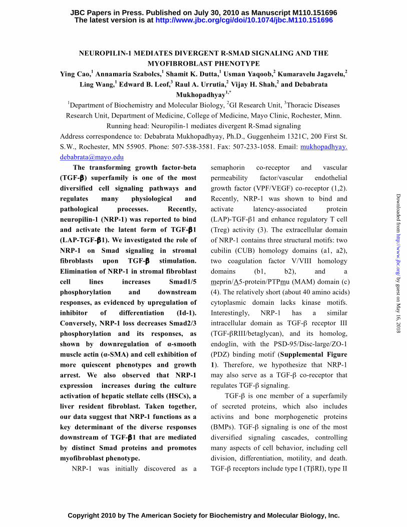
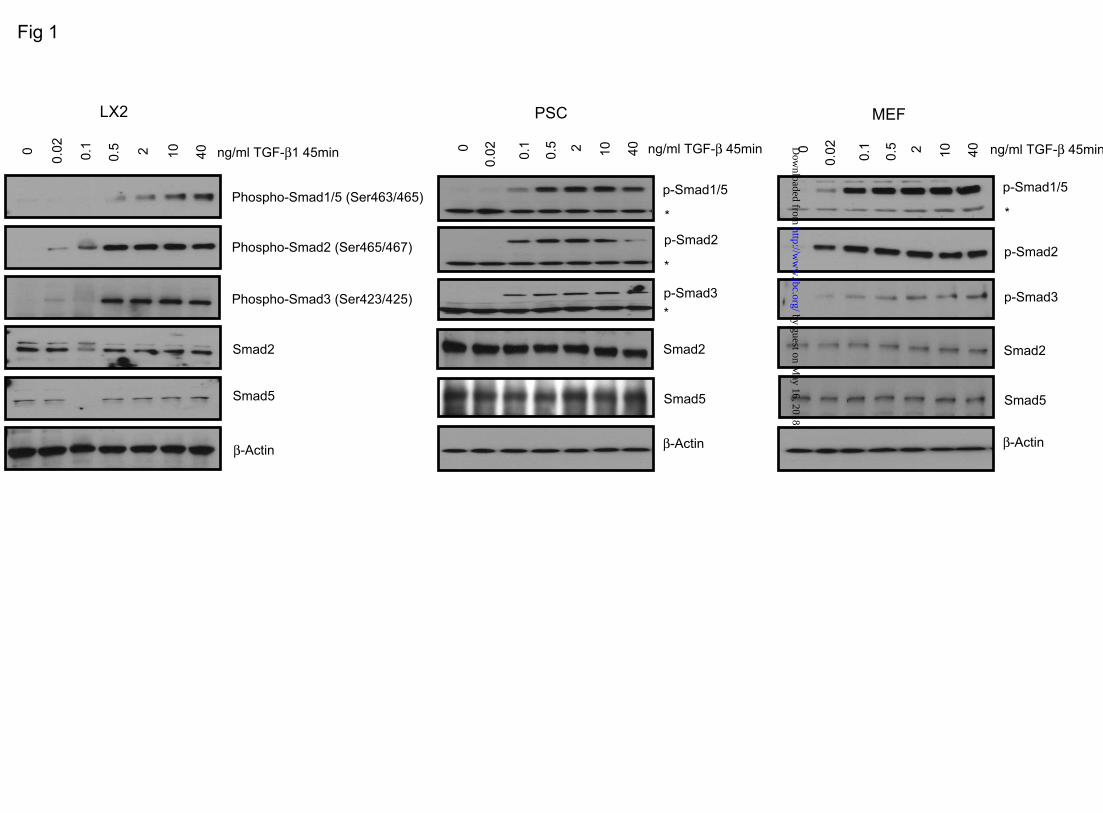
RESULTS NRP-1 controls TGF-β–induced Smad1/5 and Smad2/3 signaling balance. NRP-1 was shown to activate LAP-TGF-β1 (3). However, NRP-1 binds not only the LAP-TGF-β1, but also the active TGF-β1, indicating that NRP-1 has a role in active TGF-β1-mediated signaling (3). Hence, we hypothesized that NRP-1 might influence TGF-β-mediated signaling and downstream Smad phosphorylation. To this end, we utilized LX2 (an HSC line) cells, PSCs, and MEFs and determined the TGF-β–induced Smad activation in those cells. In order to characterize our model, we first examined Smad phosphorylation downstream of TGF-β stimulation in our cell models. TGF-β stimulation led to a concentration-dependent Smad1/5 (Ser463/465 for both Smad1 and Smad5) as well as Smad2/3 (Ser465/467 for Smad2 and Ser423/425 for Smad3) phosphorylation in each of the cell lines we studied: LX2, PSC, and MEF cells (Figure 1). Based on the finding that NRP-1 binds TGF-β1, we hypothesized that ablation of NRP-1 could globally reduce Smad activation. Surprisingly, siRNA-mediated knockdown of NRP-1 in LX2 and PSC, and NRP-1 knockout (NRP-1-/-) in MEF resulted in enhanced phosphorylation of Smad1/5, while phosphorylation of Smad2/3 was impaired (Figure 2A). NRP-1 overexpression in the NRP-1-/- MEF reversed the Smad
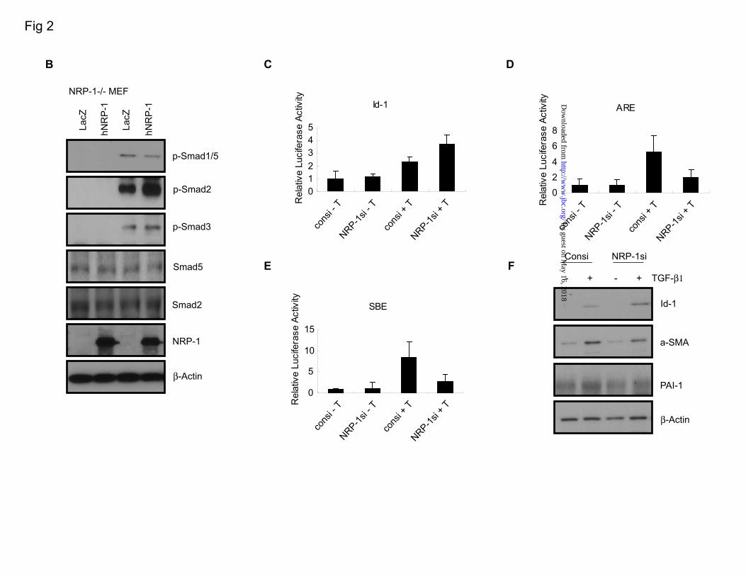
phosphorylation responses in the presence of TGF-β as suppression of Smad1/5 phosphorylation and elevation of Smad2/3 phosphorylation (Figure 2B). Consistent with these results, knockdown of NRP-1 enhanced Id-1-Luc promoter (a Smad1 reporter that contains BMP binding elements ]) activity and downregulated activin response elements (ARE-Luc, a Smad2/4-specific reporter). Smad-binding elements (SBE-Luc) , a Smad3-responsive reporter (21), was also downregulated as a result of siRNA-mediated NRP-1 knockdown (Figure 2C-E). The Id-1 protein level was also enhanced after NRP-1 knockdown. Levels of α-smooth muscle actin (α-SMA) and plasminogen activator inhibitor-1 (PAI-1), Smad2/3 response genes, were less responsive to TGF-β stimulation after NRP-1 knockdown (Figure 2F). NRP-1 associates with TGF-βRII. NRP-1 is known for the absence of any kinase domain; thus, it cannot phosphorylate Smad proteins directly. We investigated whether NRP-1 could interact with TGF-βRII as a co-receptor. Endogenous NRP-1 was co-immunoprecipitated with endogenous TGF-βRII (Figure 3A) in LX2 cells; TGF-β treatment did not increase the association between NRP-1 and TGF-βRII (Figure 3B). Knockdown of TGF-βRII with siRNA completely blocked TGF-β–mediated Smad1/5 and Smad2/3 phosphorylation, and the regulating role of NRP-1 also was eliminated (Figure 3C), indicating that NRP-1 functions through TGF-βRII–regulated Smad protein phosphorylation. NRP-1 may also directly regulate TGF-βRI to modulate Smad protein phosphorylation. Smad1/5 phosphorylation was thought to occur exclusively during BMP
by guest on May 16, 2018
http://ww
w.jbc.org/
Dow
nloaded from

6
signaling and TGF-β signaling in endothelial cells. However, recent studies indicate that TGF-β can stimulate both Smad1/5 and Smad2/3 in epithelial cells, fibroblasts, and tumor cells in a TGF-βRII-, ALK5-, and ALK2/3-dependent manner (14-16). Consistent with previous reports, we found that TGF-β can stimulate Smad1/5 and Smad2/3 phosphorylation through TGF-βRII in LX2 cells. Furthermore, the pharmacological TGF-β inhibitor SB431542 (which inhibits ALK4/5/7 but does not affect ALK1/2/3/6) and an ALK5 inhibitor (22) completely block Smad2/3 as well as Smad1/5 phosphorylation (Figure 3D), indicating that Smad2/3 and Smad1/5 phosphorylation is ALK5-dependent. Due to specificity limitations of commercially available ALK5 antibodies, we performed co-immunoprecipitation of ALK5 and NRP-1 using exogenous expression of both Flag-ALK-5 and NRP-1 in 293T cells. Our results demonstrate that NRP-1 associates with ALK-5 on TGF-β stimulation in a time-dependent manner (Figure 3E). Knockdown of the endogenous TGF-βRII in 293T cells by siRNA eliminated this association, indicating that the association between NRP-1 and ALK-5 was mediated by TGF-βRII (Figure 3F). Knocking down NRP-1 impaired BMP–induced Smad1/5 phosphorylation. The TGF-β family member BMP, through BMP receptor combinations (ALK-1, 2, 3, and 6 as a type I receptor, BMPRII as a type II receptor), has been shown to activate the Smad proteins — specifically, Smad1, 5, and 8 (8). Because betaglycan and endoglin also regulate BMP signaling (23,24), we hypothesized that NRP-1 may also influence BMP signaling. Indeed, knocking down
NRP-1 impaired BMP-9, BMP-2, and BMP-4–induced Smad1/5 phosphorylation (Figure 4). These results indicate that NRP-1 can also regulate signaling of the other TGF-β superfamily of proteins in addition to TGF-β. NRP-1 knockdown reduces cell proliferation and leads to a quiescent phenotype. We addressed the cellular effects of blocking NRP-1 expression and observed that NRP-1 knockdown significantly decreased the proliferation of LX2 cells (Figure 5A). Furthermore, we investigated whether this reduction was caused by induction of apoptosis, as we observed previously in endothelial cells that lacked NRP-1 expression (25). As demonstrated in Figure 5B, NRP-1 knockdown did not cause cell apoptosis. Lentiviral NRP-1 shRNA was used to stably knock down NRP-1 (>7days), and levels of active and quiescent stellate cell markers α-SMA and Id-1 were examined. As shown in Figure 6A, α-SMA was downregulated and Id-1 was upregulated in cells expressing NRP-1 shRNA compared with that of controls. Given the in vitro cell culture conditions, LX2 cells are in a proliferative and active stage. The downregulated α-SMA and upregulated Id-1 suggest that the cells were reversed to a relatively quiescent phenotype. Also, collagen I production, which is expressed by the active fibroblasts, was decreased after NRP-1 knockdown (Figure 6B). Taken together, these results indicate that the cells were in a less activated stage, which could be due to the accumulation effect of the stimulation of a small amount of TGF-β in the culture medium. Indeed, in the presence of TGF-β signaling inhibitor SB431542 (5 µg/mL), Id-1 and α-SMA protein levels did not change after stable knockdown of NRP-1 (7 days).
by guest on May 16, 2018
http://ww
w.jbc.org/
Dow
nloaded from

7
Moreover, we noticed that, after knockdown of NRP-1, the cells exhibited the quiescent phenotypes in the first several passages and were gradually lost during culture (cell proliferation, Id-1, and α-SMA protein levels became the same as those of the control cells), even though the NRP-1 knockdown was properly maintained by the shRNA (data not shown). This occurrence might be explained by the compensatory effects of the other signaling, as the cells are prone to be activated in the in vitro culture system. During culture activation, NRP-1 was upregulated. Lastly, we correlated culture activation of HSC with NRP-1 expression with the idea that NRP-1 expression may positively correlate with myofibroblastic activation in vitro. Primary HSCs undergo myofibroblastic activation in vitro upon extended culture, and this culture-induced activation in vitro is a good model to mimic fibroblast activation in vivo. Mouse HSCs were isolated by the collagenase/Percoll density gradient centrifugation method (26), and cells were collected for RNA each day after culture. NRP-1 level change was determined by semi-quantitative real-time PCR. The results show that NRP-1 mRNA levels were gradually upregulated during culture (Figure 7). Mouse HSCs were confirmed by α-SMA immunofluorescence staining (Supplemental Figure 2A), and the dramatically increased α-SMA level as determined by semi-quantitative real-time PCR during the culture activation confirmed that the cells were activated from the quiescent stage (Supplemental Figure 2B).
DISCUSSION We demonstrated that NRP-1, like betaglycan and endoglin, can regulate TGF-β signaling
by interacting with TGF-βRII. These proteins have similar structures: a short cytoplasmic C terminal tail containing a PDZ binding domain that binds to the GAIP-interacting protein C terminus (GIPC). NRP-1 and TβRIII are expressed in almost all kinds of cells, but levels vary. In addition to its role in neural and vascular development, NRP-1 is overexpressed in cancers and correlates with cancer progression and poor prognosis (4,27,28). Correspondingly, TβRIII expression is downregulated in most cancer types (29). Although NRP-1 and TβRIII are dysregulated in malignancies, mutated forms of the proteins have not been found in tumors. TGF-β was thought to act exclusively through TβRII and TβRI (ALK5) receptor complexes, as well as intracellular Smad2/Smad3, to mediate the signaling. In endothelial cells, which specifically express ALK1, it was shown that TGF-β, through ALK1-active Smad1/5/8, regulates cell proliferation and migration (21). Recently, TGF-β was found to activate Smad1/5 in normal and cancerous epithelial cells and fibroblasts (14-16), and all of the results indicated that TGF-β–induced Smad1/5 phosphorylation depends on ALK5. Moreover, BMP type I receptors (ALK1/2/3/6) also participated in TGF-β–induced phosphorylation of Smad1/5 (14,16). These results indicated that TGF-β, like other TGF-β family proteins, acts through multiple possible receptor combinations and regulates the complicated TGF-β signaling. While most cells express several TβRIs, it is possible that phosphorylation of Smad proteins is activated by different homodimeric TβRIs or a heterodimeric TβRI on TGF-β binding. Knocked-down TβRII blocks Smad1/5 and Smad2/3 signaling, and the regulatory
by guest on May 16, 2018
http://ww
w.jbc.org/
Dow
nloaded from

8
role of NRP-1 also inhibited. This, together with the results that NRP-1 and TβRII co-immunoprecipitate, indicates that NRP-1 regulates TGF-β signaling through TβRII. The impairment of BMP9 signaling after knockdown of NRP-1 also suggests that NRP-1, like betaglycan and endoglin, plays a role in BMP signaling, likely through the regulation of BMPRII. These findings also point out the complexity of TGF-β signaling initiation on the cell membrane. Given the cell type-specific expression patterns of TGF-β superfamily receptors, our observations regarding the novel role of NRP-1 in fibroblasts reveal an intriguing new area of investigation. Physiologically, TGF-β signaling maintains tissue homeostasis; in pathogenesis, the deregulation of TGF-β signaling causes fibrosis, tumorigenesis, and metastasis. TGF-β plays an important role in fibrotic diseases in most organs (30), as well as stromal cell activation in tumor tissue. TGF-β has a dual role in the control of fibroblast activation. On one hand, it activates Smad1/5, which controls Id-1, a functional protein that can maintain a quiescent state and be a marker for quiescent stromal cells; on the other hand, it activates Smad2/3, which induces α-SMA expression, an active stromal cell/myofibroblast marker. Id-1 is abundantly expressed in quiescent stellate cells and diminished during activation (31,32). Id-1-/- mice were susceptible to bleomycin-induced lung injury and fibrosis, and fibroblasts from Id-1-/- mice showed enhanced responses to TGF-β stimulation (33). Also, Id-1 upregulation was an early event in the fibroblast after TGF-β stimulation. Id-1, a known Smad1 response gene, was upregulated by phospho-Smad1 and can be suppressed by the Smad3/4 response gene ATF3 (34). Hence,
Id-1 induced by TGF-β may act as a negative regulator to inhibit fibrosis progression. While Smad2/3 is known for inducing α-SMA expression, Smad3 also induces ATF-3 expression. ATF-3, together with Smad3/4, inhibits Id-1 expression and overcomes the quiescent effect of Id-1. Furthermore, Smad3-/- mice were resistant to TGF-β–mediated pulmonary fibrosis (35). These findings suggest that the functions of Smad1/5/8 and Smad2/3 may counteract each other, and the fibrotic process is involved in increasing Smad2/3 signaling and diminishing Smad1/5/8 signaling. It has been shown that, during fibroblast activation, the expression of TβRI, TβRII, and TβRIII is dysregulated (36,37). It is possible that upregulation of NRP-1, as well as the TGF-β receptors, induces activation of the stellate cell by modulating TGF-β signaling. Here, we demonstrated that NRP-1 is a co-receptor of TGF-β and that it regulates the TGF-β canonical signaling in Smad proteins phosphorylation. Interestingly, in the stromal/fibroblast cell, knocking down NRP-1 upregulates TGF-β–induced Smad1/5 phosphorylation and downregulates Smad/2/3 phosphorylation. The Id-1 protein, which is transcriptionally controlled by phospho-Smad1/5, is a major protein in maintaining the quiescent state of fibroblasts. Phospho-Smad2/3 controls α-SMA expression, a marker of fibroblast activation. Thus, NRP-1 controls two aspects of TGF-β signaling: downregulation of Smad1/5 signaling, which inhibits fibrosis progression, and upregulation of Smad2/3 signaling, which promotes fibrosis; both reinforce fibrosis. During activation, the upregulated NRP-1 shifts TGF-β signaling from Smad1/5/8 to Smad2/3, from maintenance of the quiescent state to
by guest on May 16, 2018
http://ww
w.jbc.org/
Dow
nloaded from

9
activation of the cells (Figure 8). NRP-1 might also regulate TGF-β non-Smad signaling such as collagen production (17). Like endoglin, NRP-1 expression is also increased fibroblast activation. According to our results, NRP-1 upregulation worsens the fibrosis, but endoglin upregulation seems to attenuate fibrosis (38-40). The mechanism of NRP-1 upregulation during fibroblast activation is unclear. NRP-1 also functions as a cell adhesion molecule (41), and it is possible that NRP-1 is upregulated by a similar mechanism as the integrins during cell activation. TGF-β also controls NRP-1 and TβRIII expression. Endoglin is upregulated by TGF-β (40), while betaglycan and NRP-1 are downregulated by TGF-β (42,43) (Supplemental Figure 3). The mechanism through which NRP-1 controls the Smad1/5 and Smad2/3 phosphorylation counterbalance is still largely unknown. It is possible that NRP-1 binds TβRII, presents TGF-β more favorably to ALK4/5/7 than ALK1/2/3/6, or that the existence of NRP-1 in the TGF-β receptor complex changes the TβRI's conformation,
mediating phosphorylation of Smad2/3 rather than Smad1/5/8. Furthermore, NRP-1 may recruit other proteins to the complex. The cell growth arrest observed upon knockdown of NRP-1 is likely not TGF-β–dependent. In the presence of SB431542 (which inhibits TGF-β-Smad signaling), knocking down NRP-1 still induces cell growth arrest (Supplemental Figure 4). It has been previously shown that NRP-1 has TGF-β–independent functions such as acting as a semaphorin 3 (SEMA3) and VEGF co-receptor (1,2,29), mediating cell adhesion (41), and binding galectin-1 (44), forming receptor complexes with platelet-derived growth factor receptors (PDGFRs), and modulating PDGF signaling (45). It also can regulate endothelial cell survival independent of VEGF receptors (25). Finally, NRP-1 is known to promote vascular and neural development, immune responses (46), and cancer progression. TGF-β is also well known for participating in these processes. Consequently, the role of NRP-1 in the regulation of TGF-β signaling in these processes requires definition in the future.
REFERENCES 1. He, Z., and Tessier-Lavigne, M. (1997) Cell 90, 739-751 2. Soker, S., Takashima, S., Miao, H. Q., Neufeld, G., and Klagsbrun, M. (1998) Cell 92, 735-745 3. Glinka, Y., and Prud'homme, G. J. (2008) J Leukoc Biol 84, 302-310 4. Bagri, A., Tessier-Lavigne, M., and Watts, R. J. (2009) Clin Cancer Res 15, 1860-1864 5. Persson, U., Izumi, H., Souchelnytskyi, S., Itoh, S., Grimsby, S., Engstrom, U., Heldin, C. H.,
Funa, K., and ten Dijke, P. (1998) FEBS Lett 434, 83-87 6. Itoh, S., and ten Dijke, P. (2007) Curr Opin Cell Biol 19, 176-184 7. Kang, J. S., Liu, C., and Derynck, R. (2009) Trends Cell Biol 19, 385-394 8. Massague, J., and Gomis, R. R. (2006) FEBS Lett 580, 2811-2820 9. Massague, J., Seoane, J., and Wotton, D. (2005) Genes Dev 19, 2783-2810 10. Massague, J. (2008) Cell 134, 215-230 11. Davis, B. N., Hilyard, A. C., Lagna, G., and Hata, A. (2008) Nature 454, 56-61
by guest on May 16, 2018
http://ww
w.jbc.org/
Dow
nloaded from

10
12. Descargues, P., Sil, A. K., Sano, Y., Korchynskyi, O., Han, G., Owens, P., Wang, X. J., and Karin, M. (2008) Proc Natl Acad Sci U S A 105, 2487-2492
13. He, W., Dorn, D. C., Erdjument-Bromage, H., Tempst, P., Moore, M. A., and Massague, J. (2006) Cell 125, 929-941
14. Daly, A. C., Randall, R. A., and Hill, C. S. (2008) Mol Cell Biol 28, 6889-6902 15. Liu, I. M., Schilling, S. H., Knouse, K. A., Choy, L., Derynck, R., and Wang, X. F. (2009) Embo J
28, 88-98 16. Wrighton, K. H., Lin, X., Yu, P. B., and Feng, X. H. (2009) J Biol Chem 284, 9755-9763 17. Cao, S., Yaqoob, U., Das, A., Shergill, U., Jagavelu, K., Huebert, R. C., Routray, C.,
Abdelmoneim, S., Vasdev, M., Leof, E., Charlton, M., Watts, R. J., Mukhopadhyay, D., and Shah, V. H. (2010) J Clin Invest 120, 2379-2394
18. Cao, Y., Wang, L., Nandy, D., Zhang, Y., Basu, A., Radisky, D., and Mukhopadhyay, D. (2008) Cancer Res 68, 8667-8672
19. Wang, L., Zeng, H., Wang, P., Soker, S., and Mukhopadhyay, D. (2003) J Biol Chem 278, 48848-48860
20. Korchynskyi, O., and ten Dijke, P. (2002) J Biol Chem 277, 4883-4891 21. Goumans, M. J., Valdimarsdottir, G., Itoh, S., Rosendahl, A., Sideras, P., and ten Dijke, P. (2002)
Embo J 21, 1743-1753 22. Gellibert, F., Woolven, J., Fouchet, M. H., Mathews, N., Goodland, H., Lovegrove, V., Laroze, A.,
Nguyen, V. L., Sautet, S., Wang, R., Janson, C., Smith, W., Krysa, G., Boullay, V., De Gouville, A. C., Huet, S., and Hartley, D. (2004) J Med Chem 47, 4494-4506
23. Kirkbride, K. C., Townsend, T. A., Bruinsma, M. W., Barnett, J. V., and Blobe, G. C. (2008) J Biol Chem 283, 7628-7637
24. Scherner, O., Meurer, S. K., Tihaa, L., Gressner, A. M., and Weiskirchen, R. (2007) J Biol Chem 282, 13934-13943
25. Wang, L., Dutta, S. K., Kojima, T., Xu, X., Khosravi-Far, R., Ekker, S. C., and Mukhopadhyay, D. (2007) PLoS One 2, e1161
26. Vrochides, D., Papanikolaou, V., Pertoft, H., Antoniades, A. A., and Heldin, P. (1996) Hepatology 23, 1650-1655
27. Ellis, L. M. (2006) Mol Cancer Ther 5, 1099-1107 28. Klagsbrun, M., Takashima, S., and Mamluk, R. (2002) Adv Exp Med Biol 515, 33-48 29. Gatza CE, B. G. (April 2008) TGFBR3 (transforming growth factor, beta receptor III). in Atlas
Genet Cytogenet Oncol Haematol 30. Branton, M. H., and Kopp, J. B. (1999) Microbes Infect 1, 1349-1365 31. Mann, D. A., and Smart, D. E. (2002) Gut 50, 891-896 32. Vincent, K. J., Jones, E., Arthur, M. J., Smart, D. E., Trim, J., Wright, M. C., and Mann, D. A.
(2001) Gut 49, 713-719 33. Lin, L., Zhou, Z., Zheng, L., Alber, S., Watkins, S., Ray, P., Kaminski, N., Zhang, Y., and Morse,
D. (2008) Am J Pathol 173, 337-346 34. Kang, Y., Chen, C. R., and Massague, J. (2003) Mol Cell 11, 915-926 35. Bonniaud, P., Kolb, M., Galt, T., Robertson, J., Robbins, C., Stampfli, M., Lavery, C., Margetts, P.
by guest on May 16, 2018
http://ww
w.jbc.org/
Dow
nloaded from

11
J., Roberts, A. B., and Gauldie, J. (2004) J Immunol 173, 2099-2108 36. Roulot, D., Sevcsik, A. M., Coste, T., Strosberg, A. D., and Marullo, S. (1999) Hepatology 29,
1730-1738 37. Friedman, S. L., Yamasaki, G., and Wong, L. (1994) J Biol Chem 269, 10551-10558 38. Rodriguez-Barbero, A., Obreo, J., Alvarez-Munoz, P., Pandiella, A., Bernabeu, C., and
Lopez-Novoa, J. M. (2006) Cell Physiol Biochem 18, 135-142 39. Guo, B., Slevin, M., Li, C., Parameshwar, S., Liu, D., Kumar, P., Bernabeu, C., and Kumar, S.
(2004) Anticancer Res 24, 1337-1345 40. Obreo, J., Diez-Marques, L., Lamas, S., Duwell, A., Eleno, N., Bernabeu, C., Pandiella, A.,
Lopez-Novoa, J. M., and Rodriguez-Barbero, A. (2004) Cell Physiol Biochem 14, 301-310 41. Shimizu, M., Murakami, Y., Suto, F., and Fujisawa, H. (2000) J Cell Biol 148, 1283-1293 42. Hempel, N., How, T., Cooper, S. J., Green, T. R., Dong, M., Copland, J. A., Wood, C. G., and
Blobe, G. C. (2008) Carcinogenesis 29, 905-912 43. Schramek, H., Sarkozi, R., Lauterberg, C., Kronbichler, A., Pirklbauer, M., Albrecht, R., Noppert,
S. J., Perco, P., Rudnicki, M., Strutz, F. M., and Mayer, G. (2009) Lab Invest 89, 1304-1316 44. Hsieh, S. H., Ying, N. W., Wu, M. H., Chiang, W. F., Hsu, C. L., Wong, T. Y., Jin, Y. T., Hong, T.
M., and Chen, Y. L. (2008) Oncogene 27, 3746-3753 45. Ball, S. G., Bayley, C., Shuttleworth, C. A., and Kielty, C. M. (2010) Biochem J 427, 29-40 46. Romeo, P. H., Lemarchandel, V., and Tordjman, R. (2002) Adv Exp Med Biol 515, 49-54 FOOTNOTES: The authors sincerely thanks to Dr. A Kolodkin, Johns Hopkins University for generous gift of neuropilin-1flox/flox mice. This work is supported by NIH grants CA78383, CA 150190, HL072178 and the Bruce and Martha Atwater Foundation to DM.
FIGURE LEGENDS Figure 1. Smad protein phosphorylation by TGF-β1 stimulation in LX2, PSC, and MEF cells. LX2, PSC, and MEF cells were serum-starved overnight, then treated with different concentrations of TGF-β1 for 45 minutes. Antibodies against p-Smad1/5 (Ser463/465), p-Smad2 (Ser465/467), and p-Smad3 (Ser423/425) were used. Both Smad1/5 and Smad2/3 were phosphorylated by TGF-β1 stimulation in all of the cells. An asterisk (*) indicates the non-specific band. Figure 2. The effects of eliminating or overexpressing NRP-1 on Smad protein phosphorylation and the Smad target gene expression. A. Eliminating NRP-1 upregulated Smad1/5 phosphorylation and downregulated Smad2/3 phosphorylation. NRP-1 was knocked down by siRNA in LX2 and PSC. For MEF cells, wt MEF and NRP-1-/- MEF cells were used. All the cells were serum-starved overnight, then treated with 10 ng/mL TGF-β1 for the indicated time. An asterisk (*) indicates the non-specific band. B. Overexpression of NRP-1 in NRP-1-/- MEF cells downregulated Smad1/5 phosphorylation and upregulated Smad2/3 phosphorylation. NRP-1-/- MEF cells were infected with NRP-1–encoding retrovirus. LacZ retrovirus was used as a control.
by guest on May 16, 2018
http://ww
w.jbc.org/
Dow
nloaded from

12
After 36 hours, cells were serum-starved overnight, then treated with 10 ng/mL TGF-β1 for the indicated time. C-E. The effect of knocking down NRP-1 on the Smad proteins transcriptional activity in the cells measured by the luciferase promoter assay. 5x10³/well LX2 cells were plated into a 96-well plate transfected with NRP-1 siRNA for 24 to 30 hours, then serum-starved overnight. The cells were then transfected with the corresponding luciferase promoter plasmids together with pRL-TK Renilla luciferase plasmid as the internal control. One hour after the transfection, TGF-β1 was added to the corresponding well to the final concentration of 10 ng/mL. Firefly luciferase and Renilla luciferase activities were performed. C. Luc-Id-1 promoter. D. Luc-ARE co-transfected with FAST. E. Luc-SBE. F. Knocking down NRP-1 affected the Smad protein responding genes' expression. Cells were transfected with NRP-1 siRNA for 24 to 30 hours in the complete medium, then serum-starved overnight. 10 ng/mL TGF-β1 was added to the cells for 24 hours. In response to TGF-β1 stimulation, Id-1 protein (the Smad1 responding gene) expression was more highly upregulated in NRP-1 knocked-down cells than in control cells stimulated with TGF-β1. In response to TGF-β1 stimulation, α-SMA and PAI-1 protein (the Smad3 responding gene) expression was more highly downregulated in NRP-1 knocked-down cells than in control cells stimulated with TGF-β1. Figure 3. NRP-1 interacted with TGF-β receptors. A. NRP-1 bound to TGF-βRII. LX2 cells grown in the complete medium were made for cell lysate, and immunoprecipitation was performed using anti–NRP-1 antibody and Western blot for TGF-βRII. Control antibody with cell lysate and NRP-1 antibody without cell lysate were used as negative controls. B. TGF-β1 stimulation did not increase the association between NRP-1 and TGF-βRII. LX2 cells were serum-starved overnight and stimulated with 10 ng/mL TGF-β1 for 0, 15, and 45 minutes. Cell lysate was made, and immunoprecipitation was performed using anti–NRP-1 antibody and Western blot for TGF-βRII. C. siRNA knocked-down TGF-βRII eliminated the effect of NRP-1 on Smad protein phosphorylation. LX2 cells were transfected with control, NRP-1, and TGF-βRII siRNA, alone or combined, as indicated in figure. 24 to 30 hours later, cells were serum-starved overnight. 10 ng/mL TGF-β1 was added to the indicated plates for 45 minutes, and cell lysate was subjected to Western blot analysis. An asterisk (*) indicates the non-specific band. D. TGF-βRI (ALK5) inhibitors eliminated both Smad1/5 and Smad2/3 protein phosphorylation on TGF-β1 stimulation. LX2 cells were serum-starved overnight. TGF-βRI inhibitors SB431542 (10 uM) and ALK5 Inhibitor (10 uM) were added to the cells 30 minutes before TGF-β1 stimulation. An asterisk (*) indicates the non-specific band. E. NRP-1 associated with TGF-βRI (ALK5) by TGF-β1 stimulation. 293T cells were transfected with NRP-1 and Flag-ALK5–expressing plasmids. Cells were serum-starved overnight and stimulated with 10 ng/mL TGF-β1 for 0, 5, and 15 minutes. Cell lysate was made, and immunoprecipitation was performed using anti–NRP-1 antibody and Western blot for Flag tag. F. Knockdown of TGF-βRII eliminated the association between NRP-1 and TGF-βRI (ALK5) in the presence of TGF-β stimulation. 293T cells were transfected with TGF-βRII siRNA, and the following experiments were performed as described in panel E.
by guest on May 16, 2018
http://ww
w.jbc.org/
Dow
nloaded from

13
Figure 4. The effect of NRP-1 on BMP signaling. LX2 cells were transfected with control and NRP-1 siRNA, as indicated in figure. 24 to 30 hours later, cells were serum-starved overnight. 10 ng/mL BMP9 was added to the indicated plates for 0, 5, 15, and 45 minutes, and cell lysate was subjected to Western blot analysis. For BMP2 and BMP4, only the 45-minute time point was performed. BMP-induced Smad1/5 phosphorylation was decreased by NRP-1 knockdown. An asterisk (*) indicates the non-specific band. Figure 5. A. Reduced cell-proliferation capacity of NRP-1 silenced cells. LX2 cells were transfected with control and NRP-1 siRNA for 2 days, and cell proliferation was measured by [3H]-thymidine incorporation assay. B. Eliminating NRP-1 did not cause cell apoptosis. LX2 cells were transfected with control and NRP-1 siRNA for 2 days, and cell apoptosis was determined by Annexin-FITC/PI assay. Figure 6. Cells acquired the quiescent phenotype after long-term silencing of NRP-1 by shRNA. A. Increased expression of Id-1 protein and reduced α-SMA protein in the NRP-1 shRNA LX2 cells compared with the control shRNA cells. B. Collagen I protein was downregulated in the NRP-1 shRNA LX2 cells compared with the control shRNA cells. Ponceau S staining was used as the loading control. C. Id-1 and α-SMA protein levels did not change during long-term silencing of NRP-1 by shRNA in the presence of 5 µM SB431542. Figure 7. The NRP-1 level was upregulated during the culture activation of mouse HSCs, which were isolated from mouse liver using the collagenase/Percoll density gradient centrifugation method. Cells were plated in the complete medium, and total RNA from cells was isolated every day. NRP-1 mRNA was measured by semi-quantitative real-time PCR. Figure 8. The schematic illustration of NRP-1 function in regulating TGF-β signaling. TGF-β induced both Smad1/5/8 and Smad2/3 phosphorylation in the fibroblast cells. Without NRP-1 (left panel), Smad1/5/8 is more phosphorylated and Smad2/3 less phosphorylated, and the corresponding gene expression controlled by the Smad proteins (e.g., Smad1/Id-1, Smad3/a-SMA, PAI-1) caused the cell to enter a less activated state (more quiescent). With NRP-1 (right panel), Smad1/5/8 is less phosphorylated, and Smad2/3 is more phosphorylated, and the corresponding gene expression controlled by the Smad proteins caused the cell to enter a more activated state (less quiescent).
by guest on May 16, 2018
http://ww
w.jbc.org/
Dow
nloaded from


C + + SB431542 (5ug/ml)
by guest on May 16, 2018
http://ww
w.jbc.org/
Dow
nloaded from

0123456789
10
d1 d2 d3 d4 d5 d6
Rel
ativ
e m
NR
P-1
mR
NA
leve
l
by guest on May 16, 2018
http://ww
w.jbc.org/
Dow
nloaded from


MukhopadhyayLing Wang, Edward B. Leof, Raul A. Urrutia, Vijay H. Shah and Debabrata Ying Cao, Annamarie Szaboles, Shamit K. Dutta, Usman Yaqoob, Kumaravelu Jagavelu,Neuropolin-1 mediates divergent R-Smad signaling and the myofibroblast phenotype
published online July 30, 2010J. Biol. Chem.
10.1074/jbc.M110.151696Access the most updated version of this article at doi:
Alerts:
When a correction for this article is posted•
When this article is cited•
to choose from all of JBC's e-mail alertsClick here
Supplemental material:
http://www.jbc.org/content/suppl/2010/07/30/M110.151696.DC1
by guest on May 16, 2018
http://ww
w.jbc.org/
Dow
nloaded from