neurophotonics center, department of biomedical
TRANSCRIPT

The evolution of hemodynamics during stroke recovery: from early hours to subsequent weeks
Smrithi Sunil1, Sefik Evren Erdener1,2, Blaire S. Lee1, Jianbo Tang1, Sreekanth Kura1, Dmitry Postnov1,3, Xiaojun Cheng1, Kıvılcım Kılıç1, and David A. Boas1 1Neurophotonics Center, Department of Biomedical Engineering, Boston University, Boston, MA 02215, USA 2Institute of Neurological Sciences and Psychiatry, Hacettepe University, Ankara, Turkey 3Department of Biomedical Sciences, Copenhagen University, Copenhagen, Denmark Abstract
By obtaining structural and functional information such as changes in tissue properties, cerebral blood flow, and hemodynamics through optical imaging, we aim to better interpret human MRI data and guide therapeutic interventions following stroke. © 2020 Optical Society of America
Introduction
The dynamics of vascular and tissue damage following stroke are complex and highly integrative mechanisms that begin within a few minutes and lead to the loss of sensory and motor function [1]. Some spontaneous behavioral recovery is usually seen in the weeks to months following a stroke [2]. Correspondingly, various MRI techniques are used extensively to characterize and monitor stroke progression both in the acute and chronic stages [4,5]. However, the interpretations of these MR signals in terms of the underlying physiology are still poorly understood.
In the acute phase, perfusion weighted imaging (PWI) and diffusion weighted imaging (DWI) are MRI techniques used to characterize the extent of a stroke [5]. The perfusion-diffusion mismatch, indicative of “tissue at risk”, is used to guide acute interventions such as thrombolysis. Reperfusion following transient ischemia, either spontaneously or through acute interventions, have shown temporary reversal of the DWI lesion, which may depend on the extent of tissue reperfusion [6]. However, recanalization of the occluded vessel does not always lead to improved recovery. We hypothesize that recanalization is not sufficient for improved tissue outcome and that recanalization needs to be accompanied with capillary reperfusion in order to improve functional outcome. Additionally, fMRI, which shows hemodynamic responses to brain activation, is a valuable tool for longitudinal monitoring of stroke patients [7]. These signals can be very useful as an objective evaluation of recovery, comparing treatments, as well as perhaps providing a long-term predictor of better recovery. Potential alterations in neurovascular coupling, structural changes in the underlying vasculature, and changes in baseline blood flow and blood volume following a stroke can confound fMRI measurements and make the signals difficult to interpret.
Therefore, preclinical animal studies are needed to evaluate the structural and functional consequences of stroke in the acute and chronic stages of stroke recovery to better understand MRI data [8]. Towards this goal, we have optimized a translatable rodent model that more closely mimics the biology of a human stroke and allows long-term monitoring of recovery mechanisms through imaging. Utilizing multimodal imaging strategies and quantifying cerebral hemodynamics, we have identified acute and chronic prognostic indicators of functional recovery.
Results
We have optimized a chronically prepared mouse photothrombosis stroke model that induces targeted ischemic stroke in a distal pial branch of the middle cerebral artery in awake mice while simultaneously monitoring macroscopic changes to cerebral blood flow. Our approach does not involve direct illumination of cortex and therefore more appropriately represents a distal thrombotic stroke in actual patients. Figure 1A shows a schematic of the imaging system used for photothrombotic stroke. The photothrombotic setup is coupled together with laser speckle contrast imaging (LSCI) to monitor acute and chronic perfusion changes as well as intrinsic optical signal imaging (IOSI) to monitor longitudinal changes to functional activation.
In order to characterize the effects of recanalization on the perfusion-diffusion mismatch in the early hours following stroke, we monitored the evolution of infarct tissue using our photothrombosis model. We used relative cerebral blood flow (rCBF) obtained from LSCI as a metric of tissue perfusion before and hourly after photothrombosis. Additionally, we obtained tissue scattering at the same time points as an analog of the apparent
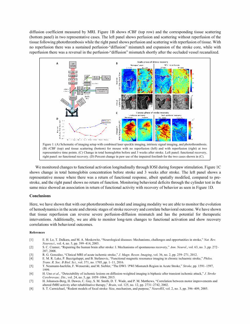
diffusion coefficient measured by MRI. Figure 1B shows rCBF (top row) and the corresponding tissue scattering (bottom panel) in two representative cases. The left panel shows perfusion and scattering without reperfusion of the tissue following photothrombosis while the right panel shows perfusion and scattering with reperfusion of tissue. With no reperfusion there was a sustained perfusion-“diffusion” mismatch and expansion of the stroke core, while with reperfusion there was a reversal in the perfusion-“diffusion” mismatch shortly after the occluded vessel recanalized.
We monitored changes to functional activation longitudinally through IOSI during forepaw stimulation. Figure 1C shows change in total hemoglobin concentration before stroke and 3 weeks after stroke. The left panel shows a representative mouse where there was a return of functional response, albeit spatially modified, compared to pre-stroke, and the right panel shows no return of function. Monitoring behavioral deficits through the cylinder test in the same mice showed an association in return of functional activity with recovery of behavior as seen in Figure 1D.
Conclusions
Here, we have shown that with our photothrombosis model and imaging modality we are able to monitor the evolution of hemodynamics in the acute and chronic stages of stroke recovery and correlate behavioral outcome. We have shown that tissue reperfusion can reverse severe perfusion-diffusion mismatch and has the potential for therapeutic interventions. Additionally, we are able to monitor long-tern changes to functional activation and show recovery correlations with behavioral outcomes.
References
[1] E. H. Lo, T. Dalkara, and M. A. Moskowitz, “Neurological diseases: Mechanisms, challenges and opportunities in stroke,” Nat. Rev. Neurosci., vol. 4, no. 5, pp. 399–414, 2003.
[2] S. C. Cramer, “Repairing the human brain after stroke: I. Mechanisms of spontaneous recovery,” Ann. Neurol., vol. 63, no. 3, pp. 272–287, 2008.
[3] R. G. González, “Clinical MRI of acute ischemic stroke,” J. Magn. Reson. Imaging, vol. 36, no. 2, pp. 259–271, 2012. [4] E. M. R. Lake, P. Bazzigaluppi, and B. Stefanovic, “Functional magnetic resonance imaging in chronic ischaemic stroke,” Philos.
Trans. R. Soc. B Biol. Sci., vol. 371, no. 1705, pp. 1–11, 2016. [5] T. Neumann-haefelin, F. Wenserski, and M. Siebler, “The DWI / PWI Mismatch Region in Acute Stroke,” Stroke, pp. 1591–1597,
1999. [6] H. Uno et al., “Detectability of ischemic lesions on diffusion-weighted imaging is biphasic after transient ischemic attack,” J. Stroke
Cerebrovasc. Dis., vol. 24, no. 5, pp. 1059–1064, 2015. [7] H. Johansen-Berg, H. Dawes, C. Guy, S. M. Smith, D. T. Wade, and P. M. Matthews, “Correlation between motor improvements and
altered fMRI activity after rehabilitative therapy,” Brain, vol. 125, no. 12, pp. 2731–2742, 2002. [8] S. T. Carmichael, “Rodent models of focal stroke: Size, mechanism, and purpose,” NeuroRX, vol. 2, no. 3, pp. 396–409, 2005.
Figure 1: (A) Schematic of imaging setup with combined laser speckle imaging, intrinsic signal imaging, and photothrombosis. (B) rCBF (top) and tissue scattering (bottom) for mouse with no reperfusion (left) and with reperfusion (right) at two representative time points. (C) Change in total hemoglobin before and 3 weeks after stroke. Left panel: functional recovery, right panel: no functional recovery. (D) Percent change in paw use of the impaired forelimb for the two cases shown in (C).
