neuropeptide y y1 receptor-mediated biodegradable ... pdf/bplp ultrasound... · neuropeptide y y1...
TRANSCRIPT

lable at ScienceDirect
Biomaterials 116 (2017) 106e117
Contents lists avai
Biomaterials
journal homepage: www.elsevier .com/locate/biomateria ls
Neuropeptide Y Y1 receptor-mediated biodegradablephotoluminescent nanobubbles as ultrasound contrast agents fortargeted breast cancer imaging
Juan Li a, 1, Yuchen Tian a, b, 1, Dingying Shan c, An Gong a, Leyong Zeng a, Wenzhi Ren a,Lingchao Xiang a, Ethan Gerhard c, Jinshun Zhao d, Jian Yang c, Aiguo Wu a, *
a Key Laboratory of Magnetic Materials and Devices & Key Laboratory of Additive Manufacturing Materials of Zhejiang Province & Division of FunctionalMaterials and Nanodevices, Ningbo Institute of Materials Technology and Engineering, Chinese Academy of Sciences, Ningbo 315201, PR Chinab Nano Science and Technology Institute, University of Science and Technology of China, Suzhou 215123, PR Chinac Department of Biomedical Engineering, Materials Research Institute, The Huck Institutes of the Life Sciences, The Pennsylvania State University, UniversityPark, PA 16802, USAd Public Health Department, Ningbo University, 818 Fenghua Road, Ningbo 315211, PR China
a r t i c l e i n f o
Article history:Received 9 April 2016Received in revised form8 November 2016Accepted 20 November 2016Available online 22 November 2016
Keywords:NanobubblesBiodegradable photoluminescent polymersUltrasound contrast agentsTargeted imagingBreast cancerNeuropeptide Y Y1 receptor
* Corresponding author.E-mail address: [email protected] (A. Wu).
1 These authors contributed equally to this work.
http://dx.doi.org/10.1016/j.biomaterials.2016.11.0280142-9612/© 2016 Elsevier Ltd. All rights reserved.
a b s t r a c t
Targeted molecular imaging has attracted great attention in cancer diagnosis and treatment. However,most clinically used ultrasound contrast agents (UCAs) are non-targeted microbubbles seldom used forcancer imaging. Here, we fabricated fluorescent nanobubbles (NBs) by encapsulation of liquid tetrade-cafluorohexane (C6F14) within biodegradable photoluminescent polymers (BPLPs) through an emulsion-evaporation process and conjugation of PNBL-NPY ligand for specific targeting of Y1 receptors overex-pressed in breast tumors. The developed PNBL-NPY modified NBs were uniform in size with good dis-persibility and photostability, presenting good ultrasound enhancement. Further, in vitro and in vivoresults indicated that the fabricated NBs exhibit high affinity and specificity to Y1 receptor-overexpressing breast cancer cells and tumors with minimal toxicity and damage to organs. Ourdeveloped PNBL-NPY-modified NBs are novel targeted UCAs for safe, efficient and specific targeted breastcancer imaging, and may provide a new nanoplatform for early cancer diagnosis and treatment in thefuture.
© 2016 Elsevier Ltd. All rights reserved.
1. Introduction
Breast cancer has become themost commonly diagnosed canceramong women [1]. Early diagnosis and monitoring is critical tocancer treatment [2,3]. Contrast agents have generated a hugescientific and economic impact on a broad range of early cancerdiagnoses [4]. Compared to commonly used magnetic resonanceimaging (MRI), positron emission tomography (PET) and computedtomography (CT), ultrasound imaging is a widely available, non-invasive and cost-effective diagnostic modality. It provides real-time imaging without the use of hazardous ionizing radiation, butthe weak difference in echogenicity between different tissues oftenhampers a clear diagnosis [5e7]. In order to better visualize specific
tissues, microbubble (MB)-based ultrasound contrast agents (UCAs)are frequently used as blood pool agents with a typical diameter of1e8 mm [8e10]. However, MBs cannot permeate through the tumorvasculature to the cellular target site to generate the desireddiagnostic and therapeutic effect [9]. Therefore, researchers arepaying more attention to developing nanobubble (NB)-based UCAsfor tumor ultrasound imaging.
Recently, NBs with various gas, liquid or solid cores and shellscomposed of polymers, such as PLA or PLGA, have been applied intumor ultrasound imaging [11e15]. It was shown that these con-structs exhibit optimal contrast enhancement abilities due to theenhanced permeation and retention (EPR) effect, thus accom-plishing passive targeting to tumors [13,16]. At the same time,improving the selectivity of contrast agents to the specific cancercells has become the focus of many researchers. Active targetingmolecules have attracted great attention for targeted moleculeimaging systems such as MRI, PET and ultrasonography [7,17e19].

J. Li et al. / Biomaterials 116 (2017) 106e117 107
Historically, the application of active targeting molecules for breastcancer targeting has demonstrated limited success due to the lowexpression of their receptors in breast tumors, such as folate re-ceptors (~29%) and integrin receptors (~26%) [20,21], or co-expression in the normal and tumor breast tissues, such as vaso-active intestinal peptide receptors and gastrin-releasing peptidereceptors [22,23]. The application of such targetingmolecules oftenresults in significant off-target effects.
Excitingly, we have recently identified a selective ligand ofneuropeptide Y Y1 receptors (Y1Rs), PNBL-NPY, which has demon-strated a high selectivity to breast cancer cells with no effects on thenormal breast cells [24]. Y1Rs are highly overexpressed in humanbreast tumors andmetastases (above 90%), while the normal breasttissues express Y2Rs only [25,26]. Therefore, the development ofY1R-based contrast agents will provide safer and more specific im-aging for breast cancer diagnosis. Until now, most of the studiesrelated to Y1Rs mainly focused on scintimammography or PET im-aging for breast cancer; however, there are no previous studiesabout the application of Y1R ligands in ultrasound imaging [27e31].
Recently, a family of biodegradable photoluminescent polymers(BPLPs) has been reported by Yang et al. [32e35]. The reactantsused to synthesize BPLPs, including citric acid, amino acids andaliphatic diols, are common compounds used in many FDA-regulated devices [32]. In contrast to photobleaching organicdyes, cytotoxic quantum dots, and conventional non-degradablefluorescent polymers, BPLPs are intrinsically fluorescent withoutconjugating any additional organic dyes or quantum dots and havedemonstrated excellent photostability, biocompatibility, and de-gradability [32,34]. By initiating the ring-opening polymerizationof L-lactide and glycolide with BPLP-cysteine (BPLP-Cys), biode-gradable photoluminescent poly-L-lactide (BPLP-PLLA) and poly(L-lactide-co-glycolide) (BPLP-PLGA) have recently been developedbased on BPLPs [34,36]. The obtained polylactone materials can beconveniently used to fabricate nanoparticles for nanoparticletracking with a variety of microscopy techniques including fluo-rescent microscopy, confocal laser scanning microscopy (CLSM)and two-photon microscopy [37].
This study aims to develop Y1R-based intrinsically fluorescentNBs as contrast agents for targeted ultrasound imaging of breastcancer. BPLP-PLLA-Cys was used for the preparation of intrinsicallyfluorescent NBs with contrast-enhanced ultrasound mode functionand in vitro fluorescent imaging with a high quantum yield (up to51%). Afterwards, a selective Y1R ligand, PNBL-NPY, was conjugatedto the NBs for the specific targeted imaging of Y1R-over expressingbreast cancer cells. The developed PNBL-NPY modified NBs werecharacterized, and their targeting ability and ultrasound-enhancingability to breast tumors, as well as the toxicity to organs, wereinvestigated via carefully designed in vitro and in vivo experiments.We have demonstrated that these unique intrinsically fluorescentNBs exhibit excellent aqueous stability, photostability, low toxicityand high contrast enhancement ability, making them extremelypromising UCAs for ultrasound imaging of Y1R-overexpressingbreast cancer.
2. Material and methods
2.1. Materials
BPLP-PLLA50-Cys was synthesized as previously described[34,36]. Dichloromethane, 1-ethyl-3-(3-dimethylaminopropyl) car-bodiimide hydrochloride (EDAC) and N-hydroxysuccinimide (NHS)were purchased from Sinopharm Chemical Reagent Co., Ltd(Shanghai, China). Polyvinyl alcohol (PVA), sodium cholate hydrate,HPLC grade acetonitrile and trifluoroacetic acid (TFA)were purchasedfrom Aladdin Industrial Inc (Shanghai, China). Dulbecco's Modified
Eagle Medium (DMEM), RPMI-1640 medium, fetal bovine serum(FBS), penicillin, streptomycin and rhodamine phalloidin (RP) werepurchased from Invitrogen™ (Carlsbad, USA). Tetradecafluorohexane(C6F14) was purchased from Sigma-Aldrich Co. LLC (Shanghai, China).[Pro30, Nle31, Bpa32, Leu34]NPY(28e36) (IIe-Asn-Pro-Nle-Bpa-Arg-Leu-Arg-Try-NH2) was synthesized by the LifeTein LLC (Beijing,China). 1, 2-distearoyl-sn-glycero-3-phosphoethanolamine-N-[car-boxy(poly ethylene glycol)-2000] (DSPE-PEG-COOH) was purchasedfrom A.V.T. Pharmaceutical Ltd. (Shanghai, China), and DiI was pur-chased from Beyotime Biotechnology (Haimen, China).
2.2. Preparation of intrinsically fluorescent BPLP-PLLA-Cysnanobubbles (BPC-NBs)
The intrinsically fluorescent BPC-NBs were prepared by modi-fying the solvent emulsion/evaporation method to obtain NBs witha polymeric shell encapsulating tetradecafluorohexane (C6F14) [13].Briefly, 0.1 g BPC was dissolved into 4 mL dichloromethane con-taining 60 mL of C6F14. The mixture was ultrasonicated for 5 min at600 W with an ultrasonic cleaner (SB 25-12DTDN, Ningbo ScientzBiotechnology Co., Ltd. China) to ensure full miscibility of the C6F14.Afterwards, the organic solution was added into 20 mL of 1.5% so-dium cholate (w/v) aqueous solution, and then the mixture wasemulsified by a superfine homogenizer (F6/10, FLUKO, China)operating with a 10 G dispersing tool (10 000 rpm, 1 min) to form apre-emulsion. For ex vivo fluorescence imaging experiments, DiIwas added to the organic solution prior to emulsification. Typically,about 10 mL a concentrated DiI solution (1.2 mgmL�1 in ethanol) wasadded to the organic solution.
The pre-emulsion was sonicated at 648 W with a vibratingmetallic tip for 1 min. The organic solvent was then evaporated bymagnetic stirring for 4 h at room temperature. After full evapora-tion of the solvents, the suspension volume was adjusted to 20 mLwith water, and then incubated with 1.5% PVA (w/v) for 5 days at4 �C to replace sodium cholate. The formed BPC-NBs were washedwith distilled water by centrifugation (10,000 g, 1 h, 4 �C). Thesupernatant was discarded and the BPC-NBs were resuspendedwith 5 mL water for the subsequent experiment.
2.3. Conjugation of PNBL-NPY to the BPC-NBs
To conjugate the PNBL-NPY ligand onto the surface of BPC-NBs,an EDAC/NHS activation technique was used [24]. Before theconjugation, BPC-NBs were modified with DSPE-PEG-COOH.1.0 mg mL�1 DSPE-PEG-COOH in ethanol was added to the BPC-NBs, and the ethanol was evaporated by magnetic stirring for4 h at room temperature, followed by washing with distilled watervia centrifugation (10,000 g,1 h, 4 �C). The carboxylic groups of BPC-NBswere conjugatedwith the amine group of PNBL-NPY. Briefly, anice cold mixture of 0.3 mL EDAC (0.3 mg mL�1 in PBS) and 0.2 mLNHS (0.3 mg mL�1 in PBS) was added into a 10 mL BPC-NB solution(1 mg mL�1 in PBS) under continuous stirring for 15 min (ice waterbath). Then 1.0 mL PNBL-NPY solution (in PBS) with different con-centrations were added dropwise into the above solution andreacted at room temperature for 16 h. The prepared BPC-NB-PNBL-NPY was centrifuged (14,000 g, 30 min) and the supernatants werestored for further analysis. Unreacted PNBL-NPY in the supernatantwere quantified with a HPLC-UV method as previously described[24]. A simple mass balance was then used to calculate the amountof PNBL-NPY conjugation on the surface of BPC-NBs.
2.4. Characterization
A particle size-zeta potential analyzer (Nano-ZS, Malvern, En-gland) was used for the measurement of particle size and zeta

J. Li et al. / Biomaterials 116 (2017) 106e117108
potential of the BPC-NB-PNBL-NPY at room temperature. Resultswere expressed as the average of the mean diameter of the NBsobtained from three measurements. High-resolution transmissionelectron microscopy (HRTEM) images were recorded by a JEOL-2100 (JEOL, Japan) instrument to get detailed structural andmorphological information of BPC-NB-PNBL-NPY. 3 mL of the BPC-NB-PNBL-NPY solution was dropped on a 300 mesh copper gridcoated with a thin layer of carbon film, and another 2 mL of 0.5%phosphotungstic acid was dropped on top of the NB droplet fornegative staining. The copper grid was dried overnight at roomtemperature before TEM analysis.
Photoluminescence spectra of BPC-NB-PNBL-NPY solutionswere acquired on a Hitachi F-4600 fluorospectrophotometer(Hitachi, Japan). Both the excitation and the emission slit widthswere set at 5 nm for all samples unless otherwise stated. Thephotostability of BPC-NB-PNBL-NPY solution was evaluated byrecording the changes of the fluorescence intensity of the samplesunder continuous excitation in the fluorospectrophotometer. Theexcitation wavelength for photostability tests was determined bythe maximum absorbance spectra of each type of sample.
2.5. Cell culture
Human breast cancer MCF-7 cell line and Mouse breast cancer4T1 cell line were cultured in Dulbecco's modified Eagle's medium(DMEM) and RPMI-1640 medium respectively, and both of themediumwere supplemented with 10 wt % fetal bovine serum (FBS),100 units mL�1 of penicillin, and 100 mgmL�1 of streptomycin. Thecells were maintained in a 37 �C incubator with 5% CO2.
2.6. Cell viability assays
Cytotoxicity of NBs to the MCF-7 and 4T1 cells were measuredby flow cytometry analysis. Both MCF-7 and 4T1 cells were treatedwith different concentrations of BPC-NB and BPC-NB-PNBL-NPY for24 h. Cells were collected and stained by propidium iodide (PI) andanalyzed by flow cytometry analysis immediately. The PI positivecells were recognized as dead cells [38]. 2.0 mL of MCF-7 (or 4T1cells) in complete DMEM medium (or RPMI-1640 medium) wereseeded into each well at 5 � 104 cells mL�1 and cultured overnight.On the following day, the culture media were replaced by freshmedium containing different concentrations of BPC-NB and BPC-NB-PNBL-NPY. After a further 24 h incubation, the cells werewashed three times with 1mL PBS to remove any absorbed free NBsand then harvested for further analysis. Cells were stained withpropidium iodide (PI) for 5 min at room temperature. The meanfluorescence intensity (MFI) of cells (1 � 104 counts) were analyzedby flow cytometry (LSRFortessa, BD, USA), where the gate wasarbitrarily set for the detection of PI (600e630 nm) with forwardand side scattering dot plots used to discriminate cellular debris.
2.7. In vitro specific targeting of BPC-NB-PNBL-NPY
The ethanol treated glass slides were put in 6-well plates andcoated with 0.01% poly-L-lysine. 2.0 mL of MCF-7 (or 4T1 cells) incomplete DMEM medium (or RPMI-1640 medium) were seededinto each well at 5 � 104 cells mL�1 and cultured overnight. On thefollowing day, the culture media were replaced by fresh mediumwith different treatments including 500 mg mL�1 BPC-NB,500 mg mL�1 BPC-NB-PNBL-NPY, or PBS. After 12 h further incu-bation, the cells were washed three times with 1 mL PBS to removeany adsorbed free NBs. For LSCM imaging, the cells were fixed with1 mL 4% formaldehyde for 30 min, then washed three times with1 mL PBS. Cells were then treated with 1 mL 0.1% triton for 5 min,washed three times with 1 mL PBS and then treated with 1 mL 1.0%
BSA for 30 min at room temperature. The actin of the cells wasstained with 200 mL rhodamine phalloidin (RP) for 30 min at roomtemperature. The samples were simultaneously excited at 405 and540 nm and the fluorescent images at emission wavelengths420e500 and 600e660 nm were observed by a LSCM (TCS SP5 II,Leica, Germany). For flow cytometry analysis, the mean fluores-cence intensity (MFI) of cells (1 � 104 counts) was analyzed by flowcytometer (LSRFortessa, BD, USA), where the gate was arbitrarilyset for the detection of blue fluorescence (425e475 nm) with for-ward and side scattering dot plots used to discriminate cellulardebris.
2.8. In vitro ultrasound imaging
In vitro ultrasound imaging of BPC-NB-PNBL-NPY was carriedout in a latex tube (inner diameter of 6 mm) which was embeddedinto 1% agarose gel as previously reported [10]. The NBs (5mgmL�1
in PBS) were injected into the latex tube simulating the blood vesseland circulated in the tube with a constant flow rate controlled by apump. Ultrasonography was performed using a M9DP-50 portableultrasonic imaging system (Mindray, Shenzhen, China) in visuali-zation mode with a transducer set to a frequency 10.0 MHz.
2.9. Animal models
Female Balb/c mice (18e20 g, 4e6 weeks old) were used in thiswork. All the experimental protocols involving animals wereapproved by the Regional Ethics Committee for Animal Experi-ments at Ningbo University (Permit No. SYXK Zhe 2013-0191). Thetumor model of murian breast cancer was established by subcu-taneous injection of 4T1 cells. Briefly, 4T1 cells (1� 106 cells for onemouse) suspended in 100 mL of serum freemediumwere inoculatedsubcutaneously in several female Balb/c mice (five weeks old) atthe back. A digital caliper was used to measure the tumor size.Tumors with a diameter of 0.8e1.2 cm were selected for subse-quent experiments.
2.10. Toxicity evaluation in vivo
For in vivo toxicity, 24 healthy Balb/c mice were randomlydivided into eight groups (3 each group), and were injected withBPC-NB-PNBL-NPY (50, 100 or 200 mg kg�1) or PBS as control viathe tail vein. Behavior observation was carried out for seven days.To further verify in vivo toxicity, the mice were sacrificed on theseventh day. Mice's blood were collected by a cardiac puncturemethod for hematological and were analyzed by blood analyzer(Sysmex XT-1800i, Japan) and Hitachi 7600-110 autoanalyzer(Hitachi, Tokyo, Japan), and the major organs including heart, liver,spleen, lung and kidney were stained with Hematoxylin and Eosin(H&E) and examined by an optical microscope (DMI3000, Leica,Germany).
2.11. In vivo ultrasound-enhanced imaging of BPC-NB-PNBL-NPY
To evaluate the ultrasound-enhanced ability of BPC-NB-PNBL-NPY, Balb/c mice bearing 4T1 cell line tumors were subdivided intwo groups, according to the administration route, with 4 animalsfor intra-tumoral injection and 15 animals for intravenous injection.BPC-NB or BPC-NB-PNBL-NPY suspensions (100 mg kg�1) wereinjected intravenously through the tail vein or intra-tumorally. Forintra-tumoral injection, images were acquired 10, 20, 30, 120, 180and 300 s after the injection. For intravenous injection, images wereacquired approximately 10, 20, 30, 60, 120, 180, 240 and 300 s postinjection. The ultrasound transducer was positioned gently on top ofthe tumor, and the space between themwas filled with an adequate
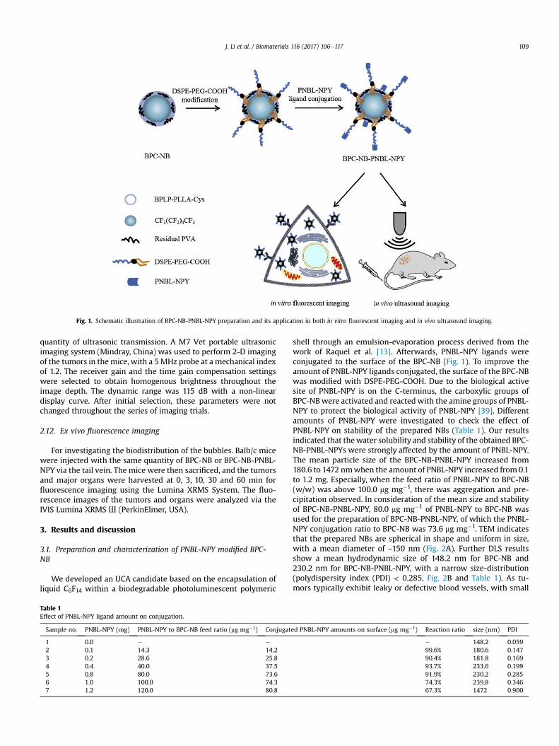
Fig. 1. Schematic illustration of BPC-NB-PNBL-NPY preparation and its application in both in vitro fluorescent imaging and in vivo ultrasound imaging.
J. Li et al. / Biomaterials 116 (2017) 106e117 109
quantity of ultrasonic transmission. A M7 Vet portable ultrasonicimaging system (Mindray, China) was used to perform 2-D imagingof the tumors in themice, with a 5MHz probe at a mechanical indexof 1.2. The receiver gain and the time gain compensation settingswere selected to obtain homogenous brightness throughout theimage depth. The dynamic range was 115 dB with a non-lineardisplay curve. After initial selection, these parameters were notchanged throughout the series of imaging trials.
2.12. Ex vivo fluorescence imaging
For investigating the biodistribution of the bubbles. Balb/c micewere injected with the same quantity of BPC-NB or BPC-NB-PNBL-NPY via the tail vein. The mice were then sacrificed, and the tumorsand major organs were harvested at 0, 3, 10, 30 and 60 min forfluorescence imaging using the Lumina XRMS System. The fluo-rescence images of the tumors and organs were analyzed via theIVIS Lumina XRMS Ш (PerkinElmer, USA).
3. Results and discussion
3.1. Preparation and characterization of PNBL-NPY modified BPC-NB
We developed an UCA candidate based on the encapsulation ofliquid C6F14 within a biodegradable photoluminescent polymeric
Table 1Effect of PNBL-NPY ligand amount on conjugation.
Sample no. PNBL-NPY (mg) PNBL-NPY to BPC-NB feed ratio (mg mg�1) Conjugat
1 0.0 e e
2 0.1 14.3 14.23 0.2 28.6 25.84 0.4 40.0 37.55 0.8 80.0 73.66 1.0 100.0 74.37 1.2 120.0 80.8
shell through an emulsion-evaporation process derived from thework of Raquel et al. [13]. Afterwards, PNBL-NPY ligands wereconjugated to the surface of the BPC-NB (Fig. 1). To improve theamount of PNBL-NPY ligands conjugated, the surface of the BPC-NBwas modified with DSPE-PEG-COOH. Due to the biological activesite of PNBL-NPY is on the C-terminus, the carboxylic groups ofBPC-NBwere activated and reactedwith the amine groups of PNBL-NPY to protect the biological activity of PNBL-NPY [39]. Differentamounts of PNBL-NPY were investigated to check the effect ofPNBL-NPY on stability of the prepared NBs (Table 1). Our resultsindicated that thewater solubility and stability of the obtained BPC-NB-PNBL-NPYs were strongly affected by the amount of PNBL-NPY.The mean particle size of the BPC-NB-PNBL-NPY increased from180.6 to 1472 nmwhen the amount of PNBL-NPY increased from 0.1to 1.2 mg. Especially, when the feed ratio of PNBL-NPY to BPC-NB(w/w) was above 100.0 mg mg�1, there was aggregation and pre-cipitation observed. In consideration of the mean size and stabilityof BPC-NB-PNBL-NPY, 80.0 mg mg�1 of PNBL-NPY to BPC-NB wasused for the preparation of BPC-NB-PNBL-NPY, of which the PNBL-NPY conjugation ratio to BPC-NB was 73.6 mg mg�1. TEM indicatesthat the prepared NBs are spherical in shape and uniform in size,with a mean diameter of ~150 nm (Fig. 2A). Further DLS resultsshow a mean hydrodynamic size of 148.2 nm for BPC-NB and230.2 nm for BPC-NB-PNBL-NPY, with a narrow size-distribution(polydispersity index (PDI) < 0.285, Fig. 2B and Table 1). As tu-mors typically exhibit leaky or defective blood vessels, with small
ed PNBL-NPY amounts on surface (mg mg�1) Reaction ratio size (nm) PDI
e 148.2 0.05999.6% 180.6 0.14790.4% 181.8 0.16993.7% 233.6 0.19991.9% 230.2 0.28574.3% 239.8 0.34667.3% 1472 0.900

Fig. 2. Characterization of BPC-NB-PNBL-NPY. (A) High-resolution transmission electron microscopy (TEM) image of BPC-NB, BPC-NB-DSPE-PEG and BPC-NB-PNBL-NPY. (B) Sizedistributions of BPC-NB, BPC-NB-DSPE-PEG and BPC-NB-PNBL-NPY in PBS at room temperature measured by dynamic light scattering (DLS) at a scattering angle of 173� (backscatterdetection).
J. Li et al. / Biomaterials 116 (2017) 106e117110
pore cutoff sizes up to 780 nm, our prepared NBs were smallenough for targeted delivery of nanoparticles to tumor sites[19,40e42]. At pH 7.4, zeta potential values were�5.5 and�3.5 mVfor BPC-NB and BPC-NB-PNBL-NPY separately. Although the zetapotential of both NBs were a bit low, but they were stable up to onemonth at 4 �C, and there was no aggregation observed. The samephenomenon has also been found in the previous literature [13].After the elimination of surfactants, zeta potential values are closeto zero for all the nanocapsules. The change of surface charge mightbe due to residual PVA present on nanocapsule surface. PVAadsorption arises from hydrogen bonding between PLGA carboxylgroups and PVA hydroxyl groups. In our study, the dispersibilityand stability of the colloidal dispersion system were not justdepend on the surface charge, but also related to the interfacialenergy. Both of the PVA and DSPE-PEG-COOH are excellent sur-factant, which can decrease the interfacial energy of the NBs.Moreover, PVA and DSPE-PEG-COOH have hydrophilic groupswhich can protect the NBs against adsorption between each otherin aqueous phase.
The maximum emission and excitation wavelengths of BPLP-PLLA-Cys solution were determined to be 434 and 365 nm,respectively (Fig. S1B). After being incorporated into nanobubbles,the excitation and the emission wavelengths of BPC-NB-PNBL-NPYdislay no significant change (Fig. S2).
Fig. 3. Cytotoxicity of BPC-NB-PNBL-NPY in MCF-7 cell line of human breast cancerand 4T1 cell line of murine breast cancer. Both MCF-7 and 4T1 cells were treated withdifferent concentration of BPC-NB and BPC-NB-PNBL-NPY for 24 h. Cells were collectedand stained by propidium iodide (PI) and analyzed by flow cytometry analysisimmediately. Data represent the mean ± SEM (n ¼ 3).
3.2. In vitro cell toxicity
The effect of BPC-NB and BPC-NB-PNBL-NPY on the viability ofhuman breast cancer MCF-7 cell line and murine breast cancer 4T1cell line were investigated after 24 h incubation (Fig. 3). For MCF-7 cells, the cell viability remained above 91.5% for BPC-NB and 93.1%for BPC-NB-PNBL-NPY when the concentrations of NBs varied from0.2 to 1.0 mg mL�1. For 4T1 cells, the cell viability was above 93.9%for BPC-NB and 89.3% for BPC-NB-PNBL-NPY at all tested concen-trations. Compared to the control group, there was about 10.7% celldeath induced by BPC-NB-PNBL-NPY. This might be due to the
toxicity of BPC-NBs (about 6%) as well as the PNBL-NPY ligand. Inour previous work, we found that the PNBL-NPY ligand itself couldinhibit the MCF-7 cell growth in a concentration depend manner,but it has no effect on the normal human breast cells MCF-10A [24].In the case of 4T1 cells, it might be happened. As both of MCF-7 and4T1 cells are cancer cells, so this inhibition of cell growth could alsobe another benefit of our NBs. These results show that both BPC-NBand BPC-NB-PNBL-NPY have good biocompatibility and causeminimal harm to the tested cells.

Fig. 4. Cellular uptake of BPC-NB and BPC-NB-PNBL-NPY in MCF-7 and 4T1 cells. (A) Laser scanning confocal microscope (LSCM) images of MCF-7 cells and 4T1 cells incubated with0.5 mg mL�1 BPC-NB and BPC-NB-PNBL-NPY for 12 h. The cytoskeletons with rhodamine phalloidin are red, and the NBs are blue. (B) Western blot analysis of NPY-Y1R expression inMCF-7 cell line of human breast cancer and 4T1 cell line of murine breast cancer. (For interpretation of the references to colour in this figure legend, the reader is referred to the webversion of this article.)
Fig. 5. Flow cytometry analysis of MCF-7 and 4T1 cells incubated with BPC-NB and BPC-NB-PNBL-NPY for 12 h. Significant analysis of mean fluorescence intensity (MFI) for MCF-7and 4T1 cells incubated with different NBs. Data represent the mean ± SEM (n ¼ 3,*P < 0.05, **P < 0.01). 1: Control; 2: BPC-NB; 3: BPC-NB-PNBL-NPY.
J. Li et al. / Biomaterials 116 (2017) 106e117 111

J. Li et al. / Biomaterials 116 (2017) 106e117112
3.3. Cellular uptake of BPC-NB-PNBL-NPY in MCF-7 and 4T1 cells
To evaluate the in vitro targeting ability of BPC-NB-PNBL-NPY,cellular uptake of the NBs was assessed using the human breastcancerMCF-7 cell line previously shown to highly express Y1Rs, andmouse breast cancer 4T1 cell line, which was proven to show acomparable expression rate of Y1Rs with MCF-7 cells in westernblot analysis (Fig. 4B). Both MCF-7 and 4T-1 cells were incubatedwith BPC-NB and BPC-NB-PNBL-NPY for 12 h, and the LSCM im-aging results are shown in Fig. 4A (samples were simultaneouslyexcited at 405 and 540 nm). The cytoskeletons with rhodaminephalloidin (RP) (EX 540 nm, EM 600e660 nm) are red, while theintrinsically fluorescent BPC-NB and BPC-NB-PNBL-NPY (EX405 nm, EM 420e460 nm) are blue. LSCM images show that PNBL-NPY modified BPC-NB are more internalized into both MCF-7 and4T1 cells than unmodified BPC-NB. Further flow cytometry analysis(EX 355 nm, EM 425e475 nm) were used to quantify the meanfluorescence intensity (MFI) of NBs treatedMCF-7 or 4T1 cells. Fig. 5
Fig. 6. (A) Histological analysis of mice main organs. Mice were injected intravenously withdays. Then the mice were sacrificed and the main organs were collected for the test. (Scale bData are expressed as the mean ± SEM (n ¼ 3).
shows that the MFI of BPC-NB-PNBL-NPY treated cells are muchstronger than BPC-NB, which is 68.1% (P < 0.01) for MCF-7 cells, and108% (P < 0.01) for 4T1 cells (Fig. 5). Taken together, the aboveresults indicate that PNBL-NPY modification could significantlyimprove the delivery of BPC-NB into the MCF-7 and 4T1 cells. Ourfindings agree well with previous reports that PNBL-NPY has theability to improve internalization of nanoparticles into breastcancer cells [24].
3.4. In vitro ultrasound imaging of BPC-NB-PNBL-NPY
In vitro ultrasonic imaging was performed to confirm thefeasibility of the BPC-NB-PNBL-NPY as UCAs. In Fig. 7, it is observedthat the latex tube's lumen and wall is very clear before injection ofBPC-NB and BPC-NB-PNBL-NPY. After injection, the lumen of thelatex tube is brightened for both BPC-NB and BPC-NB-PNBL-NPY.This indicates that our prepared NBs have the potential to act asultrasound enhanced contrast agents.
PBS, 50 mg kg�1, 100 mg kg�1 or 200 mg kg�1 of BPC-NB-PNBL-NPY and held for sevenar ¼ 20 mm).(B) Hematological analysis and (C) blood biochemical analysis of the mice.

J. Li et al. / Biomaterials 116 (2017) 106e117 113
3.5. Toxicity evaluation in vivo
As a potential in vivo contrast agent, the toxicity of BPC-NB-PNBL-NPY must be evaluated in vivo. In this study, histologicalanalysis was used to evaluate the toxicity of the NBs in vivo ac-cording to previous study. Healthy Blab/c mice were injectedintravenously with either PBS, 50, 100 or 200 mg kg�1 of BPC-NB-PNBL-NPY. Over a one week period, various behaviors of micesuch as eating, drinking, excretion, activity and neurological statuswere observed. There is no significant difference in the above be-haviors between control and BPC-NB-PNBL-NPY groups. After oneweek, the mice were sacrificed and the main organs were analyzed.Fig. 6 A shows histological analysis of the organs including heart,liver, spleen, lung and kidney. There is no detectable tissue damageor other pathologies such as necrosis, inflammation, or pulmonaryfibrosis when comparing the BPC-NB-PNBL-NPY group with thecontrol group. Fig. 6B and C indicates blood analysis and hemato-logical analysis of the mice including white blood cell (WBC), redblood cell (RBC), platelet (PLT), and hemoglobin (HGB), hematocrit(HCT), mean corpuscular volume (MCV), mean corpuscular hemo-globin concentration (MCHC), red blood cell distribution width(RDW-CV), platelet distribution width (PDW-CV), and meanplatelet volume (MPV). Number and distribution changes of bloodcell are an important indicator of health status. As demonstrated inFig. 6 B, there is no significant difference between control anddifferent concentrations of BPC-NB-PNBL-NPY groups except PLT,the increase of PLT suggested a possible effect of BPC-NB-PNBL-NPYon blood coagulation, causing damage of platelets. Furthermore,blood biochemical analysis was carried out by blood autoanalyzer.Six important hepatic indicators for liver functions (direct bilirubin,DBIL; albumin, ALB; globin, GLOB; alkaline phosphatase, ALP;gamma glutamyl transpeptidase, GGT), three indicators for kidneyfunctions (urea nitrogen, UREA; creatinine, CREA; uric acid, URCA).
Fig. 7. In vitro ultrasound images obtained in a nonlinear imaging mode (THI) at 10 MHz an
As shown in Fig. 6 C, there is no significant difference betweencontrol and different concentrations of BPC-NB-PNBL-NPY groupsexcept AST, UREA and CREA. The decreased AST value indicated theBPC-NB-PNBL-NPY has no effect on the liver function, but theincreased UREA and CREA values might suggested the effect of ourNBs on the kidney function at the high dose (200 mg kg�1). How-ever, further histological analysis results showed that there was nodetectable tissue damage was observed. The pathologist suggestedthat the changes of UREA and CREA might also because of otherunexpected reason, such as the physiological state changes of themice themselves. In addition, more population need to be collectedand analyzed in the future to investigate the detail reason of thisphenomenon.
3.6. In vivo ultrasound-enhanced imaging of tumors
In vivo ultrasound imaging was assessed in Balb/c mice bearing4T1 cell line tumors after either an intra-tumoral or an intravenousinjection of BPC-NB or BPC-NB-PNBL-NPY. Fig. 8 presents ultra-sound images of tumors before and after the intra-tumoral injec-tion of BPC-NB or BPC-NB-PNBL-NPY. Before injection tumorsappear dark whereas after injection tumors present significantcontrast enhancement for both types of NBs. The enhancementlasts about 300 s and similar enhancement was observed for allmice included in this trial, but no significant difference could bedetected between BPC-NB and BPC-NB-PNBL-NPY.
To evaluate the selective imaging ability of BPC-NB-PNBL-NPYtowards breast tumors, an intravenous injection of the NBs wasadministrated and the imaging characteristics in tumors wereanalyzed. As shown in Fig. 9A and B, both PNBL-NPY unmodifiedand modified BPC-NB exhibited ultrasound enhancement ability attumor sites 30 s after injection. However, the contrast enhance-ment of BPC-NB-PNBL-NPY (96.4 ± 19.4) was significantly higher
d 10.3 mW cm�2 after injection with 5 mL of BP-NB or BP-NB-PNBL-NPY (5 mg mL�1).

Fig. 8. (A)Y1R-overexpression transplanted tumor in Balb/c mouse model. (B) Conventional ultrasound images of tumor (in red frame). (C) Contrast enhanced ultrasound imaging oftumor before and after intra-tumoral injection of BPC-NB or BPC-NB-PNBL-NPY (100 mg kg�1) at different time points (10, 20, 30, 120, 180 and 300 s). (For interpretation of thereferences to colour in this figure legend, the reader is referred to the web version of this article.)
J. Li et al. / Biomaterials 116 (2017) 106e117114
than BPC-NB (48.0 ± 10.2) at 30 s after injection (P < 0.05). Theenhanced contrast of BPC-NB-PNBL-NPY reached a peak at 60 s(121.6 ± 19.8), and kept in a higher level until 300 s. However, theenhanced contrast of BPC-NB at tumor sites decreased rapidly after240 s. These findings can be explained by the assumption that the
Fig. 9. In vivo ultrasound-enhanced imaging of BPC-NB-PNBL-NPY. (A) Contrast enhanced ulNPY (100 mg kg�1) at different time points (10, 20, 30, 60, 120, 180, 240 and 300 s). (B and(n ¼ 3,*P < 0.05, **P < 0.01).
NBs easily passed through the tumor vessels, arrived at the tumorsite, or even targeted the Y1Rs in the case of BPC-NB-PNBL-NPY.Additionally, the contrast enhancement of NBs could also beobserved in the background around tumors from 30 to 120 s. Thismight be due to the increased circulation of NBs in the blood vessels
trasound imaging of tumor after intravenous injection of PBS, BPC-NB or BPC-NB-PNBL-C) Time-gray scale intensity curves and AUC analysis. Data represent the mean ± SEM

Fig. 10. (A) Ex vivo fluorescence imaging results for BPC-NB and BPC-NB-PNBL-NPY group at 0, 3, 10, 30 and 60min. In each image: 1. heart; 2. liver; 3. spleen; 4. lung; 5. kidney; 6.tumor. (B) ROI analysis of heart, liver, spleen, spleen, kidney and tumor in ex vivo fluorescence imaging for the two groups at 0, 3, 10, 30 and 60 min.
J. Li et al. / Biomaterials 116 (2017) 106e117 115
around tumors. After 300 s, the enhanced background signaldecreased slowly, while the contrast enhancement of BPC-NB-PNBL-NPY could still be observed at the tumor sites. This mightbe due to the selective targeting of BPC-NB-PNBL-NPY on the Y1Rsof tumors, while the NBs in the blood vessels around the tumortissues were cleared away quickly. To further compare the contrastenhancement of the NBs as a function of imaging time, area underthe curve (AUC) plots from 10 to 300 s were analyzed by PhoenixWinNonlin 6.4 software (Certara, USA). In Fig. 9C, it indicated thatenhancements induced by the BPC-NB-PNBL-NPY (AUC:33,121 ± 3046) were significantly stronger than the enhancement
induced by the BPC-NB (AUC: 23,315 ± 2089) (P < 0.05). Theseresults suggest that PNBL-NPY modified BPC-NB can more easilyarrive at tumors, pass through the tumor capillaries and target Y1R-overexpressing tumor cells. The high affinity between PNBL-NPYand Y1Rs on tumor cells allows more BPC-NB-PNBL-NPY toremain at the tumor site and coalesce to form MBs, which exhibit ahigher scattering cross-section than that of BPC-NB and producesuperior contrast enhancement [19,43].
In previous studies, NBs with different formulations demon-strated excellent ultrasound imaging enhancement in vivo. Histo-logical examination has demonstrated that NBs can pass through

J. Li et al. / Biomaterials 116 (2017) 106e117116
the endothelial gaps of tumors. These results have been attributedto EPR effects [15,44,45]. However, most cases of the EPR effects ofNBs have previously been correlated with poor in vivo tumorselectivity during intravenous delivery [13,15]. For example, NBsprepared by Raquel et al. produced extensive and strong ultrasoundimaging enhancement by intra-tumoral injection but cannot bedetected by intravenous injection [13]. The typical solutionemployed to achieve tumor selectivity is to conjugate specific tar-geted molecules (such as ligands or antibodies) to the surface ofNBs, which can target to specific receptors or antigens expressed onthe tumors [15,19,46,47]. For example, NBs-affibody has been pre-pared for human epidermal growth factor receptor type 2 (HRE2)-targeted ultrasound imaging, providing good ultrasound-enhanced signal [19]. Although the affibody was able to mimicthe function of an antibody with a lowmolecular weight of 14 kDa,it was still composed of 58 amino acid residues. However, thePNBL-NPY ligand that we used here only consists of 9 amino acids,which might provide higher labeling efficiency with a lower syn-thesis cost compared to large peptides [48,49]. In addition, the sizeof BPC-NB-PNBL-NPY (~230 nm) was much smaller than the NBs-affibody (~478 nm), but still displayed good ultrasound enhance-ment. Moreover, due to the use of photoluminescent polymers, ourfluorescent BPC-NBs could also be explored for photoacoustic im-aging of tumors in the future [50,51]. Therefore, the PNBL-NPYmodified NBs may provide an alternative in the early diagnosisand treatment of Y1R-overexpressing breast cancers.
3.7. Ex vivo fluorescence imaging
To further confirm the targeted selectivity of BPC-NB-PNBL-NPYtoward tumors, we performed ex vivo fluorescence imaging ex-periments. The fluorescence signal intensity of ex vivo imaging is areflection of the BPC-NB-PNBL-NPY retained inside the organsbecause of the low or even lack of autofluorescence observed inex vivo images [52]. Tumors and major organs from the miceinjected with the BPC-NB or BPC-NB-PNBL-NPY groups were har-vested to acquire fluorescence images at different time points. Asshown in Fig. 10A, the fluorescence was obviously distributed in thetumors, livers and kidneys, but less fluorescence signal wasdetected in hearts and spleens. A region of interest (ROI) analysiswere performed on the ex vivo fluorescence images to semi-quantitatively analyze the DiI uptake in each organs. Fig. 10B indi-cated that the BPC-NB-PNBL-NPY induced fluorescence in tumorswere much higher than the BPC-NBs after the injection 3 min(5.9 � 107 vs 3.7 � 107) to 60 min (2.3 � 107 vs 2.0 � 107). Theseresults confirmed that the targeted selectivity of BPC-NB-PNBL-NPY was distinct at 3 min after intravenous injection, which wasconsistent with the results of in vivo ultrasound imaging. Takentogether, the ex vivo imaging results further confirmed the target-ability of BPC-NB-PNBL-NPY to Y1Rs-over expressing tumors.
4. Conclusion
In this work, we have developed Y1R-based biodegradablephotoluminescent NBs as UCAs with excellent dispersibility, sta-bility and biocompatibility. The hydrodynamic size of our BPC-NB-PNBL-NPY could be under 240 nm in diameter, favoring extrava-sation. The in vitro fluorescence spectra and CLSM experimentshow that the BPC-NB-PNBL-NPY exhibits bright and stable auto-fluorescence, facilitating tracking of their interaction with variouscells. Further, we demonstrated that the BPC-NB-PNBL-NPY ex-hibits high affinity and specificity to Y1R-overexpressing breastcancer cells and tumors, decreasing toxicity while providing goodultrasound enhancement in both in vitro and in vivo experiments.These results indicate that the fabricated Y1R-based NBs have the
potential to be excellent UCAs in Y1R positive breast cancer imagingand might also provide a potential nanoplatform for Y1R positivebreast cancer treatment.
Acknowledgments
This work was supported by the Natural Science Foundation ofChina (Grant No. 51303196 to Juan Li and U1432114 to Aiguo Wu),Science Technology Department of Zhejiang Province (2016C33093,to Juan Li), Special Program for Applied Research on SuperComputation of the NSFC-Guangdong Joint Fund (the secondphase) (to Juan Li and Aiguo Wu), and USA National Institutes ofHealth award (NCI CA182670, to Jian Yang). The authors are gratefulto the Dr. Saijun Chen and Dr. Minxia Zeng from the Department ofUltrasound Diagnosis, Ningbo No. 2 Hospital for their help in ul-trasound imaging. The author also thanks Mr. Yong Zeng fromMindray Medical International Limited for their technical supportand kind provision of equipment and Mr. Xuzhe Zhang from BeijingDryas Pharma-Tech Co., Ltd. for his help of AUC analysis. In the end,the author wants to thank Lijia Luo, Jie Feng, Yuanzhi Xia and RuifenZou for their help in animal experiments.
Appendix A. Supplementary data
Supplementary data related to this article can be found at http://dx.doi.org/10.1016/j.biomaterials.2016.11.028.
References
[1] L.A. Torre, F. Bray, R.L. Siegel, J. Ferlay, J. Lortet-Tieulent, A. Jemal, Globalcancer statistics, 2012, CA Cancer J. Clin. 65 (2015) 87e108.
[2] M.A. Richards, The national awareness and early diagnosis initiative in En-gland: assembling the evidence introduction, Br. J. Cancer 101 (2009) S1eS4.
[3] M.P. Coleman, D. Forman, H. Bryant, J. Butler, B. Rachet, C. Maringe, et al.,Cancer survival in Australia, Canada, Denmark, Norway, Sweden, and the UK,1995-2007 (the international cancer benchmarking partnership): an analysisof population-based cancer registry data, Lancet 377 (2011) 127e138.
[4] R. Weissleder, Molecular imaging in cancer, Science 312 (2006) 1168e1171.[5] S.R. Wilson, P.N. Burns, Microbubble-enhanced US in body imaging: what
role? Radiology 257 (2010) 24e39.[6] F. Kiessling, S. Fokong, P. Koczera, W. Lederle, T. Lammers, Ultrasound
microbubbles for molecular diagnosis, therapy, and theranostics, J. Nucl. Med.53 (2012) 345e348.
[7] V. Sanna, G. Pintus, P. Bandiera, R. Anedda, S. Punzoni, B. Sanna, et al.,Development of polymeric microbubbles targeted to prostate-specific mem-brane antigen as prototype of novel ultrasound contrast agents, Mol. Pharm. 8(2011) 748e757.
[8] S. Hernot, A.L. Klibanov, Microbubbles in ultrasound-triggered drug and genedelivery, Adv. Drug Deliv. Rev. 60 (2008) 1153e1166.
[9] K.W. Ferrara, M.A. Borden, H. Zhang, Lipid-shelled vehicles: engineering forultrasound molecular imaging and drug delivery, Acc. Chem. Res. 42 (2009)881e892.
[10] A. Gong, X.H. Ma, L.C. Xiang, W.Z. Ren, Z.Y. Shen, A.G. Wu, Improved doubleemulsion technology for fabricating autofluorescent microcapsules as novelultrasonic/fluorescent dual-modality contrast agents, Colloid Surf. B 116(2014) 561e567.
[11] E. Pisani, E. Fattal, J. Paris, C. Ringard, V. Rosilio, N. Tsapis, Surfactant depen-dent morphology of polymeric capsules of perfluorooctyl bromide: influenceof polymer adsorption at the dichloromethane-water interface, J. ColloidInterf. Sci. 326 (2008) 66e71.
[12] E. Pisani, N. Tsapis, J. Paris, V. Nicolas, L. Cattel, E. Fattal, Polymeric nano/mi-crocapsules of liquid perfluorocarbons for ultrasonic imaging: physical char-acterization, Langmuir 22 (2006) 4397e4402.
[13] R. Díaz-L�opez, N. Tsapis, M. Santin, S.L. Bridal, V. Nicolas, D. Jaillard, et al., Theperformance of PEGylated nanocapsules of perfluorooctyl bromide as an ul-trasound contrast agent, Biomaterials 31 (2010) 1723e1731.
[14] J.S. Xu, J.W. Huang, R.G. Qin, G.H. Hinkle, S.P. Povoski, E.W. Martin, et al.,Synthesizing and binding dual-mode poly (lactic-co-glycolic acid) (PLGA)nanobubbles for cancer targeting and imaging, Biomaterials 31 (2010)1716e1722.
[15] T.H. Yin, P. Wang, R.Q. Zheng, B.W. Zheng, D. Cheng, X.L. Zhang, et al.,Nanobubbles for enhanced ultrasound imaging of tumors, Int. J. Nanomed. 7(2012) 895e904.
[16] N. Rapoport, Z.G. Gao, A. Kennedy, Multifunctional nanoparticles forcombining ultrasonic tumor imaging and targeted chemotherapy, J. Natl.Cancer Inst. 99 (2007) 1095e1106.

J. Li et al. / Biomaterials 116 (2017) 106e117 117
[17] I.U. Khan, A.G. Beck-Sickinger, Targeted tumor diagnosis and therapy withpeptide hormones as radiopharmaceuticals, Anti Cancer Agents Med. Chem. 8(2008) 186e199.
[18] K. Temming, R.M. Schiffelers, G. Molema, R.J. Kok, RGD-based strategies forselective delivery of therapeutics and imaging agents to the tumour vascu-lature, Drug Resist. Update 8 (2005) 381e402.
[19] H.L. Yang, W.B. Cai, L. Xu, X.H. Lv, Y.B. Qiao, P. Li, et al., Nanobubble-Affibody:novel ultrasound contrast agents for targeted molecular ultrasound imagingof tumor, Biomaterials 37 (2015) 279e288.
[20] W. Xia, P.S. Low, Folate-targeted therapies for Cancer, J. Med. Chem. 53 (2010)6811e6824.
[21] Q. Chen, H.J. Millar, F.L. McCabe, C.D. Manning, R. Steeves, K. Lai, et al., Alphavintegrin-targeted immunoconjugates regress established human tumors inxenograft models, Clin. Cancer Res. Off. J. Am. Assoc. Cancer Res. 13 (2007)3689e3695.
[22] J.C. Reubi, U. L€aderach, B. Waser, J.-O. Gebbers, P. Robberecht, J.A. Laissue,Vasoactive intestinal peptide/pituitary adenylate cyclase-activating peptidereceptor subtypes in human tumors and their tissues of origin, Cancer Res. 60(2000) 3105e3112.
[23] M. Gugger, J.C. Reubi, Gastrin-releasing peptide receptors in non-neoplasticand neoplastic human breast, Am. J. Pathol. 155 (1999) 2067e2076.
[24] J. Li, Z.Y. Shen, X.H. Ma, W.Z. Ren, L.C. Xiang, A. Gong, et al., Neuropeptide Y Y-1 receptors meditate targeted delivery of anticancer drug with encapsulatednanoparticles to breast Cancer cells with high selectivity and its potential forbreast Cancer therapy, ACS Appl. Mater. Interfaces 7 (2015) 5574e5582.
[25] M. K€orner, J.C. Reubi, NPY receptors in human cancer: a review of currentknowledge, Peptides 28 (2007) 419e425.
[26] J.C. Reubi, M. Gugger, B. Waser, J.C. Schaer, Y-1-mediated effect of neuro-peptide Y in cancer: breast carcinomas as targets, Cancer Res. 61 (2001)4636e4641.
[27] S. Hofmann, S. Maschauer, T. Kuwert, A.G. Beck-Sickinger, O. Prante, Synthesisand in vitro and in vivo evaluation of an (18)F-Labeled neuropeptide Yanalogue for imaging of breast Cancer by PET, Mol. Pharm. 12 (2015)1121e1130.
[28] N. Braekeveldt, C. Wigerup, D. Gisselsson, S. Mohlin, M. Merselius, S. Beckman,et al., Neuroblastoma patient-derived orthotopic xenografts retain metastaticpatterns and geno- and phenotypes of patient tumours, Int. J. Cancer 136(2015) E252eE261.
[29] C. Morgat, A.K. Mishra, R. Varshney, M. Allard, P. Fernandez, E. Hindie, Tar-geting neuropeptide receptors for Cancer imaging and therapy: perspectiveswith bombesin, neurotensin, and Neuropeptide-Y receptors, J. Nucl. Med. 55(2014) 1650e1657.
[30] M. Memminger, M. Keller, M. Lopuch, N. Pop, G. Bernhardt, E. von Angerer, etal., The neuropeptide Y Y-1 receptor: a diagnostic Marker? Expression in MCF-7 breast Cancer cells is down-regulated by antiestrogens in vitro and in xe-nografts, PLoS One 7 (2012) 11.
[31] I.U. Khan, D. Zwanziger, I. Bohme, M. Javed, H. Naseer, S.W. Hyder, et al.,Breast-Cancer diagnosis by neuropeptide Y analogues: from synthesis toclinical application, Angew. Chem Int Ed. 49 (2010) 1155e1158.
[32] D. Gyawali, S.Y. Zhou, R.T. Tran, Y. Zhang, C. Liu, X.C. Bai, et al., Fluorescenceimaging enabled biodegradable photostable polymeric micelles, Adv. Healthc.Mater. 3 (2014) 182e186.
[33] J. Yang, Y. Zhang, S. Gautam, L. Liu, J. Dey, W. Chen, et al., Development ofaliphatic biodegradable photoluminescent polymers, Proc. Natl. Acad. Sci. U. S.A. 106 (2009) 10086, 2009;106:11818.
[34] Z.W. Xie, Y. Zhang, L. Liu, H. Weng, R.P. Mason, L.P. Tang, et al., Development ofintrinsically photoluminescent and photostable polylactones, Adv. Mater. 26(2014) 4491e4496.
[35] A.S. Wadajkar, T. Kadapure, Y. Zhang, W.N. Cui, K.T. Nguyen, J. Yang, Dual-imaging enabled Cancer-Targeting nanoparticles, Adv. Health. Mater. 1 (2012)450e456.
[36] J. Hu, J. Guo, Z. Xie, D. Shan, E. Gerhard, G. Qian, et al., Fluorescence imagingenabled poly(lactide-co-glycolide), Acta Biomater. 29 (2016) 307e319.
[37] G.J. Chen, L.W. Wang, T. Cordie, C. Vokoun, K.W. Eliceiri, S.Q. Gong, Multi-functional self-fluorescent unimolecular micelles for tumor-targeted drugdelivery and bioimaging, Biomaterials 47 (2015) 41e50.
[38] J. Fan, Y. Sun, S. Wang, Y. Li, X. Zeng, Z. Cao, et al., Inhibition of autophagyovercomes the nanotoxicity elicited by cadmium-based quantum dots, Bio-materials 78 (2016) 102e114.
[39] I. B€ohme, J. Stichel, C. Walther, K. M€orl, A.G. Beck-Sickinger, Agonist inducedreceptor internalization of neuropeptide Y receptor subtypes depends onthird intracellular loop and C-terminus, Cell. Signal. 20 (2008) 1740e1749.
[40] J. Kim, Y.M. Lee, Y. Kang, W.J. Kim, Tumor-homing, size-tunable clusterednanoparticles for anticancer therapeutics, Acs Nano 8 (2014) 9358e9367.
[41] A. Albanese, P.S. Tang, W.C.W. Chan, The effect of nanoparticle size, shape, andsurface chemistry on biological systems, Annu. Rev. Biomed. Eng. 14 (2012)1e16.
[42] H. Kim, D. Lee, J. Kim, T.I. Kim, W.J. Kim, Photothermally triggered cytosolicdrug delivery via endosome disruption using a functionalized reduced gra-phene oxide, Acs Nano 7 (2013) 6735e6746.
[43] N. Deshpande, A. Needles, J.K. Willmann, Molecular ultrasound imaging:current status and future directions, Clin. Radiol. 65 (2010) 567e581.
[44] H.-P. Tong, L.-F. Wang, Y.-L. Guo, L. Li, X.-Z. Fan, J. Ding, et al., Preparation ofprotamine cationic nanobubbles and experimental study of their physicalproperties and in vivo contrast enhancement, Ultrasound Med. Biol. 39 (2013)2147e2157.
[45] H. Wu, N.G. Rognin, T.M. Krupka, L. Solorio, H. Yoshiara, G. Guenette, et al.,Acoustic characterization and pharmacokinetic analyses of new nanobubbleultrasound contrast agents, Ultrasound Med. Biol. 39 (2013) 2137e2146.
[46] S.M. Stieger, P.A. Dayton, M.A. Borden, C.F. Caskey, S.M. Griffey, E.R. Wisner, etal., Imaging of angiogenesis using Cadence™ contrast pulse sequencing andtargeted contrast agents, Contrast Media Mol. Imaging 3 (2008) 9e18.
[47] G.E.R. Weller, M.K.K. Wong, R.A. Modzelewski, E.X. Lu, A.L. Klibanov,W.R. Wagner, et al., Ultrasonic imaging of tumor angiogenesis using contrastmicrobubbles targeted via the tumor-binding peptide arginine-arginine-leucine, Cancer Res. 65 (2005) 533e539.
[48] D. Zwanziger, I. Bohme, D. Lindner, A.G. Beck-Sickinger, First selective agonistof the neuropeptide Y-1-receptor with reduced size, J. Pept. Sci. 15 (2009)856e866.
[49] J. Li, Y. Tian, A. Wu, Neuropeptide Y receptors: a promising target for cancerimaging and therapy, Regen. Biomater. 2 (2015) 215e219.
[50] X.D. Wang, Y.J. Pang, G. Ku, X.Y. Xie, G. Stoica, L.H.V. Wang, Noninvasive laser-induced photoacoustic tomography for structural and functional in vivo im-aging of the brain, Nat. Biotechnol. 21 (2003) 803e806.
[51] H.F. Zhang, K. Maslov, G. Stoica, L.H.V. Wang, Functional photoacoustic mi-croscopy for high-resolution and noninvasive in vivo imaging, Nat. Biotechnol.24 (2006) 848e851.
[52] J. Gao, K. Chen, R. Xie, J. Xie, Y. Yan, Z. Cheng, et al., In vivo tumor-targetedfluorescence imaging using near-infrared non-cadmium quantum dots, Bio-conjug. Chem. 21 (2010) 604e609.