neuron-specific enolase as an index of … · the journal of neuroscience neuron-specific enolase...
TRANSCRIPT

0270-6474/83/0305-0915$02.00/O Copyright 0 Society for Neuroscience Printed in U.S.A.
‘The Journal of Neuroscience Vol. 3, No. 5, pp. 915-923
May 1983
NEURON-SPECIFIC ENOLASE AS AN INDEX OF NEURONAL REGENERATION AND REINNERVATION’
TAKAAKI KIRINO,*, ’ MILTON W. BRIGHTMAN,*, WOLFGANG H. OERTEL,$ 4 DONALD E. SCHMECHEL,$ 5 AND PAUL J. MARANGOSt 6
* Laboratory of Neuropathology and Neuroanatomical Sciences and $ Laboratory of Clinical Sciences, National Institutes of
Health, Bethesda, Maryland 20205
Received August 3,1982; Revised October 18, 1982; Accepted November 9,1982
Abstract
Neuron-specific enolase (NSE) is a glycolytic isoenzyme which is located in central and peripheral neurons and neuroendocrine cells. Another enolase isoenzyme, non-neuronal enolase (NNE), occurs in glial cells. The purpose of this study was to follow any changes in NSE and/or NNE in cranial motor neurons after separation of their cell bodies from their axon terminals. One hypoglossal nerve in the rat and the cynomolgus monkey was thus crushed or cut and, after a given period, the brains were perfusion fixed. Immunocytochemistry, using anti-rat NSE and NNE or anti-human NSE and NNE, was performed on Vibratome-sectioned specimens of the hypoglossal nuclei. In the rat, NSE immunostaining decreased in the affected neurons 2 to 10 days following axonal injury. The change was greatest on the 10th day. Twenty days following nerve crush, NSE staining began to recover on the operated side and by the 45th day had returned to normal levels. NSE changes in the monkey were similar to those in the rat. In rats, where the nerve was cut and the proximal stump was translocated to a normally innervated muscle to inhibit re-formation of synaptic contacts, the NSE remained low for 60 days after nerve injury. As NSE levels fell during degeneration, there was a slight increase in NNE in some of the monkey specimens but not in others; the NNE alterations were, therefore, equivocal. The results demonstrate that the content of NSE in neurons serves as a molecular marker of axon injury, regeneration, and target reinnervation.
Neuron-specific enolase (NSE; EC 4.2.1.11) is an acidic, soluble protein (Marangos and Schmechel, 1980; Zomzely-Neurath and Walker, 1980) which functions as a glycolytic isoenzyme (Bock and Dissing, 1975) and is homologous to bovine 14-3-2 protein characterized by Moore and McGregor (1965). Immunocytochemical sur- veys have demonstrated that NSE is confined solely to neurons (Pickel et al., 1976), not only central ones but those of dorsal root ganglia, autonomic ganglia, and some peripheral neuroendocrine cells as well. NSE can be used, therefore, as an identifying molecular marker for all types of neurons in uiuo and in vitro (Schmechel et al., 1978a).
’ We are grateful to Virginia K. Weise and Alexandra M. Parma of the Laboratory of Clinical Sciences, National Institutes of Health, for their competent technical assistance.
’ Present address: Department of Neurosurgery, University of Tokyo Hospital, Tokyo, Japan.
’ To whom requests for reprints should be sent.
4 Present address: Neurologische Klinik, Munich, West Germany. ’ Present address: Neurology Department, Duke University Medical
School, Durham, NC 27710.
’ Present address: Laboratory of Clinical Psychobiology, National Institutes of Health, Bethesda, MD 20205.
Glial cells, on the other hand, have a different enolase isoenzyme. This glial or non-neuronal enolase (NNE) is thought to be identical to liver enolase (Marangos and Schmechel, 1980). NNE has been demonstrated within astrocytes, oligodendrocytes, ependyma, and choroid plexus epithelia and can thus serve as a marker for glial cells (Schmechel et al., 1978a).
In the developing nervous system, however, this simple enzyme dichotomy: NSE - neurons, NNE - glia, is not applicable. According to biochemical assays, NNE is dominant at first in developing brain and, later, NSE becomes ascendant (Marangos et al., 1978a). Cerebellar granule cells have immunocytochemically detectable NNE while they are still in the external granular layer, but they acquire their normal NSE content as they migrate down to the internal granular layer and form synapses. In other words, neurons switch from NNE to NSE as they differentiate (Schmechel et al., 1980b). The NSE antigen is thus an excellent marker for neuronal differentiation.
Neurons undergo a certain sequence of morphological and metabolic changes following axonal injury (Lieber- man, 1971; Grafstein and McQuarrie, 1978). It is believed that neurons shift, after axonal injury, to the immature
915
916 Kirino et al. Vol. 3, No. 5, May 1983
“growing phase” from the differentiated “secretory phase” (Jacobson, 1978). It might be assumed that de- differentiation takes place following axotomy and that differentiation resumes during nerve regeneration. The purpose of these experiments was 3-fold: to see whether the level of NSE changes in response to axotomy, whether there is a shift from NSE to NNE that would thus resemble a dedifferentiated or developmental state, and whether the restoration of NSE levels depends on the axon’s reinnervation of its target.
Materials and Methods
With radioimmunoassay of NNE, a modest change in the neuronal content of this enzyme could well be masked by the NNE content of surrounding glial cells. Moreover, the relatively densely stained NSE of the ventral part of the hypoglossal nucleus, regardless of treatment, could easily be included in variable amounts within a punch sample for radioimmunoassay and could mask changes in levels of NSE. Thus, in order to detect changes in neuronal NNE and NSE, immunocytochemistry rather than radioimmunoassay for these enzymes was used.
Antisera against purified rat NSE and NNE or human NSE and NNE were prepared in New Zealand White rabbits as previously described (Marangos et al., 1975a, b). No cross-reactivity of anti-NSE with NNE or anti- NNE with NSE was observed (Marangos et al., 1978b).
Changes in NSE content were estimated by comparing the operated side with the immediately adjacent unop- erated side in each section. The slides were coded by one observer and two other observers (W. 0. and D. S.) evaluated the sections without knowledge of their source.
Rats. Adult (45-day-old) male Osborne-Mendel rats were anesthetized with fluothane. The right hypoglossal nerve (12th nerve) trunk was exposed beneath the pos- terior belly of the digastric muscle, freed, and manually crushed with a fine forceps (Dumont stainless steel, no. 5) for 1 min. Animals were fixed by perfusion fixation 2, 5, 10, 20, 30, and 45 days following this operative proce- dure. Sham-operated animals, serving as the controls, were fixed 10 days after the operation. At least four animals were included in each group, and a total of 42 rats were used. In another group of 10 adult, male rats of the same strain, the 12th nerve trunk was cut rather than crushed. In five of them, the severed stumps were anas- tomosed with 10-O nylon suture. In the other five rats, the distal stump (1.0 mm) was removed and the proximal trunk was inserted into the posterior belly of the digastric muscle, the normal innervation of which was left intact, and sealed into place with fascia using a 10-O suture. These 10 animals were fixed 60 days later. In a third group of thirteen g-day-old pups, the 12th nerve was cut and the severed ends were simply left apposed. They were fixed 2, 5, 10, 20, and 30 days after axotomy.
The fixative consisted of 4% paraformaldehyde, 1% glutaraldehyde, 0.2% picric acid, and 2% sucrose in 0.1 M
sodium acetate buffer, pH 6.0 (Schmechel et al., 1980a). Two hours after perfusion fixation, the brains were re- moved from the skull and kept in the same fixative at 4°C for 3 hr then transferred to Tris-buffered saline (TBS; 150 mM NaCl in 50 mM Tris-HCl buffer, pH 7.6). The medulla oblongata was cut into 25-pm-thick sections
on a Vibratome. From each animal, 40 to 60 Vibratome sections containing the hypoglossal nucleus (12th nerve nucleus) were processed for immunocytochemistry using the unlabeled antibody, peroxidase-antiperoxidase (PAP) method7 (Sternberger, 1979). The specimens were mounted on chrome/alum-coated glass slides after stain- ing and observed with a Zeiss Photomicroscope III. Some sections were embedded in Araldite 502; semithin sec- tions were then cut and lightly stained with toluidine blue.
Monkeys. Since antibody of human NNE reacts with monkey antigen more vigorously than anti-rat NNE does with rat antigen, the experiment was repeated in six adult cynomolgus monkeys (Macaca fascicularis). The right 12th nerve was cut at the level of the anterior belly of the digastric muscle. At 10, 15, and 20 days following nerve section, the brains were perfusion fixed with the same fixative composition as that used for the rats. The medulla oblongata was removed and processed for im- munocytochemistry in the same way, using the antisera against human NSE and NNE. Four different “bleeds” of antisera against human NNE were used for each animal.
Results
Rats. In sham-operated rats, the bilateral 12th nerve nuclei were symmetrically stained for NSE. Stainability was almost even throughout the rostral-caudal extent of the 12th nerve nucleus but the ventral portion, where 12th nerve neurons are most crowded, was slightly but consistently darker. While the neuronal perikaryon was densely colored, the cell nucleus was unstained. The perikaryal cytoplasm was stained inhomogeneously and had a reticular pattern (Fig. 1). In the neuropil around the neurons were scattered numerous fine granular struc- tures which were mostly dendrites and a few axons (Fig. 1). There was a marked variation in staining among these myelinated axons and dendrites (Fig. 1). A myelinated axon could be stained, whereas its immediate neighbor was not. In a single axon or dendrite, the degree of staining diminished in its more distal extent. Glial and endothelial cells were completely devoid of staining (Fig. 1). The 12th nerve neurons, in general, were more lightly stained for NSE than were the reticular neurons at the same level of the medulla oblongata. These staining properties were maintained on the unoperated side through the experiment (Fig. 6). In the dorsal part of the 12th nerve nucleus, there were always a few fusiform
7 The method was as follows: (I ) 5 min in 10% methanol and 3%
H202 in TBS. (2) 5 min in 0.25% Triton X-100 in TBS. (3) 60 min in a solution containing 10% normal sheep serum (NSS) and 0.1 M crystal- line rx-lysine in TBS. (4) 2 hr in the solution of anti-rat NSE (I:4000 or l:SOOO), anti-rat NNE (1:400 or l:lOOO), or anti-human NSE (1:400
or 1:800), anti-human NNE (1:400 or 1800) at room temperature for 16 to 18 hr at 4°C. Antisera were dissolved in 2% NSS in TBS. Normal rabbit serum, at the same dilution, was used as controls. (5) 30 min in the solution of sheep anti-rabbit IgG (1:80) in TBS with 2% NSS. (6) 30 min in the solution of PAP (1:lOO) in TBS with 2% NSS. (7) 5 to 10 min in the solution containing 0.01% Hz02 and 0.05% diaminobenzidine
in TBS. Between each step, the specimens were washed several times with TBS.

The Journal of Neuroscience Neuron-specific Enolase as an Index of Reinnervation
Figure 1. Focal variations in the levels of immunocytochemically detected NSE of the intact rat hypoglossal nucleus. The cytoplasm of the neurons is filled with reaction product. There is marked variation in staining between neighboring axons and dendrites (small arrowheads). In a single neuronal process, staining fades proximodistally (large arrowheads). Glial cells (G) and endothelial (E) are devoid of staining. Plastic embedded specimen, lightly counterstained with toluidine blue. Magnification x 800.
neurons as densely reactive for NSE as the reticular neurons (Figs. 2 and 4).
The first recognizable change in stainability was noted 2 days after the 12th nerve crush when the content of NSE on the operated side decreased slightly. Five days following operation, the neurons and, to a lesser degree, their processes on the affected side were definitely more lightly stained than were those on the unoperated side.
The greatest change in NSE content was noticed 10 days after nerve crush (Fig. 2). In the affected 12th nerve nucleus, distinction between the perikarya and their cell nuclei was less marked due to a decreased staining in the perikarya (Fig. 7). At this stage, the neuronal cell bodies had become slightly swollen (Fig. 7). The cell nuclei of some neurons were often stained exclusively on the af- fected side (Fig. 2). Axons and dendrites of the neuropil also had a decreased stainability (Fig. 7). As a result, the affected nucleus appeared far lighter than the intact side at low magnification. Although almost all neurons had lost their normal stainability for NSE, a few small, fusi- form neurons, horizontally disposed, remained darkly stained (Fig. 2). These neurons, therefore, do not send their axons to the 12th nerve and are probably interneu- rons.
Twenty days after the 12th nerve was crushed, the difference between the bilateral 12th nerve nuclei in NSE staining was only slight. However, a part of the affected 12th nerve nucleus had a decreased immunostaining and included a few scattered, lightly stained neurons contain- ing nuclei that were darker than the cytoplasm. Thirty days following the operation, differences in NSE content between both sides were hardly discernible at low mag- nification, but a few neurons still remained lightly stained. By 45 days, the affected side was no longer distinguishable from the unaffected, opposite side (Fig. 3). The NSE content had decreased rapidly and simul- taneously in most neurons, but returned to near normal levels rather slowly and asynchronously.
In rats whose 12th nerve had been cut and then anas- tomosed, the 12th nerve nucleus on the operated side was only minimally shrunken. Sixty days following sur- gery, NSE stainability had recovered to almost normal levels; only a very few neurons remained lightly stained (Fig. 5). In those rats where the proximal cut end of the 12th nerve had been inserted into the normally inner- vated digastric muscle in order to prevent synapse for- mation, almost all of the hypoglossal neurons on the affected side had a marked depletion of NSE 60 days

Kirino et al. Vol. 3, No. 5, May 1983
Figures 2 to 5. The right half is the side of operation. Magnification x 130. Figure 2. Ten days following 12th nerve crush. The affected side shows overall decrease in staining. Not only neuronal
perikarya but dendrites and axons, which appear as dark granules on the normal side, are only faintly stained. Cell nuclei are distinctly stained only on the affected side (arrows). A small neuron remains still intensely stained (arrowhead). See Figures 6 and 7 for enlargements.
Figure 3. Forty-five days after operation, both sides of 12th nerve nucleus show similar stainability for NSE. No definite shrinkage of the nucleus followed crush injury in adult rats.

The Journal of Neuroscience Neuron-specific Enolase as an Index of Reinnervation
Figure 4. Sixty days after 12th nerve severance. The nerve’s proximal stump was relocated to the digastric muscle so that tongue reinnervation was prevented. The affected nucleus was slightly atrophied. Marked reduction of NSE staining on the operated side persists. A few small neurons (arrowheads) are intensely stained.
Figure 5. Sixty days after 12th nerve cut. The 12th nerve has been anastomosed immediately after severance. The degree of atrophy on the operated side is less than that in Figure 4. NSE staining looks normal on the affected side except for one neuron which is lighter and has nuclear staining (arrow).
after operation (Fig. 4). The 12th nerve nucleus was marked shrinkage on the operated side, but the surviving moderately shrunken, while the denervated side of the 12th nerve neurons were normally stained for NSE 30 tongue had become severely atrophied. days after 12th nerve interruption (Fig. 8).
In young pups, the 12th nerve nucleus underwent Every other section in most of the specimens was also
Kirino et al. Vol. 3, No. 5, May 1983
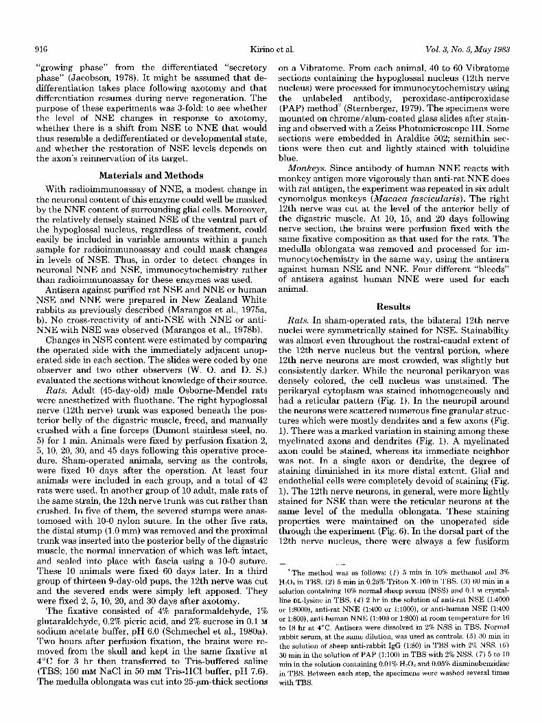
Figures 6 and 7. Ten days following nerve crush (rat). Magnification X 450. Figure 6. Control side. Perikarya and cell processes are stained intensely, whereas the cell nuclei are relatively pale. Figure 7. Operated side. Staining is generally reduced. The nuclei of a few cells are stained (arrows) only in the affected 12th
nerve nucleus. Neuronal processes are thinner.
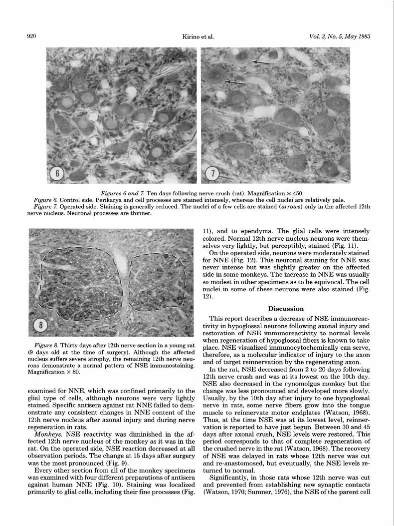
Figure 8. Thirty days after 12th nerve section in a young rat (9 days old at the time of surgery). Although the affected nucleus suffers severe atrophy, the remaining 12th nerve neu- rons demonstrate a normal pattern of NSE immunostaining. Magnification x 80.
examined for NNE, which was confined primarily to the glial type of cells, although neurons were very lightly stained. Specific antisera against rat NNE failed to dem- onstrate any consistent changes in NNE content of the 12th nerve nucleus after axonal injury and during nerve regeneration in rats.
Monkeys. NSE reactivity was diminished in the af- fected 12th nerve nucleus of the monkey as it was in the rat. On the operated side, NSE reaction decreased at all observation periods. The change at 15 days after surgery was the most pronounced (Fig. 9).
Every other section from all of the monkey specimens was examined with four different preparations of antisera against human NNE (Fig. 10). Staining was localized primarily to glial cells, including their fine processes (Fig.
ll), and to ependyma. The glial cells were intensely colored. Normal 12th nerve nucleus neurons were them- selves very lightly, but perceptibly, stained (Fig. 11).
On the operated side, neurons were moderately stained for NNE (Fig. 12). This neuronal staining for NNE was never intense but was slightly greater on the affected side in some monkeys. The increase in NNE was usually so modest in other specimens as to be equivocal. The cell nuclei in some of these neurons were also stained (Fig. 12).
Discussion
This report describes a decrease of NSE immunoreac- tivity in hypoglossal neurons following axonal injury and restoration of NSE immunoreactivity to normal levels when regeneration of hypoglossal fibers is known to take place. NSE visualized immunocytochemically can serve, therefore, as a molecular indicator of injury to the axon and of target reinnervation by the regenerating axon.
In the rat, NSE decreased from 2 to 20 days following 12th nerve crush and was at its lowest on the 10th day. NSE also decreased in the cynomolgus monkey but the change was less pronounced and developed more slowly. Usually, by the 10th day after injury to one hypoglossal nerve in rats, some nerve fibers grow into the tongue muscle to reinnervate motor endplates (Watson, 1968). Thus, at the time NSE was at its lowest level, reinner- vation is reported to have just begun. Between 30 and 45 days after axonal crush, NSE levels were restored. This period corresponds to that of complete regeneration of the crushed nerve in the rat (Watson, 1968). The recovery of NSE was delayed in rats whose 12th nerve was cut and re-anastomosed, but eventually, the NSE levels re- turned to normal.
Significantly, in those rats whose 12th nerve was cut and prevented from establishing new synaptic contacts (Watson, 1970; Sumner, 1976), the NSE of the parent cell

The Journal of Neuroscience Neuron-specific Enolase as an Index of Reinnervation
Figures 9 to 12. NSE and NNE staining in the cynomolgus monkey 15 days after unilateral 12th nerve severance. Operated sides are on the right in Figures 9 and 10.
Figure 9. There is a definite decrease in staining on the operated side. Magnification x 40. Figure 10. There is no distinct difference between the two sides at this magnification (X 40), partly because of the background
staining of glial processes. Figure 11. Magnification of Figure 10 (normal side). Glial processes appear as a densely stained network surrounding neurons
and their processes which are faintly stained. Magnification x 350. Figure 12. Magnification of Figure 10 (affected side). Increased NNE staining in 12th nerve neurons. The nuclei of some
neurons are densely stained (arrows). In other parts of the hypoglossal nucleus, there was no increase in NNE staining. Magnification x 350.
bodies remained at a low level. The full recovery of NSE stein, 1975, for summary). The specific activities of some content seemed to depend on a more or less complete glycolytic enzymes are reduced about 30%, 10 days after reformation of myoneural contacts. sectioning of the postganglionic axons of the superior
Parallel studies on NNE immunoreactivity in monkeys cervical ganglion (SCG). This decrement was believed to revealed in some specimens a discernible rise in NNE be part of a general decline in protein concentration staining intensity concurrent with the decline in NSE (Harkonen and Kaufman, 1974). In a sensory system- immunoreactivity. Nevertheless, the staining of regen- the fourth order neurons in cortical barrel fields-the erating neurons for NNE in other rat and monkey spec- activity of several enzymes also decreased after injury to imens was not appreciably greater than normal. This the first order neurons (Wong-Riley and Welt, 1980; variable response makes questionable the reality of the Dietrich et al., 1981). This decrease has been attributed minor increase in NNE staining. to a decline in metabolic activity because of disuse.
One probable reason for the decline in NSE may be a Other studies indicate that axon injury may lead to redirection of synthesis away from NSE to other proteins. heightened protein synthesis. Ten days after axon injury, Reports on patterns of protein synthesis after axonal during the “outgrowth’ phase of axon regeneration, syn- injury, however, have revealed disparate results. On one thesis of protein in the rabbit’s hypoglossal nucleus was hand, enzyme proteins, such as those involved in trans- enhanced (Brattgard et al., 1957; Agranoff et al., 1980). mitter metabolism, decrease after axotomy (see Graf- In the SCG, both augmented and diminished synthetic

922 Kirino et al. Vol. 3, No. 5, May 1983
rates of different proteins accompanied nerve regenera- tion. Axon injury lead to augmented synthesis of 16 unidentified proteins but a diminished production of 5 others. Synthesis of actin was heightened, whereas tublin was statistically insignificant from control levels (Hall et al., 1978). It remains to be shown whether a dichotomy of synthesis exists whereby structural proteins are pro- duced at a higher rate than are certain enzymes.
The functional significance of the transient decline in NSE has yet to be determined. NSE comprises almost 3% of all soluble proteins in the neuron (Marangos and Schmechel, 1980). The reason for this excess of enzyme is not known. Since NSE is apparently not a rate-limiting enzyme in the glycolytic pathway (Zomzely-Neurath and Walker, 1980), it is conceivable that a decreased amount of NSE is still sufficient to meet the requirements for augmented metabolism. In such cases, however, we have to postulate that intact neurons have a glycolytic enzyme far in excess of that which is needed, or that NSE has a different metabolic role in normal neurons. Such a dif- ferent role might be indicated by the point-to-point vari- ation in NSE staining between neighboring axons and dendrites and within a given process, where the decre- ment in staining proceeds distally. Moreover, a slight increase in NNE levels, if it does take place, may be sufficient to meet, temporarily, the metabolic demands of the regenerating neuron.
Neurons lose their normal electrical activity after ax- otomy and shift from a mature “secretory” form to a “growing” form (Jacobson, 1978). During this stage, en- ergy metabolism apparently increases to meet the re- quirements for augmented synthesis (Singer and Mahler, 1979), yet the NSE level diminishes.
The fall in NSE content of damaged neurons may be related to a reduced depolarization. After axon injury, the presynaptic terminals impinging upon the soma and dendrites of the affected neurons are reversibly displaced by intervening microglial processes (Blinzinger and Kreutzberg, 1968). The degree of depolarization and, consequently, the amount of chloride ion entering such neurons would be reduced. It has been proposed that NSE, which withstands a high concentration of chloride without becoming denatured, is a stable form of enolase adapted to neuronal activity (Marangos and Schmechel, 1980). Since there would be a fall in the amount of chloride entering axon-damaged neurons, the require- ment for NSE would be less than that of normal neurons and could account for the decrement in NSE.
The causes of the fall in NSE can only be surmised. The amount of enzyme within the soma at any one time may depend on the transient balance between the rate st which the enzyme is synthesized and its transport from the cell body (Cragg, 1970). An alternative, unsubstan- tiated possibility for the decline in NSE is an accelerated, anterograde transport of NSE out of the soma and den- drites. A substantial degradation of the enzyme, in re- sponse to axon trauma, might also conceivably lead to the decline in NSE. Another possible, but unlikely reason for the fall in NSE could be swelling of the soma as a form of “axon reaction” (Lieberman, 1971) and resultant dilution of NSE; however, decreased staining was also detected in dendrites and unswollen neurons. The appar- ently artifactitious migration of NSE into the cell nucleus
of affected neurons may also deplete the cytoplasm of some of its NSE. This redistribution may be due to the nuclear membrane having been rendered permeable to the antibody after aldehyde fixation of such a neuron.
In the monkey fetus, the cerebellar granule cells con- tain NNE as a dominant enolase isoenzyme until they have completed their migration and, presumably, estab- lished synapses with Purkinje dendrites. At that time, there is a switch from NNE to NSE (Schmechel et al., 1980b). A switch of enolase isoenzyme in damaged hy- poglossal neurons would represent a metabolic “de- differentiation” and during regeneration, a “redif- ferentiation.” This change in protein species would be concordant with that in chromatolytic, facial nerve neu- rons where there is a reappearance of certain proteins that had been found in immature but not older hamsters (Griffith and La Velle, 1971). In contrast, two-dimen- sional electrophoretic protein patterns of regenerating SCG neurons show that no new proteins, which would suggest a reversion to an immature pattern, are synthe- sized during regeneration (Hall et al., 1978). The latter study, however, lacks resolution at the cellular level, i.e., the SCG protein pattern represents proteins of neuronal and non-neuronal elements. Second, this model may not be comparable to hypoglossal motor neurons, as SCG neurons may belong to the group of peripheral nerve cells which contain both NNE and NSE (Schmechel et al., 1978b). In such a case, electrophoretic protein patterns might more likely reveal a change in NNE/NSE ratio than a new protein spot.
Because of only a minor increase of NNE immuno- reactivity and the low signal to background ratio of NNE staining in hypoglossal neurons following axon injury, we presently cannot conclude that the NNE level during regeneration was an appreciable one, signifying a return to the isoenzyme characteristic of younger neurons.
Our findings of a marked decline and subsequent re- turn to a normal level of NSE immunoreactivity in hypoglossal neurons after axonal injury are reproducible with respect to time course and degree, and correspond to the known time course and extent of hypoglossal nerve regeneration. Furthermore, there is suggestive evidence that immunocytochemically detectable levels of NSE are produced only when cerebellar granule cells have estab- lished their full complement of synaptic connections (Schmechel, et al., 1980b). Also, chick ventral horn neu- rons express NSE immunoreactivity, only after a critical level of functional activity has developed (Maxwell et al., 1982). On the basis of the present study, in conjunction with the two above reports, we propose NSE as a reliable enzymatic indicator of axonal injury, regeneration, and, in particular, of target innervation and reinnervation.
References Agranoff, B. W., C. B. Smith, and L. Sokoloff (1980) Regional
protein synthesis in rat brain after hypoglossal axotomy. Trans. Am. Sot. Neurochem. 11: 95.
Blinzinger, K., and G. Kreutzberg (1968) Displacement of syn- aptic terminals from regenerating motoneurons by microglial cells. Z. Zellforsch. 85: 145-157.
Bock, E., and J. Dissing (1975) Demonstration of enolase activ- ity connected to the brain-specific protein 14-3-2. Stand. J. Immunol. (Suppl. 2) 4: 31-36.
Brattgard, S. O., J. E. Edstrom, and H. Hyden (1957) The
The Journal of Neuroscience Neuron-specific Enolase as an Index of Reinnervation 923
chemical changes in regenerating neurons. J. Neurochem. 1: 316-325.
Cragg, B. G. (1970) What is the signal for chromatolysis? Brain Res. 23: 1-21.
Dietrich, W. D., D. Durham, 0. H. Lowry, and T. A. Woolsey (1981) Quantitative histochemical effects of whisker damage on single identified cortical barrels in the adult mouse. J. Neurosci. 1: 929-935.
Grafstein, B. (1975) The nerve cell body response to axotomy. Exp. Neurol. 48: 32-51.
Grafstein, B., and I. G. McQuarrie (1978) Role of the nerve cell body in axonal regeneration. In Neuronal Plasticity, C. W. Cotman, ed., pp. 155-195, Raven Press, New York.
Griffith, A., and A. La Velle (1971) Developmental protein changes in normal and chromatolytic facial nerve nuclear region. Exp. Neurol. 33: 360-371.
Hall, M. E., D. L. Wilson, and G. C. Stone (1978) Changes in protein metabolism following axonotomy: A two-dimensional analysis. J. Neurobiol. 9: 353-366.
Harkonen, M. H. A., and F. C. Kaufman (1974) Metabolic alterations in the axotomized superior cervical ganglion of the rat. I. Energy metabolism. Brain Res. 65: 127-139.
Jacobson, M. (1978) Developmental Neurobiology, Ed. 2, Plenum Press, New York.
Lieberman, A. R. (1971) The axon reaction: A review of the principal features of perikaryal responses to axon injury. Int. Rev. Neurobiol. 14: 49-124.
Marangos, P. J., and D. E. Schmechel(l980) The neurobiology of the brain enolases. In Essays in Neurochemistry and Neuropharmacology, M. B. H. Youdim, W. Lovenberg, D. F. Sharman, and J. R. Lagnado, eds., vol. 4, pp. 212-247, John Wiley and Sons, New York.
Marangos, P. J., C. Zomzely-Neurath, D. C. M. Luk, and C. York (1975a) Isolation and characterization of the nervous system specific protein 14-3-2 from rat brain. J. Biol. Chem. 250: 1884-1901.
Marangos, P. J., C. Zomzely-Neurath, and C. York (197513) Immunological studies of a nerve specific protein (NSP). Arch. Biochem. Biophys. 170: 289-293.
Marangos, P. J., A. M. Parma, D. E. Schmechel, and F. K. Goodwin (1978a) The developmental profile of neuronal and glial enolase in rat brain. Sot. Neurosci. Abstr. 4: 120.
Marangos, P. J., A. P. Zis, R. L. Clark, and F. K. Goodwin (197813) Neuronal, non-neuronal and hybrid forms of enolase in brain: Structural, immunological and functional compari- sons. Brain Res. 150: 117-133.
Maxwell, G. D., M. C. Whitehead, S. M. Connolly, and P. J.
Marangos (1982) Development of neuron-specific enolase immuno-reactivity in avian nervous tissue in vivo and in vitro. Dev. Brain Res. 3: 401-418.
Moore, B. W., and D. McGregor (1965) Chromatographic and electrophoretic fractionation of soluble proteins of brain and liver. J. Biol. Chem. 240: 1647-1653.
Pickel, V. M., D. J. Reis, P. J. Marangos, and C. Zomzely- Neurath (1976) Immunocytochemical localization of nervous system specific protein (NSP-R) in rat brain. Brain Res. 105: 184-187.
Schmechel, D. E., P. J. Marangos, A. P. Zis, M. Brightman, and F. K. Goodwin (1978a) The brain enolases as specific markers of neuronal and glial cells. Science 199: 313-315.
Schmechel, D. E., P. J. Marangos, and M. Brightman (1978b) Neuron-specific enolase is a molecular marker for peripheral and central neuroendocrine cells. Nature 276: 834-836.
Schmechel, D. E., M. W. Brightman, and J. L. Baker (1980a) Localization of neuron-specific enolase in mouse spinal neu- rons grown in tissue culture. Brain Res 181: 391-400.
Schmechel, D. E., M. W. Brightman, and P. J. Marangos (1980b) Neurons switch from non-neuronal enolase to neu- ron-specific enolase during differentiation. Brain Res. 190: 195-214.
Singer, P. A., and S. Mahler (1979) The time course of increased glucose utilization in hypoglossal nucleus neurons during regeneration. Sot. Neurosci. Abstr. 5: 683.
Sternberger, L. W. (1979) Immunocytochemistry, Ed. 2, John Wiley and Sons, New York.
Sumner, B. E. H. (1976) Quantitative ultrastructural observa- tions on the inhibited recovery of the hypoglossal nucleus from the axotomy response when regeneration in the hypo- glossal nerve is prevented. Exp. Brain Res. 26: 141-150.
Watson, W. E. (1968) Observations on the nucleolar and total cell body nucleic acid of injured nerve cells. J. Physiol. (Lond.) 196: 655-676.
Watson, W. E. (1970) Some metabolic responses of axotomized neurones to contact between their axons and denervated muscle. J. Physiol. (Lond.) 210: 321-343.
Wong-Riley, M. T. T., and C. Welt (1980) Histochemical changes in cytochrome oxidase of cortical barrels after vi- brissal removal in neonatal and adult mice. Proc. Natl. Acad. Sci. U. S. A. 77: 2333-2337.
Zomzely-Neurath, C. E., and W. A. Walker (1980) Nervous system-specific proteins: 14-3-2 protein, neuron-specific eno- lase, and S-100 protein. In Proteins of the Nervous System, R. A. Bradshaw and D. M. Schneider, eds., Ed. 2, pp. l-57, Raven Press, New York.