neurodegenerative diseases: quantitative predictions of
TRANSCRIPT

10.1261/rna.034777.112Access the most recent version at doi: 2013 19: 129-140 originally published online December 21, 2012RNA
Davide Cirillo, Federico Agostini, Petr Klus, et al. interactions
RNA−Neurodegenerative diseases: Quantitative predictions of protein
Material
Supplemental
http://rnajournal.cshlp.org/content/suppl/2012/12/12/rna.034777.112.DC1.html
References
http://rnajournal.cshlp.org/content/19/2/129.full.html#ref-list-1
This article cites 55 articles, 14 of which can be accessed free at:
Open Access
Open Access option.RNAFreely available online through the
ServiceEmail Alerting
click here.right corner of the article or
Receive free email alerts when new articles cite this article - sign up in the box at the top
http://rnajournal.cshlp.org/subscriptions go to: RNATo subscribe to
Copyright © 2013 RNA Society
Cold Spring Harbor Laboratory Press on February 18, 2014 - Published by rnajournal.cshlp.orgDownloaded from Cold Spring Harbor Laboratory Press on February 18, 2014 - Published by rnajournal.cshlp.orgDownloaded from
Neurodegenerative diseases: Quantitative predictionsof protein–RNA interactions
DAVIDE CIRILLO,1,2,3 FEDERICO AGOSTINI,1,2,3 PETR KLUS,1,2 DOMENICA MARCHESE,1,2
SILVIA RODRIGUEZ,1,2 BENEDETTA BOLOGNESI,1,2 and GIAN GAETANO TARTAGLIA1,2,4
1Centre for Genomic Regulation (CRG), 08003 Barcelona, Spain2Universitat Pompeu Fabra (UPF), 08003 Barcelona, Spain
ABSTRACT
Increasing evidence indicates that RNA plays an active role in a number of neurodegenerative diseases. We recently introduceda theoretical framework, catRAPID, to predict the binding ability of protein and RNA molecules. Here, we use catRAPIDto investigate ribonucleoprotein interactions linked to inherited intellectual disability, amyotrophic lateral sclerosis,Creutzfeuld-Jakob, Alzheimer’s, and Parkinson’s diseases. We specifically focus on (1) RNA interactions with fragile X mentalretardation protein FMRP; (2) protein sequestration caused by CGG repeats; (3) noncoding transcripts regulated by TARDNA-binding protein 43 TDP-43; (4) autogenous regulation of TDP-43 and FMRP; (5) iron-mediated expression ofamyloid precursor protein APP and α-synuclein; (6) interactions between prions and RNA aptamers. Our results are instriking agreement with experimental evidence and provide new insights in processes associated with neuronal function andmisfunction.
Keywords: protein–RNA interactions; noncoding RNA; neurodegeneration; autogenous regulation; RNA aptamers
INTRODUCTION
Although neurodegenerative diseases are traditionally de-scribed as protein disorders leading to amyloidosis (Dobson1999; Rubinsztein 2006), very recent evidence indicates thatprotein–RNA associations are involved in a number ofneuropathies (Anthony and Gallo 2010). In Huntington’sdisease, ataxias, and myotonic dystrophy, primary trans-cripts containing expanded trinucleotide regions form intra-nuclear foci where proteins are sequestered and inactivated(Kryshtafovych et al. 2005). In Creutzfeldt-Jakob disease,the RNA ability to interact with proteins facilitates con-version of the α-helix-rich prion protein (PrPC) into its infec-tious β-structure-rich insoluble conformer (PrPSc) (Deleaultet al. 2003). In Alzheimer’s disease (AD), translation ofaggregation-prone proteins is regulated by iron-dependentribonucleoprotein interactions (Bartzokis et al. 2000). Inmany cases, aggregation of RNA-binding proteins (RBP)is triggered by defective transcription and affects processingof RNA molecules, which leads to progressive cell death(Fiesel and Kahle 2011). For a number of RBPs such asTAR DNA-binding protein 43 (TDP-43) and fragile X
mental retardation protein (FMRP), regulation of expressionlevels requires interaction with autogenous mRNA (Johnsenet al. 1982; Schaeffer et al. 2001; Kim et al. 2010; Ayala et al.2011).We recently introduced the catRAPID method to predict
interactions between protein and RNA molecules (Bellucciet al. 2011). Our approach exploits physico-chemical proper-ties of nucleotide and amino acid chains such as secondarystructure, hydrogen bonding, and van der Waals’ propensi-ties to predict protein–RNA associations with a confidenceof 80% or higher (Bellucci et al. 2011). Here, we usecatRAPID to investigate processes that involve changes inprotein and RNA metabolism. We aim to understand the in-volvement of coding and noncoding RNAs (ncRNAs) in neu-rodegeneration. Indeed, a number of transcripts have beenfound to be up- or down-regulated in many neuropatholo-gies, but their mechanisms of action and association withproteins are still uncharacterized (Salta and De Strooper2012). We are also interested in exploring the ability of pro-teins to bind to artificial RNA molecules. In fact, the use ofRNA aptamers could open up new therapeutic avenues forthe cure of neurodegenerative diseases (Lee et al. 2006).In this study, we introduce an algorithm to estimate the
specificity of associations (“interaction strength”) and amethod to identify binding regions within polypeptide andnucleotide regions (“interaction fragments”). The new algo-rithms available at http://tartaglialab.crg.cat were developed
3These authors contributed equally to this work.4Corresponding authorE-mail [email protected] published online ahead of print. Article and publication date are at
http://www.rnajournal.org/cgi/doi/10.1261/rna.034777.112.
BIOINFORMATICS
RNA (2013), 19:129–140. Published by Cold Spring Harbor Laboratory Press. Copyright © 2013 RNA Society. 129
Cold Spring Harbor Laboratory Press on February 18, 2014 - Published by rnajournal.cshlp.orgDownloaded from

to characterize ribonucleoprotein interactions in detail andprovide help for future experimental design.
RESULTS
Here we use catRAPID (Bellucci et al. 2011; Agostini et al.2012) to investigate a number of protein–RNA associationsinvolved in neuronal function and misfunction: (1) pro-tein–RNA interactions associated with fragile X syndrome;(2) protein sequestration in CGG aggregates; (3) the TDP-43 noncoding interactome; (4) FMRP and TDP-43 autoge-nous regulation; (5) iron-mediated translation of APP andα-synuclein; (6) prion proteins and RNA aptamers.
Protein–RNA interactions associated with fragileX syndrome
FragileX syndrome (FXS) is themost common inherited formofmental retardation,which affects∼1:4000males and1:7000females. The fragile Xmental retardation-1 gene FMR-1 is lo-cated in the X chromosome, and its mutations are responsiblefor the development of FXS (Pieretti et al. 1991). One of themain mechanisms underlying FXS onset is the repeat codonexpansion (>200 CGG repeats) in the 5′ untranslated portionof the gene, which results in hypermethylation and trans-criptional silencing of the FMR-1 region. FMRP, the proteincodified by FMR-1, is an RNA-binding protein that functionsprimarily as a regulator of translation in neurons.
Recently, Davidovic et al. (2011) investigated the meta-bolomic signature associated with FXS. By taking advan-
tage of UV-cross-linking and immunoprecipitation assays(CLIP), the investigators studied FMRP interactions witha number of mRNAs including superoxide dismutase 1SOD1, Ras-related protein RAB3A, calcium/calmodulin-de-pendent protein kinase type II α chain CAMK2A, and post-synaptic density protein 95 PSD95 (Davidovic et al. 2011;Subramanian et al. 2011). Using catRAPID we investigatethe ability of these four transcripts to interact with FMRP(Methods: Interaction Propensity).According to our calculations, SOD1, RAB3A, CAMK2A,
and PSD95 have high propensity to bind to FMRP (interac-tion strengths in the range of from 71% to 85%) (Fig. 1A–D; Supplemental Table 1; Methods: Interaction Strength)and interact more strongly than control RNA (RNA interac-tion strength in the range of from 76% to 99%; SupplementalFig. 1A–D; Methods: Interaction Strength). In agreementwith previous studies (Schaeffer et al. 2001), we find thatFMRP has negligible propensity to bind to nucleotides230–884 of transcript NM_002024.5, which serves as a neg-ative control (Fig. 1E; Supplemental Table 1).It has been shown that the RGG box of FMRP interacts
with the 5′ of SOD1 (Bechara et al. 2009) and that the APPbinds to FMRP through a G-quadruplex motif (Westmarkand Malter 2007). To assess whether catRAPID is able toidentify regions involved in protein–RNA recognition, weintroduced a procedure called “fragmentation” that in-volves division of polypeptide and nucleotide sequencesinto fragments and prediction of their interaction propensi-ties (Methods: Interaction Fragments). We find that SOD1fragments 71–122 nt and 76–127 nt overlap with the
FIGURE 1. FMRP associations with coding transcripts. Prediction of FMRP association with (A) SOD1, (B) CAMK2A, (C) RAB3A, (D) PSD-95mRNAs, and (E) negative control RNA. The interaction strength is the propensity to bind with respect to a reference set of protein–RNA associations(black area in the score distribution; Supplemental Table 1).
Cirillo et al.
130 RNA, Vol. 19, No. 2
Cold Spring Harbor Laboratory Press on February 18, 2014 - Published by rnajournal.cshlp.orgDownloaded from

experimentally identified region located at nucleotides 86–157 and have the highest propensities to bind to FMRP(Fig. 2A,B; Bechara et al. 2009). As for APP, we predict thatFMRP binds to nucleotides 650–751 and 751–852 (Fig. 2C,D; Supplemental Fig. 2A,B), which is in the experimentallyvalidated region encompassing nucleotides 694–846 (nu-cleotides 699–796 show the highest experimental affinity)(Westmark and Malter 2007). In agreement with experimen-tal evidence, we predict that the FMRP RGG box and the kiss-ing domain KH2 are involved in RNA binding (Fig. 2A,C;Darnell et al. 2001, 2005). Our performances are truly re-markable, especially if we consider that the probability of se-lecting one experimentally validated interaction by chance is10−4 (Fig. 2A,C; Methods: Interaction Fragments).Our analysis indicates that catRAPID predicts FMRP
associations with SOD1, RAB3A, CAMK2A, and PSD95(Davidovic et al. 2011; Subramanian et al. 2011) and identifiesinteracting regionswithin protein (e.g., theRGGboxdomain)and RNA (e.g., the APP G-quadruplex region) domains(Westmark andMalter 2007; Bechara et al. 2009). Intriguing-ly, our results suggest that mutations in the RNA-bindingdomains of FMRP could affect its ability to bind to SOD1and APP, which could have consequences for the onset ofAD and amyotrophic lateral sclerosis (ALS) (Table 1).
Protein sequestration in CGG aggregates
Fragile X-associated tremor/ataxia syndrome (FXTAS) is arecently identified neurodegenerative disorder that affectsmainly older adult males who carry a pre-mutation codonexpansion (55–200 CGG repeats) in the 5′-untranslated re-gion of FMR-1 (Hagerman and Hagerman 2004; Oostraand Willemsen 2009). While FXS is caused by protein defi-ciency, FXTAS is characterized by an increase up to eightfoldin the levels of FMR-1 and displays little effect on FMRPexpression (Tassone et al. 2000; Kenneson et al. 2001; Pri-merano et al. 2002).Recently, Sellier et al. (2010) investigated the ability of CGG
expansions to sequester RBPs. The investigators identifiedseveral proteins that colocalize with RNA aggregates: Muscle-blind-likeMBNL1andheterogeneousnuclear ribonucleopro-tein hnRNP-G colocalize during the initial phases of CGGgranule formation, while a number of heterogeneous ri-bonucleoproteins (hnRNP-A1, hnRNP-A2/B1, hnRNP-C,hnRNP-D, hnRNP-E, and hnRNP-C) coaggregate at latetimepoints indying cells (Sellier et al. 2010). FMRP,CUGBP1,and PURα do not colocalize with CGG aggregates (Sellieret al. 2010). Interestingly, SRC-associated in mitosis 68 kDaSAM68 is essential for the recruitment of other proteins, but
FIGURE 2. FMRP interactions with SOD1 and APP. Interaction maps of FMRP binding to (A) SOD1 and (C) APP (FMRP secondary structure el-ements are displayed next to the “protein residue index” axis; blue areas indicate experimentally validated interactions). RNA interaction profiles forFMRP interactions with (B) SOD1 and (D) APP (blue lines indicate experimentally identified binding regions) (Supplemental Table 1).
Protein–RNA interactions and neurodegeneration
www.rnajournal.org 131
Cold Spring Harbor Laboratory Press on February 18, 2014 - Published by rnajournal.cshlp.orgDownloaded from
does not interact with CGG repeats, suggesting that otherRBPs could mediate its sequestration (Sellier et al. 2010).
According to our calculations, CGG repeats have a strongpropensity to sequester MBNL1 and hnRNP-G and all theproteins (hnRNP-A1, hnRNP-A2/B1, hnRNP-C, hnRNP-D, hnRNP-E, and hnRNP-C) that colocalize with CGG re-peats (Fig. 3A; Table 1; Methods: Sequestration Propensity).We also find poor sequestration propensity for FMRP andCUGBP1, while we predict that PURα interacts with CGG re-peats (Fig. 3A). It should be noted that PURα colocalizes withcytoplasmic CGG repeats in flies (Jin et al. 2007) but not inmammalian cells, where is strictly nuclear (Sellier et al.2010). Hence, the subcellular localization of PURα mightprevent its physical interaction with RNA aggregates.
In agreement with experimental evidence (Itoh et al. 2002;Sellier et al. 2010), we predict that SAM68 does not interactwith CGG repeats (Fig. 3A) or negative controls β-actin1291–1417 nt (Fig. 3B) and hnRNPA2/B1 1638–1754 nt(Fig. 3D). In contrast, we find strong interaction propensityfor positive controls β-actin fragment 1398–1504 nt (Fig.
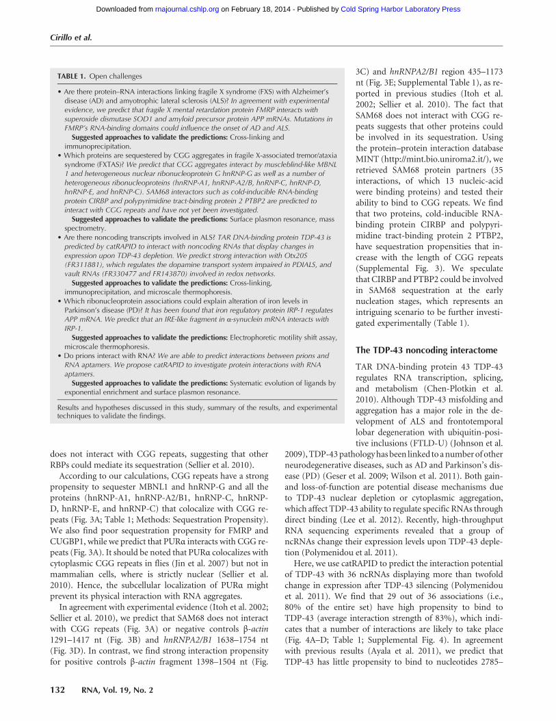
3C) and hnRNPA2/B1 region 435–1173nt (Fig. 3E; Supplemental Table 1), as re-ported in previous studies (Itoh et al.2002; Sellier et al. 2010). The fact thatSAM68 does not interact with CGG re-peats suggests that other proteins couldbe involved in its sequestration. Usingthe protein–protein interaction databaseMINT (http://mint.bio.uniroma2.it/), weretrieved SAM68 protein partners (35interactions, of which 13 nucleic-acidwere binding proteins) and tested theirability to bind to CGG repeats. We findthat two proteins, cold-inducible RNA-binding protein CIRBP and polypyri-midine tract-binding protein 2 PTBP2,have sequestration propensities that in-crease with the length of CGG repeats(Supplemental Fig. 3). We speculatethat CIRBP and PTBP2 could be involvedin SAM68 sequestration at the earlynucleation stages, which represents anintriguing scenario to be further investi-gated experimentally (Table 1).
The TDP-43 noncoding interactome
TAR DNA-binding protein 43 TDP-43regulates RNA transcription, splicing,and metabolism (Chen-Plotkin et al.2010). Although TDP-43 misfolding andaggregation has a major role in the de-velopment of ALS and frontotemporallobar degeneration with ubiquitin-posi-tive inclusions (FTLD-U) (Johnson et al.
2009),TDP-43pathologyhasbeen linked to anumberof otherneurodegenerative diseases, such as AD and Parkinson’s dis-ease (PD) (Geser et al. 2009; Wilson et al. 2011). Both gain-and loss-of-function are potential disease mechanisms dueto TDP-43 nuclear depletion or cytoplasmic aggregation,which affect TDP-43 ability to regulate specific RNAs throughdirect binding (Lee et al. 2012). Recently, high-throughputRNA sequencing experiments revealed that a group ofncRNAs change their expression levels upon TDP-43 deple-tion (Polymenidou et al. 2011).Here, we use catRAPID to predict the interaction potential
of TDP-43 with 36 ncRNAs displaying more than twofoldchange in expression after TDP-43 silencing (Polymenidouet al. 2011). We find that 29 out of 36 associations (i.e.,80% of the entire set) have high propensity to bind toTDP-43 (average interaction strength of 83%), which indi-cates that a number of interactions are likely to take place(Fig. 4A–D; Table 1; Supplemental Fig. 4). In agreementwith previous results (Ayala et al. 2011), we predict thatTDP-43 has little propensity to bind to nucleotides 2785–
TABLE 1. Open challenges
• Are there protein–RNA interactions linking fragile X syndrome (FXS) with Alzheimer’sdisease (AD) and amyotrophic lateral sclerosis (ALS)? In agreement with experimentalevidence, we predict that fragile X mental retardation protein FMRP interacts withsuperoxide dismutase SOD1 and amyloid precursor protein APP mRNAs. Mutations inFMRP’s RNA-binding domains could influence the onset of AD and ALS.Suggested approaches to validate the predictions: Cross-linking and
immunoprecipitation.• Which proteins are sequestered by CGG aggregates in fragile X-associated tremor/ataxiasyndrome (FXTAS)? We predict that CGG aggregates interact by muscleblind-like MBNL1 and heterogeneous nuclear ribonucleoprotein G hnRNP-G as well as a number ofheterogeneous ribonucleoproteins (hnRNP-A1, hnRNP-A2/B, hnRNP-C, hnRNP-D,hnRNP-E, and hnRNP-C). SAM68 interactors such as cold-inducible RNA-bindingprotein CIRBP and polypyrimidine tract-binding protein 2 PTBP2 are predicted tointeract with CGG repeats and have not yet been investigated.Suggested approaches to validate the predictions: Surface plasmon resonance, mass
spectrometry.• Are there noncoding transcripts involved in ALS? TAR DNA-binding protein TDP-43 ispredicted by catRAPID to interact with noncoding RNAs that display changes inexpression upon TDP-43 depletion. We predict strong interaction with Otx20S(FR311881), which regulates the dopamine transport system impaired in PDIALS, andvault RNAs (FR330477 and FR143870) involved in redox networks.Suggested approaches to validate the predictions: Cross-linking,
immunoprecipitation, and microscale thermophoresis.• Which ribonucleoprotein associations could explain alteration of iron levels inParkinson’s disease (PD)? It has been found that iron regulatory protein IRP-1 regulatesAPP mRNA. We predict that an IRE-like fragment in α-synuclein mRNA interacts withIRP-1.Suggested approaches to validate the predictions: Electrophoretic motility shift assay,
microscale thermophoresis.• Do prions interact with RNA? We are able to predict interactions between prions andRNA aptamers. We propose catRAPID to investigate protein interactions with RNAaptamers.Suggested approaches to validate the predictions: Systematic evolution of ligands by
exponential enrichment and surface plasmon resonance.
Results and hypotheses discussed in this study, summary of the results, and experimentaltechniques to validate the findings.
Cirillo et al.
132 RNA, Vol. 19, No. 2
Cold Spring Harbor Laboratory Press on February 18, 2014 - Published by rnajournal.cshlp.orgDownloaded from

3268 of transcript NM_007375.3, which is a negative control(Fig. 4E; Supplemental Table 1).When compared with a con-trol set of RNAmolecules, 84% of ncRNAs (30 out of 36 tran-scripts) are predicted to have significantly higher propensityto bind to TDP-43, which indicates great specificity for theseinteractions (Methods: Interaction Strength; SupplementalFig. 5; Strong et al. 2007).
Two of the most interacting targets are natural antisensetranscripts participating in the regulation of gene expression(Supplemental Table 1): RevΔ5ase (FR033920/LIT3360, in-teraction strength = 99%), which modulates docosahexaeno-ic acid levels preventing cytoplasmic accumulation of TDP-43 (Tremblay et al. 2011) and Otx2OS (FR311881/LIT3411,interaction strength = 99%), which regulates the dopamine
FIGURE 3. CGG protein sequestration. Depending on the spatial position with respect to CGG aggregates, proteins are classified as (1) colocalizing(MBNL1 and hnRNP-G, black); (2) colocalizing in the late stage granules (inset); (3) noncolocalizing (FMRP, CUGBP1, and PURα, star), and (4) non-binding (SAM68, italics). (A) In agreement with experimental results, we observe that colocalizing proteins have strong propensity to be sequestered byCGG repeats (MBNL1, hnRNP-G, and proteins in the inset). SAM68 interacting partners (CIRBP and PTBP2, gray) are found to bind to the CGG re-peats. (A,B,D)Wepredict that SAM68does not interactwithCGGrepeats aswell as negative controlsβ-actin1291–1417nt andhnRNPA2/B11638–1754nt. (C,E) SAM68 interacts with positive controls β-actin fragment 1398–1504 nt and hnRNPA2/B1 region 435–1173 nt (Supplemental Table 1).
FIGURE 4. TDP-43 associations with ncRNAs. Predictions of TDP-43 interactions with (A) FR033920; (B) FR311881; (C) FR330477; (D) FR143870,and (E) negative control RNA (Supplemental Material). (A,B) Natural antisense transcripts FR033920 and FR311881 regulate docosahexaenoic acidlevels and dopamine transport, relatively. (C,D) vault RNAs FR330477 and FR143870 could be implicated in redox regulatory networks (Table 1).
Protein–RNA interactions and neurodegeneration
www.rnajournal.org 133
Cold Spring Harbor Laboratory Press on February 18, 2014 - Published by rnajournal.cshlp.orgDownloaded from
transport system impaired in PD/ALS patients (Buttarelliet al. 2006). Other ncRNAs predicted to highly interactwith TDP-43 are vault RNAs (FR330477/LIT2028, interac-tion strength = 90%, and FR143870/LIT2029, interactionstrength = 92%) (Supplemental Table 1; Kedersha andRome 1986). The interaction between TDP-43 and vaultRNAs might be implicated in redox regulatory networks, assuggested by preliminary experimental evidence (Iwashitaet al. 2010; Fiesel et al. 2011). Although controversial hypoth-eses exist on the mechanisms by which TDP-43 contributesto neurodegeneration, catRAPID could help in the identifica-tion of protein–RNA interactions dysregulated in disease.The analysis of TDP-43 noncoding interactome shows thatcatRAPID is extremely powerful to investigate protein–ncRNA associations inferred from changes in regulatory net-works (Table 1).
FMRP and TDP-43 autogenous regulation
Autogenous regulation of gene expression involves interac-tion between protein and RNA encoded by the same gene(Lee et al. 2012). Regulation of expression levels through au-togenous associations has been reported for a number ofRNA processing factors, including serine–arginine rich pro-teins SC35 and SF2, and heterogeneous ribonucleoproteinmembers PTB and hnRNP-L (Johnsen et al. 1982; Kimet al. 2010). Here, we focus on the autogenous regulationof FMRP and TDP-43, because impairment of this feedbackmechanism could have consequences for the onset of FXSand ALS.
Coimmunoprecipitation assays indicate that a region locat-ed at position 1557–1658 nt in the 3′ UTR of FMR-1 is crucialfor autogenous interaction (Schaeffer et al. 2001). UsingcatRAPID to compute the interaction scores between proteinand RNA fragments (Methods: Interaction Fragments), wepredict that FMR-1 region 1587–1660 nt, overlapping withthe experimental region 1557–1658 nt, has high propensityto interact with the RGG domain of FMRP (Fig. 5A–C;Supplemental Fig. 6A; Schaeffer et al. 2001). As for TDP-43,CLIP experiments performed in HEK293 cells indicate thatthe protein self-associates at the 3′ UTR of its pre-mRNATARDBP (Ayala et al. 2011). catRAPID correctly identifiesthe two experimentally validated RNA regions located at po-sitions 301–744 nt and 745–1060 nt (Fig. 5D–F; SupplementalFig. 6B; Ayala et al. 2011). The algorithm also identifies twoRNA recognition motifs (RRM1 and RRM2) and the gly-cine-rich C-terminal domain of TDP-43 (Fig. 5D; Lagier-Tourenne and Cleveland 2009). Our predictions indicatethat the C-terminal region spanning residues 321–366 ofTDP-43 is directly involved in autogenous regulation, inagreement with previous experimental observations (Ayalaet al. 2011). We speculate that many other RNA-bindingproteins have evolved similar mechanisms of autogenousregulation.
Iron-mediated expression of APP and α-synuclein
Iron regulatory protein 1, IRP-1, is a cytosolic iron sensorthat binds to an iron-sulfur cluster and acts as aconitasewhen cellular iron levels are high (Philpott et al. 1994). Ifiron levels are low, IRP-1 binds to stem–loop structures calledIron-Responsive Elements (IREs, conserved nucleotides5′CAGU/AGN3′) in target mRNA species, and regulates up-take, storage, and transport of iron ions. Binding of IRP-1 toan iron–sulfur cluster impairs its ability to interact with RNA.An electrophoretic motility shift assay (REMSA) has beenused to probe the interaction between IRP-1 and the iron-re-sponsive element (IRE-Type II) within the 5′ UTR of APPmRNA (Rogers et al. 2002). Importantly, IRE-Type II se-quence shows high similarity with the IRE in the mRNA offerritin H-subunit (Supplemental Fig. 7) that is regulatedby IRP-induced translational repression in case of irondeficiency.We use catRAPID to predict the interaction potential of
IRP-1 with both ferritin and APP transcripts (Methods:Interaction Fragments) (Rogers et al. 2002). Our methodcorrectly predicts that the 5′ UTRs of ferritin (SupplementalFig. 8) and APP (Fig. 6A,B) interact with IRP-1 in several re-gions of the protein sequence, which is consistent with exper-imental evidence (Rogers et al. 2002; Walden et al. 2006). Asfor the APP transcript, nucleotides 25–76 show the highestinteraction propensity and overlap with the experimentalsegment at nucleotides 40–95 (Fig. 6A,B; Supplemental Fig.8). We note that the IRE-binding surface of IRP-1 does notconsist of canonical RNA-binding motifs such as those ofother ribonucleoproteins, and that IRE binding is mediatedby residues from each of the four IRP-1 domains (Waldenet al. 2006). Recently, Cho et al. (2010) showed that disrup-tion of the CAGAGC motif leads to strong reduction in theIRP-1 propensity to bind to APP IRE (Supplemental Table1; Supplemental Fig. 9A). We use catRAPID to carry out anexhaustive analysis of all the mutations in the CAGAGCsite (4095 sequence variants) and discovered that 87% ofthe mutations strongly reduce IRP-1 binding ability (Supple-mental Fig. 9A–C). Intriguingly, shuffling the IRE sequence ispredicted to abrogate IRP-1 interactions in 95 out 100 cases(Supplemental Fig. 9B,C). The same effect is observed forTDP-43 interactions with ncRNAs, where shuffling reducesthe interaction propensity in about 80 out of 100 cases(Supplemental Fig. 9D–F).Interestingly, one RNA stem–loop within the 5′ UTR of
human α-synuclein transcript has been predicted to be struc-turally related to the IRE element present in ferritin mRNA(Supplemental Fig. 7; Olivares et al. 2009). According toour calculations, the IRE-containing fragment of α-synucleinmRNA (nucleotides 190–252) has the highest propensity tobind to IRP-1 (Fig. 6C; Supplemental Fig. 8B). Moreover,we predict that IRP-1 domain 4, containing the largestnumber of binding sites (Walden et al. 2006), interactswith the IRE-containing fragment (interaction strength =
Cirillo et al.
134 RNA, Vol. 19, No. 2
Cold Spring Harbor Laboratory Press on February 18, 2014 - Published by rnajournal.cshlp.orgDownloaded from

FIGURE 5. FMRP and TDP-43 self-regulation. (A) FMRP-FMR-1 and (D) TDP-43-TARDBP interaction maps (blue areas indicate experimentally validatedinteractions, protein secondary structure elements are displayed next to the “protein residue index” axis); (B) FMR-1 and (E) TARDBP RNA interaction profiles(blue lines indicate experimentally identified binding regions; Methods: Interaction Fragments); interaction strengths with (C) nucleotides 1557–1658 of FMR-1and (F) nucleotides 495–555 of TARDBP (Supplemental Table 1).
Protein–R
NAinteractions
andneurodegeneration
www.rnajournal.org
135
C
old Spring H
arbor Laboratory Press
on February 18, 2014 - P
ublished by rnajournal.cshlp.org
Dow
nloaded from

85%) (Fig. 6D). Our predictions suggest that IRP-1 could beinvolved in regulating α-synuclein, which might have impli-cations for the alteration of iron levels found in PD (Table 1).
Prions and RNA aptamers
Mammalian prions (PrP) are infectious agents causingneurodegenerative diseases (Prusiner 1998). To date, prioninfectivity is attributed to conversion of the soluble PrPc
into an aggregation-prone structural isoform PrPSc (Panet al. 1993). The exact physiological function of PrPc remainselusive; however, there is an increasing understanding ofthe molecular mechanisms underlying PrPc pathologicalconversion and its interactions with other biological macro-molecules. Among these, cellular adhesion molecules, nucle-ic acids, basal membrane molecules, and sulfated glycanshave been reported to interact with PrPc and to induce ormodulate conversion into β-sheet-rich structures that sharesmany features with infectious PrPSc (Silva et al. 2011). It hasbeen proposed that PrPc could undergo specific structural re-arrangements modulated by binding with specific nucleic ac-ids molecules, such as highly structured RNAs (Deleault et al.2003) or RNA aptamers (Mercey et al. 2006).
Several studies showed that residues 90–141 are crucial forthe conversion from natural to pathological form (Tartagliaet al. 2005, 2008). Proske et al. (2002) discovered an RNAaptamer, DP7, which binds with great affinity to an epitopelocated within residues 90–141 of hamster PrP (highly con-served in mouse and human sequences) (SupplementalTable 1). Here we estimate the binding ability of DP7 to ham-ster PrP using catRAPID (Methods: Interaction Fragments).Based on our calculations, the fragment located at residues104–155 shows the highest propensities to bind to DP7 andhas the largest overlap with the experimental region spanningresidues 90–141 (Fig. 7A; Proske et al. 2002). In agreementwith experimental evidence, we predict that full-length ham-ster PrP and DP7 aptamer are highly interacting (interactionstrength = 85%) (Fig. 7B; Proske et al. 2002). We also reportvery high interaction propensity between residues 90 and 141of hamster PrP and DP7 aptamer (interaction strength =99%; RNA interaction strength = 100%), which is consistentwith experimental findings (Methods: Interaction Score)(Fig. 7C; Proske et al. 2002). In agreement with Proskeet al. (2002), we predict high interaction propensities be-tween DP7 and mouse and human PrP (Supplemental Fig.10A–D).
FIGURE 6. IRP-1 interactions with APP and α-synucleinmRNA. (A) Interactionmap of IRP-1 with APP (secondary structure elements are displayedat the “protein residue index” axis; blue areas indicate experimentally validated interactions). RNA-interaction profiles for IRP-1 associations with (B)APP and (C) α-synuclein mRNA (blue lines indicate experimentally identified binding regions) (Supplemental Table 1). (D) Interaction strength forIRP-1 domain 4 region (amino acids 661–889) and putative IRE fragment in α-synuclein transcript (nucleotides 190–252) (Supplemental Table 1).
Cirillo et al.
136 RNA, Vol. 19, No. 2
Cold Spring Harbor Laboratory Press on February 18, 2014 - Published by rnajournal.cshlp.orgDownloaded from
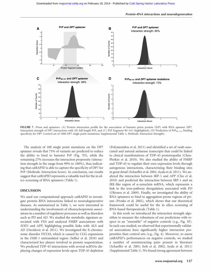
The analysis of 100 single point mutations on the DP7aptamer reveals that 73% of variants are predicted to reducethe ability to bind to hamster PrP (Fig. 7D), while theremaining 27% increases the interaction propensity (interac-tion strength in the range from 99% to 100%), thus indicat-ing that catRAPID is able to capture the specificity of DP7 forPrP (Methods: Interaction Score). In conclusion, our resultssuggest that catRAPID represents a valuable tool for the in sil-ico screening of RNA aptamers (Table 1).
DISCUSSION
We used our computational approach catRAPID to investi-gate protein–RNA interactions linked to neurodegenerativediseases. As summarized in Table 1, we were interested inunderstanding the involvement of ribonucleoprotein associ-ations in a number of regulatory processes as well as disorderssuch as PD and AD. We studied the metabolic signature as-sociated with FXS and analyzed FMRP associations withSOD1 and APP, highlighting possible links with ALS andAD (Davidovic et al. 2011). We investigated the X-chromo-some disorder FXTAS, which is caused by CGG expansionsin the FMR-1 untranslated region (Sellier et al. 2010) andcharacterized key players involved in protein sequestration.We predicted TDP-43 interactions with several ncRNAs dis-playing changes of expression levels upon TDP-43 depletion
(Polymenidou et al. 2011) and identified a set of vault-asso-ciated and natural antisense transcripts that could be linkedto clinical manifestations of TDP-43 proteinopathy (Chen-Plotkin et al. 2010). We also studied the ability of FMRPand TDP-43 to regulate their own expression levels throughautogenous interactions, characterizing their binding sitesin great detail (Schaeffer et al. 2001; Ayala et al. 2011). We an-alyzed the interaction between IRP-1 and APP (Cho et al.2010) and predicted the interaction between IRP-1 and anIRE-like region of α-synuclein mRNA, which represents alink to the iron-pathway deregulation associated with PD(Olivares et al. 2009). Finally, we investigated the ability ofRNA aptamers to bind to aggregation-prone regions of pri-ons (Proske et al. 2002), which shows that our theoreticalframework could be useful for the in silico screening ofRNA-based therapeuticals (Table 1).In this work we introduced the interaction strength algo-
rithm to measure the robustness of our predictions with re-spect to an “ensemble” of negative controls (e.g., Fig. 5F).In each case studied, we observed that experimentally validat-ed associations have significantly higher interaction pro-pensities than control sets (e.g., Fig. 4). Moreover, to assesscatRAPID’s performances on negative controls, we studieda number of noninteracting pairs present in literature(Schaeffer et al. 2001; Itoh et al. 2002; Ayala et al. 2011)(Supplemental Table 1).We found strong agreement between
FIGURE 7. Prion and aptamers. (A) Protein interaction profile for the association of hamster prion protein (PrP) with RNA aptamer DP7.Interaction strength of DP7 interactions with (B) full-length PrP, and (C) PrP fragment 90–141 (highlighted); (D) Prediction of PrP90–141 bindingspecificity for DP7 (control set of 1000 DP7 single point mutations; Supplemental Table 1; Methods: Interaction Strength).
Protein–RNA interactions and neurodegeneration
www.rnajournal.org 137
Cold Spring Harbor Laboratory Press on February 18, 2014 - Published by rnajournal.cshlp.orgDownloaded from

experimental results and our calculations (Figs. 1E, 3B,D, 5E,7D), which indicates that the use of control sets is very im-portant to achieve accurate predictions. As shown in Figures1B,D, 6B,C, 7A, the interaction fragments algorithm isable to identify binding regions in both protein and RNAmolecules.
We propose catRAPID for predictions of protein–RNA as-sociations, to flag putative interactions and select candidatesfor experimental studies (Table 1). Our method allows pro-cessing of a large amount of protein–RNA pairs and canlead to finding previously unknown interactions. Due tothe vastly increased analysis throughput, even whole pro-tein–RNA networks could be soon investigated without theneed to focus on small subsets. Our methodology providesa significant amount of new information on protein–RNAassociations, discovery of which would not be possible witha purely experimental workflow due to the sheer volume.Most importantly, our approach works on the intersectionof protein and RNA biology and will help to bridge the gapbetween the two disciplines.
MATERIALS AND METHODS
Interaction propensity
We use the catRAPID method to predict protein–RNA interactions(Bellucci et al. 2011). In catRAPID, the contributions of secondarystructure, hydrogen bonding, and van der Waals’ are combined to-gether into the “interaction profile”:
|Fxl = aS|Sxl+ aH |Hxl+ aW |Wxl (1)The “interaction propensity” π is defined as the inner product be-
tween the protein propensity profile |Cpl and the RNA propensityprofile |Crl weighted by the “interaction matrix” I:
p = kC p|I |Crl (2)The interaction matrix I as well as the parameters αS, αH, and αW
were derived using aMontecarlo procedure under the condition thatinteraction propensities π take maximal values for associations inthe positive training set and minimal values for associations in thenegative training set:
I :max kC p|I|Crl ∀ { r,p} [ { positive training set}min kC p|I|Crl ∀ { r,p} [ {negative training set}
{(3)
The catRAPID method was trained to predict interactionpropensities of protein–RNA pairs in the range of from 50 to750 amino acids and 50 to 1500 nt. The algorithm to compute theinteraction propensity with respect to the negative training set (dis-criminative power) is available at www.tartaglialab.crg.cat/catrapid.html.
Interaction strength
The concept of interaction strength is introduced to compare the in-teraction propensity of a protein–RNA pair with a reference set thathas little propensity to bind (random associations between polypep-tide and nucleotide sequences). For each protein–RNA pair under
investigation, we use a “reference set” of 102 protein and 102 RNAmolecules. To assess the strength of a particular association, wecompute its interaction propensity π and compare with the interac-tion propensities p̃ of the reference set (total of 104 nonredundantprotein–RNA pairs). Using the interaction propensity distributionof the reference set, we generate the “interaction score”:
Interaction Score = p− m
s
m = 1L
∑Li=1
p̃i
s2 = 1L
∑Li=1
(p̃i − m)2
⎧⎪⎪⎪⎨⎪⎪⎪⎩
(4)
The number of interactions is Λ = 104. From the distribution ofinteraction propensities we compute the “interaction strength”:
Interaction Strength = P(p̃ ≤ p)= cumulative distribution function (cdf) (5)
Reference sequences have the same lengths as the pair of interestto guarantee that the interaction strength does not depend onprotein and RNA lengths. The “protein interaction strength”and the “RNA interaction strength” are special cases of the inter-action strength in which only the protein or the RNA sequence israndomized to generate a reference set. For instance, the RNA inter-action strength used for the analysis of the TDP43 interactome is theRNA-binding ability of a protein with respect to a pool of 100 pro-teins. The algorithm to compute the interaction strength is availableat http://tartaglialab.crg.cat/catrapid.strength.html.
Interaction fragments
In some cases, protein or RNA sequences exceed the size compatiblewith our computational requirements, and catRAPID could not beused to calculate the interaction propensity. To overcome this lim-itation, we developed a procedure called “fragmentation,” whichinvolves division of polypeptide and nucleotide sequences intofragments, followed by prediction of the interaction propensities.The analysis of fragments is particularly useful to identify regions in-volved in the binding (e.g., self-interactions of TDP-43 and FMRP).The fragmentation approach is based on the division of protein andRNA sequences into overlapping segments:
(kb + 1
2) f kb = 1,2, . . . ,b
km f km = 1,2, . . . ,m
l − (ke + 1
2) f ke =1,2, . . . ,e
⎧⎪⎪⎪⎨⎪⎪⎪⎩
(6)
Where kb, km, and ke indicate the position of fragments, f is theirlength and l is the overall sequence length. The number of total frag-ments is b +m + e = t≤ 100 (limited by catRAPID sequence restric-tions). The maximum number of protein–RNA interactions is 104,which implies that the ability to identify an experimentally validatedinteraction by chance is 10−4. The list of all the protein–RNA frag-ment associations is called “interaction map.” “Protein and RNA in-teraction profiles” are bidimensional projections of the interactionmap onto the protein or RNA positions, respectively. A variant ofthe fragmentation algorithm developed to analyze protein interac-tions with long transcripts has been described in a recent paper
Cirillo et al.
138 RNA, Vol. 19, No. 2
Cold Spring Harbor Laboratory Press on February 18, 2014 - Published by rnajournal.cshlp.orgDownloaded from

(Agostini et al. 2012). The algorithms to compute interaction frag-ments are available at http://tartaglialab.crg.cat/catrapid.fragments.html.
CGG sequestration propensity
The CGG sequestration propensity is calculated as the interactionstrength multiplied by the protein concentration:
CGGSequestration Propensity =, Interaction Strength
.×a log(Abundance) (7)Where,Interaction Strength. = 1/( f − i+ 1)∑f
k=i InteractionStrength (k) is the average interaction strength for CGG repeatsranging from i = 20 to f = 200 and α = 1/log(Abundance)max is thenormalization factor. Protein abundances are retrieved from thedatabase http://pax-db.org.
SUPPLEMENTAL MATERIAL
Supplemental material is available for this article.
ACKNOWLEDGMENTS
We thank Professor R. Guigo’, Dr. B. Lehner, and Viola Tartaglia forstimulating discussions. Our work was supported by the SpanishMinistry of Economy and Competitiveness (SAF2011-26211). Weare grateful to the Programa de Ayudas FPI del Ministerio deEconomia y Competitividad—BES-2012-052457. This project wassupported by a grant from “la Caixa” to Petr Klus.
Received June 8, 2012; accepted November 16, 2012.
REFERENCES
Agostini F, Cirillo D, Bolognesi B, Tartaglia GG. 2012. X-inactivation:Quantitative predictions of protein interactions in the Xist network.Nucleic Acids Res doi: 10.1093/nar/gks968.
Anthony K, Gallo J-M. 2010. Aberrant RNA processing events in neuro-logical disorders. Brain Res 1338: 67–77.
Ayala YM, De Conti L, Avendaño-Vázquez SE, Dhir A, Romano M,D’Ambrogio A, Tollervey J, Ule J, Baralle M, Buratti E, et al. 2011.TDP-43 regulates its mRNA levels through a negative feedbackloop. EMBO J 30: 277–288.
BartzokisG, SultzerD,Cummings J,Holt LE,HanceDB,HendersonVW,Mintz J. 2000. In vivo evaluation of brain iron in Alzheimer diseaseusing magnetic resonance imaging. Arch Gen Psychiatry 57: 47–53.
Bechara EG, Didiot MC, Melko M, Davidovic L, Bensaid M, Martin P,Castets M, Pognonec P, Khandjian EW,Moine H, et al. 2009. A nov-el function for fragile X mental retardation protein in translationalactivation. PLoS Biol 7: e16.
Bellucci M, Agostini F, Masin M, Tartaglia GG. 2011. Predicting proteinassociations with long noncoding RNAs. Nat Methods 8: 444–445.
Buttarelli FR, Circella A, Pellicano C, Pontieri FE. 2006. Dopaminetransporter immunoreactivity in peripheral bloodmononuclear cellsin amyotrophic lateral sclerosis. Eur J Neurol 13: 416–418.
Chen-Plotkin AS, Lee VM-Y, Trojanowski JQ. 2010. TAR DNA-bindingprotein 43 in neurodegenerative disease. Nat Rev Neurol 6: 211–220.
Cho H-H, Cahill CM, Vanderburg CR, Scherzer CR, Wang B, Huang X,Rogers JT. 2010. Selective translational control of the Alzheimer am-yloid precursor protein transcript by iron regulatory protein-1. J BiolChem 285: 31217–31232.
Darnell JC, Jensen KB, Jin P, Brown V, Warren ST, Darnell RB. 2001.Fragile X mental retardation protein targets G quartet mRNAs im-portant for neuronal function. Cell 107: 489–499.
Darnell JC, Fraser CE, Mostovetsky O, Stefani G, Jones TA, Eddy SR,Darnell RB. 2005. Kissing complex RNAs mediate interaction be-tween the Fragile-X mental retardation protein KH2 domain andbrain polyribosomes. Genes Dev 19: 903–918.
Davidovic L, Navratil V, Bonaccorso CM, Catania MV, Bardoni B,Dumas M-E. 2011. A metabolomic and systems biology perspectiveon the brain of the fragile X syndromemousemodel.Genome Res 21:2190–2202.
Deleault NR, Lucassen RW, Supattapone S. 2003. RNA molecules stim-ulate prion protein conversion. Nature 425: 717–720.
Dobson CM. 1999. Protein misfolding, evolution and disease. TrendsBiochem Sci 24: 329–332.
Fiesel FC, Kahle PJ. 2011. TDP-43 and FUS/TLS: Cellular functions andimplications for neurodegeneration. FEBS J 278: 3550–3568.
Fiesel FC, Schurr C, Weber SS, Kahle PJ. 2011. TDP-43 knockdown im-pairs neurite outgrowth dependent on its target histone deacetylase6. Mol Neurodegener 6: 64.
Geser F, Martinez-Lage M, Kwong LK, Lee VM-Y, Trojanowski JQ.2009. Amyotrophic lateral sclerosis, frontotemporal dementia andbeyond: The TDP-43 diseases. J Neurol 256: 1205–1214.
Hagerman PJ, Hagerman RJ. 2004. The fragile-X premutation: Amatur-ing perspective. Am J Hum Genet 74: 805–816.
Itoh M, Haga I, Li Q-H, Fujisawa J. 2002. Identification of cellularmRNA targets for RNA-binding protein Sam68. Nucleic Acids Res30: 5452–5464.
Iwashita K, Ikeda R, Takeda Y, Sumizawa T, Furukawa T, Yamaguchi T,Akiyama S, Yamada K. 2010. Major vault protein forms complexeswith hypoxia-inducible factor (HIF)-1α and reduces HIF-1α level inACHN human renal adenocarcinoma cells. Cancer Sci 101: 920–926.
Jin P, Duan R, Qurashi A, Qin Y, Tian D, Rosser TC, Liu H, Feng Y,Warren ST. 2007. Pur α binds to rCGG repeats and modulates re-peat-mediated neurodegeneration in a Drosophila model of FragileX Tremor/Ataxia Syndrome. Neuron 55: 556–564.
Johnsen M, Christensen T, Dennis PP, Fiil NP. 1982. Autogenous con-trol: Ribosomal protein L10-L12 complex binds to the leader se-quence of its mRNA. EMBO J 1: 999–1004.
Johnson BS, Snead D, Lee JJ, McCaffery JM, Shorter J, Gitler AD. 2009.TDP-43 is intrinsically aggregation-prone, and amyotrophic lateralsclerosis-linked mutations accelerate aggregation and increase toxic-ity. J Biol Chem 284: 20329–20339.
Kedersha NL, Rome LH. 1986. Isolation and characterization of a novelribonucleoprotein particle: Large structures contain a single speciesof small RNA. J Cell Biol 103: 699–709.
Kenneson A, Zhang F, Hagedorn CH,Warren ST. 2001. Reduced FMRPand increased FMR1 transcription is proportionally associated withCGG repeat number in intermediate-length and premutation carri-ers. Hum Mol Genet 10: 1449–1454.
KimHD, Kim T-S, Joo YJ, Shin H-S, Kim S-H, Jang C-Y, Lee CE, Kim J.2010. RpS3 translation is repressed by interaction with its ownmRNA. J Cell Biochem 110: 294–303.
KryshtafovychA, Venclovas C, Fidelis K,Moult J. 2005. Progress over thefirst decade of CASP experiments. Proteins 61 (Suppl 7): 225–236.
Lagier-Tourenne C, Cleveland DW. 2009. Rethinking ALS: The FUSabout TDP-43. Cell 136: 1001–1004.
Lee JF, Stovall GM, Ellington AD. 2006. Aptamer therapeutics advance.Curr Opin Chem Biol 10: 282–289.
Lee EB, Lee VM-Y, Trojanowski JQ. 2012. Gains or losses: Molecularmechanisms of TDP43-mediated neurodegeneration. Nat RevNeurosci 13: 38–50.
Mercey R, Lantier I, Maurel M-C, Grosclaude J, Lantier F, Marc D. 2006.Fast, reversible interaction of prion protein with RNA aptamers con-taining specific sequence patterns. Arch Virol 151: 2197–2214.
Olivares D, Huang X, Branden L, Greig NH, Rogers JT. 2009.Physiological and pathological role of α-synuclein in Parkinson’sdisease through iron mediated oxidative stress; The role of a putativeiron-responsive element. Int J Mol Sci 10: 1226–1260.
Protein–RNA interactions and neurodegeneration
www.rnajournal.org 139
Cold Spring Harbor Laboratory Press on February 18, 2014 - Published by rnajournal.cshlp.orgDownloaded from

Oostra BA, Willemsen R. 2009. FMR1: A gene with three faces. BiochimBiophys Acta 1790: 467–477.
Pan KM, Baldwin M, Nguyen J, Gasset M, Serban A, Groth D,Mehlhorn I, Huang Z, Fletterick RJ, Cohen FE. 1993. Conversionof α-helices into β-sheets features in the formation of the scrapie pri-on proteins. Proc Natl Acad Sci 90: 10962–10966.
Philpott CC, Klausner RD, Rouault TA. 1994. The bifunctional iron-re-sponsive element binding protein/cytosolic aconitase: The role of ac-tive-site residues in ligand binding and regulation. Proc Natl Acad Sci91: 7321–7325.
Pieretti M, Zhang FP, Fu YH, Warren ST, Oostra BA, Caskey CT,Nelson DL. 1991. Absence of expression of the FMR-1 gene in fragileX syndrome. Cell 66: 817–822.
Polymenidou M, Lagier-Tourenne C, Hutt KR, Huelga SC, Moran J,Liang TY, Ling S-C, Sun E, Wancewicz E, Mazur C, et al. 2011.Long pre-mRNA depletion and RNA missplicing contribute toneuronal vulnerability from loss of TDP-43. Nat Neurosci 14:459–468.
Primerano B, Tassone F, Hagerman RJ, Hagerman P, Amaldi F, Bagni C.2002. Reduced FMR1 mRNA translation efficiency in fragile X pa-tients with premutations. RNA 8: 1482–1488.
Proske D, Gilch S, Wopfner F, Schätzl HM,Winnacker E-L, FamulokM.2002. Prion-protein-specific aptamer reduces PrPSc formation.Chembiochem 3: 717–725.
Prusiner SB. 1998. Prions. Proc Natl Acad Sci 95: 13363–13383.Rogers JT, Randall JD, Cahill CM, Eder PS, Huang X, Gunshin H,
Leiter L, McPhee J, Sarang SS, Utsuki T, et al. 2002. An iron-respon-sive element type II in the 5′-untranslated region of the alzheimer’samyloid precursor protein transcript. J Biol Chem 277: 45518–45528.
Rubinsztein DC. 2006. The roles of intracellular protein-degradationpathways in neurodegeneration. Nature 443: 780–786.
Salta E, De Strooper B. 2012. Non-coding RNAs with essential roles inneurodegenerative disorders. Lancet Neurol 11: 189–200.
Schaeffer C, Bardoni B, Mandel J-L, Ehresmann B, Ehresmann C,Moine H. 2001. The fragile X mental retardation protein binds spe-cifically to its mRNA via a purine quartet motif. EMBO J 20:4803–4813.
Sellier C, Rau F, Liu Y, Tassone F, Hukema RK, Gattoni R, Schneider A,Richard S, Willemsen R, Elliott DJ, et al. 2010. Sam68 sequestrationand partial loss of function are associated with splicing alterations inFXTAS patients. EMBO J 29: 1248–1261.
Silva JL, Vieira TCRG, Gomes MPB, Rangel LP, Scapin SMN,Cordeiro Y. 2011. Experimental approaches to the interaction ofthe prion protein with nucleic acids and glycosaminoglycans:Modulators of the pathogenic conversion. Methods 53: 306–317.
Strong MJ, Volkening K, Hammond R, Yang W, Strong W, Leystra-Lantz C, Shoesmith C. 2007. TDP43 is a human low molecularweight neurofilament (hNFL) mRNA-binding protein. Mol CellNeurosci 35: 320–327.
Subramanian M, Rage F, Tabet R, Flatter E, Mandel J-L, Moine H. 2011.G-quadruplex RNA structure as a signal for neuritemRNA targeting.EMBO Rep 12: 697–704.
Tartaglia GG, Cavalli A, Pellarin R, Caflisch A. 2005. Prediction of aggre-gation rate and aggregation-prone segments in polypeptide sequenc-es. Protein Sci 14: 2723–2734.
Tartaglia GG, Pawar AP, Campioni S, Dobson CM, Chiti F,Vendruscolo M. 2008. Prediction of aggregation-prone regions instructured proteins. J Mol Biol 380: 425–436.
Tassone F, Hagerman RJ, Loesch DZ, Lachiewicz A, Taylor AK,Hagerman PJ. 2000. Fragile X males with unmethylated, full muta-tion trinucleotide repeat expansions have elevated levels of FMR1messenger RNA. Am J Med Genet 94: 232–236.
Tremblay C, St-Amour I, Schneider J, Bennett DA, Calon F. 2011.Accumulation of transactive response DNA binding protein 43 inmild cognitive impairment and Alzheimer disease. J NeuropatholExp Neurol 70: 788–798.
Walden WE, Selezneva AI, Dupuy J, Volbeda A, Fontecilla-CampsJC, Theil EC, Volz K. 2006. Structure of dual function iron regula-tory protein 1 complexed with ferritin IRE-RNA. Science 314:1903–1908.
Westmark CJ, Malter JS. 2007. FMRP mediates mGluR5-dependenttranslation of amyloid precursor protein. PLoS Biol 5: e52.
Wilson AC, Dugger BN, Dickson DW, Wang D-S. 2011. TDP-43 in ag-ing and Alzheimer’s disease - a review. Int J Clin Exp Pathol 4:147–155.
Cirillo et al.
140 RNA, Vol. 19, No. 2
Cold Spring Harbor Laboratory Press on February 18, 2014 - Published by rnajournal.cshlp.orgDownloaded from