neurocognitive research in post-stroke ... - ppk.elte.hu
TRANSCRIPT
1
EOTVOS LORAND UNIVERSITY FACULTY OF EDUCATION AND PSYCHOLOGY
DOCTORAL SCHOOL OF EDUCATION
Head of the Doctoral School: Anikó Zsolnai, Ph.D., Professor, DSc.
SPECIAL EDUCATION MODULE
Head of the Doctoral Module: Klára Marton, Ph.D., Professor
Izabella Szöllősi
Neurocognitive Research in Post-Stroke Aphasia:
Comparison of Linguistic and Nonlinguistic Cognitive Control Functions
THESES OF PHD DISSERTATION
Supervisor: Klára Marton, Ph.D., Professor
Opponents:
Zsuzsanna Kövi, Ph.D., Associate Professor
Dezső Németh, Ph.D., Professor
2021
2
1. Introduction
Even though our knowledge of post-stroke aphasia is continuously growing, a
comprehensive and unified model is still missing for an integrative explanation of the
heterogeneity of the produced symptoms.
The theoretical framework of the current research is based on the cognitive control model
(Cohen, 2017), which has a solid neurological foundation (Frontal–Cingular–Parietal network),
and involves the executive function system (Niendam, et al., 2012). To manage complex tasks
the cognitive control system is responsible for the synchronization of its processes such as
working memory, attentional control or proactive interference control. It means that the
cognitive control system directs the activations of representations in working memory, sustains
these activations, and reduces interference in order to achieve goal-directed behavior.
The control of both behavior and language can be characterized as parts of a continuum of
automatic and controlled processes (Cohen, Dunbar, & McClelland, 1990, Botvinick & Cohen,
2014; Code, 2005). First, behavioral control processes, in a particular task, can be determined
along a continuum from automatic to fully controlled. In general, the less control is required by
a task, the more likely the behavior relies on automatic processes. In contrast, the more complex
a task, the more control is needed for appropriate performance; in this case, behavior relies
more on cognitive control processes. The magnitude and complexity of control depends on
whether a conflict or interference is present in the actual task or whether additional attentional
resources are needed (Kane, Conway, Hambrick, & Engle, 2008; Cohen, 2017). Second,
similarly to the cognitive control model, language performance can also be evaluated along a
continuum of control. Along this continuum, language performance depends on the
accessibility of language control functions, language modalities and processes of language
levels (Code, 2005).
Post-stroke aphasia is traditionally viewed as an acquired language impairment, due to a
focal brain lesion (American Speech-Language-Hearing Association, 2020; Bánréti, 2014;
Osmánné, 1994). It means that language disturbances may involve a various degree of the
impairment in language production, language processing, as well as in language control
(Haarmann, Davelaar, & Usher, 2003; Kolk, 1999). A person with aphasia has often relatively
intact nonlinguistic cognitive skills, such as memory and executive function (American Speech-
Language-Hearing Association, 2020).
In some studies, transcortical motor aphasia (TMA) is treated as a type of nonfluent aphasia,
(Kertesz, 1979), whereas others consider this dysfunction as an executive function disorder
3
(Ardila, 2010). Based on these observations, dissociative relations might be assumed among
individuals with Broca’s aphasia and TMA on the basis of their linguistic and nonlinguistic
abilities (Ardila, 2010; Bánréti, 2014). In case of similar linguistic and nonlinguistic profiles in
cognitive control functions, these two nonfluent groups of aphasia would only differ in severity
(Buckingham, 1999). This result would support the view that in severe aphasia both domain-
general and linguistic control functions are impaired, while in individuals with mild aphasia
both their behavior and communication are better supported by their language and cognitive
control functions (Code, 2005; Buckingham, 1999).
Contrary to this, during the last decades, there has been mounting evidence that individuals
with aphasia also demonstrate nonlinguistic processing deficits (Kasselimis, 2015), which
might contribute to their language deficits (Nozari & Novick, 2017; Purdy, 2002; Kuzmina &
Weekes, 2017). There might be a number of nonlinguistic impairments behind the linguistic
symptoms that can be associated with different language processes and elements (Kuzmina &
Weekes, 2017; Novick, Trueswell, & Thompson-Shill, 2005; Nozari & Schwartz, 2012; Ye &
Zhou, 2009). As noted before, cognitive control is a complex mental system which is
responsible for the execution of goal-directed behavior according to the changing environment
(Diamond, 2013; Cohen, 2017). It is unclear whether language performance is supported by
more domain general (Nozari & Schwartz, 2012) or domain specific functions (Hula & McNeil,
2008) .
Our next assumption is based on both research evidence and clinical experience, namely
that the stroke itself might result in a slowing of cognitive operations, which might be
independent from the aphasia-specific slowing (Alderman, 2016). Since aphasic symptoms
often derive from a stroke, the resulting slowing in information processing might be the
cumulative result of stroke-specific and aphasia-specific slowing, which can be detected in both
linguistic and nonlinguistic domains (Yoo, 2017).
In sum, there is no clear evidence of the role of domain-general cognitive dysfunctions in
language performance deficits (American Speech-Language-Hearing Association, 2020;
McNeil, Hula, & Sung, 2011; Nozari & Schwartz, 2012). Moreover, it is not clear whether
cognitive control deficits are specific to the linguistic domain in post-stroke aphasia (Hula &
McNeil, 2008) or extend to the nonlinguistic domain too (Rodd, Johnsrude, & Davis, 2010).
Finally, there are only a few results about the effects of stroke in information processing (Su,
Wuang, Lin, & Su, 2015) and it is not clear whether slow processing speed in people with
aphasia is specific to this population exhibiting language disorders, or originates from the stroke
itself.

4
2. Purpose of the Research
The goal of the present study was to test the impact of differential cognitive control deficits
on language performance in individuals with different types of nonfluent aphasia by comparing
performances of individuals with post-stroke aphasia and control groups in specific tasks of
information processing. Cognitive control tasks included vigilance, selective attention,
response inhibition task, as well as linguistic and nonlinguistic working memory tasks which
examined resistance to distractor and to proactive interference and memory updating processes.
Performance profiles in these specific cognitive control functions reveal a complex picture
about the underlying mental mechanisms of post-stroke nonfluent aphasia.
Hypothesis:
1) There are differences between the performances of individuals with Broca’s aphasia and
TMA in both nonlinguistic (domain-general) and linguistic (domain-specific) cognitive
control functions (Schumacher, Halai, & Lambon Ralph, 2019; Kuzmina & Weekes,
2017; Murray, 1999). Participants with Broca’s aphasia demonstrate lower performance
in domain-specific cognitive control tasks than participants with TMA. However, we
hypothesize the opposite profile in domain-general control functions (Jefferies &
Lambon Ralph, 2006; Hula & McNeil, 2008).
2) There are differences between the performances of individuals with both Broca’s
aphasia and TMA in domain-general cognitive control tasks compared to the stroke and
the control groups (Kasselimis, 2015).
3) Individuals in the stroke group demonstrate lower performance in domain-general
cognitive control tasks than individuals in the control group, however there is no
difference between these groups in linguistic cognitive control functions (Alderman,
2016; Yoo, 2017).
3. Methods
3.1. Participants
Twenty-two participants with aphasia (PWA) – 9 with Broca’s aphasia, and 13 with TMA
– were recruited from the National Medical Rehabilitation Center (Budapest). One of the
control groups comprised 13 participants who had had an ischemic stroke attack, but
demonstrated no language disorder. They were participants of the same institute as PWA. The
second control group included 13 healthy, age-matched control participants who had no aphasia
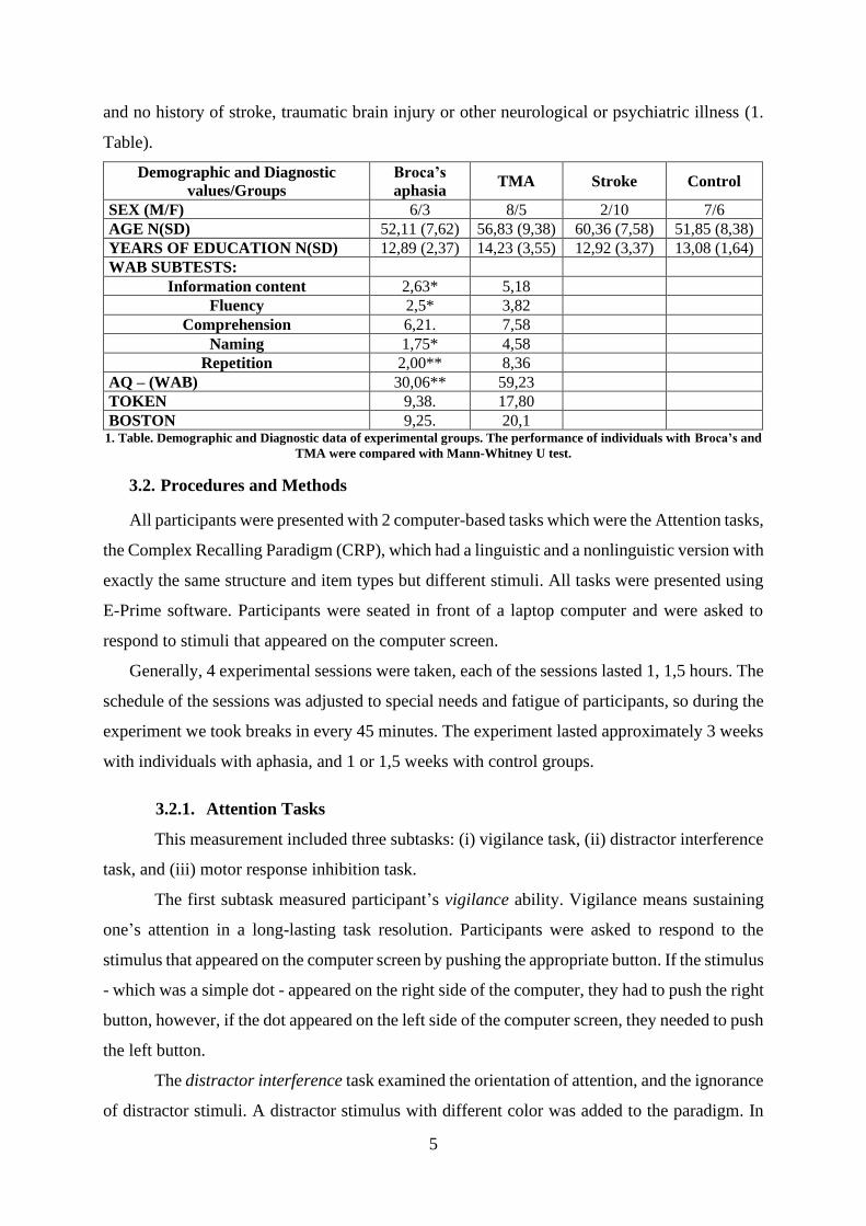
5
and no history of stroke, traumatic brain injury or other neurological or psychiatric illness (1.
Table).
Demographic and Diagnostic
values/Groups
Broca’s
aphasia TMA Stroke Control
SEX (M/F) 6/3 8/5 2/10 7/6
AGE N(SD) 52,11 (7,62) 56,83 (9,38) 60,36 (7,58) 51,85 (8,38)
YEARS OF EDUCATION N(SD) 12,89 (2,37) 14,23 (3,55) 12,92 (3,37) 13,08 (1,64)
WAB SUBTESTS:
Information content 2,63* 5,18
Fluency 2,5* 3,82
Comprehension 6,21. 7,58
Naming 1,75* 4,58
Repetition 2,00** 8,36
AQ – (WAB) 30,06** 59,23
TOKEN 9,38. 17,80
BOSTON 9,25. 20,1
1. Table. Demographic and Diagnostic data of experimental groups. The performance of individuals with Broca’s and
TMA were compared with Mann-Whitney U test.
3.2. Procedures and Methods
All participants were presented with 2 computer-based tasks which were the Attention tasks,
the Complex Recalling Paradigm (CRP), which had a linguistic and a nonlinguistic version with
exactly the same structure and item types but different stimuli. All tasks were presented using
E-Prime software. Participants were seated in front of a laptop computer and were asked to
respond to stimuli that appeared on the computer screen.
Generally, 4 experimental sessions were taken, each of the sessions lasted 1, 1,5 hours. The
schedule of the sessions was adjusted to special needs and fatigue of participants, so during the
experiment we took breaks in every 45 minutes. The experiment lasted approximately 3 weeks
with individuals with aphasia, and 1 or 1,5 weeks with control groups.
3.2.1. Attention Tasks
This measurement included three subtasks: (i) vigilance task, (ii) distractor interference
task, and (iii) motor response inhibition task.
The first subtask measured participant’s vigilance ability. Vigilance means sustaining
one’s attention in a long-lasting task resolution. Participants were asked to respond to the
stimulus that appeared on the computer screen by pushing the appropriate button. If the stimulus
- which was a simple dot - appeared on the right side of the computer, they had to push the right
button, however, if the dot appeared on the left side of the computer screen, they needed to push
the left button.
The distractor interference task examined the orientation of attention, and the ignorance
of distractor stimuli. A distractor stimulus with different color was added to the paradigm. In

6
this task two dots (a target and a distractor) appeared on the screen at the same time in each
trial. Participants needed to ignore the distractor stimuli.
In the motor response inhibition task selective attention and motor response inhibition
abilities were measured. Participants were asked to respond to the target by pressing the
response button and to the distractor by withholding this automatic response and pressing the
start button. Thus, individuals needed to suppress their automatic motor response and press a
different button.
3.2.2. Complex Recalling Paradigm (CRP)
The non-verbal CRP task measured individual’s attention, recollection and updating of
working memory, and proactive interference control. It included two subtasks and used abstract
figures that are difficult to name as stimuli: (1) Baseline task, and (2) Cue task.
(1) We used the Baseline task as a reference to the Cue task. The baseline task included
two item types: true target and new distractor stimulus. Participants were asked whether an
individual item appeared in the set of three stimuli that was introduced in the previous probe.
(2) In the Cue task we used the same item types as in the baseline task, however, a cue was
presented after the stimuli. The role of the cue, which was checkerboard, was to direct
individual’s attention to the position of the target item in the original stimuli set. Participants
were asked whether the item (target or distractor) had an identical position in the stimulus set
to the position of the cue.
The paradigm included five item manipulations; however, only three were included in this
research. Beyond the previously introduced two types of stimuli (true target and new distractor),
interference stimuli supplemented the stimulus set.
• true target: the function of these stimuli was to measure attention and working
memory storage. Participants needed to maintain the activation of the target stimuli
to make a decision about the probe, and the correct response was to accept the true
target stimuli.
• new distractor: the function of these stimuli was to measure attention and working
memory recollection based on newness and familiarity. Participants needed to make
a decision based on familiarity, and the correct response was to reject new
distractors.
• interference item: to be able to respond correctly to an interfering item it is important
to recognize the difference between relevant and irrelevant stimuli and to ignore
irrelevant ones.
7
3.2.3. Linguistic tasks
In addition to condition and stimulus manipulation in the CRP task, the types of the
stimuli were manipulated as well. The items in the linguistic CRP task were prototypical
pictures (Rossion & Pourtois, 2004). The words collected were high frequency (1000-10000),
everyday nouns composed of two syllables in Hungarian.
Pictures represent linguistic representations through their semantic content and they are
useful in facilitating semantic processing (Bohnemeye, 2014). Images as representational
referents elicit language mechanisms, unlike abstract figures. Thus, images as stimuli are
convenient to measure manipulations on linguistic representations. The structure, number of
trials, item types (target, distractor, interference) did not differ between the linguistic and
nonlinguistic tasks.
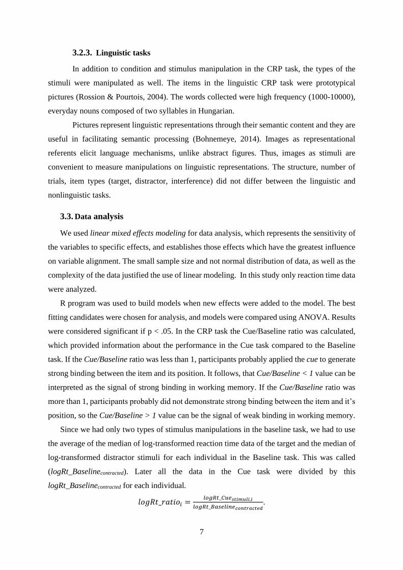
3.3. Data analysis
We used linear mixed effects modeling for data analysis, which represents the sensitivity of
the variables to specific effects, and establishes those effects which have the greatest influence
on variable alignment. The small sample size and not normal distribution of data, as well as the
complexity of the data justified the use of linear modeling. In this study only reaction time data
were analyzed.
R program was used to build models when new effects were added to the model. The best
fitting candidates were chosen for analysis, and models were compared using ANOVA. Results
were considered significant if p < .05. In the CRP task the Cue/Baseline ratio was calculated,
which provided information about the performance in the Cue task compared to the Baseline
task. If the Cue/Baseline ratio was less than 1, participants probably applied the cue to generate
strong binding between the item and its position. It follows, that Cue/Baseline < 1 value can be
interpreted as the signal of strong binding in working memory. If the Cue/Baseline ratio was
more than 1, participants probably did not demonstrate strong binding between the item and it’s
position, so the Cue/Baseline > 1 value can be the signal of weak binding in working memory.
Since we had only two types of stimulus manipulations in the baseline task, we had to use
the average of the median of log-transformed reaction time data of the target and the median of
log-transformed distractor stimuli for each individual in the Baseline task. This was called
(logRt_Baselinecontracted). Later all the data in the Cue task were divided by this
logRt_Baselinecontracted for each individual.
𝑙𝑜𝑔𝑅𝑡_𝑟𝑎𝑡𝑖𝑜𝑖 =𝑙𝑜𝑔𝑅𝑡_𝐶𝑢𝑒𝑠𝑡𝑖𝑚𝑢𝑙𝑖,𝑖
𝑙𝑜𝑔𝑅𝑡_𝐵𝑎𝑠𝑒𝑙𝑖𝑛𝑒𝑐𝑜𝑛𝑡𝑟𝑎𝑐𝑡𝑒𝑑.
8
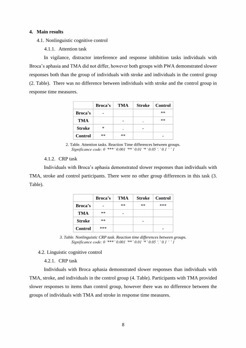
4. Main results
4.1. Nonlinguistic cognitive control
4.1.1. Attention task
In vigilance, distractor interference and response inhibition tasks individuals with
Broca’s aphasia and TMA did not differ, however both groups with PWA demonstrated slower
responses both than the group of individuals with stroke and individuals in the control group
(2. Table). There was no difference between individuals with stroke and the control group in
response time measures.
Broca’s TMA Stroke Control
Broca’s - **
TMA - . **
Stroke * . -
Control ** ** -
2. Table. Attention tasks. Reaction Time differences between groups.
Significance code: 0 ‘***’ 0.001 ‘**’ 0.01 ‘*’ 0.05 ‘.’ 0.1 ‘ ’ 1
4.1.2. CRP task
Individuals with Broca’s aphasia demonstrated slower responses than individuals with
TMA, stroke and control participants. There were no other group differences in this task (3.
Table).
Broca’s TMA Stroke Control
Broca’s - ** ** ***
TMA ** -
Stroke ** -
Control *** -
3. Table. Nonlinguistic CRP task. Reaction time differences between groups.
Significance code: 0 ‘***’ 0.001 ‘**’ 0.01 ‘*’ 0.05 ‘.’ 0.1 ‘ ’ 1
4.2. Linguistic cognitive control
4.2.1. CRP task
Individuals with Broca aphasia demonstrated slower responses than individuals with
TMA, stroke, and individuals in the control group (4. Table). Participants with TMA provided
slower responses to items than control group, however there was no difference between the
groups of individuals with TMA and stroke in response time measures.

9
Broca’s TMA Stroke Control
Broca’s - * *** ***
TMA * - .
Stroke *** -
Control *** . -
4. Table. Linguistic CRP task. Reaction time differeneces between groups.
5. Discussion
This section has the following structure. (1) Comparison of performances between the
groups of individuals with Broca’s aphasia and TMA; (2) Comparison of performances between
the groups of nonfluent aphasia with the group of individuals with a stroke and the neurotypical
control participants; (3) Comparison between the groups with individuals with a stroke and the
neurotypical controls.
5.1. Nonlinguistic cognitive control
5.1.1. Comparison of performances between the groups of individuals with Broca’s
aphasia and TMA
In this section we present together the interpretation of results in the attention and the CRP
tasks. Results in both attention task and CRP task contradicted our hypothesis that stated that
individuals with TMA would demonstrate weaker performances in these domain-general
cognitive control functions including vigilance, selective attention, working memory updating,
conflict resolution and proactive interference control compared to the group of individuals with
Broca’s aphasia, based on Ardila’s definition of TMA (Ardila, 2010). Instead of this profile,
individuals with Broca’s aphasia demonstrated slower responses than individuals with TMA.
The assumed impairment of cognitive control functions in TMA cannot be observed neither
in attention nor in working memory updating or proactive interference control compared to
Broca’s aphasia. It can be inferred that the accessibility of these cognitive control functions
might correspond to the severity of aphasia (Marinelli, Spaccavento, Craca, Marangolo, &
Angelelli, 2017), so we assume that the symptoms of severe Broca’s aphasia are aligned with
more severely impaired subfunctions of cognitive control compared to TMA.
This result is in agreement with previous studies, where severe aphasia symptoms were
related to the impairment of more subfunctions of cognitive control, like short-term memory,
or working memory (Potagas, Kasselimis, & Evdokimidis, 2011). Taking into account that in
10
these studies authors did not differentiate nonfluent aphasias (they focused on AQ), our research
might supplement these results with the idea that mobilizing working memory processes can be
slower in only severe nonfluent aphasia.
The continuity hypothesis of language performance (Buckingham, 1999; Code, 1989; Code,
2018) can help us to explain this result. The difference between performances in Broca’s and
TMA groups depends on the impairment of language modalities and domain-general cognitive
control functions. The more severe the aphasia, the more domain-general cognitive control
function is damaged (Potagas, Kasselimis, & Evdokimidis, 2011; Kuzmina & Weekes, 2017).
In Broca’s aphasia significant amounts of language and domain-general cognitive control
functions are impaired compared to TMA. In contrast, there are fewer language deficits and
fewer cognitive control dysfunctions in individuals with TMA. This leads to the assumption
that a more flexible cognitive control system is available in people with TMA.
5.1.2. Comparison of performances among the groups of nonfluent aphasia,
individuals with a stroke without aphasia, and the neurotypical control participants
We begin with the interpretation of the performance pattern of participants with Broca’s
aphasia. In both the attention task and the CRP task participants with Broca’s aphasia showed
slower response times compared to the stroke and the control groups.
The slower maintenance of attentional focus seems to be independent of the general slowing
effect due to the stroke itself, based on the differences between reaction time of individuals with
Broca’s aphasia and the stroke group. The impairment of the orientation of attention together
with the maintenance of stimulus representations might be responsible for a lower performance
in attention tasks in severe nonfluent aphasia. We can infer that besides the language symptoms,
the impairment of selective focused attention and the impairment of motor response resolution
might be present in severe nonfluent aphasia. Our results are in accordance with the results of
previous studies (Lee & Pyun, 2014; LaCroix, Tully, & Rogalsky, 2020, Ewans, 2014; Murray,
1999).
The higher response time of individuals with Broca’s aphasia compared to the stroke and
control groups in the CRP task can be established based on the model of cognitive control
(Cohen, 2017). The results of pair-wised comparisons of reaction times in the proactive
interference condition between the participants with Broca’s aphasia and the individuals with
stroke and the neurotypical control participants suggest that resistance to proactive interference
is impaired in Broca’s aphasia. This deficit in proactive interference control may reflect that
these individuals maintain the activation of working memory representations longer than
11
needed, even when those representations are already irrelevant, therefore show a delayed and/or
weaker activation of the relevant representations (Oberauer, 2002; Oberauer, Süß, Wilhelm, &
Sander, 2008). The working memory capacity is limited by resistance to proactive interference
(Pettigrew & Martin, 2016; Wilhelm, Hildebrandt, & Oberauer, 2013). Participants with
Broca’s aphasia, demonstrated slowness in short term maintenance of working memory
representations, which might be one of the consequences of the impairment of resistance to
proactive interference. We found impaired resistance to proactive interference in our previous
research in group of individuals with heterogenous aphasia types. Our current results
supplement this finding in the group of Broca’s aphasia.
Additionally, we also found evidence for the impairment of suppressing simultaneously
activated representations. The weak resistance to this type of interference can be explained by
weak binding between items and their physical position (Beaman, 2004). The weak binding is
indicated by the Cue/Baseline ratio, which was less than 1 in the groups of TMA, stroke and
controls. This implies that they correctly applied the cue, which helped them to maintain the
bindings between items and their positions. This was not observed in individuals with Broca’s
aphasia, in fact the Cue/Baseline ratio was more than 1.
The proper application of the cue strengthens the binding between the stimulus and its
position, thereby stronger representations might develop, which decrease both the interference
effect and the possibility of forgetting (Oberauer, Süß, Wilhelm, & Sander, 2008). Individuals
with Broca’s aphasia could not benefit from the cue, which indicates weak binding between the
stimuli and the context. Weak binding does not allow appropriate resistance to interference
(Oberauer, 2002), which is responsible for the appearance of weak memory traces. An intense
interference effect appeared, because representations that should have been suppressed due to
task demands, still had high activation levels. The dual mechanism of suppression of irrelevant
representations and the increase of activation of relevant representations seems to be impaired
in individuals with Broca’s aphasia, which directly leads to further limitations in working
memory capacity.
We conclude, that it is possible that weaker resistance to interference results in weakness
of working memory processes and attentional capacity (Meier & Kane, 2017; Hasher, Lustig,
& Zacks, 2007). However, it should be added that these dysfunctions interact with weak
language functions as well, as attested in the current study by the performance of participants
with Broca’s aphasia. This is in line with previous studies where authors established conflict
resolution impairment in lexical access in this population (Nozari & Schwartz, 2012; Ye &
Zhou, 2009). However, our results display new insights into the profile of cognitive control
12
functions in post-stroke nonfluent aphasia, and prove that domain-general cognitive control
functions are impaired in Broca’s aphasia.
Individuals with TMA demonstrated slower response time compared to the stroke and
control groups only in the attention tasks. This profile contradicts our hypothesis (we expected
slower performance in the TMA group than that in the stroke group in all non-linguistic tasks).
We can conclude, that in nonfluent aphasia there is a slowness in vigilance, in orientation of
attention, as well as selective attention (Murray, 2012; LaCroix, Tully, & Rogalsky, 2020). As
an explanation, slowness in orientation of attention and in selective attention might be
responsible for slow decision-making processes (Kane, Bleckely, Conway, & Engle, 2001). The
slow decision making might increase uncertainty in response selection of a task. This leads to
unsuccessful ignorance or inhibition of irrelevant and distracting stimulus in nonfluent aphasia
(Szöllősi, Lukács, & Zakariás, 2015).
Participants with TMA demonstrated similar performance in the CRP task to participants
with stroke. The performance of individuals with TMA indicates that despite their attentional
deficit, stroke-affected individuals with mild language disorder are able to mobilize their
cognitive resources in ways similar to stroke affected participants with no language disorder.
This result contradicts our hypothesis, because slower performance was expected in the TMA
group compared to the stroke and the control groups. This pattern allows the interpretation that
performance in the CRP task in individuals with TMA might be related to stroke-specific
slowing rather than aphasia.
Their similar performance was only displayed in the complex working memory tasks,
however, in the simple vigilance tasks they demonstrated poorer performance. Previous studies
provided evidence of attentional deficits in aphasia, and found a correlation between attentional
control functions and the severity of aphasia (Lee & Pyun, 2014). Our findings suggest that
there is no difference between severe and mild nonfluent aphasia in attentional control,
however, they significantly differ in managing working memory representations. Participants
with TMA might mobilize cognitive resources more effectively, indicated by their similar
performance to the stroke group, and higher performance than the Broca’s group.
Mild language disorder is accompanied by flexible cognitive capacity, which is manifested
in effective maintenance and manipulation of representations. Previous studies implied that the
improvement of language functions is correlated with the improvement of general cognitive
abilities, which proves the interrelation between language and attention, cognitive control
functions, and working memory (Seniów, Litwin, & Lesniak, 2009, Kang, Jeong, Moon, Lee,
& Lee, 2016).
13
5.1.3. Comparison between the groups of individuals with a stroke and their
neurotypical peers
In both the attention task and the CRP task the group of individuals with stroke showed
similar reaction times to the control group. Despite of previous observations about the slowing
effect of vascular stroke in information processing (Alderman, 2016; Su, Wuang, Lin, & Su,
2015), which can influence the adequate mobilization of cognitive control functions, in the
current study this slowing did not appear to contribute neither to simple attentional functions,
nor to working memory updating and proactive interference control. Previous papers have
described psychomotor slowing due to stroke without aphasia (Su, Wuang, Lin, & Su, 2015),
although there was no difference between these groups in simple attentional tasks.
Even though we only focused on the reaction time results in this study, we must note that
participants with stroke demonstrated lower accuracy compared to the control group. Based on
the accuracy differences it is likely that the well-documented psychomotor slowing in
individuals with stroke is related to complex decision-making processes of attentional control
and to the maintenance of response representations (Yoo, 2017). The results may also reflect a
trade-off between speed of processing and accuracy.
Our results indicate that stroke influences the response selection and decision-making
processes of the stimuli based on their familiarity and newness. An analysis of stimulus content,
which requires the maintenance of the activation of representations for access to that content,
should precede decision making. An inefficient maintenance of activation of representations
might result in less accurate performance compared to the control group. Although when a task
demands complex processes (conflict resolution, or proactive interference control), stroke-
affected participants are able to activate and flexibly mobilize proper cognitive resources in
time, in order to compensate for their limited short-term memory functions.
5.2. Linguistic cognitive control
5.2.1. Comparison of performances between the groups of individuals with Broca’s
aphasia and TMA
Participants with Broca’s aphasia demonstrated slower responses than individuals with
TMA in the linguistic CRP task. This suggests that the slowness that we have observed in
nonlinguistic working memory updating, conflict resolution, and proactive interference in
Broca’s aphasia may be present in the linguistic domain as well (Kuzmina & Weekes, 2017).
Presumably, linguistic control functions are not selectively impaired in Broca’s aphasia, or in
TMA. It is more likely that participants with Broca’s aphasia -with a severe language disorder-
14
show a more severe deficit in domain-general cognitive control than individuals with TMA.
The control of language functions is at least in part connected to domain-general cognitive
control, as shown by previous studies (Nozari, Swartz, 2011; Kuzmina, 2017; Christensen,
Wright, & Ratiu, 2018; Rodd, Johnsrude, & Davis, 2010). Thus, high language performance is
likely to be associated with intact general cognitive control, and higher cognitive flexibility.
To summarize, the dysfunction of domain-general cognitive control has been
demonstrated in all types of nonfluent aphasia in previous research (Kuzmina & Weekes, 2017).
According to these findings, overlapping linguistic and nonlinguistic control processes were
emphasized, however, the authors did not differentiate between types of nonfluent aphasia. The
current research supports the separate nature of control functions in nonfluent aphasias. For
participants with severe aphasia, it is more difficult to mobilize cognitive control functions for
goal achievement. While in mild aphasia, cognitive mechanisms which are activated in response
to verbal and nonverbal stimuli, interact with each other, while in in Broca’s aphasia the
cognitive control system seems to be more fragmented.
5.2.2. Comparison of performances among the groups of nonfluent aphasia, individuals
with a stroke without aphasia, and the neurotypical control participants
In this section we begin our discussion with the performance of individuals with Broca’s
aphasia compared to performance of individuals with stroke and control group in the CRP task.
In line with our predictions, linguistic control functions in Broca’s aphasia were slower
compared to those of individuals with stroke and the control group.
The results can be explained based on the cognitive control model (Cohen, 2017). Slower
responses of individuals with Broca’s aphasia compared to the stroke and control groups seems
to stem from impaired interference control. This impairment in participants with Broca’s
aphasia might be related to their difficulties in decision making with regards to the relevance
of the stimuli and to suppressing irrelevant representations (Nozari & Schwartz, 2012; Piai,
Roelofs, Acheson, & Takashima, 2013). These individuals demonstrate difficulties in shifting
their attention level from one representation to the other in order to produce a correct response.
Decreased activation of representations leads to weak memory traces; as a result, participants
forget the information easier (Engle, 2018; Oberauer, Süß, Wilhelm, & Sander, 2008). Our
findings are aligned with previous results that emphasize cognitive control deficits in nonfluent
aphasia (Ivanova, Dragoy, Kuptsova, Ulicheva, & Laurinavichyute, 2015)
Similar to their performance in resistance to proactive interference in the nonlinguistic
tasks, the Cue/Baseline ratio was more than 1 in the group of individuals with Broca’s aphasia
15
in linguistic task as well. It is possible that, the binding of linguistic stimuli to the context is
impaired which is reflected in the incorrect use of the cue. This might be associated with a low
activation of representations and together with poor binding, resistance to interference is
diminished or slowed down. The same pattern was observed in the nonlinguistic tasks. We can
conclude that cognitive operations of resisting interference are slow in both linguistic and
nonlinguistic tasks in Broca’s aphasia. This impairment blocks the production of increased
linguistic memory traces, which may result in slow decision-making in Broca’s aphasia.
Overall, domain-general cognitive control functions seem to be impaired in Broca’s aphasia
(Schumacher, Halai, & Lambon Ralph, 2019; Kuzmina & Weekes, 2017; Murray, 1999).
Participants without aphasia demonstrated normal general attentional control and proactive
interference control functions. Moreover, the lexical-semantic information might facilitate
coding processes of representations in these groups. A combination of these factors leads to an
effective supervisory mechanism. Although lexical-semantic information probably helps to
manipulate representations in working memory in Broca’s aphasia, due to severe impairment
in domain-general cognitive control functions, this facilitation is not sufficient to decrease the
group differences in the CRP tasks.
Our results of participants with TMA demonstrated similar reaction times to the stroke
group. However, they displayed slower responses compared to the neurotypical control group.
Most deficits in individuals with TMA can be explained by the effect of the stroke which is
indicated by the results that suggest no difference in reaction time between the stroke and the
TMA groups in most tasks. Instead of the impact of aphasia, which is mild in this group, a
stroke-specific deficit contributes to their weaker attentional and linguistic cognitive control.
The stroke-specific slowing related to the impairment of maintaining working memory
activations, might influence participants’ slower performance compared to the control group.
Goal oriented behavior is supervised at higher levels (Meier & Kane, 2017; Diamond,
2013), which in healthy individuals is composed of strongly interacting mechanisms. Accessing
linguistic information is a well-organized process which can be impeded due to severe
dysfunction of the cognitive system, which affects the success of communication (Purdy, 2002;
Ye & Zhou, 2009), however, different cognitive resources are more accessible for participants
with mild than with more severe language disorders.
16
5.2.3. Comparison between the groups of individuals with a stroke and their
neurotypical peers
There was no difference in the reaction time data in the CRP task between the stroke and
the control groups in the language control functions. In other words, in the stroke group the
speed of processing that is involved in managing the activation of representations based on
familiarity and newness seems to be intact. The above-mentioned stroke-specific slowing did
not affect the response times of participants who had no aphasia.
We do not present accuracy data in this study, however we found accuracy differences in
performance between individuals with stroke and the control group, similar to the nonlinguistic
tasks. We mention this accuracy result for the same reason as in nonlinguistic tasks, because
based on the reaction time data one might misinterpret the performance of individuals with
stroke, as they demonstrate no difference in speed of processing compared to the controls.
However, accuracy data imply that the process of comparison between the representations
based on familiarity and newness seems to be impaired. This dysfunction indicates that
participants often choose rejection incorrectly instead of acceptance of the targets, or they
accept distractor stimuli instead of rejecting them. Again, there is a tradeoff between accuracy
and speed of processing in the stroke group.
These results fit the cognitive control model if we consider stroke-specific slowing (Yoo,
2017) as a delay in sustained activation of representation for the access of mental processes
(Cohen, 2017). We suppose that psychomotor slowing, which is a well-documented symptom
after a stroke (Alderman, 2016), is related to the deficit of keeping representations active in
short-term memory. This process might be delayed, which promotes impaired decision-making
processes (Kane, et al., 2004). This stroke-specific domain-general short-term memory
dysfunctions influence cognitive processes in aphasia as well. However, it is important to note
that in severe language disorders aphasia-specific dysfunctions even in domain-general
cognitive control functions (working memory updating, conflict resolution, and proactive
interference control) are present as well. This cumulated effect of stroke-specific and aphasia-
specific dysfunctions can be responsible for the poor performance of individuals in Broca’s
aphasia in all tasks compared to the control group.
6. Limitations
The generalization of the results has some limitations because of the relatively small sample
sizes and the heterogeneity of participants in the stroke group.
The reason for small sample sizes is that we followed strict inclusion criteria in recruiting
17
participants. This was needed to allow us to answer the research questions of this study. We
selected members of groups according to language profiles, while finding the suitable
participants with Broca’s and TMA aphasia created major difficulties. In addition to the
language criteria compliance and contra-indication criteria needed to be considered (e.g.,
neglect syndrome, internal medical status), which caused a further decrease in the possible
number of participants involved.
Another limitation can be the composition of the stroke group. We involved participants
with both right and left hemisphere stroke. Early in our research we planned to recruit
individuals with dominant (left) hemisphere stroke without language disorder, however, we
found only a very low number of participants. As a compromised solution we extended the
criteria of the stroke group and we involved participants who demonstrated stroke in their right
hemisphere as well. Individuals in the stroke group were identical in their preserved language
abilities. Participants with right hemisphere stroke had use their dominant hand for button press,
whereas all other participants (those with left hemisphere stroke, participants with aphasia, and
the neurotypical control group) used their non-dominant hand. Although the results of our pilot
study found no difference in the most important conditions between the performances of
individuals with left and right hemisphere stroke, we need to identify this pattern as a limitation.
It can be identified as a further scientific limitation that, despite the data cleaning, variables
typically did not show a normal distribution, which was probably the result of leaving the salient
values in the patterns. We considered it important to keep the heterogeneity of the performances
of the different aphasia groups, and show a more natural picture of their behavior. Thus, leaving
these values in the dataset, the number of suitable statistical probes decreased, and mostly non-
parametric or regressive and correlation probes were used. To overcome this limitation, we also
used hierarchical linear modeling, because this method is less sensitive to the sample size.
7. Conclusion
Current research focused on the involvement of linguistic and domain-general cognitive
control functions in post-stroke nonfluent aphasias. We measured these functions in two groups
of nonfluent aphasias and compared them to stroke- affected participants without aphasia, and
a neurotypical control group.
In concordance with previous studies (Ewans, 2014; LaCroix, Tully, & Rogalsky, 2020),
our findings indicate that attentional control functions are impaired in nonfluent aphasia. This
impairment is independent on the negative effects of stroke, and of the severity of aphasia. This
conclusion is proved by the result, i.e., significantly poorer performance in both Broca’s and
18
TMA aphasia compared to the stroke group, and the equal performance in the groups of
aphasias. Cognitive control seems to be impaired differently depending on the effect of stroke
and the severity of aphasia. In addition to the influence of stroke, impairments in cognitive
control are also present in aphasia (Potagas, Kasselimis, & Evdokimidis, 2011), hence it is
important to conclude that language performance is affected by cognitive control functions.
Maintenance and orientation of attention are crucial in the process of generating and later
keeping the appropriate activation of representations in working memory (Kane, Conway,
Hambrick, & Engle, 2008). Slow attentional control functions can be associated with slow
working memory, which is responsible for the management of activation of representations
(Stedron, Sahni, & Munakata, 2005), although this relationship is typical in individuals with
Broca’s aphasia only. The delayed activation process results in more representations being
active at the same time, which leads to a conflict between these simultaneously activated
representations. The inhibition of irrelevant representations requires cognitive control functions
(Botvinick, Carter, Braver, Barch, & Cohen, 2001; Cohen, Botvinick, & Carter, 2000).
We found impaired interference control in both linguistic and nonlinguistic domains, which
leads us to conclude that domain-general cognitive control deficits are present in Broca’s
aphasia. As a result of this deficit both the suppression of irrelevant representations and the
shifting to the following relevant stimuli become diminished or delayed (Oberauer, Süß,
Wilhelm, & Sander, 2008). The deficit in interference control might contribute to the limited
working memory capacity in Broca’s aphasia. Overall domain-general cognitive control
dysfunctions are parallel to severe language disorders in Broca’s aphasia, which makes the
updating and manipulation of working memory representations slow or impaired (Kuzmina &
Weekes, 2017).
Our results support the interrelation between language performance and the cognitive
control system (Buckingham, 1999; Code, 2018). This interaction between the language system
and cognitive control system was supported by their neural organization in previous studies.
The distributive neural activation pattern of language system shares neural regions with
cognitive domains (Hickok & Poeppel, 2007, Blumstein & Amso, 2013). The similar neural
correlates (Left Inferior Frontal Gyrus, Anterior Cingular Cortex) of these mental systems
determine the overlaps in functional organization as well.
Nonfluent aphasias can be distinguished by both the characteristics of individual’s language
disturbances and, as our findings suggest, by the functions of domain-general cognitive control.
Since we found differences in profiles in both the linguistic and nonlinguistic abilities of
individuals with Broca’s aphasia and TMA, we can conclude that these aphasia types differ
19
from each other in the severity of cognitive and language functions (Buckingham, 1999).
Individuals with TMA show slow attentional control functions, which possibly prevent the
selection and activation of representations in working memory, although the results indicate
relatively intact linguistic and nonlinguistic working memory storage, conflict resolution and
proactive interference control processes.
In sum, the results show that the continuum of language control and behavioral cognitive
control processes (Code, 1989; Code, 2005, Kane, Conway, Hambrick, & Engle, 2008; Cohen,
2017) determine parallel linguistic and nonlinguistic abilities. The severity of aphasia interacts
with the severity of cognitive control dysfunctions (Buckingham, 1999; Code, 1989). This
means that the better the language abilities, the more effective the cognitive control functions.
Contrary to our prediction, the existence of selective language control behind aphasia (Hula
& McNeil, 2008; Jefferies, Hoffman, Jones, & Lambon Ralph, 2008; Hula, McNeil, & Sung,
2007) was not proved in this research. The language symptoms of post-stroke nonfluent aphasia
interact with attentional control disorders (Kuzmina &Weekes, 2017; Murray, 2012), short-
term memory dysfunctions (Potagas, Kasselimis, & Evdokimidis, 2011), dysfunctions of
monitoring and updating processes in working memory (Nozari & Novick, 2017) and
impairment of proactive interference control (Novick, Trueswell, & Thompson-Shill, 2005),
although only in severe aphasia. In mild aphasia, such as in TMA, attentional control interacts
with language symptoms. Other discrepancies compared with neurotypical groups potentially
originate from stroke-specific slowing.
Our goal was to investigate the question of whether the impairment of cognitive control
functions in post-stroke aphasia is independent from the effect of stroke, as well as what general
influence stroke itself has on the cognitive control system.
Based on our results, stroke has an independent effect on information processing, especially
on maintaining the activation of representations. This weakness might be interpreted as
psychomotor slowing in previous studies (Alderman, 2016). This deficit can be detected
differently in the performance of individuals with nonfluent aphasia (Yoo, 2017). The larger
number of group differences indicates that in Broca’s aphasia, which represents a severe
language disorder, stroke-specific and aphasia-specific slowness and impairments are jointly
responsible for the poor performance in cognitive tasks.
In contrast to this, in TMA, which represents a mild language disorder, stroke-specific
slowing seems to result in similar symptoms to those of stroke affected participants without
aphasia, based on their similar performance in most cognitive control tasks. We assume that
aphasia-specific slowing does not have a significant effect on the performance of individuals
20
with TMA due to their mild language disorder, and their relatively intact cognitive control sub-
functions, which jointly result in similar performance compared to neurotypical participants in
many cognitive tasks.
To sum up: a better definition of aphasia is a cognitive disorder with prominent linguistic
deficits rather than a language-specific disturbance (Kuzmina, 2017).
8. Clinical Relevance
In international practice, there are growing number of nonlinguistic therapies which, besides
verbal reconstructive techniques (Seniów, Litwin, & Lesniak, 2009), focus on the improvement
of cognitive functions as well. Despite the fact that in clinical practice more and more emphasis
is being placed on evidence-based therapies, in Hungary there are still too few clinical methods
dedicated to systematic investigations.
The results of the present research show that therapeutic success in the improvement of
communication abilities of post-stroke aphasia can increase significantly if we take into account
cognitive control functions, including proactive interference, attentional control and working
memory processes. For example, n-back training, which targets working memory updating
could be an efficient method to improve these domain-general cognitive abilities. Previous
research has shown that as a result of n-back training, general language scores of participants
with aphasia improved (Zakariás, Keresztes, Marton, & Wartenburger, 2018).
Other effective methods would be the computer-based cognitive control tasks, which are
traditionally used as experimental tasks; however, their usefulness can be experienced as
training tasks as well. To improve attentional control functions training with attentional tasks
would be a useful, in addition to traditional language therapies (Helm-Estabrooks, 2002; Zhou,
Lu , Zhang, Sun, & Li, 2018).
21
Publications
Szöllősi I. & Marton K. (2018). Monitorozás és implicit tanulás afáziában. Gyógypedagógiai szemle- A
magyar gyógypedagógusok Egyesületének folyóirata. 2 : (XLVI. évfolyam) pp.109-126.
Szöllősi I. & Marton K. (2016). Interference control in aphasia. PSYCHOLOGIA HUNGARICA
CAROLIENSIS, 4:(1) pp. 169-187.
Szöllősi I., Lukács Á., & Zakariás L. (2015). A végrehajtó funkciók zavara afáziában. Magyar
Pszichológiai Szemle, 70:(2/4) pp. 349-369.
Bibliography
Alderman, S. (2016). Information Processing Speed Impairment After Stroke, a Descriptive Study. UT
SON Dissertations (Open Access), 10.
American Speech-Language-Hearing Association. (2020). Retrieved 2020, from www.asha.org:
https://www.asha.org/PRPSpecificTopic.aspx?folderid=8589934663§ion=Overview
Ardila, A. (2010). A proposed reinterpretation and reclassification of aphasic syndromes. Aphasiology,
24(3), 363-394.
Bánréti, Z. (. (1999). Nyelvi struktúrák és az agy. Neurolingvisztikai tanulmányok. Budapest: Corvina.
Beaman, C. (2004). The Irrelevant Sound Phenomenon Revisited: What Role for Working Memory
Capacity? Journal of Experimental Psychology: Learning, Memory, and Cognition., 30(5),
1106–1118.
Blumstein, S., & Amso, D. (2013). Dynamic Functional Organization of Language: Insights From
Functional Neuroimaging. Perspect Psychol Sci., 8(1), 44-48.
Bohnemeye, J. (2014). A practical epistemology for semantic elicitation in the field and. In R. Bochnak,
& L. Matthewson, Methodologies in semantic fieldwork. (pp. 13-46.). Oxford:: Oxford
University Press.
Botvinick, M. M., & Cohen, J. D. (2014). The Computational and Neural Basis of Cognitive Control:
Charted Territory and New Frontiers. Cognitive Science, 38, 1249–1285.
Botvinick, M. M., Carter, C. S., Braver, T. S., Barch, D. M., & Cohen, J. D. (2001). Conflict Monitoring
and Cognitive Controll. Psychological Review, 108(3), 624-652.
Buckingham, H. W. (1999). Freud's Continuity Thesis. Brain and Language, 69(1), 76-92.
Code, C. (2005). First in, last out? The evolution of aphasic lexical speech automatisms to agrammatism
and the evolution of human communication. Interaction Studies, 6(2), 311–334.
Cohen, J. D. (2017). Cognitive Control. Core Constructs and Current Considerations. In T. Egner, The
Wiley handbook of cognitive control (pp. 3-29.). Chichester, West Sussex, UK: John Wiley &
Sons.
Cohen, J. D., Dunbar, K., & McClelland, J. L. (1990). On the controll of automatic processes. A parallel
disrtibuted processing model of the Stroop effect. Psychological review, 97(3), 332-361.
Diamond, A. (2013). Executive Functions. Annual Review of Psychology, 64, 135-168.
Engle, R. W. (2018). Working Memory and Executive Attention: A Revisit. Perspectives on
Psychological Science, 13(2), 190-193.
Ewans, W. S. (2014). Executive Attention deficits in aphasia: case studies. The Aphasiology Archive.
Clinical Aphasiology Conference .
Haarmann, H. J., Davelaar, E. J., & Usher, M. (2003). Individual Differences in Semantic Short-term
Memory Capacity and Reading Comprehension. Journal of Memory and Language, 48, 320-
345.
Haarmann, J., & Kolk, H. H. (1999). A Broca afázia valós idejű (on-line) érzékenysége az alany-ige
egyeztetés megsértésére: a szintaktikai komplexitás és az idő szerepe. In Z. Bánréti, A nyelvi
struktúrák és az agy. Nurolingvisztikai tanulmányok. (pp. 136-164.). Budapest: Corvina.
Hasher, L., Lustig, C., & Zacks, R. T. (2007). Inhibitory mechanisms and the control of attention. In A.
Conway, C. Jarrold, M. Kane, & J. Towse, Variation in working memory (pp. 227-249). New
York, NY: Oxford University Press.
22
Helm-Estabrooks, N. (2002). Cognition and aphasia: a discussion and a study. Journal of
Communication Disorders, 35, 171-186.
Hickok, G., & Poeppel, D. (2007). The cortical organization of speech processing. Nature Reviews
Neuroscience, 8, 393-402.
Hula, W. D., & McNeil, M. (2008). Models of Attention and Dual-task Performance as Explanatory
Constructs in Aphasia. Seminars in Speech and Language, 29(3), 169-187.
Ivanova, M. V., Dragoy, O. V., Kuptsova, S. V., Ulicheva, A. S., & Laurinavichyute, A. K. (2015). The
contribution of working memory to language comprehension: differential effect of aphasia type.
Aphasiology, 29(6), 645-664.
Jefferies, E., & Lambon Ralph, M. A. (2006). Semantic impairment in stroke aphasia versus semantic
dementia: a case-series comparison. Brain: a journal of neurology, 129(8), 2132-2147.
Kane, M. J., Bleckely, K. M., Conway, A. R., & Engle, R. W. (2001). A Controlled-Attention View of
Working-Memory Capacity. Journal of Experimental Psychology: General, 130(2).
Kane, M. J., Conway, A. R., Hambrick, D. Z., & Engle, R. W. (2008). Variation in working memory
capacity as variation in executive attention and control. In A. R. Conway, C. Jarrold, M. J. Kane,
A. Miyake, & J. N. Towse, Variation in Working Memory (pp. 21-49.). Oxford University Press.
Kane, M. J., Hambrick, D. Z., Tuholski, S. W., Wilhelm, O., Payne, T. W., & Engle, R. W. (2004). The
Generality of Working Memory Capacity: A Latent-Variable Approach to Verbal and
Visuospatial Memory Span and Reasoning. Journal of Experimental Psychology: General,
133(2), 189-217.
Kang, E. K., Jeong, H. S., Moon, E. R., Lee, Y. Y., & Lee, K. J. (2016). Cognitiv and Language Function
in Aphasic Patients Assessed With the Korean Version of Mini-Mental Status Examination. Ann
Rehabil Med, 40(1), 152-161.
Kasselimis, D. S. (2015). Working Memory and Aphasia. International Journal of Neurology Research,
1(4), 188-190.
Kertesz, A. (1979). Aphasia and associated disorders. New York: Grune & Stratton.
Kolk, H. (1999). Az agrammatikus beszédprodukció időalapú megközelítése. In Z. Bánréti, Nyelvi
struktúrák és az agy. Neurolingvisztikai tnulmányok. (pp. 164-191.). Budapest: Corvina.
Kuzmina, E., & Weekes, B. S. (2017). Role of cognitive control in language deficits in different types
of aphasia. Aphasiology, 31(7), 765-792.
LaCroix, A., Tully, M., & Rogalsky, C. (2020). Assessment of alerting, orienting, and executive control
in persons with aphasia using the Attention Network Test. Aphasiology, 1-16.
Lee, B., & Pyun, S.-B. (2014). Characteristics of Cognitive Impairment in Patients With Post-stroke
Aphasia. Annals of Rehabilitation Medicine, 38(6), 759–765.
Marinelli, C. V., Spaccavento, S., Craca, A., Marangolo, P., & Angelelli, P. (2017). Different Cognitive
Profiles of Patients with Severe Aphasia. Behavioural Neurology, 15 pages.
McNeil, M. R., Hula, W., & Sung, J. E. (2011). The role of memory and attention in aphasic language
performance. In J. Guendouzi, F. Loncke, & M. Z. Williams, The handbook of psycholinguistic
and cognitive processes. Hove, East Sussex: Psychology Press.
Meier, M. E., & Kane, M. J. (2017). Attentional Control and Working Memory Capacity. In T. Egner,
The Wiley Handbook of Cognitive Control (pp. 50-64.). Chichester, West Sussex, UK: John
Wiley & Sons.
Murray, L. L. (1999). Attention and aphasia: Theory, research and clinical implications. Aphasiology,
13, 91-112.
Murray, L. L. (2012). Attention and Other Cognitíve Deficits in Aphasia: Presence and Relation to
language and Communication Measures. American Journal of Speech-Language Pathology, 21,
51-64.
Niendam, T. A., Laird, A. R., Ray, K. L., Dean, Y. M., Glahn, D. C., & Carter, C. S. (2012). Meta-
analytic evidence for a superordinate cognitive control network subserving diverse executive
functions. Cognitive, Affective, & Behavioral Neuroscience volume, 12, 241–268.
Novick, J. M., Trueswell, J. C., & Thompson-Shill, S. L. (2005). Cognitive control and parsing:
Reexamining the role f Broca's area in sentence comprehension. Cognitive, Affective &
Behavioral Neuroscience, 5(3), 263-281.
23
Nozari, N., & Schwartz, M. F. (2012). Fluency of speech depends on executive abilities: Evidence for
two levels of conflict in Speech Production. Procedia-Social and Behavioral Sciences, 61, 183-
184.
Oberauer, K. (2005). Binding and Inhibition in Working Memory: Individual and Age Differences in
Short-Term Recognition. Journal of Experimental Psychology: General, 134(3), 368-387.
Oberauer, K., Süß, H.-M., Wilhelm, O., & Sander, N. (2008). Individual Differences in Working
Memory Capacity and Reasoning. In A. R. Conway, C. Jarrold, M. J. Kane, A. Miyake, & J. A.
Towse, Variation in Working Memory (pp. 49-76). New York: Oxford University Press.
Osmánné, J. S. (1994). Az afáziák neurolingvisztikai alapjai. Budapest: Nemzeti Tankönyvkiadó.
Potagas, C., Kasselimis, D., & Evdokimidis, I. (2011). Short-term and working memory impairments in
aphasia. Neuropsychologia, 49(10), 2874-8.
Purdy, M. (2002). Executive function ability in persons with aphasia. APHASIOLOGY, 16(4/5/6), 549–
557.
Rodd, J. M., Johnsrude, I. S., & Davis, M. H. (2010). The role of domain-general frontal systems in
language comprehension: Evidence from dual-task interference and semantic ambiguity. Brain
and Language, 115(3), 182-188.
Rossion, B., & Pourtois, G. (2004). Revisiting Snodgrass and Vanderwart's object pictorial set:The role
of surface detail in basic-level object recognition. Perception, 33, 217-236.
Schumacher, R., Halai, A. D., & Lambon Ralph, M. A. (2019). Assessing and mapping language,
attention and executive multidimensional deficits in stroke aphasia. Brain, 142, 3202–3216.
Seniów, J., Litwin, M., & Lesniak, M. (2009). The relationship between non-linguistic cognitive deficits
and language recovery in. Journal of the Neurological Sciences, 283, 91–94.
Stedron, J., Sahni, S., & Munakata, Y. (2005). Common mechanisms for working memory and attention:
The case of perseveration with visible solutions. Journal of Cognitive Neuroscience, 17, 623-
631.
Su, C.-Y., Wuang, Y.-P., Lin, Y.-H., & Su, Y.-H. (2015). The Role of Processing Speed in Post-Stroke
Cognitive Dysfunction. Archives of Clinical Neuropsychology, 1-13.
Szöllősi, I., & Marton, K. (2016). Interference control in aphasia. PSYCHOLOGIA HUNGARICA
CAROLIENSIS, 4(1), 169-187.
Szöllősi, I., Lukács, Á., & Zakariás, L. (2015). A végrehajtó funkciók zavara afáziában. Magyar
Pszichológiai Szemle, 70(2/4.), 349-369.
WHO. (2003). A funkcióképesség, fogyatékosság és egészség nemzetközi osztályozása. Egészségügyi
Világszervezet.
Wilhelm, O., Hildebrandt, A., & Oberauer, K. (2013). What is working memory capacity, and how can
we measure it? Front Psychol., 4(433).
Ye, Z., & Zhou, X. (2009). Conflict control during sentence comprehension. NeuroImage, 48, 280-290.
Ye, Z., & Zhou, X. (2009). Executive control in language processing. Neuroscience and Behavioral
Review, 33, 1168-1177.
Yoo, H. (2017). Processing Speed Among Adult Stroke Survivors with Left-Hemisphere Damage with
and without Aphasia and Normal Healthy Controls. University of Pittsburgh: Doctoral Thesis.
Zakariás, L., Keresztes, A., Marton, K., & Wartenburger, I. (2018). Positive effects of a computerised
working memory and executive function training on sentence comprehension in aphasia.
Neuropshychological Rehabilitation, 28(3), 2369-386.
Zhou, Q., Lu , X., Zhang, Y., Sun, Z., & Li, J. (2018). Telerehabilitation Combined Speech-Language
and Cognitive Training Effectively Promoted Recovery in Aphasia Patients. Front. Psychol.,
9(2312).
24