neurocognitive and somatic components of temperature
TRANSCRIPT
Neurocognitive and Somatic Componentsof Temperature Increases during g-
Tummo Meditation: Legend and RealityThe Harvard community has made this
article openly available. Please share howthis access benefits you. Your story matters
Citation Kozhevnikov, Maria, James Elliott, Jennifer Shephard, and KlausGramann. 2013. Neurocognitive and somatic components oftemperature increases during g-tummo meditation: legend andreality. PLoS ONE 8(3): e58244.
Published Version doi:10.1371/journal.pone.0058244
Citable link http://nrs.harvard.edu/urn-3:HUL.InstRepos:11180396
Terms of Use This article was downloaded from Harvard University’s DASHrepository, and is made available under the terms and conditionsapplicable to Other Posted Material, as set forth at http://nrs.harvard.edu/urn-3:HUL.InstRepos:dash.current.terms-of-use#LAA
Neurocognitive and Somatic Components ofTemperature Increases during g-Tummo Meditation:Legend and RealityMaria Kozhevnikov1,2*, James Elliott1,3, Jennifer Shephard4, Klaus Gramann5,6
1 Psychology Department, National University of Singapore, Singapore, 2 Martinos Center for Biomedical Imaging, Department of Radiology, Harvard Medical School, Charlestown,
Massachusetts, United States of America, 3 Department of Psychological and Brain Sciences, University of California Santa Barbara, Santa Barbara, California, United States of
America, 4 Division of Social Science, Harvard University, Cambridge, Massachusetts, United States of America, 5 Biological Psychology and Neuroergonomics, Berlin Institute of
Technology, D-Berlin, Germany, 6 Swartz Center for Computational Neuroscience, University of California San Diego, La Jolla, California, United States of America
Abstract
Stories of g-tummo meditators mysteriously able to dry wet sheets wrapped around their naked bodies during a frigidHimalayan ceremony have intrigued scholars and laypersons alike for a century. Study 1 was conducted in remotemonasteries of eastern Tibet with expert meditators performing g-tummo practices while their axillary temperature andelectroencephalographic (EEG) activity were measured. Study 2 was conducted with Western participants (a non-meditatorcontrol group) instructed to use the somatic component of the g-tummo practice (vase breathing) without utilization ofmeditative visualization. Reliable increases in axillary temperature from normal to slight or moderate fever zone (up to38.3uC) were observed among meditators only during the Forceful Breath type of g-tummo meditation accompanied byincreases in alpha, beta, and gamma power. The magnitude of the temperature increases significantly correlated with theincreases in alpha power during Forceful Breath meditation. The findings indicate that there are two factors affectingtemperature increase. The first is the somatic component which causes thermogenesis, while the second is theneurocognitive component (meditative visualization) that aids in sustaining temperature increases for longer periods.Without meditative visualization, both meditators and non-meditators were capable of using the Forceful Breath vasebreathing only for a limited time, resulting in limited temperature increases in the range of normal body temperature.Overall, the results suggest that specific aspects of the g-tummo technique might help non-meditators learn how toregulate their body temperature, which has implications for improving health and regulating cognitive performance.
Citation: Kozhevnikov M, Elliott J, Shephard J, Gramann K (2013) Neurocognitive and Somatic Components of Temperature Increases during g-TummoMeditation: Legend and Reality. PLoS ONE 8(3): e58244. doi:10.1371/journal.pone.0058244
Editor: Andrej A. Romanovsky, St. Joseph’s Hospital and Medical Center, United States of America
Received August 15, 2012; Accepted February 4, 2013; Published March 29, 2013
Copyright: � 2013 Kozhevnikov et al. This is an open-access article distributed under the terms of the Creative Commons Attribution License, which permitsunrestricted use, distribution, and reproduction in any medium, provided the original author and source are credited.
Funding: This research was supported by the National University of Singapore. The funders had no role in study design, data collection and analysis, decision topublish, or preparation of the manuscript.
Competing Interests: The authors have declared that no competing interests exist.
* E-mail: [email protected]
Introduction
The g-tummo meditative practice targeted at controlling ‘‘inner
energy’’ is described by Tibetan practitioners as one of the most
sacred spiritual practices in the Indo-Tibetan traditions of
Vajrayana Buddhism and Bon. It is also called ‘‘psychic heat’’
practice since it is associated with descriptions of intense sensations
of bodily heat in the spine [1–3]. Little is known about the specifics
of the g-tummo technique. Monasteries maintaining an extensive
practice of g-tummo are quite rare and located mostly in the
remote Chinese provinces of Qinghai and Sichuan (also known as
eastern Tibet). Eyewitness accounts describe g-tummo practition-
ers as being able to generate sufficient heat to dry wet sheets
wrapped around their naked bodies, producing a visible amount of
steam, while sitting or walking in the freezing cold of the
Himalayas [4], [5].
The only attempts to study the physiological effects of g-tummo
have been made by Benson and colleagues [6], [7] who researched
Indo-Tibetan Yogis in the Himalayas and in India. The authors
reported that three g-tummo meditators showed a dramatic
increase of up to 8.3uC in peripheral body temperature (fingers
and toes), more modest skin temperature increases of 1.9uC in the
navel and lumbar regions, and no increase in rectal temperature.
Unfortunately, these findings have subsequently been distorted in
reports in other sources, possibly due to confusion between
Fahrenheit and Centigrade scales or lack of clear specification
regarding the anatomical sites of temperature measurement,
leading to general claims of temperature increases during g-
tummo ranging from ‘‘… up to 15 degrees only within a few
moments of concentration’’ [3] to ‘‘17 degrees in peripheral body
temperature’’ [8].
There is currently no evidence, however, indicating that
temperatures are elevated beyond the normal range during g-
tummo meditation. The visual effect of steaming sheets reported
by eye-witnesses of the g-tummo ceremony cannot be taken as
evidence of elevated body temperature. Wet sheets wrapped
around a practitioner’s body would steam and dry due to the
significant temperature difference between the wet sheets (heated
by a human body) and the cold air outside, even if the
practitioners simply maintain their normal body temperature. As
impressive as the peripheral body temperature increases during g-
tummo meditation reported by Benson et al. might seem, they
PLOS ONE | www.plosone.org 1 March 2013 | Volume 8 | Issue 3 | e58244

were in the range of normal body temperature (finger and toe
temperatures increased from 22uC to 33uC). Furthermore, they
did not exceed the peripheral body temperature increases reported
in clinical studies of (non-meditating) individuals who were able to
increase hand or finger temperature by up to 11.7uC during
biofeedback alone or in combination with hypnosis, mental
imagery, or autogenic training [9–11]. Subsequent clinical
research, however, reported that such peripheral temperature
increases were primarily mediated by somatic (e.g., altered
respiration and/or tensing and contracting of muscles) but not
cognitive factors [12].
The g-tummo practice involves both somatic and neurocogni-
tive components. The somatic component involves specialized
breathing techniques as well as isometric exercises (i.e. exercises
performed in static positions, rather than incorporating a range of
motion) involving muscle tensing and contraction. The neurocog-
nitive component involves meditative visualization requiring the
generation and maintenance of mental images of flames at specific
locations in the body accompanied by intense sensations of bodily
heat in the spine. The questions remain as to whether the g-
tummo practice is indeed associated with elevated body temper-
ature, and whether these temperature increases are due to
cognitive (e.g., attention, mental imagery) or merely somatic,
components of the practice. Thus, the goals of the current research
were 1) to explore systematically the temperature increases and
neural (EEG) activity associated with g-tummo practices; and 2) to
investigate the contribution of neurocognitive versus somatic
components of the g-tummo practice to the temperature increases
associated with the practice. First, we conducted a study in remote
monasteries of eastern Tibet with ten expert meditators perform-
ing g-tummo practices while their axillary body temperature and
electroencephalographic (EEG) activity were measured. Second, to
further investigate the contribution of somatic versus neurocogni-
tive components of the g-tummo practice, we conducted an
additional study with eleven Western participants (non-meditators)
instructed to use the somatic component of the g-tummo practice
without utilization of meditative visualization.
If it is true that g-tummo meditators can elevate their body
temperature beyond normal as a result of g-tummo meditation,
this would have a number of important theoretical and practical
implications. Recent studies report that raising body temperature
might be an effective way to boost immunity and treat infectious
diseases and immunodeficiencies [13–15] as well as to induce
synaptic plasticity in the hippocampus [16]. It has been long
recognized that increased body temperature (in the zone of a slight
fever) is associated with higher alertness, faster reaction time, and
better cognitive performance on tasks such as visual attention and
working memory [17–19]. Thus, an understanding of the
mechanisms underlying body temperature increases during g-
tummo practice could lead to the development of effective self-
regulatory techniques in ‘‘ordinary’’ individuals (e.g., non-medi-
tators) to regulate their neurocognitive functions and fight
infectious diseases.
Methods
Study 1In order to access experienced g-tummo meditators, the first
author travelled to Gebchak nunnery, which is the only nunnery in
Tibet with a tradition of extensive g-tummo practice. Gebchak
nunnery is located close to Nangchen town, in the Qinghai
province of China (above 4200 m altitude) and is very remote and
isolated. Gebchak Wangdrak Rinpoche, the abbot of Gebchak
nunnery, helped with all the logistical arrangements and the
recruitment of meditators (N = 10, 7 females) from Gebchak
nunnery and other local monasteries, such as Chobdrak monastery
(Barom Kagyu linage), Yachengar monastery (Nygma linage), and
Kala Ring-go (Karma Kagyu). One meditator was tested in her
hermitage, one in the guest room of Gebchak nunnery, and the
rest were tested in a typical unheated mud-and-brick house in
Nangchen. The room temperature in these locations during
January, when the testing was done, is usually around 0uC. A
Tibetan-English interpreter translated all instructions into Tibetan
before each testing sessions began. The research was approved by
National University of Singapore institutional review board. All
the participants provided their written consent to participate in the
study.
Participants ranged in age from 25 to 52 years, and their g-
tummo meditation experience ranged from 6 to 32 years. The
monasteries the participants were recruited from vary slightly in
the emphasis they place on different stages of the g-tummo
practice. Some, but not all, of these monasteries test their
practitioners’ capabilities at the end of a three-year retreat with
a ceremony where the practitioners dry wet sheets. As a testament
to the importance of the g-tummo practice at Gebchak nunnery,
this ceremony is held annually, at dawn, and all of the experienced
practitioners walk slowly for a few hours around the nunnery
complex in 225uC to 230uC weather, wearing only short skirts
and shoes and a wet sheet draped around their naked torsos.
Two types of g-tummo practice. The g-tummo practice is
characterized by a special breathing technique, ‘‘the vase’’,
accompanied by isometric muscle contractions, where after
inhalation, during a period of holding their breath (apnea), the
practitioners contract both abdominal and pelvic muscles so that
the protruding lower belly takes the shape of a vase or pot [1].
Oral tradition, as confirmed by our extensive interviews with g-
tummo practitioners, differentiates between at least two main types
of g-tummo practice, Forceful Breath (FB) and Gentle Breath (GB).
These types of g-tummo practice differ not only in terms of the
breathing technique involved, but also in their goals and the
content of visualization. Although both FB and GB are based on
the ‘‘vase’’ breathing technique, FB is forceful and vigorous, while
GB is gentle and without any strain. Whereas the goal of FB is to
raise ‘‘psychic heat’’, the goal of GB is to maintain it. During FB,
attention is focused on visualizing a rising flame that starts below
the navel and with each breath rises up to the crown of the head,
whereas GB is accompanied by visualization of the entire body
being filled with a surging sensation of bliss and heat. It is
physically difficult for practitioners to use the complex FB
technique for extended periods of time or while walking around;
therefore it is GB that is used during the ceremony of drying wet
sheets.
Procedure. We recorded EEG activity of the meditators as
well as their peripheral (left fifth finger) and core body temperature
(left armpit) during g-tummo practices in four conditions:1) Baseline
FB (BFB), during which the participants were asked to breathe and
perform (specifically, contract abdominal and pelvic muscles as
well as maintain hand and body positions) exactly the same way
they perform during FB but without meditative visualization; 2)
Baseline GB (BGB), during which the participants were asked to
breathe and perform exactly the same way they perform during
GB but without meditative visualization; 3) Meditation FB (MFB),
during which the participants were asked to perform their usual FB
meditation practice, including vase breathing and visualization;
and 4) Meditation GB (MGB), during which the participants were
asked to perform their usual GB meditation practice including vase
breathing and visualization. Participants’ eyes remained opened
during all the conditions.
Temperature Increases during g-Tummo Meditation
PLOS ONE | www.plosone.org 2 March 2013 | Volume 8 | Issue 3 | e58244

Due to differences in the meditators’ experience in FB and GB
practices, and their time availability, only four participants
completed all conditions in the following continuous sequence:
BFB, BGB, MFB, and MGB. Two of the remaining participants
performed the BFB followed by MFB conditions, and four
completed BGB followed by MGB. The duration of FB and GB
varied between participants; while a few were not able to perform
FB continuously for more than 6–7 min, others were able to
continue for up to 50 min. Thus, we asked the participants to
perform each specific practice for as long as they felt comfortable;
however, both baseline conditions (BFB and BGB) were limited to
10 min.
Most experiments were conducted during the first half of the
day (8:00 am23:00 pm). During data acquisition, all participants
wore a wireless sensor headset (B-Alert Headset Model 600B,
ABM), which consists of head and host units for bi-directional
transmission of digitized physiological signals, and a sensor headset
cap with sensors at Fz, Cz, POz, F3, F4, C3, C4, P3, and P4 and
referenced to linked mastoids. The signals were communicated
using a 2.4 to 2.5 GHz radio transmitter. A standard Class 1
Bluetooth dongle was used as the receiving base unit affixed to the
PC workstation. EEG was recorded with a digitization rate of
256 Hz.
We also measured apnea duration (in sec) during BFB and MFB
by recording the breathing sounds with a microphone placed near
the practitioners during their practice. Both inhalation and
exhalation of the ‘‘forceful’’ type of vase breathing are character-
ized by specific sounds. Inhalation is relatively long and loud.
Exhalation is fast and forceful and is accompanied by distinct
‘‘huh!’’ sound. During data analysis, the waveform of the audio
signal was analyzed using audio-analysis software (Goldwave v5,
Goldware, Inc) to determine the beginning of each inhalation and
exhalation. The time period between each inhalation and
exhalation was then measured, and the average was computed.
We did not measure the apnea duration during GB since
breathing is more natural during this practice.
Participants’ body temperature was measured using small disk
thermometers of 5 mm in diameter, attached to the body with
adhesive tape. The thermometer was connected to a computer
through a USB high precision 8-channel temperature measure-
ment device from the Measurement Computing Corporation. The
temperature data were sampled at a rate of 100 Hz. The sensor’s
operating temperature range is from 235uC to 120uC; the
maximum error is 0.001uC. One thermometer was placed on the
left fifth finger to measure peripheral body temperature and
another one under the armpit to measure core body temperature
at least 10 min prior to taking any measurements to allow the
temperature to stabilize. Although not as precise as an internally
taken rectal or oral measurement of core body temperature [20],
axillary measurements are less intrusive. Importantly, they are not
affected by muscle contractions (e.g., anal sphincter), or the airflow
through the mouth, during the vase breathing.
Study 2Participants and procedure. Eleven Western participants
(10 females) who had experience in breathing and isometric
exercise in different branches of yoga (e.g., hatha yoga, kundalini
yoga) and martial arts (e.g. kung fu) participated in this study
(Mexp = 13. 6 years, range from 7230 years). The participants did
not have any experience in Tibetan meditation practices. Their
age ranged from 46 to 70 years old, Mage = 52.64. The
experiments were conducted in one of the studios in the Shakti
Yoga School (Mapplewood, NJ) during the first half of the day
(9:00 am – 3:00 pm).
First the participants, in groups of 3–4, were given detailed
instructions on how to perform BFB (vase breathing with
corresponding isometric exercises) followed by a 30 min training
session. Then, all the participants were tested individually in a 45–
60 min session, during which they were requested 1) to rest for 15–
20 min; 2) to perform BFB as long as they felt comfortable; 3) to
rest again for another 15–20 min. During the session, participants’
axillary temperature was measured using a small disk thermometer
(Measurement Computing Corporation), attached to the left
armpit of the participants with adhesive tape, similar to the one
used in Study 1. In addition, similar to Study 1, we also measured
participants’ apnea duration during BFB exercises by recording
the breathing sounds with a microphone placed nearby.
Two additional Western participants (2 females, Mage = 46.3)
were invited on a different occasion. One of the participants had
extensive experience in hatha yoga; she was tested in a session
similar to the other participants, however, she was asked to
perform BFB twice. That is, after completion of her first BFB
session, she was asked to rest until she felt comfortable enough to
continue with the second round of BFB, followed by a final 15 min
rest. The second participant had 7 years experience in g-tummo
practices, and she was tested in a session similar to the first
participant, however, instead of a second round of BFB, she was
asked to perform MFB.
Results
Study 1Temperature changes. For each participant, temperature
data were averaged over 15 sec intervals. The results indicated
peripheral (finger) temperature increases between 1.2uC to 6.8uCduring different conditions (see Figure 1 for a representative
participant measured at the left fifth finger during MGB; while the
peripheral temperature underwent pronounced changes, axillary
body temperature remained constant at around 36.6uC). The
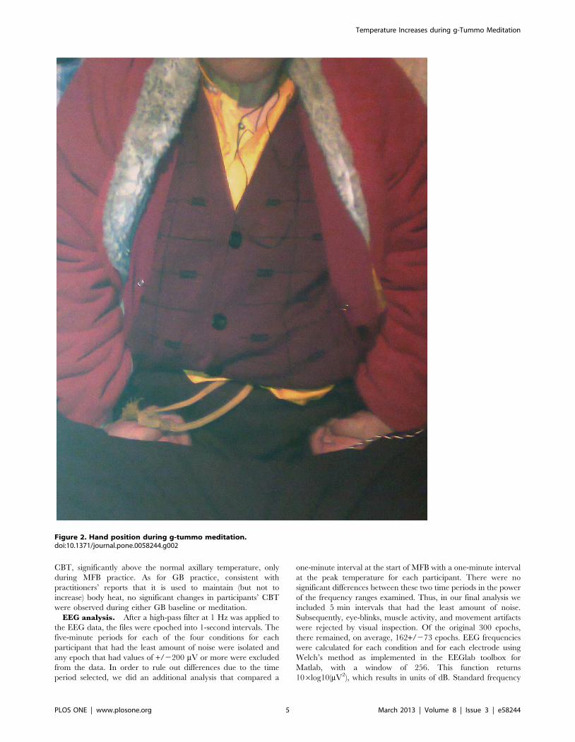
peaks of the peripheral temperature increases were associated with
changes in hand mudra positions (symbolic gestures used in
meditation), e.g., tensing the hand muscles as well as pressing the
fists against the inguinal crease (on the femoral artery) during
particular meditative periods (Figure 2). This suggests that the
peripheral temperature increases are primarily the result of
increased peripheral blood flow due to peripheral muscular action
(and proximity of the femoral artery) rather than a result of
psychological (caused by meditation) or physiological (caused by
breathing or isometric techniques) states. Since hand movements
constitute an integral part of g-tummo, it was impossible to
eliminate the effect of these factors on peripheral temperature
increases. Thus, in all further analyses we focus on core body
temperature (CBT) data from the armpit sensors only.
Table 1 presents the data for axillary temperature for each of
the participants at the beginning and at the end of each condition
(BFB, BGB, MFB, MGB) they performed. Parametric statistics
were used to analyze the temperature data despite the small
sample size used in the current study, since the temperature data
in the population fit a normal distribution [21]. The average initial
CBT of all the participants was 36.49uC (SD = 0.21), which is not
significantly different from the normal axillary temperature of
36.6uC in the healthy population [one sample two-tailed
t(9) = 1.71, p = 0.12]. The average temperature at the end of
BFB was 36.9uC (SD = 0.32), which is only marginally above the
normal axillary temperature: t(5) = 2.52, p = .053. During BFB, the
maximum CBT increase was 1.14uC (participant #3) and the
maximum temperature reached was 37.45uC (participant #4). By
the end of MFB, the average temperature increased to 37.6uC
Temperature Increases during g-Tummo Meditation
PLOS ONE | www.plosone.org 3 March 2013 | Volume 8 | Issue 3 | e58244

(SD = 0.52), which is significantly above the normal axillary
temperature [t(5) = 4.77, p = .005]. The maximum CBT increase
from the beginning of the experiment to the end of MFB was
2.2uC (participant #3), and the maximum temperature reached
was 38.30uC (participant #5). No elevation in CBT was observed
among the four participants who performed only the GB practice.
The final average CBT of those four participants after BGB and
MGB was 36.42uC (SD = 0.14).
Figure 3 shows the CBT increases for three representative
participants who completed the sequence of all four conditions
(BFB, BGB, MFB, and MGB).
During FB (either BFB or MFB), participants’ CBTs typically
exhibited a step-like pattern, with a period of steady temperature
increase followed by a plateau or equilibrium phase corresponding
to the ‘‘temperature saturation point’’, above which the partici-
pants were not able to raise their CBT further despite their efforts
to continue with FB. This pattern of CBT increases is very similar
to that usually observed during induction of systemic hyperthermia
(i.e., deliberate heating of a patient’s body to achieve an elevated
core temperature for therapeutic purposes), where the equilibrium
phase indicates the beginning of heat losses due to physiological
mechanisms (e.g. vasodilation, evaporation) limiting the rate of
heating that can be achieved, and thus protecting the body from
excessively high temperatures [22]. For example, one of the six
participants who performed FB reached the equilibrium phase
very quickly during the end of BFB (participant #4, Figure 3) and
despite her continuous efforts, she was not able to raise it further
during MFB, whereas two participants reached the equilibrium
only at the end of the MFB practice (e.g., participant #1). Three
other participants exhibited two equilibrium plateaus, one at the
end of BFB and another at the end of MFB (e.g., participant #3,
Figure 3), suggesting that the meditative visualization they
performed might have dampened the physiological mechanisms
leading to the heat losses.
To quantify the effective period of steady CBT increases for
those participants who exhibited temperature equilibrium phases
during either BFB or MFB, we defined the rise time (DTr) as the
time taken for the CBT to rise from 10% to 90% of its final value
(see Figure 4). This is similar to the definition of rise time in signal
control theory, describing the transition time for a signal that
changes from one level to another [23], [24]. For all the BFB and
MFB conditions where the participant CBT did not reach
equilibrium phase by the end of the condition, the rise time was
taken as time for the CBT to increase from 10% to 100%.
Furthermore, to quantify the rate of steady CBT increases, we
conducted linear regression analyses (CBT increases were
regressed on the rise time for BFB and MFB and on the overall
condition time for BGB and MGB) for each participant separately.
All linear regressions for each participant for each of the above
four time periods were significant (all R squares .0.88, all ps
,0.001). In order to account for possible variations between
participants, we treated regression coefficients as random vari-
ables, and used a linear mixed effect (MIXED) model to estimate
the population regression lines. The regression intercepts and
slopes d (representing the rate of axillary temperature change per
unit time) are presented in Table 2 for BFB, BGB, MFB, and
MGB separately. The regression slopes were significantly different
from zero for BFB and MFB (ps ,0.05) only. The variance of
intercept, the variance of slopes, and the covariance between the
intercepts and the slopes were non-significant.
In summary, the results suggest that although CBT increases
during BFB were not as dramatic as during MFB, participants
were able to produce body heat, utilizing only the somatic
component of the FB practice (breathing and isometric tech-
niques). However, the meditators were able to reach an elevated
Figure 1. Representative temperature data taken at armpit and left fourth finger of one of the Study 1 participants during BGB andMGB.doi:10.1371/journal.pone.0058244.g001
Temperature Increases during g-Tummo Meditation
PLOS ONE | www.plosone.org 4 March 2013 | Volume 8 | Issue 3 | e58244
CBT, significantly above the normal axillary temperature, only
during MFB practice. As for GB practice, consistent with
practitioners’ reports that it is used to maintain (but not to
increase) body heat, no significant changes in participants’ CBT
were observed during either GB baseline or meditation.
EEG analysis. After a high-pass filter at 1 Hz was applied to
the EEG data, the files were epoched into 1-second intervals. The
five-minute periods for each of the four conditions for each
participant that had the least amount of noise were isolated and
any epoch that had values of +/2200 mV or more were excluded
from the data. In order to rule out differences due to the time
period selected, we did an additional analysis that compared a
one-minute interval at the start of MFB with a one-minute interval
at the peak temperature for each participant. There were no
significant differences between these two time periods in the power
of the frequency ranges examined. Thus, in our final analysis we
included 5 min intervals that had the least amount of noise.
Subsequently, eye-blinks, muscle activity, and movement artifacts
were rejected by visual inspection. Of the original 300 epochs,
there remained, on average, 162+/273 epochs. EEG frequencies
were calculated for each condition and for each electrode using
Welch’s method as implemented in the EEGlab toolbox for
Matlab, with a window of 256. This function returns
106log10(mV2), which results in units of dB. Standard frequency
Figure 2. Hand position during g-tummo meditation.doi:10.1371/journal.pone.0058244.g002
Temperature Increases during g-Tummo Meditation
PLOS ONE | www.plosone.org 5 March 2013 | Volume 8 | Issue 3 | e58244

ranges were then defined: theta (4.5–7.5 Hz), alpha (8.5–12.5 Hz),
beta (13–25 Hz), and gamma (35–45 Hz), and power values
within those ranges were averaged. Oscillatory EEG data has been
shown to be normally distributed [25], and therefore standard
parametric tests were used.
The most pronounced differences in brain activity for medita-
tive as compared to baseline condition were observed for Forceful
Breath. There was a significant increase in the alpha power band
during FB meditation (BFBa = 3.88 dB, MFBa = 5.28 dB;
F(1,5) = 15.9, p = .01) revealing a dominantly parieto-occipital
topography. A significant power increase was also observed for
beta (BFBb = 0.44 dB, MFBb = 1.01 dB; F(1,5) = 20.59, p,.01) as
well as a strong tendency toward a significant power increase in
the gamma frequency band (MBFc = 22.06 dB,
BFBc = 22.83 dB; F(1,5) = 5.3, p = .06). Both beta and gamma
increases were most pronounced at lateral frontal sites. There was
no significant difference in theta frequency band between BFB and
MFB (BFBh = 28.04 dB, MFBh = 27.73 dB).
For Gentle Breath, only beta activity demonstrated a significant
increase in power between BGB and MGB (BGBb = 20.14 dB,
MGBb = 0.96 dB; F(1,7) = 7.87, p,.05). In contrast to FB, there
was no significant difference between BGB and MGB in alpha
(BGBa = 4.17 dB, MGBa = 5.51 dB; F(1,7) = 2.45, p = 0.16) or in
gamma power (MBFc = 23.41 dB, BFBc = 22.22 dB;
Table 1. Initial (t0) and final (t1) CBT for each of the four practices.
Participant BFB BGB MFB MGB
t0 t1 t0 t1 t0 t1 t0 t1
1 36.56 36.79 36.79 37.00 37.03 37.44 37.44 37.46
2 36.49 36.66 36.68 36.77 37.09 37.12 37.05 37.09
3 36.00 37.14 37.16 37.29 37.30 38.21 38.21 38.03
4 36.56 37.45 37.45 37.49 37.48 37.50 37.49 37.49
5 36.82 37.00 – – 37.07 38.30 – –
6 36.53 36.58 – – 36.86 37.12 – –
7 – – 36.54 36.46 – – 35.98 36.25
8 – – 36.49 36.49 – – 36.51 36.5
9 – – 36.56 36.56 – – 36.57 36.57
10 – – 36.31 36.36 – – 36.35 36.36
doi:10.1371/journal.pone.0058244.t001
Figure 3. CBT increases for Study 1 participants #1, #3, and #4 during BFB, BGB, MFB, and MGB performed in a continuoussequence. Since the duration of each of the four practices varied from participant to participant, to simplify the figure presentation, the duration ofeach practice is rescaled from 0 to 1, with t0 the starting point of each practice, and t1 the ending point.doi:10.1371/journal.pone.0058244.g003
Temperature Increases during g-Tummo Meditation
PLOS ONE | www.plosone.org 6 March 2013 | Volume 8 | Issue 3 | e58244

F(1,7) = 2.23, p = 0.18). Also, there was no difference in theta
power (BGBh = 24.05 dB, MGBh = 24.75 dB).
To investigate potential relationships of power in distinct
frequency bands and CBT increases, we correlated the increases
in alpha, beta, and gamma power during MFB over BFB with the
magnitude of the CBT increase during MFB. Interestingly, there
was a significant linear relationship (Figure 5) between the
increase in alpha power during MFB over BFB and the CBT
increases during MFB (R square = 0.82, p,0.01). None of the
other relationships were significant.
To investigate whether there was a similar relationship between
increases in alpha power and CBT increases during BFB, we
conducted an additional analysis that compared alpha power of
the practitioners at a one-minute interval at the start of BFB with a
one-minute interval at the end of BFB. There were no significant
differences between these two time periods in the alpha power:
F(1,5) = 1.95, p = 0.22, suggesting that, in contrast to MFB, CBT
increases during BFB are not related to the alpha band activity.
In summary, the results suggest that besides differences in CBT
changes, Forceful and Gentle Breath meditation are associated
with divergent brain state dynamics. Furthermore, the higher the
increases in alpha power developed by participants during FB
meditation, the larger their increases in CBT during FB
meditation, while the CBT increases during BFB were achieved
without any changes in alpha power. This suggests that different
mechanisms may be affecting CBT increases during MFB versus
BFB, and that meditative visualization characterized by significant
increases in alpha power might uniquely contribute to overall
CBT increases beyond the contribution of the vase breathing
technique. Further analyses were conducted to investigate different
factors contributing to CBT increases during BFB and MFB.
Factors contributing to CBT increases. The CBT increas-
es (Dt) during BFB and MFB could be described according to a
linear regression equation, as Dti < diDTri, where di represents the
regression coefficient (the rate of CBT change per unit time) and
DTri, represents the rise time of the CBT for condition i (mean ds
and DTrs for each condition i are given in Table 2). Further
analyses were conducted to investigate factors contributing to the
overall CBT increase (Dt) by affecting 1) the rate of CBT increase
(d), and 2) the CBT rise time (DTr).
First, we computed the average length of time each participant
held his/her breath (apnea duration) during BFB
[Mean = 1.58(0.52) min, range 1.30–2.50 min] and MFB
[Mean = 1.50(0.45) min, range 1.15–2.30 min], and correlated
the apnea duration during BFB and MFB with dBFB, dMFB,
DTBFB, and DTMFB. There was a significant positive correlation
between the apnea duration during BFB and dBFB (r = 0.91;
p,.05). There was also a trend toward a correlation between the
apnea duration during MFB and dMFB (r = 0.72; p = 0.1). All
other correlations were non-significant. This suggests that apnea
Figure 4. CBT rise time for Study 1 participant #4 during BFB. The rise time is taken as the time during which the CBT rises from 10% to 90%of its maximum value.doi:10.1371/journal.pone.0058244.g004
Table 2. Regression coefficients d, intercepts, duration DTand rise time DTr.
d 6C/min (SD)Intercept
6C(SD) DT min (SD) DTr min (SD)
BFB 0.146(0.072) 36.09(0.26) 5.91 (1.37) 5.71 (1.31)
BGB 0.011(0.008) 36.82(0.21) 6.37 (1.16) –
MFB 0.030(0.009) 37.02(0.32) 19.41 (15.69) 18.76 (15.14)
MGB 0.002(0.005) 37.02(0.43) 21.50 (13.10) –
doi:10.1371/journal.pone.0058244.t002
Temperature Increases during g-Tummo Meditation
PLOS ONE | www.plosone.org 7 March 2013 | Volume 8 | Issue 3 | e58244

duration, during which the practitioners hold their breath while
concurrently maintaining isometric muscle tension, is related to
the rate of the CBT increase, thus contributing to the overall CBT
increases during g-tummo practices.
Second, we correlated the increases in alpha power during MFB
over BFB with dBFB, dMFB, DTrMFB, and DTrMFB. There was
a significant linear relationship between the increase in alpha
power during MFB over BFB and the DTrMFB (r = 0.94, p,0.01);
that is, those who showed a greater increase in alpha power during
meditation were capable of prolonging their CBT rise time
without reaching equilibrium, thus reaching higher overall CBT
increases. None of other relationships were significant.
In addition, for those participants who performed FB, we
correlated their age (Mage = 44. 16 (SD = 5.49) y.o., range 38 251
y.o.) as well as their experience in g-tummo (Mexp = 12.
50(SD = 3.93) years, range 19–30 years) with their dBFB, dMFB,
DTrMFB, and DTrMFB. None of the relationships were
significant. However, the lack of correlations is not surprising,
taking into account that all the practitioners who performed FB
were of similar middle age and all of them had extensive g-tummo
experience, including at least three three-year g-tummo retreats.
In summary, our findings indicate that the two parameters,
apnea duration and increases in alpha power achieved during
meditative visualization are significant predictors of the overall
CBT increases during FB practice. The apnea duration is
significantly related to the rate of CBT increase. The increase in
alpha power developed during FB meditation is related to the
CBT rise time, that is, it predicts how long the meditators are
capable of sustaining their CBT increases without reaching
equilibrium.
Study 2The CBT changes for all the participants showed a similar
pattern, which involved 1) a 10 min period of thermometer
heating under the arm until it reflected an accurate axillary
temperature; 2) a baseline (rest) period of relatively constant CBT
until the beginning of BFB; 3) a steady CBT increase during BFB,
followed by a plateau (equilibrium) where the temperature was
maintained as long as the participant continued with BFB; and 3) a
cool-down phase to the baseline temperature during the final 20
minutes.
The CBT changes during baseline (rest) and BFB for two
representative participants are shown in Figure 6. Similar to
Study 1, in order to quantify the effective period of steady
temperature increase during BFB before equilibrium was reached
we used the rise time (DTr) as the time taken for the CBT to rise
from 10% to 90% of its final value. To quantify the rate of CBT
increase, we conducted linear regression analyses (CBT increase
was regressed on the overall rest time for baseline period and on
the rise time for the BFB practice) for each participant separately.
All linear regressions for each participant for each of the above
periods were significant (all R squares .0.92; ps ,0.001).
Furthermore, the linear mixed effect (MIXED) model was
conducted to estimate the population regression slopes represent-
ing the rate of axillary temperature changes. The estimated
regression slopes were d= 0.006uC/min for the baseline period
before BFB and d= 0.091uC/min for the period of BFB practice.
The regression slopes were significantly different from zero for the
BFB time period only (p,0.05).
Although the estimated regression slope was not significantly
different from zero for the baseline period (and in fact, 7
participants showed relatively constant CBT during the baseline,
such as participant #2, Fig. 6), there were four participants who
exhibited some noticeable gradual increase of their CBT during
the rest (e.g., participant #1, Fig. 6). The average rate of the CBT
increase for these four participants during this time period was
d= 0.009uC/min, which is comparable to the rate of CBT
increase of up to 0.5uC/hour due to CBT circhoral rhythmic
fluctuations [21], [26]. Thus, the observed increases during
baseline rest seem to reflect daily CBT oscillations. The rate of
these CBT increases, however, is 10 times less than the rate of
CBT increases observed during BFB practice.
Figure 5. CBT increases during FB meditation in Study 1as a function of alpha power increases during MFB over BFB (solid linerepresents regression).doi:10.1371/journal.pone.0058244.g005
Temperature Increases during g-Tummo Meditation
PLOS ONE | www.plosone.org 8 March 2013 | Volume 8 | Issue 3 | e58244

The increases in CBT during BFB were significant [paired-
samples, two-tailed t(10) = 3.196, p = 0.01]. The average initial
CBT of all the participants before BFB was 36.38uC (SD = 0.23),
while the average temperature at the end of BFB reached 36.99uC(SD = 0.13), only marginally above the normal axillary tempera-
ture of 36.6uC in the healthy population [one sample two-tailed
t(10) = 2.02, p = 0.07]. The maximum CBT reached during BFB
was 37.02uC. The average apnea duration of the participants was
30.38 sec (SD = 6.35), ranging from 19 to 41 sec; the apnea
duration correlated with the rate of CBT increase during BFB:
r = 0.60, p = 0.050. Independent sample t-tests (two-tailed) con-
ducted to compare the CBTs of the Western participants in Study
2 with those of 6 Tibetan meditators in Study 1 before and after
BFB revealed no significant differences between the two groups’
initial CBTs, t(15) = 0.85, p = 0.40 or their CBT at the end of the
BFB, t(15) = 0.39, p = 0.69. However, the final CBTs reached by
the Tibetan practitioners at the end of MFB were significantly
higher than the final CBT of the Western participants at the end of
BFB, t(15) = 3.92, p = 0.001.
The cool-down phase started immediately after completion of
BFB (average rate of cool-down is d= 0.034uC/min); all the
Western non-meditator participants returned to their baseline
CBT during the next 20 minutes. This is in contrast to Tibetan
practitioners performing GB (Study 1) who did not show any
decreases in their CBTs during either BGB or MGB (d= 0.011uC/
min and d= 0.002uC/min respectively), and were able to maintain
an elevated body temperature throughout the whole duration of
MGB (on average 21.50 min). This further confirms that the GB
practice facilitates maintenance of body temperature.
Figure 7A presents data from the Western participant who
performed BFB twice. As can be seen in the figure, the participant
raised her CBT to 36.96uC during the first BFB session, then
stopped when she felt uncomfortable, and after about 10 min rest
(accompanied by a gradual decrease in CBT), she started the
second session of BFB. However, she was not able to raise her
CBT any higher during the second BFB session, even though she
started the session at a higher baseline temperature. This contrasts
with the Western g-tummo practitioner who performed BFB
followed by MFB (Figure 7B). This participant reached a similar
CBT of 36.95uC by the end of the BFB session, but was able to
raise her CBT further during MFB up to the zone of slight fever
(37.03uC).
In summary, the results of Study 2 indicate that the BFB
technique brings about significant increases in CBTs not only in
meditators but also in those individuals who do not have any prior
experience in meditation. The rate of these increases is much
higher than the rate of the temperature increases due to CBT
circhoral rhythmic fluctuations. However, the overall CBT
increases due to the BFB technique are limited and in the range
of normal body temperature. The data further suggest that the
meditative visualization involved in MFB allows the practitioner to
reach CBTs higher than can be reached using breathing and
isometric techniques alone.
Discussion
The findings of our research indicate that there are two distinct
types of g-tummo practice, FB and GB, each characterized by
different temperature patterns and neural (EEG) correlates. The
temperature data bear out the practitioners’ claims that FB
meditation is used to increase body heat, while GB meditation
seems to facilitate the maintenance of an elevated body
temperature. Indeed, reliable CBT increases were observed during
the FB component of g-tummo practice, not only via FB
meditation but also via FB vase breathing alone. However, the
results of Studies 1 and 2 also suggest that that the neurocognitive
component (‘‘internalized attention’’ on visual images) of the MFB
practice may facilitate elevation in CBT beyond the range of
normal body temperature (into the fever zone), whereas the CBT
Figure 6. CBT increases during baseline and BFB in Study 2.doi:10.1371/journal.pone.0058244.g006
Temperature Increases during g-Tummo Meditation
PLOS ONE | www.plosone.org 9 March 2013 | Volume 8 | Issue 3 | e58244

increases during FB vase breathing alone were limited, and did not
exceed the range of normal body temperature.
The results of the EEG analysis also indicate that FB and GB
meditation were associated with different brain states in the
practitioners. The EEG data showed significant increases in alpha
and beta, and a marginally significant increase in gamma power
during MFB, whereas during MGB only beta increased. Although
the potential functional role of beta-band oscillations is not yet well
understood, beta activity is associated with focused executive
attention, and it seems to be related to the maintenance of one’s
current sensorimotor or cognitive state [27]. Thus, significant
increases in beta, as observed in the present research as well as in
the study by Benson et al [7] might correspond to enhancement of
attentional processes during both FB and GB meditation
compared to their corresponding baselines. As for the increased
gamma activity observed during FB meditation, studies on
meditation consider it a signature of ‘‘samadhi’’ (deep meditative
states of consciousness), but the regions of increase have varied,
with recent studies reporting in some cases a frontally distributed
increase in gamma [28], and in other cases an increase in gamma
at posterior and occipital electrodes [29]. Our data showed that
gamma had the highest power at frontal areas during the FB
meditation (however, the frontal distribution of gamma effects
should be considered with caution as these high frequency changes
might be confounded with activity of supraficial muscles and eye
movements [30].
Of particular interest is the increase in the power of the alpha
frequency band which was observed during FB meditation and its
significant relation to the CBT increases. Contrary to an early
view that alpha represented an idling process in the visual system,
evidence has begun to accumulate in support of a role for alpha
oscillations as a general inhibitory mechanism in the brain [31],
[32]. Specifically, increased alpha activity in occipito-parietal areas
has been linked to the successful suppression of visual input, which
could disturb the maintenance of visual working memory
representations [33], [34]. This is consistent with a more general
observation that an inward shift of attentional focus toward mental
activity (as in mental rotation or other visual-spatial imagery tasks)
is typically accompanied by increases in posterior alpha power
[35], [36], suggesting that alpha might be working to decrease the
distractibility of ‘‘external’’ sensory events to aid concentration on
the mental activity. Similarly, meditation research has suggested
that increases in alpha power correspond to ‘‘internalization of
attention’’ (internally directed attention) during task performance
as compared to baseline [37], which seems to be a plausible
interpretation of our results. During FB meditation, practitioners
are supposed to direct their attention to internally generated
sensory information (visual imagery, heat perception). In contrast,
there were no significant increases in alpha power during vase
breathing alone, suggesting that these increases could be
specifically attributed to the meditative component of the practice.
We suggest that the increase in alpha power observed in our study
reflects the meditators’ ability to focus attention on relevant
internal states, specifically on visual images of flames and
sensations of heat, while inhibiting external sensory information
for extended periods.
The central finding of our study is that there are at least two
separate factors that affect CBT increases during FB meditation.
The first factor is related to the somatic component of the FB
practice, specifically to the effectiveness of the vase breathing
technique that affects the rate of CBT increase. Indeed, the results
of studies 1 and 2 showed a significant correlation between the rate
of CBT increases and apnea duration, during which the
practitioners hold their breath while concurrently maintaining
isometric muscle tension. The rate of these CBT increases was
significantly higher than that due to circhoral rhythmic fluctua-
tions in CBT, and it was similar to the rate of CBT increases of
0.02–0.17uC per min during the use of noninvasive systemic
hyperthermia methodologies (e.g., immersing a patient’s body in
hot water or wax, wrapping the body in a blanket or suit through
which heated water is pumped, irradiating with IR energy) [22].
The second factor is related to the neurocognitive component of
the FB practice, specifically to the amount of ‘‘internalization of
attention’’ or quality of meditative visualization, as reflected by
increased alpha power during FB meditation. This factor seems to
determine the rise time of CBT, that is, how long the meditators are
capable of continuing to raise their CBT beyond the range of
normal body temperature without reaching an equilibrium phase.
Indeed, the results of Study 1 showed that the greater the increase
in alpha power achieved during FB meditation, the longer the
CBT rise time, leading to higher CBTs.
Both factors work in conjunction to maximize the temperature
increase. That is, the FB somatic component (vase breathing)
causes thermogenic effects, while the neurocognitive component
(meditative visualization) seems to be key for facilitating a
sustained increase in body temperature for longer periods, possibly
due to mitigating physiological mechanisms leading to heat loss. In
systemic hyperthermia treatments, to prevent heat loss, and thus
sustain CBT increases for longer periods, different insulation
techniques are used (e.g., wrapping a patient body in reflective
blankets, foil, or plastic films [22]). In the case of FB meditation,
one of the possible mechanisms preventing heat loss could be the
mental imagery of flames and heat. Indeed, previous research has
Figure 7. CBT increases during FB as performed by a Westernnon-meditator and a Western g-tummo practitioner. The verticaldashed lines indicate the beginning/end of different phases of thepractice; (A, Western non-meditator): rest, BFB, rest, BFB, rest, and (B,Western g-tummo pract itioner): rest, BFB, rest, MFB, rest.doi:10.1371/journal.pone.0058244.g007
Temperature Increases during g-Tummo Meditation
PLOS ONE | www.plosone.org 10 March 2013 | Volume 8 | Issue 3 | e58244

regarded mental imagery as a potentially effective technique in
influencing peripheral body temperature, blood flow, and local
vasodilation [38–41]. Thus, it is possible that the mental imagery
component of FB meditation minimizes heat loss, and thus
prolongs the CBT rise time by similar mechanisms (changes in
blood flow, reduced vasodilation). Without the accompanying
meditative visualization, vase breathing might not be very effective
and result in only limited CBT increases. At the same time,
without an effective FB breathing technique, even small CBT
increases, if possible at all, might require significantly longer
meditation periods.
One of the questions arising from this study is why breathing
and isometric exercises have been chosen by Tibetan meditators as
a mean of thermogenesis, instead of dynamic movements (e.g.,
physical exercising, running). Different types of breathing and
isometric techniques have been used for thousand years not only in
Tibetan traditions but also in static holds in certain branches of
yoga or oriental martial arts. What is common to all these
practices is that they require focused attention on the internal
mental states, which is difficult to do during rigorous dynamic
exercise. Indeed, according to the g-tummo practitioners’ reports,
FB meditation requires significant concentration, and cannot be
performed in conjunction with running or even walking. For these
reasons, during the ceremony of drying wet sheets, while walking
outside, g-tummo meditators do not perform FB, but GB
meditative visualization, which is much less effortful.
A limitation affecting the generalizability our findings is the
small sample size due to the sacredness of the practice and
difficulties in accessing g-tummo practitioners. Despite this
limitation, we were able, for the first time, to document reliable
CBT increases during the FB type of g-tummo practice, all within
the slight to moderate fever zone, validating the legends of the
extraordinary capacity of g-tummo meditators to elevate their
body temperature beyond normal. However, the results also
suggest that temperature increases during g-tummo meditation are
neither solely a by-product of meditation nor its goal, but instead
may be a means to facilitate the achievement of ‘‘deep meditative
states’’. The g-tummo meditators may use the CBT increases as a
vehicle to enhance their attention and focus their meditative
performance (which may in turn facilitate a further increase in
their temperature through meditative visualization). Future studies
of experts in g-tummo meditation who are capable of elevating
and maintaining elevated CBT may offer promising research
insights and approaches to investigating mechanisms of CBT
regulation. Because many variables underlying neuronal function-
ing (e.g., transport via ion-selective channels, amplitude and
duration of single-unit spikes) are temperature-dependent [42],
[43], possibility of self-regulation of CBT may have a direct effect
on self-regulating and maximizing neurocognitive activity. If
future studies show that it is possible to self regulate CBT, by
mastering vase breathing in conjunction with guided mental
imagery without extensive meditation experience, it will open a
wide range of possible medical and behavior interventions, such as
adapting to and functioning in hostile (cold) environments,
improving resistance to infections, boosting cognitive performance
by speeding response time, and reducing performance problems
associated with decreased body temperature as reported in human
factor studies of shift work and continuous night operations [44],
[45].
Acknowledgments
We thank Gebchak Wangdrak Rinpoche for his help in participant
recruitment and all the arrangements for the study and to Ani Chozom for
her English-Tibetan translation and help in running experiments. We also
want to thank Geshe Gelek Jinpa for his consultation on the g-tummo
practice. Finally, special thanks to Anna Wrinkler who helped to recruit
Western participants for Study 2 as well as to the team of Advanced Brain
Monitoring, who provided constant consultation during the EEG studies to
resolve difficulties in running the equipment in unusually cold weather
conditions.
Author Contributions
Conceived and designed the experiments: MK. Performed the experi-
ments: MK. Analyzed the data: JE JS KG. Wrote the paper: MK.
References
1. Evans-Wentz WY (2002) Tibetan Yoga and Secret Doctrines (Pilgrims
Publishing, Varanisa, India).
2. Mullin GH (1997) Readings on Six Yogas of Naropa (Snow Lion Publication,
Ithaca, NY).
3. Mullin GH (1996) Tsongkhapa’s Six Yogas of Naropa (Snow Lion Publication,
Ithaca, NY).
4. David-Neel A (1971) Magic and Mystery in Tibet (Dover Publications, New
York).
5. Govinda Lama Anagarika (1988) Way of White Clouds (Shambhala Publica-
tions,).
6. Benson H, Lehmann JW, Malhotra MS, Goodman RF, Hopkins J, et al. (1982)
Body temperature changes during the practice of g-tummo yoga. Nature 295:
234–236.
7. Benson H, Malhotra MS, Goldman RF, Jacobs GD, Hopkins PJ (1990) Three
case reports of the metabolic and electroencephalographic changes during
advanced Buddhist meditation techniques. Behav. Med 16: 90–95.
8. Cromie WJ (2002) Meditation changes temperatures: Mind controls body in
extreme experiments. Harvard University Gazette.
9. Bird EI, Colborne GR (1980) Rehabilitation of an electrical burn patient
through thermal biofeedback. Biofeedback Self. Regul 5: 283–288.
10. Willerman L, Skeen JT, Simpson JS (1976) Retention of learned temperature
changes during problem solving. Percept. Mot. Skills 43: 995–1002.
11. Taub E, Emurian CS (1976) Feedback-aided self-regulation of skin temperature
with a single feedback locus: I. Acquisition and reversal training. Biofeedback
Self. Regul I: 147–168.
12. King NJ, Montgomery RM (1981) The self-control of human peripheral (finger)
temperature: An exploration of somatic manoeuvres as aids to biofeedback
training. Behav. Ther 12: 263–27.
13. Singh V, Aballay A (2006) Heat-shock transcription factor (HSF)-1 pathway
required for Caenorhabditis elegans immunity. Proc Natl Acad Sci USA 103:
13092–13097.
14. Kluger MJ, Kozak W, Conn CA, Leon LR, Soszynski D (1996) The adaptivevalue of fever. Infect. Dis. Clin. North. Am 10: 1–20.
15. Mackowiak PA (1994) Fever: Blessing or Curse? A Unifying Hypothesis Ann.
Intern. Med June 15: 1037–1040.
16. Masino SA, Dunwiddie TV (2000) A transient increase in temperature induces
persistent potentiation of synaptic transmission in rat hippocampal slices.
Neuroscience 101: 907–912.
17. Kleitman N, Jackson DP (1950) Body temperature and performance under
different routines. J. Appl. Physiol 3: 309–328.
18. Holland RL, Sayer JA, Keatinge WR, Davis HM, Peswani R (1985) Effects ofraised body temperature on reasoning, memory, and mood. J. Appl. Physiol 59:
1823–1827.
19. Wright KP Jr, Hull JT, Czeisler CA (2002). Relationship between alertness,performance and body temperature in humans. Am. J. Physiol. Regul. Integr.
Comp. Physiol 283: R1370–R1377.
20. Kelly G (2006) Body temperature variability (Part 1): a review of the history ofbody temperature and its variability due to site selection, biological rhythms,
fitness, and aging. Altern. Med. Rev 11: 278–93.
21. Mackowiak PA, Wasserman SS, Levine MM (1992) A critical appraisal of 98.6degrees F, the upper limit of the normal body temperature, and other legacies of
Carl Reinhold August Wunderlick. J. Am. Med. Assoc 268: 1578–1580.
22. Rowe-Horwege RW (2006) Hyperthermia, Systemic. Encyclopedia of MedicalDevices and Instrumentation.
23. Levine WS (1996) The control handbook. Boca Raton, FL: CRC Press.
24. Nise NS (2008) Control Systems Engineering (Fifth ed). John Wiley & Sons.
25. Kiebel SJ, Tallon-Baudry C, Friston KJ (2005) Parametric analysis of oscillatoryactivity as measured with EEG/MEG. Hum. Brain Mapp 26: 170–177.
26. Lindalwy G, Dowse HB, Burgoon PW, Kolka MA, Stephenson LA (1999)
Persistent circhoral ultradian rhythm is identified in human core temperature.Chronobio. Internat.16: 69–78.
27. Engel AK, Fries P (2010) Beta-band oscillations – signalling the status quo. Curr.Opin. Neurobiol 20: 156–165.
Temperature Increases during g-Tummo Meditation
PLOS ONE | www.plosone.org 11 March 2013 | Volume 8 | Issue 3 | e58244

28. Lutz A, Greischar LL, Rawlings NB, Ricard M, Davidson RJ (2004) Long-term
meditators self-induce high-amplitude gamma synchrony during mentalpractice. Proc. Natl. Acad. Sci USA 101: 16369–16373.
29. Cahn BR, Delorme A, Polich J (2010) Occipital gamma activation during
Vipassana meditation. Cogn. Process 11: 39–56.30. Whitham EM, Pope KJ, Fitzgibbon SP, Lewis T, Clark CR, et al. (2007) Scalp-
electrical recording during paralysis: quantitative evidence that EEG frequenciesabove 2-Hz are contaminated by EMG. Clin. Neurophysiol 118: 1877–1888.
31. Klimesch W, Sauseng P, Hanslmayr S (2007) EEG alpha oscillations: the
inhibition-timing hypothesis. Brain Res. Rev 53: 63–88.32. Jensen O, Mazaheri A (2010) Shaping functional architecture by oscillatory
alpha activity: gating by inhibition. Front. Hum. Neurosci 4: 186.33. Worden MS, Foxe JJ, Wang N, Simpson GV (2000) Anticipatory biasing of
visuospatial attention indexed by retinotopically specific alpha-band electroen-cephalography increases over occipital cortex. J Neurosci 20:RC63.
34. Sauseng P, Klimesch W, Gerloff C, Hummel FC (2009) Spontaneous locally
restricted EEG alpha activity determines cortical excitability in the motor cortex.Neuropsychologia 47: 284–288.
35. Jensen O, Gelfand J, Kounios J, Lisman JE (2002) Oscillations in the alpha band(9–12 Hz) increase with memory load during retention in a short-term memory
task. Cereb. Cortex 12: 877–882.
36. Cooper NR, Croft RJ, Dominey SJJ, Burgess AP, Gruzelier JH (2003) Paradoxlost? Exploring the role of alpha oscillations during externally vs. internally
directed attention and the implications for idling and inhibition hypotheses. Int.J. Psychophysiol 47: 65–74.
37. Aftanas LI, Golocheikine SA (2001) Human anterior and frontal midline theta
and lower alpha reflect emotionally positive state and internalized attention:
High resolution EEG investigation of meditation. Neurosci. Lett 310: 57–60.
38. McGuik J, Fitzgerald D, Firedman PS, Oakley D, Salmon P (1998) The effect of
guided imagery in a hypnotic context on forearm blood flow. Contemp Hypn
15: 101–108.
39. Lee LH, Olness K (1996) Effect of self-induced mental imagery on autonomic
reactivity in children. J Dev Behav Pediatr 17: 323–327.
40. Serra D, Parris CR, Carper E, Homel P, Fleishman SB, et al. (2012) Outcomes
of guided imagery in patients receiving radiation therapy for breast cancer.
Clin J Oncol Nurs: 16: 617–23.
41. Moore LE, Wiesner SL (1996) Hypnotically induced vasodilation in the
treatment of repetitive strain injuries. Am J Clin Hypn: 39: 97–104.
42. Thompson SM, Musakawa LM, Rince DA (1985). Temperature dependence of
intrinsic membranes properties and synaptic potentials in hippocampal CA1
neurons in vitro. J. Neurosc 5: 817–824.
43. Erickson CA, Jung MW, McNaughton BL, Barnes CA (1996). Contribution of
single-unit spike waveform changes to temperature induced alterations in
hippocampal population spikes. Exp. Brain. Res 107: 348–360.
44. Colquhoun WP, Folkard S (1985) in Hours of Work: Temporal Factors in Work-
Scheduling, eds Folkard S, Monk TH (Wiley, New York), 253–261.
45. Campbell SS (1995) Effects of timed bright-light exposure on shift-work
adaptation in middle-aged subjects. Sleep 18: 408–416.
Temperature Increases during g-Tummo Meditation
PLOS ONE | www.plosone.org 12 March 2013 | Volume 8 | Issue 3 | e58244