neurobiologyofdisease ... · semble, exhibit lewy bodies and degenerate in pd (marras and...
TRANSCRIPT

Neurobiology of Disease
Rapamycin Protects against Neuron Death in In Vitro andIn Vivo Models of Parkinson’s Disease
Cristina Malagelada,1 Zong Hao Jin,1 Vernice Jackson-Lewis,2 Serge Przedborski,1,2,3 and Lloyd A. Greene1,3
Departments of 1Pathology and Cell Biology and 2Neurology and the 3Center for Motor Neuron Biology and Disease, Columbia University, New York,New York 10032
We report that rapamycin, an allosteric inhibitor of certain but not all actions of the key cellular kinase mammalian target of rapamycin(mTOR), protects neurons from death in both cellular and animal toxin models of Parkinson’s disease (PD). This protective actionappears to be attributable to blocked translation of RTP801/REDD1/Ddit4, a protein that is induced in cell and animal models of PD andin affected neurons of PD patients and that causes neuron death by leading to dephosphorylation of the survival kinase Akt. In support ofthis mechanism, in PD models, rapamycin spares phosphorylation of Akt at a site critical for maintenance of its survival-promotingactivity. The capacity of rapamycin to provide neuroprotection in PD models appears to arise from its selective suppression of some butnot all actions of mTOR, as indicated by the contrasting finding that Torin1, a full catalytic mTOR inhibitor, is not protective and inducesAkt dephosphorylation and neuron death.
IntroductionParkinson’s disease (PD) is characterized by neurodegenerationof specific populations of central and peripheral neurons, includ-ing those in the substantia nigra pars compacta (SNpc) andsympathetic ganglia (Fahn, 1998; Dauer and Przedborski, 2003;Marras and Lang, 2008). Current treatments principally amelio-rate clinical manifestations of the disease rather than the under-lying neuron degeneration and death. This reflects an incompleteunderstanding of the pathways that lead to neuron loss in PD(Levy et al., 2009).
A SAGE screen (Ryu et al., 2005) designed to uncover novelparticipants (and potential therapeutic targets) in PD-relatedneuron death revealed RTP801 (REDD1; Ddit4) as a gene that ishighly induced in a cellular toxin model of PD. RTP801 proteinand transcripts are induced by a variety of stresses relevant to PD,including oxidative stress, DNA damage, mitochondrial dys-function, and endoplasmic reticulum stress (Ellisen et al., 2002;Shoshani et al., 2002; Wang et al., 2003; Schwarzer et al., 2005) as wellas in various cellular PD toxin models (Malagelada et al., 2006). Inaddition, RTP801 is induced in dopaminergic SN neurons in amouse 1-methyl-4-phenyl-1,2,3,6-tetrahydropyridine (MPTP)model of PD and in dopaminergic SN neurons of PD patients(Malagelada et al., 2006).
RTP801 has variable effects on survival of non-neuronalcells but induces apoptotic death of all neuronal types tested todate (Shoshani et al., 2002; Malagelada et al., 2006, 2008). Signifi-cantly, RTP801 knockdown protects from death in cellular modelsof PD (Malagelada et al., 2006). RTP801 promotes neuron death bysuppressing activation of mammalian target of rapamycin (mTOR)(Malagelada et al., 2006), a kinase that regulates key cellular func-tions (Fingar and Blenis, 2004; Swiech et al., 2008). This mTORblockade in turn suppresses activation of Akt, a protein kinase with amajor role in maintaining neuron survival (Dudek et al., 1997;Franke et al., 1997a,b), and it is this action that appears to underlie, atleast in part, the proapoptotic actions of PD toxins and RTP801(Malagelada et al., 2008). In consonance with this mechanism, SNneurons of PD patients possess substantially lower levels of phos-phorylated (but not total) Akt (Malagelada et al., 2008).
Rapamycin is a macrolide that specifically inhibits certainmTOR actions by allosterically modulating access of a subset ofsubstrates to the mTOR catalytic site (Sabers et al., 1995; Snyderet al., 1998; Pong and Zaleska, 2003). Thus, in contrast toRTP801, which apparently inhibits all mTOR actions by sup-pressing its activation by the Rheb GTPase (Brugarolas et al.,2004; Sofer et al., 2005), rapamycin blocks only a subset of mTORactivities (Lorenz and Heitman, 1995; Choo et al., 2008; Thoreenet al., 2009). One relevant consequence is that rapamycin sup-presses mTOR-dependent translation of some classes of mRNAsbut not others (Choo et al., 2008).
Here, we report that rapamycin blocks RTP801 induction inboth in vitro and in vivo toxin models of PD. This appears tooccur at the level of mTOR-controlled translation. Consistentwith the role of RTP801 in mediating neuron death, rapamycin isalso protective in such models. The contrasting actions ofRTP801 and rapamycin in neuron death and survival appear tobe attributable to their differential actions on mTOR and onregulating Akt phosphorylation.
Received Aug. 12, 2009; revised Oct. 30, 2009; accepted Nov. 30, 2009.This work was supported in part by Department of Defense Grants DAMD 17-03-1, W81XWH-08-1-0465, and
W81XWH-08-1-0522, National Institutes of Health/National Institute of Neurological Disorders and Stroke GrantsAG21617, NS062180, NS064191, NS11766, NS38370, and NS042269, the Parkinson’s Disease Foundation, theThomas Hartman Foundation, Muscular Dystrophy Association/Wings Over Wall Street, and the American Parkin-son’s Disease Foundation. We thank Dr. Delphine Prou and Luís Parada for helpful advice, Dr. Gary Chiang for mTORconstructs, and Dr. D.M. Sabatini for providing Torin1.
Correspondence should be addressed to Cristina Malagelada, Department of Pathology and Cell Biology, Colum-bia University, College of Physicians and Surgeons, 630 West 168th Street, New York, NY 10032. E-mail:[email protected].
DOI:10.1523/JNEUROSCI.3944-09.2010Copyright © 2010 the authors 0270-6474/10/301166-10$15.00/0
1166 • The Journal of Neuroscience, January 20, 2010 • 30(3):1166 –1175

Materials and MethodsAntibodies, plasmids, and materialsAnti-RTP801 antiserum was purchased from Millipore Bioscience Re-search Reagents or from Proteintech Group. Anti-ERK1 antibody wasobtained from Santa Cruz Biotechnology. Anti-horseradish peroxidasesecondary antibodies were obtained from Pierce. Antibodies againstphospho-Ser473–Akt, phospho-Thr308 –Akt, total Akt, phospho-Ser2448 –mTOR, phospho-Ser235/236 –S6, phospho-Thr37/46 – 4EBP1,phospho-Ser371–p70S6kinase, and p70S6kinase were obtained fromCell Signaling Technology. Tyrosine hydroxylase (TH) polyclonal anti-body was purchased from Calbiochem EMD Biosciences. Donkey anti-rabbit or anti-mouse secondary antibodies conjugated with Alexa 488 orAlexa 568 were purchased from Invitrogen.
RTP801 constructs were generated as described previously (Malagelada etal., 2006). All newly made constructs were verified by DNA sequencing.6-OHDA was purchased from Sigma or Tocris Bioscience. Rapamycinwas purchased from Calbiochem or LC Laboratories for cell culturetreatment or in vivo experiments, respectively. Cycloheximide was pur-chased from Calbiochem EMD Biosciences. FK506 was purchased fromAxxora. pcDNA3 wild-type (WT) mTOR was a kind gift from Dr. GaryG. Chiang (Burnham Institute for Medical Research, La Jolla, CA)(Chiang and Abraham, 2005). Torin1 was a kind gift from Dr. D. M.Sabatini (Harvard University, Cambridge, MA).
Cell culturePC12 cells were cultured and treated with nerve growth factor (NGF) asdescribed previously (Greene and Tischler, 1976) For NGF treatment,the cells were cultured in RPMI 1640 medium (Cellgro) supplementedwith 1% horse serum, penicillin/streptomycin, and 50 ng/ml recombi-nant human NGF (a kind gift from Genentech) for 8 –10 d. Medium waschanged every other day and immediately before treatments. 6-OHDAand 1-methyl-4-phenylpyridinium (MPP�) were prepared before use in10 and 100 mM stocks, respectively, and diluted in medium to the indi-cated final concentrations. Neonatal rat superior cervical ganglion sym-pathetic neurons were cultured as described previously (Ryu et al., 2002;Malagelada et al., 2006, 2008). Treatments with 6-OHDA were per-formed at day 7 in vitro, and cell viability was assessed 24 h later asreported previously (Malagelada et al., 2006). Values represent themean � SEM of at least three different experiments. Evaluation of pro-portions of neurite-bearing cells was performed as described previously(Greene and Tischler, 1976) by counting the proportion of cells that havethe neurites at least twice as long as the diameter of their soma.
Quantitative reverse transcription-PCREach sample of total RNA was isolated from neuronal PC12 cells by usingTRI reagent (Molecular Research Center). cDNA was transcribed fromtotal RNA with First-Strand cDNA Synthesis for Quantitative RT-PCRkit (Marlingen Biosciences). The primers used for quantitative-PCR am-plification of RTP801 were 5�-GCTCTGGACCCCAGTCTAGT-3� and5�-GGGACAGTCCTTCAGTCCTT-3�.
Equal amounts of cDNA template were used for each quantitative PCRanalysis of RTP801 and A-tubulin. Quantitative PCR was performedusing a Cepheid SmartCycler following the specifications of the manu-facturer. A-tubulin was used for normalization of RTP801 transcripts.cDNA was added to a 25 �l volume reaction mix containing Ready-to-Go Beads (GE Healthcare) or OmniMix HS master mix (Cepheid) andSYBR Green I (Invitrogen) together with appropriate primers at 0.2 �M each.Analyses of amplification curves of real-time fluorescence and of meltingcurves were performed as described previously (Troy et al., 2001).
RTP801 half-lifeCultures of neuronal PC12 cells were treated with 1 �M rapamycin for 1 hand then with 1 mM MPP� for 8 h in the continued presence of rapamy-cin. The cultures were then treated with 1 �M cycloheximide for 0, 5, or10 min and then harvested and subjected to Western immunoblotting forRTP801 and ERK1. The relative optical densities of the RTP801 signalswere determined and normalized to that for ERK1. There were no evidenteffects of cycloheximide on ERK1 expression levels over the 5–10 min courseof the experiment. The half-life of RTP801 protein under each condition was
calculated from the slope derived by plotting the natural log of values forrelative RTP801 expression versus time of cycloheximide treatment and fit-ting the data to a straight line using least-squares analysis.
TransfectionNeuronal PC12 cells were transfected with Lipofectamine 2000 (Invitro-gen) according to the instructions of the manufacturer. At 48 h aftertransfection, cells were treated with 6-OHDA or MPP�. At 24 h after thetreatments, viable transfected (as judged by expression of fluorophore)neuronal PC12 cells were scored by strip counting under an epifluores-cence microscope (Malagelada et al., 2006).
In vivo MPTP treatments in miceRapamycin preparation. Rapamycin (LC Laboratories) was dissolved in0.2 ml of 100% ethanol and then diluted 100-fold with 40% propyleneg-lycol to obtain a final concentration of 0.75 mg/ml (Zhou et al., 2009).
Animals and MPTP regimens. Ten-week-old, male, C57BL mice(Charles River Laboratories) were divided into four groups of 3–10 mice(MPTP/rapamycin, MPTP/vehicle, saline/rapamycin, and saline/vehi-cle) and were subjected to either an acute or a subacute MPTP regimen.
Acute regimen. For additional details, see the study by Jackson-Lewis etal. (1995). Animals from each group received one intraperitoneal injec-tion of MPTP-HCl (18 mg/kg free base suspended in saline; Sigma-Aldrich) or saline every 2 h for a total of four doses over an 8 h period in1 d and were killed at the indicated time points after the last MPTP/salineinjection. Rapamycin (7.5 mg/kg) (Zhou et al., 2009) or vehicle wasadministered intraperitoneally daily for 2 d before the first MPTP/salineinjection and continued for 4 d after the last MPTP/saline injection. Onthe day of the MPTP/saline injections, rapamycin/vehicle was adminis-trated 30 min before first MPTP/saline injection.
Subacute regimen. For additional details, see the study by Tatton andKish (1997). Animals from each group received one intraperitoneal in-jection of MPTP-HCl daily (30 mg/kg free base in saline) for 5 d and werekilled at the indicated time points after the last MPTP injection. Rapa-mycin/vehicle was administered intraperitoneally daily, starting 2 d be-fore the first MPTP/saline injection and continuing for 4 d after the lastMPTP/saline injection. Rapamycin/vehicle was administrated 30 minbefore each MPTP/saline injection.
Quantification of dopamine neurodegeneration. The total numbers ofTH-positive (TH �) SNpc neurons were determined in the acute modelat 7 d after the last MPTP or vehicle injection by unbiased stereologyusing the optical fractionator method (StereoInvestigator; MBF Bio-science). Quantification of the numbers of condensed chromatin SN cellsin the subacute model were assessed 2 d after the last MPTP injection inthe subacute regimen as described previously (Vila et al., 2001).
Western blot analysis. Whole-cell extracts or mouse midbrain extractswere analyzed as described previously (Malagelada et al., 2006).
StatisticsAll experiments were performed at least in triplicate, and results arereported as means � SEM. Student’s t test was performed as unpaired,two-tailed sets of arrays and presented as probability ( p) values. ANOVAwith Bonferroni’s multiple comparison test or the Newman–Keuls testwas performed to compare multiple groups with the control group.
ResultsmTOR protects against 6-OHDA-induced deathOur past studies have indicated that PD toxins induce RTP801and that RTP801 in turn causes neuron death by suppressingactivation of mTOR (Malagelada et al., 2006, 2008). If this mech-anism is correct, then restoring mTOR activity should preventneuronal death induced by a PD-mimicking toxin such as6-OHDA. Because 6-OHDA reduces but does not completelyblock mTOR activity (Malagelada et al., 2006), we reasoned thatWT mTOR overexpression should provide a sufficient level of theactivated kinase to confer neuroprotection. Accordingly, overex-pression in neuronally differentiated PC12 cells of WT mTOR(Edinger et al., 2003) protected from 6-OHDA (Fig. 1a). Con-
Malagelada et al. • Rapamycin Is Protective in Parkinson’s Disease Models J. Neurosci., January 20, 2010 • 30(3):1166 –1175 • 1167

versely, overexpression of a kinase-inactive form of mTOR failed to rescuethe cells from 6-OHDA (data not shown).Immunostaining for phosphorylatedepitopes that are targets of mTOR or of itstarget, p70S6 kinase, confirmed that over-expression maintained mTOR kinase ac-tivity in presence of 6-OHDA (Fig. 1b–d).
To further assess the role of mTOR inneuronal survival, we treated cultureswith 250 nM Torin1, a highly selectiveATP-competitive inhibitor of all mTORactivities (Thoreen et al., 2009). This in-duced death of 22 � 4% of the cells by 24 h(**p � 0.01 vs control untreated cells; re-sult represents mean � SEM of three in-dependent experiments, done at least intriplicate). Immunoblot analysis ofmTOR-dependent phosphorylations at5 h of Torin1 treatment confirmed inhibi-tion of mTOR activity at this time (Fig.1e). However, there was no inhibition at24 h (data not shown), suggesting thatTorin1 was either ineffective or unstableafter more prolonged times. This may ex-plain why Torin1 was not more effectivein inducing death. Together, these find-ings indicate that activated mTOR pro-vides neuroprotection, whereas mTORinhibition induces neuronal death.
Rapamycin blocks RTP801 inductionby PD toxinsIf mTOR activity is required for neuron sur-vival and this is inhibited by RTP801, thenstrategies that suppress RTP801 inductionshould be neuroprotective. Because thereare often reciprocal control pathways be-tween mTOR and its regulators (Huang andManning, 2009), we assessed whether par-tial inhibition of mTOR function might af-fect RTP801 expression. We therefore testedthe effect of rapamycin on RTP801 induc-tion in 6-OHDA-treated PC12 cell cultures.As shown in Figure 2a, we found inhibitionat 0.1 and 1 �M (but not 20 nM) rapamycin. Western immunoblot-ting confirmed that phosphorylation of p70S6 kinase at Ser371,which is mTOR dependent and rapamycin sensitive (Burnett et al.,1998), was also suppressed (Fig. 2a,b). Finally, rapamycin alsoblocked RTP801 induction in neuronal PC12 cells by a second PD-mimetic toxin, MPP� (Fig. 2c).
Sympathetic neurons, which neuronal PC12 cells closely re-semble, exhibit Lewy bodies and degenerate in PD (Marras andLang, 2008; Orimo et al., 2008). Significantly, RTP801 is inducedand mediates death of 6-OHDA-treated cultured sympatheticneurons (Malagelada et al., 2006, 2008). Rapamycin also sup-presses RTP801 induction in these neurons (Fig. 2d).
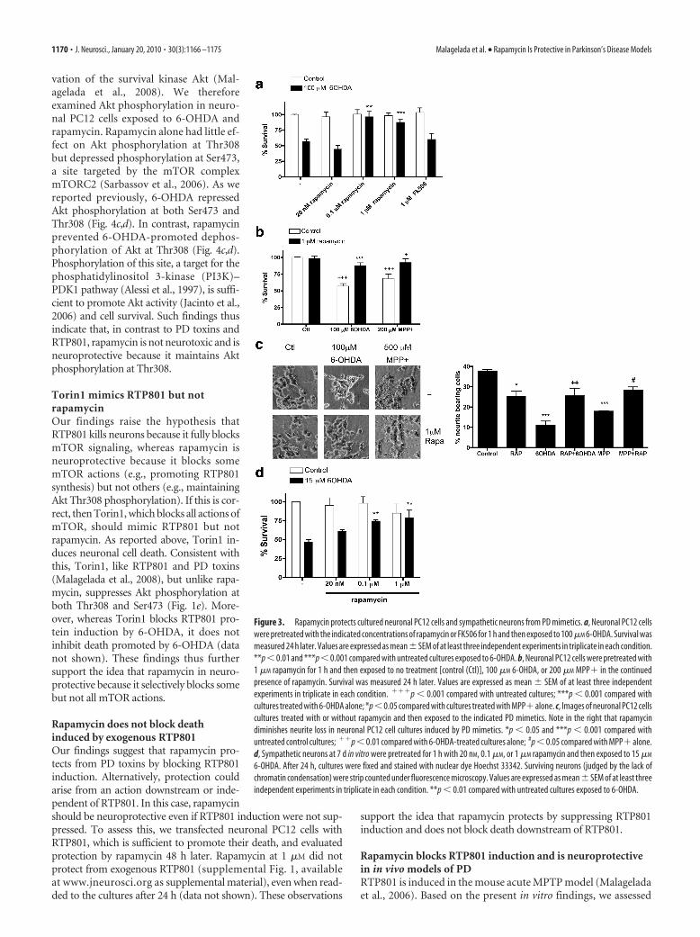
Rapamycin protects from PD toxinsBecause RTP801 mediates death of neuronal PC12 cells and sympa-thetic neurons triggered by PD toxins, we next assessed whetherrapamycin, which suppresses RTP801 induction, is protective. Inparallel with effects on RTP801 induction, 100 nM and 1 �M, but not
20 nM, rapamycin protected neuronal PC12 cells from 6-OHDA(Figs. 1a, 3a). Rapamycin also effectively suppressed death inducedby MPP� (Fig. 3b). Importantly, rapamycin protected not only cellbodies but also neurites (Fig. 3c). Note that rapamycin alone slightlydecreased the length of neurites (Costantini and Isacson, 2000;Zheng et al., 2002), a result most likely attributable to partial inhibi-tion of mTOR by rapamycin and its consequent effects on Akt activ-ity and protein synthesis. To extend our findings to neurons,sympathetic neurons were pretreated with rapamycin and then as-sessed for survival after 6-OHDA treatment. Here also, rapamycinprotected from death (Fig. 3d).
FK506 does not block RTP801 induction or protect from 6-OHDARapamycin binds the immunophilin FK506-binding protein-12(FKBP12), and this complex can mediate the interference ofrapamycin with mTOR activities (Sabers et al., 1995). FK506, likerapamycin, is a macrolide that binds FKBP12 (Avramut andAchim, 2002). Although FK506 does not block mTOR, it and
Figure 1. mTOR overexpression protects from 6-OHDA. a, Neuronal PC12 cells were transfected with either enhanced greenfluorescent protein (eGFP)/pcDNA3 (control) or eGFP/pcDNA3 WT mTOR and, after 48 h, were treated with or without 50 �M
6-OHDA. Replicate cultures were pretreated for 1 h with 1 �M rapamycin as indicated before 6-OHDA treatment and maintainedwith rapamycin for the duration of the experiment. One day later, transfected viable cells were scored under fluorescence micros-copy. Values are expressed as mean � SEM of at least three independent experiments in triplicate in each condition. ***p � 0.01compared with cells transfected with eGFP/pcDNA3 empty vector; **p � 0.01 compared with cells transfected with eGFP/pcDNA3empty vector exposed to 50 �M 6OHDA. b– d, Neuronal PC12 cells were transfected with eGFP/pcDNA3 or eGFP/pcDNA3 WT mTORvector. After 48 h, cultures were treated with 50 �M 6-OHDA for 24 h. Cultures were then immunostained with antibodies tophopsho-Ser2448 –mTOR, phospho-Ser235/236 –S6, and phospho-Thr37/46 – 4EBP1, respectively, along with antiserum to GFPto visualize transfected GFP � cells. Proportions of transfected cells positive for phospho-mTOR, phospho-S6, or phospho-4EBP1were scored under fluorescence microscopy. Values represent means � SEM for three independent experiments done in triplicate.*p � 0.05 compared with cells transfected with pcDNA3 empty vector treated with 6-OHDA. e, Neuronal PC12 cells were exposedto 250 nM or 1 �M Torin1 for 5 h. Cell extracts were then collected and subjected to SDS-PAGE and Western immunoblotting.Membranes were probed with the indicated immunoreagents and reprobed with an antibody for total Akt, as a loading control.M.W., Molecular weight; Ct, control.
1168 • J. Neurosci., January 20, 2010 • 30(3):1166 –1175 Malagelada et al. • Rapamycin Is Protective in Parkinson’s Disease Models

derivatives are reportedly neuroprotective(Snyder et al., 1998; Pong and Zaleska,2003), thus raising the possibility that it isimmunophilin interactionrather thanmTORinhibition that accounts for the protective ac-tions of rapamycin in PD models. However,100 �M FK506 did not block RTP801 induc-tionby6-OHDA(Fig.2b, right)nordiditpro-tect against 6-OHDA (Fig. 3a).
Rapamycin appears to suppressRTP801 translationWe next explored the mechanism by whichrapamycin blocks RTP801 induction by PDtoxins. There are three means by which thiscould occur: inhibition of transcription,increased turnover, and suppression oftranslation. Rapamycin did not inhibit in-duction of RTP801 transcripts by MPP�and somewhat enhanced RTP801 mRNAlevels (Fig. 4a). We also assessed whetherrapamycin regulates RTP801 protein byaffecting its rate of turnover. RTP801 pro-tein is unstable, with a half-life of 2–5 minin mouse embryo fibroblasts (Kimball et al.,2008). Neuronal PC12 cells were treatedwith or without MPP� and/or rapamycinfor 8 h and then with cycloheximide for5–60 min. Relative RTP801 protein expres-sion was then assessed by Western immu-noblotting, and the data from the 5 and 10min points were used to calculate RTP801protein half-life under each condition (Fig.4b) (supplemental Table 1, available atwww.jneurosci.org as supplemental mate-rial). MPP� did not appreciably affect thehalf-life of RTP801 protein, and, in the pres-ence of MPP� and rapamycin, the half-lifewas somewhat increased rather than re-duced (from 5–7 to �10 min). Consistentwith the inhibition by RTP801 of mTOR ac-tivity and the short half-life of RTP801 pro-tein, cycloheximide restored mTOR activityin cells treated with MPP�, as indicated byrestoration of mTOR-dependent mTORphosphorylation at Ser2448 (data notshown) (Kimball et al., 2008).
Together, thesedata indicate thatrapamy-cin does not prevent RTP801 protein induc-tion by either inhibiting RTP801 transcriptionor enhancing RTP801 turnover. Eliminationof these possibilities thus indicates that rapa-mycin blocks RTP801 protein induction byinhibiting its translation. This is consistentwith the capacity of rapamycin to suppressmTOR-dependent translation of a subset ofcellular transcripts (Choo et al., 2008).
Rapamycin prevents 6-OHDA-promotedAkt dephosphorylation at Thr308Our findings indicate that PD toxins andRTP801 trigger neuron death by blockingmTOR-dependent phosphorylation/acti-
Figure 2. Rapamycin blocks upregulation of RTP801 protein by PD mimetics. a, Neuronal PC12 cells were pretreated with orwithout 0.1 or 1 �M rapamycin (top) or with 20 nM rapamycin (bottom) and exposed to 100 �M 6-OHDA for 8 h. Cell extracts weresubjected to SDS-PAGE and Western immunoblotting. Membranes were probed with the indicated immunoreagents and reprobedwith antibodies for ERK1/2 or total Akt as loading controls. Right panels show quantification (means � SEM) from three or moreindependent experiments. RTP801 relative expression (normalized to ERK1/2): *p � 0.05 versus untreated control (Ct) cultures;��p � 0.01, ���p � 0.001 versus 6-OHDA-treated cultures. phospho-S6K relative expression (normalized to ERK1/2): **p �0.01 versus untreated control cultures; �p � 0.05 versus 6-OHDA-treated cultures. b, Neuronal PC12 cells were pretreated with orwithout 1 �M rapamycin or 1 �M FK506 and then exposed to 100 �M 6-OHDA for 8 h. Cell extracts were subjected to SDS-PAGE andWestern immunoblotting. Membranes were probed with the indicated immunoreagents and reprobed with antibodies for totalS6K and ERK1. The vertical white line between lanes in b (bottom) indicates elimination of irrelevant intervening lanes in the blot.c, Neuronal PC12 cells were pretreated with or without 1 �M rapamycin and then exposed to 100 �M 6-OHDA or 500 �M MPP�for 8 h. Cell extracts were subjected to SDS-PAGE and Western immunoblotting. Membranes were probed with the indicatedimmunoreagents and reprobed with an antibody for total ERK1 as loading control. Right panel shows quantification (means �SEM; normalized to ERK1/2) from three or more independent experiments. RTP801 relative expression: *p � 0.05, **p � 0.01versus untreated control cultures; ��p � 0.01 versus 6-OHDA-treated cultures; ##p � 0.01 versus MPP�-treated cultures.d, Sympathetic neuron cultures were pretreated with or without 1 �M rapamycin and then exposed to 10 �M 6-OHDA for 16 h. Cellextracts were analyzed by Western immunoblotting with the indicated immunoreagents. Right panel shows quantification(means � SEM; normalized to ERK1/2) from three or more independent experiments. RTP801 relative expression: *p � 0.05versus untreated control (Ctl) cultures. The vertical white line between lanes indicates elimination of irrelevant intervening lanesin the blot.
Malagelada et al. • Rapamycin Is Protective in Parkinson’s Disease Models J. Neurosci., January 20, 2010 • 30(3):1166 –1175 • 1169
vation of the survival kinase Akt (Mal-agelada et al., 2008). We thereforeexamined Akt phosphorylation in neuro-nal PC12 cells exposed to 6-OHDA andrapamycin. Rapamycin alone had little ef-fect on Akt phosphorylation at Thr308but depressed phosphorylation at Ser473,a site targeted by the mTOR complexmTORC2 (Sarbassov et al., 2006). As wereported previously, 6-OHDA repressedAkt phosphorylation at both Ser473 andThr308 (Fig. 4c,d). In contrast, rapamycinprevented 6-OHDA-promoted dephos-phorylation of Akt at Thr308 (Fig. 4c,d).Phosphorylation of this site, a target for thephosphatidylinositol 3-kinase (PI3K)–PDK1 pathway (Alessi et al., 1997), is suffi-cient to promote Akt activity (Jacinto et al.,2006) and cell survival. Such findings thusindicate that, in contrast to PD toxins andRTP801, rapamycin is not neurotoxic and isneuroprotective because it maintains Aktphosphorylation at Thr308.
Torin1 mimics RTP801 but notrapamycinOur findings raise the hypothesis thatRTP801 kills neurons because it fully blocksmTOR signaling, whereas rapamycin isneuroprotective because it blocks somemTOR actions (e.g., promoting RTP801synthesis) but not others (e.g., maintainingAkt Thr308 phosphorylation). If this is cor-rect, then Torin1, which blocks all actions ofmTOR, should mimic RTP801 but notrapamycin. As reported above, Torin1 in-duces neuronal cell death. Consistent withthis, Torin1, like RTP801 and PD toxins(Malagelada et al., 2008), but unlike rapa-mycin, suppresses Akt phosphorylation atboth Thr308 and Ser473 (Fig. 1e). More-over, whereas Torin1 blocks RTP801 pro-tein induction by 6-OHDA, it does notinhibit death promoted by 6-OHDA (datanot shown). These findings thus furthersupport the idea that rapamycin in neuro-protective because it selectively blocks somebut not all mTOR actions.
Rapamycin does not block deathinduced by exogenous RTP801Our findings suggest that rapamycin pro-tects from PD toxins by blocking RTP801induction. Alternatively, protection couldarise from an action downstream or inde-pendent of RTP801. In this case, rapamycinshould be neuroprotective even if RTP801 induction were not sup-pressed. To assess this, we transfected neuronal PC12 cells withRTP801, which is sufficient to promote their death, and evaluatedprotection by rapamycin 48 h later. Rapamycin at 1 �M did notprotect from exogenous RTP801 (supplemental Fig. 1, availableat www.jneurosci.org as supplemental material), even when read-ded to the cultures after 24 h (data not shown). These observations
support the idea that rapamycin protects by suppressing RTP801induction and does not block death downstream of RTP801.
Rapamycin blocks RTP801 induction and is neuroprotectivein in vivo models of PDRTP801 is induced in the mouse acute MPTP model (Malageladaet al., 2006). Based on the present in vitro findings, we assessed
Figure 3. Rapamycin protects cultured neuronal PC12 cells and sympathetic neurons from PD mimetics. a, Neuronal PC12 cellswere pretreated with the indicated concentrations of rapamycin or FK506 for 1 h and then exposed to 100 �M 6-OHDA. Survival wasmeasured 24 h later. Values are expressed as mean�SEM of at least three independent experiments in triplicate in each condition.**p � 0.01 and ***p � 0.001 compared with untreated cultures exposed to 6-OHDA. b, Neuronal PC12 cells were pretreated with1 �M rapamycin for 1 h and then exposed to no treatment [control (Ctl)], 100 �M 6-OHDA, or 200 �M MPP� in the continuedpresence of rapamycin. Survival was measured 24 h later. Values are expressed as mean � SEM of at least three independentexperiments in triplicate in each condition. ���p � 0.001 compared with untreated cultures; ***p � 0.001 compared withcultures treated with 6-OHDA alone; *p�0.05 compared with cultures treated with MPP�alone. c, Images of neuronal PC12 cellscultures treated with or without rapamycin and then exposed to the indicated PD mimetics. Note in the right that rapamycindiminishes neurite loss in neuronal PC12 cell cultures induced by PD mimetics. *p � 0.05 and ***p � 0.001 compared withuntreated control cultures; ��p � 0.01 compared with 6-OHDA-treated cultures alone; #p � 0.05 compared with MPP� alone.d, Sympathetic neurons at 7 d in vitro were pretreated for 1 h with 20 nM, 0.1 �M, or 1 �M rapamycin and then exposed to 15 �M
6-OHDA. After 24 h, cultures were fixed and stained with nuclear dye Hoechst 33342. Surviving neurons (judged by the lack ofchromatin condensation) were strip counted under fluorescence microscopy. Values are expressed as mean� SEM of at least threeindependent experiments in triplicate in each condition. **p � 0.01 compared with untreated cultures exposed to 6-OHDA.
1170 • J. Neurosci., January 20, 2010 • 30(3):1166 –1175 Malagelada et al. • Rapamycin Is Protective in Parkinson’s Disease Models

whether rapamycin blocks RTP801 induction and is neuroprotective invivo. Rapamycin (7.5 mg/kg) (Zhou et al., 2009) was administered tomiceintraperitoneallyonceaday,2dbeforeMPTP.Thiswasthehighestdose that could be used without major toxicity when administeredalong with MPTP, apparently attributable to peripheral effects. An-imals then received four MPTP injections at 2 h intervals. Rapamy-cin was continued on the day of treatment and for the next 4 d.Additional animals received vehicle alone, rapamycin alone, orMPTP alone. RTP801 levels increase in dopaminergic midbrainneurons by 24 h after acute MPTP treatment (Malagelada et al.,2006), and we therefore killed one group of animals at this time andexamined RTP801 levels in ventral midbrain homogenates by West-ern immunoblotting. MPTP increased RTP801 protein levels inmidbrains by an average of 55% compared with vehicle-treated an-imals (Fig. 5a). Rapamycin had no effect on basal RTP801 but fullyblocked the increase promoted by MPTP (Fig. 5a).
We also measured Akt phosphorylation in ventral midbrainhomogenates obtained from animals subjected to the various treat-
ments (supplemental Fig. 2, available atwww.jneurosci.org as supplemental mate-rial). Although MPTP-insensitive glial cellsand TH-negative (TH�) neurons in theventral midbrain should contribute to thetotal and phospho-Akt signals, phosphory-lation of Akt at Thr308 showed a tendencyto decrease in response to MPTP, and therewas a significant increase in phospho-Thr308–Akt in animals treated with MPTPplus rapamycin compared with thosetreated with MPTP alone. This apparentprotection of phospho-Thr308–Akt byrapamycin is consistent with our in vitrofindings.
Additional animals were killed 7 d afterthe last MPTP injection, and numbers ofTH� neurons in the SNpc were assessedby unbiased stereology. MPTP alone re-duced the numbers of TH� neurons by�60% (Fig. 5b). Rapamycin substantiallyblocked this effect but had no significanteffect on its own. Figure 5b shows repre-sentative sections at comparable levels ofthe SNpc from each condition and illus-trates the protection with rapamycin.
To assess the possibility that the differ-ences in numbers of SNpc TH� neuronsamong the various experimental groupswere attributable to merely a loss of ex-pression of the phenotypic marker THrather than to neuron death, we alsocounted by unbiased stereology the totalnumbers of TH�/Nissl� neurons in theSNpc (determined by staining with cresylviolet). This total number (which repre-sents nondopaminergic neurons insensi-tive to MPTP plus MPTP-sensitivedopaminergic neurons that have lost THexpression) did not show a significantchange among the various treatments(supplemental Fig. 3, available at www.jneurosci.org as supplemental material),indicating that protection by rapamycinwas attributable to effects on survival
rather than on preservation of TH expression. In addition, anal-ysis of control and treated striata confirmed that rapamycin didnot affect the capacity of MPTP treatment to deplete levels ofdopamine (DA) and its metabolites (data not shown) and that itdid not interfere with MPTP metabolism or MPP� uptake (stri-atal levels 90 min after one intraperitoneal injection of 30 mg/kgMPTP: 13.7 � 1.0 �g/g for MPTP alone, 17.6 � 1.3 �g/g forMPTP plus rapamycin; n � 3; p � 0.07 by Student’s t test).
We additionally investigated the effects of rapamycin in thesubacute MPTP model in which higher doses of MPTP are ad-ministered daily for 5 d, resulting in maximal induction of apo-ptotic profiles in the SN between 2 and 4 d after the last injection(Tatton and Kish, 1997). Rapamycin or vehicle was administered30 min before MPTP. Mice were killed 2 d after the last MPTPinjection, and SNpc apoptotic cells were assessed by quantitativestereology for numbers of condensed nuclei after Nissl staining.Rapamycin alone had no significant effect on numbers of con-densed nuclei, whereas MPTP greatly increased this population
Figure 4. Effects of rapamycin on RTP801 mRNA, RTP801 turnover, and Akt phosphorylation. a, Neuronal PC12 cells werepretreated with 1 �M rapamycin and then exposed to 1 mM MPP� for 8 h. RNA was extracted, and reverse transcription-PCR wasperformed to quantify the number of copies of cDNA coding for RTP801 under the indicated conditions. Values represent mean �SEM of at least four independent experiments. *p � 0.05 versus control (Ctl); **p � 0.01 versus control (Ctl). b, Neuronal PC12cells were pretreated with 1 �M rapamycin and then exposed to 1 mM MPP� for 8 h. Then they were treated with or without 1 �M
cycloheximide (CHX) for 10 or 60 min. Cell extracts were collected immediately after and subjected to SDS-PAGE and Westernimmunoblotting. Membranes were probed with the indicated immunoreagents and reprobed with antibodies for total ERK1 andAkt. Densitometry values represent mean � SEM of at least three independent experiments. *p � 0.05 versus control (�) and�p � 0.05 versus MPP� alone. M.W., Molecular weight. c, Neuronal PC12 cells were pretreated with or without 1 �M rapamycinand exposed to 100 �M 6-OHDA for 16 h. Cell extracts were analyzed by SDS-PAGE and Western immunoblotting. Membranes wereprobed with anti-phospho-Thr308 –Akt, anti-phospho-Ser473–Akt, and anti-RTP801 antibodies. Membranes were reprobedwith antibody against total Akt as loading control. Films were scanned and densitometries (NIH ImageJ) were expressed asarbitrary units versus total Akt loading control for the two phospho-Akt residues (d) and RTP801 (e). Values represent mean�SEMof at least three independent experiments in each condition. #p � 0.05 versus phospho-Ser473–Akt in control extracts (Ct); *p �0.05 versus phospho-Thr308 –Akt in control extracts (Ct); �p � 0.05 versus phospho-Thr308 –Akt in 100 �M 6-OHDA-treatedcells.
Malagelada et al. • Rapamycin Is Protective in Parkinson’s Disease Models J. Neurosci., January 20, 2010 • 30(3):1166 –1175 • 1171

(Fig. 5c). In contrast, cotreatment with rapamycin reduced byhalf the numbers of condensed nuclei in MPTP-treated animals.
DiscussionRTP801 is induced in cell and animal models of PD and in dopa-minergic SN neurons of PD patients. Moreover, RTP801 overex-pression is sufficient to kill neurons (Shoshani et al., 2002;Malagelada et al., 2006, 2008), and interference with RTP801expression protects from PD toxins (Malagelada et al., 2006).Here, we sought means to interfere with RTP801 induction andassessed whether and how this would provide neuroprotection incell and animal PD models. We used sympathetic neurons andneuronal PC12 cells for mechanism studies because the formershow Lewy body formation and functional impairment in PD(Marras and Lang, 2008; Orimo et al., 2008), whereas the latterclosely model sympathetic neurons and their susceptibility to PDtoxins and provide ample material for experimental analysis(Greene and Tischler, 1976; Malagelada and Greene, 2008).
The only described action for RTP801 is repression of mTORactivation through the TSC1/TSC2 tuberous sclerosis complex(Brugarolas et al., 2004; DeYoung et al., 2008). In line with this,RTP801 represses mTOR-dependent actions in neuronal cellssuch as phosphorylation of S6 kinase, 4EBP1, and Akt, andneuronal death caused by RTP801 or 6-OHDA requires TSC2(Malagelada et al., 2006, 2008). Our findings that mTOR overex-pression protects from 6-OHDA and that the mTOR inhibitorTorin1, like RTP801, induces death and blocks Akt phosphory-lation at Ser473 and Thr308, further supporting a mechanismwhereby RTP801 promotes neuron death by inhibiting mTORand Akt signaling.
We observed that rapamycin suppresses RTP801 induction byPD toxins. RTP801 is an unstable protein (Kimball et al., 2008),and we found a half-life of 5– 8 min in neuronal PC12 cells.Interference with either the transcription, translation, or proteinstability of RTP801 would therefore cause its rapid depletion.Our data indicate that rapamycin does not reduce either the tran-scription or protein stability of RTP801 and thus favor a mecha-nism whereby rapamycin inhibits the translation of RTP801transcripts. This is consistent with the well described actions ofrapamycin on protein translation (Terada et al., 1994). Recentfindings indicate that rapamycin can differentially inhibitmTOR-dependent translation promoted by p70S6 kinase versuscap-dependent translation regulated by 4EBP1 (Choo et al.,2008). p70S6 kinase appears to selectively control translation ofmRNAs with structured 5� untranslated regions (Fingar andBlenis, 2004). This would explain why rapamycin suppresses in-duction of endogenous RTP801 but not synthesis of exogenousRTP801 driven by a heterologous promoter.
RTP801 knockdown protects cultured neuronal cells from avariety of PD toxins (Malagelada et al., 2006). We therefore rea-soned that, if rapamycin blocks RTP801 induction, it should alsobe neuroprotective. In consonance with this, rapamycin blocksdeath of neuronal PC12 cells and sympathetic neurons inducedby 6-OHDA and MPP� and of SN neurons in living mice in twodifferent MPTP protocols. In contrast, rapamycin did not sup-press death promoted by exogenous RTP801, ruling out a mech-anism whereby rapamycin inhibits death downstream of RTP801induction.
Schwarzer et al. (2005) reported on the basis of an array studythat rapamycin reduces the basal level RTP801 transcripts in aprostate cell line. This contrasts with the present study in whichrapamycin did not reduce either basal or induced levels ofRTP801 message.
Figure 5. Rapamycin prevents RTP801 protein upregulation by MPTP in vivo and pro-tects substantia nigral neurons from MPTP in living mice. a, Mice were injected withvehicle, rapamycin, MPTP, or rapamycin plus MPTP, as described in Materials and Methodsfor the acute regimen, and killed after 24 h. Ventral midbrains were dissected and homog-enized in lysis buffer, and the homogenates were subjected to SDS-PAGE and Westernimmunoblotting. Membranes were probed with RTP801 antibody and reprobed with anantibody for total ERK1/2 as a loading control. Film resulting from the immunoblot wasscanned, and relative densities of RTP801 bands were normalized in each case to thedensities of the corresponding ERK1/2 signals. Values (in arbitrary units) for RTP801 areexpressed as mean � SEM for at least three mice per condition. *p � 0.01 versus miceinjected with vehicle; �p � 0.01 versus mice injected with MPTP. M.W., Molecularweight. b, Mice subjected to the acute regimen as indicated were killed 7 d after the lastMPTP injection. Midbrains were cryosectioned, immunostained with tyrosine hydroxylaseantibody, and subjected to Nissl staining. Unbiased stereology was used to determine thetotal numbers of TH-positive neurons in the substantia nigra. Results represent mean �SEM of total number of TH-positive cells per substantia nigra (right and left) for at leastseven mice per each condition. **p � 0.001 versus mice injected with vehicle; ��p �0.01 versus mice injected with rapamycin plus MPTP. Bottom shows representative mi-crographs of SN stained with TH antibody in comparable sections for each condition. c,Mice subjected to the subacute MPTP regimen as indicated were killed 2 d after the lastinjection. Midbrains were cryosectioned, immunostained lightly with tyrosine hydroxy-lase antibody, and subjected to Nissl staining to visualize condensed nuclei (with frag-mented chromatin). Results represent mean � SEM of total number of apoptotic nucleiper midbrain for at least four mice per each condition. ***p � 0.001 versus mice injectedwith MPTP alone.
1172 • J. Neurosci., January 20, 2010 • 30(3):1166 –1175 Malagelada et al. • Rapamycin Is Protective in Parkinson’s Disease Models

Rapamycin and RTP801 both inhibit mTOR signaling. Thisraises the issue as to why the former is neuroprotective whereas thelatter promotes neuron death. The resolution appears to lie inthe distinction in mechanisms and consequent actions of the twomolecules. RTP801 acts upstream of mTOR to block its activation bythe Rheb GTPase (Sofer et al., 2005; DeYoung et al., 2008). Thissuppresses most, if not all, mTOR activities. In contrast, rapamycinallosterically affects mTOR–substrate interactions and selectivelyblocks some mTOR actions but not others in a cell-type specificmanner (Edinger et al., 2003; Choo et al., 2008; Thoreen et al., 2009).Relevant examples include rapamycin-resistant mTOR-dependentphosphorylation of Akt (Jacinto et al., 2004; Sarbassov et al., 2004)(Fig. 4) and cap-dependent mRNA translation (Choo et al., 2008;Choo and Blenis, 2009). These considerations suggest that rapamy-cin, unlike RTP801, spares mTOR-dependent activities in neuronsneeded for survival. This is consistent with findings that rapamycindoes not impede neuron survival, dendritic growth and complexity,or action potentials (Ruegg et al., 2007). Additional support comesfrom our observation that Torin1, which, like RTP801 and in con-trast to rapamycin, appears to block all actions of mTOR (Thoreen etal., 2009), promoted neuronal cell death.
An important aspect of the neuroprotective role of rapamycinin PD models appears to be its effects on Akt. Akt plays a major
role in neuron survival (Franke et al.,1997a,b), and active Akt protects SN neu-rons from 6-OHDA in vivo (Ries et al.,2006) and neuronal PC12 cells from6-OHDA and overexpressed RTP801(Malagelada et al., 2008). Whereas Aktphosphorylation at both Ser473 andThr308 provides maximum catalytic ac-tivity, phosphorylation at Thr308 (the siteregulated by growth factors through PI3Ksignaling) is sufficient to activate the ki-nase and to maintain survival (Jacinto etal., 2006). Our studies indicate that oneRTP801 action responsible for neurondeath is inhibition of mTOR-dependentAkt phosphorylation at both Ser473 andThr308 (Malagelada et al., 2008). This ap-pears relevant to the pathophysiology ofPD because DA neurons from PD patientshave greatly reduced expression ofphospho-Ser473 and phospho-Thr308 –Akt but not of total Akt (Malagelada et al.,2008). In this context, it is significant thatrapamycin blocks RTP801 protein induc-tion and preserves Akt phosphorylation atThr308, even in the presence of a PDtoxin.
The capacity of rapamycin to preserveThr308 phosphorylation may reflect thewell described pathway in which mTOR-activated p70S6 kinase phosphorylates,and consequentially destabilizes, IRS-1, ascaffold protein involved in growth factoractivation of PI3K and Akt (Shah et al.,2004). By blocking mTOR-dependentp70S6K activation (Chung et al., 1992;Price et al., 1992), rapamycin leads to ele-vated IRS-1 and enhances PI3K signalingand Akt phosphorylation at Thr308 (Ha-ruta et al., 2000; Jacinto et al., 2006; Wan
et al., 2007). In agreement with this, as in other systems (Hartleyand Cooper, 2002; Tremblay et al., 2005), rapamycin maintainedIRS-1 expression in our cultures, even in the presence of PDtoxins (C. Malagelada, unpublished results).
Our findings highlight suppression of RTP801 induction as alikely mechanism by which rapamycin protects neurons from PDtoxins. However, there are additional mechanisms by whichrapamycin may confer neuroprotection. One is induction of au-tophagy. Autophagy has been suggested to be neuroprotective byenhancing clearance of harmful protein aggregates (Ravikumaret al., 2004, 2006; Sarkar and Rubinsztein, 2008; Sarkar et al.,2009). In particular, rapamycin protects in Huntington’s diseasemodels, and evidence suggests that this is attributable to stimu-lation of autophagy (Sarkar et al., 2009). Conversely, autophagyoccurs in PD neurons (Anglade et al., 1997), and MPP� inducesautophagy in SH-SY5Y neuroblastoma cells (Zhu et al., 2007).Stimulation of macroautophagy also contributes to neuron deathin an �-synuclein PD model (Xilouri et al., 2009). Thus, it isunclear whether or not rapamycin-stimulated autophagy wouldbe protective in the context of PD. Additionally, it was reportedthat rapamycin protects macroautophagy-deficient fibroblastsfrom aggregated mutant huntingtin fragments by inhibitingmTOR-dependent protein synthesis (Wyttenbach et al., 2008).
Figure 6. Scheme of hypothesized mechanisms by which neuron cell death and survival are regulated by mTOR and RTP801signaling. a, Under basal conditions, RTP801 gene and protein expression are low. mTOR activity is unaffected and promotesrapamycin-sensitive and -insensitive translation as well as phosphorylation of Akt at Ser473 and Thr308. Cell survival is favored.b, In the presence of Torin1, all mTOR activities including Akt phosphorylation at Ser473 and Thr308 and rapamycin-sensitiveand -insensitive translation are blocked. RTP801 expression is low. Cell death is favored because of inhibition of Akt phosphoryla-tion/activation. c, In the presence of PD toxins, RTP801 gene and protein expression are induced. This leads to RTP801-dependentinhibition of mTOR activation and consequently to inhibition of Akt phosphorylation at Ser473 and Thr308. Cell death is favored.Although mTOR inhibition will affect RTP801 translation, the increased half-life of RTP801 protein in the presence of PD toxins andmutual feedback relationship between mTOR activity and RTP801 translation likely account for the sustained elevation of RTP801levels in the presence of the toxins. d, Rapamycin does not affect induction of RTP801 transcripts by PD toxins but blocksmTOR-dependent RTP801 protein synthesis (most likely through inhibition of mTORC1-dependent p70S6 kinase activation) andphosphorylation of Akt at Ser473 (most likely through inhibition of mTORC2). Other activities of mTOR are left intact such ascap-dependent translation. Phosphorylation of Akt at Thr308 is also spared, leading to cell survival. This sparing of Thr308 phos-phorylation may reflect inhibition by rapamycin of mTOR-dependent p70S6 kinase activation and of RTP801 synthesis. In addition,inhibition of p70S6 kinase activity may enhance Akt Thr308 phosphorylation by leading to stabilization of IRS-1 and to enhancedPI3K activity.
Malagelada et al. • Rapamycin Is Protective in Parkinson’s Disease Models J. Neurosci., January 20, 2010 • 30(3):1166 –1175 • 1173

Such observations support the hypothesis that rapamycin confersneuroprotection by interfering with RTP801 translation.
In summary, rapamycin confers neuroprotection from PDtoxins both in vitro and in vivo. Our data support a model inwhich rapamycin blocks translation of RTP801, thereby relievingRTP801 inhibition of mTOR and, consequently, of the survival-promoting kinase Akt (Fig. 6). The neuroprotective properties ofrapamycin arise from its capacity to block some actions of mTOR(such as p70S6K activation) but not others (such as regulation ofAkt Thr308 phosphorylation). Such findings, along with the ele-vation of RTP801 that occurs in human PD-affected neurons,support the idea of targeting RTP801 as a therapeutic strategy tosuppress neuron death in PD.
ReferencesAlessi DR, James SR, Downes CP, Holmes AB, Gaffney PR, Reese CB, Cohen
P (1997) Characterization of a 3-phosphoinositide-dependent proteinkinase which phosphorylates and activates protein kinase Balpha. CurrBiol 7:261–269.
Anglade P, Vyas S, Javoy-Agid F, Herrero MT, Michel PP, Marquez J, Mouatt-Prigent A, Ruberg M, Hirsch EC, Agid Y (1997) Apoptosis and autoph-agy in nigral neurons of patients with Parkinson’s disease. HistolHistopathol 12:25–31.
Avramut M, Achim CL (2002) Immunophilins and their ligands: insightsinto survival and growth of human neurons. Physiol Behav 77:463– 468.
Brugarolas J, Lei K, Hurley RL, Manning BD, Reiling JH, Hafen E, Witters LA,Ellisen LW, Kaelin WG Jr (2004) Regulation of mTOR function in re-sponse to hypoxia by REDD1 and the TSC1/TSC2 tumor suppressor com-plex. Genes Dev 18:2893–2904.
Burnett PE, Barrow RK, Cohen NA, Snyder SH, Sabatini DM (1998) RAFT1phosphorylation of the translational regulators p70 S6 kinase and 4E-BP1.Proc Natl Acad Sci U S A 95:1432–1437.
Chiang GG, Abraham RT (2005) Phosphorylation of mammalian target ofrapamycin (mTOR) at Ser-2448 is mediated by p70S6 kinase. J Biol Chem280:25485–25490.
Choo AY, Blenis J (2009) Not all substrates are treated equally: implicationsfor mTOR, rapamycin-resistance and cancer therapy. Cell Cycle8:567–572.
Choo AY, Yoon SO, Kim SG, Roux PP, Blenis J (2008) Rapamycin differen-tially inhibits S6Ks and 4E-BP1 to mediate cell-type-specific repression ofmRNA translation. Proc Natl Acad Sci U S A 105:17414 –17419.
Chung J, Kuo CJ, Crabtree GR, Blenis J (1992) Rapamycin-FKBP specifi-cally blocks growth-dependent activation of and signaling by the 70 kd S6protein kinases. Cell 69:1227–1236.
Costantini LC, Isacson O (2000) Immunophilin ligands and GDNF en-hance neurite branching or elongation from developing dopamine neu-rons in culture. Exp Neurol 164:60 –70.
Dauer W, Przedborski S (2003) Parkinson’s disease: mechanisms and mod-els. Neuron 39:889 –909.
DeYoung MP, Horak P, Sofer A, Sgroi D, Ellisen LW (2008) Hypoxia regu-lates TSC1/2-mTOR signaling and tumor suppression through REDD1-mediated 14-3-3 shuttling. Genes Dev 22:239 –251.
Dudek H, Datta SR, Franke TF, Birnbaum MJ, Yao R, Cooper GM, Segal RA,Kaplan DR, Greenberg ME (1997) Regulation of neuronal survival bythe serine-threonine protein kinase Akt. Science 275:661– 665.
Edinger AL, Linardic CM, Chiang GG, Thompson CB, Abraham RT (2003)Differential effects of rapamycin on mammalian target of rapamycin sig-naling functions in mammalian cells. Cancer Res 63:8451– 8460.
Ellisen LW, Ramsayer KD, Johannessen CM, Yang A, Beppu H, Minda K,Oliner JD, McKeon F, Haber DA (2002) REDD1, a developmentally reg-ulated transcriptional target of p63 and p53, links p63 to regulation ofreactive oxygen species. Mol Cell 10:995–1005.
Fahn S (1998) Medical treatment of Parkinson’s disease. J Neurol245:P15–P24.
Fingar DC, Blenis J (2004) Target of rapamycin (TOR): an integrator ofnutrient and growth factor signals and coordinator of cell growth and cellcycle progression. Oncogene 23:3151–3171.
Franke TF, Kaplan DR, Cantley LC (1997a) PI3K: downstream AKTionblocks apoptosis. Cell 88:435– 437.
Franke TF, Kaplan DR, Cantley LC, Toker A (1997b) Direct regulation of the
Akt proto-oncogene product by phosphatidylinositol-3,4-bisphosphate. Sci-ence 275:665–668.
Greene LA, Tischler AS (1976) Establishment of a noradrenergic clonal lineof rat adrenal pheochromocytoma cells which respond to nerve growthfactor. Proc Natl Acad Sci U S A 73:2424 –2428.
Hartley D, Cooper GM (2002) Role of mTOR in the degradation of IRS-1:regulation of PP2A activity. J Cell Biochem 85:304 –314.
Haruta T, Uno T, Kawahara J, Takano A, Egawa K, Sharma PM, Olefsky JM,Kobayashi M (2000) A rapamycin-sensitive pathway down-regulates in-sulin signaling via phosphorylation and proteasomal degradation of in-sulin receptor substrate-1. Mol Endocrinol 14:783–794.
Huang J, Manning BD (2009) A complex interplay between Akt, TSC2 andthe two mTOR complexes. Biochem Soc Trans 37:217–222.
Jacinto E, Loewith R, Schmidt A, Lin S, Ruegg MA, Hall A, Hall MN (2004)Mammalian TOR complex 2 controls the actin cytoskeleton and is rapa-mycin insensitive. Nat Cell Biol 6:1122–1128.
Jacinto E, Facchinetti V, Liu D, Soto N, Wei S, Jung SY, Huang Q, Qin J, Su B(2006) SIN1/MIP1 maintains rictor-mTOR complex integrity and regu-lates Akt phosphorylation and substrate specificity. Cell 127:125–137.
Jackson-Lewis V, Jakowec M, Burke RE, Przedborski S (1995) Time courseand morphology of dopaminergic neuronal death caused by the neuro-toxin 1-methyl-4-phenyl-1,2,3,6-tetrahydropyridine. Neurodegenera-tion 4:257–269.
Kimball SR, Do AN, Kutzler L, Cavener DR, Jefferson LS (2008) Rapid turn-over of the mTOR complex 1 (mTORC1) repressor REDD1 and activa-tion of mTORC1 signaling following inhibition of protein synthesis. J BiolChem 283:3465–3475.
Levy OA, Malagelada C, Greene LA (2009) Cell death pathways in Parkin-son’s disease: proximal triggers, distal effectors, and final steps. Apoptosis14:478 –500.
Lorenz MC, Heitman J (1995) TOR mutations confer rapamycin resistanceby preventing interaction with FKBP12-rapamycin. J Biol Chem270:27531–27537.
Malagelada C, Greene LA (2008) PC12 cells as a model for Parkinson’s dis-ease research. In: Parkinson’s disease: molecular and therapeutic insightsfrom experimental models (Przedborski S, Nass R, eds), pp 375–389.Amsterdam: Elsevier.
Malagelada C, Ryu EJ, Biswas SC, Jackson-Lewis V, Greene LA (2006)RTP801 is elevated in Parkinson brain substantia nigral neurons andmediates death in cellular models of Parkinson’s disease by a mechanisminvolving mammalian target of rapamycin inactivation. J Neurosci26:9996 –10005.
Malagelada C, Jin ZH, Greene LA (2008) RTP801 is induced in Parkinson’sdisease and mediates neuron death by inhibiting Akt phosphorylation/activation. J Neurosci 28:14363–14371.
Marras C, Lang A (2008) Invited article: changing concepts in Parkinsondisease: moving beyond the decade of the brain. Neurology70:1996 –2003.
Orimo S, Uchihara T, Nakamura A, Mori F, Kakita A, Wakabayashi K,Takahashi H (2008) Axonal alpha-synuclein aggregates herald centrip-etal degeneration of cardiac sympathetic nerve in Parkinson’s disease.Brain 131:642– 650.
Pong K, Zaleska MM (2003) Therapeutic implications for immunophilinligands in the treatment of neurodegenerative diseases. Curr Drug TargetsCNS Neurol Disord 2:349 –356.
Price DJ, Grove JR, Calvo V, Avruch J, Bierer BE (1992) Rapamycin-induced inhibition of the 70-kilodalton S6 protein kinase. Science257:973–977.
Ravikumar B, Vacher C, Berger Z, Davies JE, Luo S, Oroz LG, Scaravilli F,Easton DF, Duden R, O’Kane CJ, Rubinsztein DC (2004) Inhibition ofmTOR induces autophagy and reduces toxicity of polyglutamine expan-sions in fly and mouse models of Huntington disease. Nat Genet36:585–595.
Ravikumar B, Berger Z, Vacher C, O’Kane CJ, Rubinsztein DC (2006) Rapamy-cin pre-treatment protects against apoptosis. Hum Mol Genet 15:1209–1216.
Ries V, Henchcliffe C, Kareva T, Rzhetskaya M, Bland R, During MJ, KholodilovN, Burke RE (2006) Oncoprotein Akt/PKB induces trophic effects in mu-rine models of Parkinson’s disease. Proc Natl Acad Sci U S A103:18757–18762.
Ruegg S, Baybis M, Juul H, Dichter M, Crino PB (2007) Effects of rapamycinon gene expression, morphology, and electrophysiological properties ofrat hippocampal neurons. Epilepsy Res 77:85–92.
1174 • J. Neurosci., January 20, 2010 • 30(3):1166 –1175 Malagelada et al. • Rapamycin Is Protective in Parkinson’s Disease Models
Ryu EJ, Harding HP, Angelastro JM, Vitolo OV, Ron D, Greene LA (2002)Endoplasmic reticulum stress and the unfolded protein response in cel-lular models of Parkinson’s disease. J Neurosci 22:10690 –10698.
Ryu EJ, Angelastro JM, Greene LA (2005) Analysis of gene expressionchanges in a cellular model of Parkinson disease. Neurobiol Dis 18:54 –74.
Sabers CJ, Martin MM, Brunn GJ, Williams JM, Dumont FJ, Wiederrecht G,Abraham RT (1995) Isolation of a protein target of the FKBP12-rapamycin complex in mammalian cells. J Biol Chem 270:815– 822.
Sarbassov DD, Ali SM, Kim DH, Guertin DA, Latek RR, Erdjument-Bromage H,Tempst P, Sabatini DM (2004) Rictor, a novel binding partner of mTOR,defines a rapamycin-insensitive and raptor-independent pathway that regu-lates the cytoskeleton. Curr Biol 14:1296–1302.
Sarbassov DD, Ali SM, Sengupta S, Sheen JH, Hsu PP, Bagley AF, MarkhardAL, Sabatini DM (2006) Prolonged rapamycin treatment inhibitsmTORC2 assembly and Akt/PKB. Mol Cell 22:159 –168.
Sarkar S, Rubinsztein DC (2008) Small molecule enhancers of autophagyfor neurodegenerative diseases. Mol Biosyst 4:895–901.
Sarkar S, Ravikumar B, Floto RA, Rubinsztein DC (2009) Rapamycinand mTOR-independent autophagy inducers ameliorate toxicity ofpolyglutamine-expanded huntingtin and related proteinopathies. CellDeath Differ 16:46 –56.
Schwarzer R, Tondera D, Arnold W, Giese K, Klippel A, Kaufmann J (2005)REDD1 integrates hypoxia-mediated survival signaling downstream ofphosphatidylinositol 3-kinase. Oncogene 24:1138 –1149.
Shah OJ, Wang Z, Hunter T (2004) Inappropriate activation of the TSC/Rheb/mTOR/S6K cassette induces IRS1/2 depletion, insulin resistance,and cell survival deficiencies. Curr Biol 14:1650 –1656.
Shoshani T, Faerman A, Mett I, Zelin E, Tenne T, Gorodin S, Moshel Y, ElbazS, Budanov A, Chajut A, Kalinski H, Kamer I, Rozen A, Mor O, Keshet E,Leshkowitz D, Einat P, Skaliter R, Feinstein E (2002) Identification of anovel hypoxia-inducible factor 1-responsive gene, RTP801, involved inapoptosis. Mol Cell Biol 22:2283–2293.
Snyder SH, Sabatini DM, Lai MM, Steiner JP, Hamilton GS, Suzdak PD(1998) Neural actions of immunophilin ligands. Trends Pharmacol Sci19:21–26.
Sofer A, Lei K, Johannessen CM, Ellisen LW (2005) Regulation of mTORand cell growth in response to energy stress by REDD1. Mol Cell Biol25:5834 –5845.
Swiech L, Perycz M, Malik A, Jaworski J (2008) Role of mTOR in physiologyand pathology of the nervous system. Biochim Biophys Acta 1784:116–132.
Tatton NA, Kish SJ (1997) In situ detection of apoptotic nuclei in the substantianigra compacta of 1-methyl-4-phenyl-1,2,3,6-tetrahydropyridine-treatedmice using terminal deoxynucleotidyl transferase labelling and acridine or-ange staining. Neuroscience 77:1037–1048.
Terada N, Patel HR, Takase K, Kohno K, Nairn AC, Gelfand EW (1994)Rapamycin selectively inhibits translation of mRNAs encoding elonga-tion factors and ribosomal proteins. Proc Natl Acad Sci U S A91:11477–11481.
Thoreen CC, Kang SA, Chang JW, Liu Q, Zhang J, Gao Y, Reichling LJ, Sim T,Sabatini DM, Gray NS (2009) An ATP-competitive mammalian target ofrapamycin inhibitor reveals rapamycin-resistant functions of mTORC1.J Biol Chem 284:8023–8032.
Tremblay F, Gagnon A, Veilleux A, Sorisky A, Marette A (2005) Activationof the mammalian target of rapamycin pathway acutely inhibits insulinsignaling to Akt and glucose transport in 3T3-L1 and human adipocytes.Endocrinology 146:1328 –1337.
Troy CM, Rabacchi SA, Hohl JB, Angelastro JM, Greene LA, Shelanski ML(2001) Death in the balance: alternative participation of the caspase-2and -9 pathways in neuronal death induced by nerve growth factor depri-vation. J Neurosci 21:5007–5016.
Vila M, Jackson-Lewis V, Vukosavic S, Djaldetti R, Liberatore G, Offen D,Korsmeyer SJ, Przedborski S (2001) Bax ablation prevents dopaminergicneurodegeneration in the 1-methyl-4-phenyl-1,2,3,6-tetrahydropyridinemouse model of Parkinson’s disease. Proc Natl Acad Sci U S A 98:2837–2842.
Wan X, Harkavy B, Shen N, Grohar P, Helman LJ (2007) Rapamycin in-duces feedback activation of Akt signaling through an IGF-1R-dependentmechanism. Oncogene 26:1932–1940.
Wang Z, Malone MH, Thomenius MJ, Zhong F, Xu F, Distelhorst CW(2003) Dexamethasone-induced gene 2 (dig2) is a novel pro-survivalstress gene induced rapidly by diverse apoptotic signals. J Biol Chem278:27053–27058.
Wyttenbach A, Hands S, King MA, Lipkow K, Tolkovsky AM (2008) Ame-lioration of protein misfolding disease by rapamycin: translation or auto-phagy? Autophagy 4:542–545.
Xilouri M, Vogiatzi T, Vekrellis K, Park D, Stefanis L (2009) Abberantalpha-synuclein confers toxicity to neurons in part through inhibition ofchaperone-mediated autophagy. PLoS ONE 4:e5515.
Zheng WH, Kar S, Quirion R (2002) FKHRL1 and its homologs are newtargets of nerve growth factor Trk receptor signaling. J Neurochem80:1049 –1061.
Zhou J, Blundell J, Ogawa S, Kwon CH, Zhang W, Sinton C, Powell CM,Parada LF (2009) Pharmacological inhibition of mTORC1 suppressesanatomical, cellular, and behavioral abnormalities in neural-specific Ptenknock-out mice. J Neurosci 29:1773–1783.
Zhu JH, Horbinski C, Guo F, Watkins S, Uchiyama Y, Chu CT (2007) Reg-ulation of autophagy by extracellular signal-regulated protein kinasesduring 1-methyl-4-phenylpyridinium-induced cell death. Am J Pathol170:75– 86.
Malagelada et al. • Rapamycin Is Protective in Parkinson’s Disease Models J. Neurosci., January 20, 2010 • 30(3):1166 –1175 • 1175