neurobiologyofdisease ... · pdf filetreatment of tko animals with drugs affecting dopamine...
TRANSCRIPT
Neurobiology of Disease
Functional Alterations to the Nigrostriatal System in MiceLacking All Three Members of the Synuclein Family
Sabina Anwar,2* Owen Peters,1* Steven Millership,1* Natalia Ninkina,1,4 Natalie Doig,5 Natalie Connor-Robson,1
Sarah Threlfell,2 Gurdeep Kooner,1 Robert M. Deacon,3 David M. Bannerman,3 J. Paul Bolam,5 Sreeganga S. Chandra,6
Stephanie J. Cragg,2 Richard Wade-Martins,2 and Vladimir L. Buchman1
1School of Biosciences, Cardiff University, Cardiff CF10 3AX, United Kingdom, 2Department of Physiology, Anatomy and Genetics, 3Department ofExperimental Psychology, and Oxford Parkinson’s Disease Centre, University of Oxford, Oxford OX1 3QX, United Kingdom, 4Institute of PhysiologicallyActive Compounds, Russian Academy of Sciences, Chernogolovka 142432, Moscow Region, Russian Federation, 5Medical Research Council AnatomicalNeuropharmacology Unit, Department of Pharmacology, and Oxford Parkinson’s Disease Centre, University of Oxford, Oxford OX1 3TH, United Kingdom,and 6Program in Cellular Neuroscience, Neurodegeneration and Repair, Department of Neurology, Department of Molecular Cellular and DevelopmentalBiology, Yale University, New Haven, Connecticut 06536
The synucleins (�, �, and �) are highly homologous proteins thought to play a role in regulating neurotransmission and are foundabundantly in presynaptic terminals. To overcome functional overlap between synuclein proteins and to understand their role in pre-synaptic signaling from mesostriatal dopaminergic neurons, we produced mice lacking all three members of the synuclein family. Theeffect on the mesostriatal system was assessed in adult (4- to 14-month-old) animals using a combination of behavioral, biochemical,histological, and electrochemical techniques. Adult triple-synuclein-null (TKO) mice displayed no overt phenotype and no change in thenumber of midbrain dopaminergic neurons. TKO mice were hyperactive in novel environments and exhibited elevated evoked release ofdopamine in the striatum detected with fast-scan cyclic voltammetry. Elevated dopamine release was specific to the dorsal not ventralstriatum and was accompanied by a decrease of dopamine tissue content. We confirmed a normal synaptic ultrastructure and a normalabundance of SNARE (soluble N-ethylmaleimide-sensitive factor attachment protein receptor) protein complexes in the dorsal striatum.Treatment of TKO animals with drugs affecting dopamine metabolism revealed normal rate of synthesis, enhanced turnover, andreduced presynaptic striatal dopamine stores. Our data uniquely reveal the importance of the synuclein proteins in regulating neu-rotransmitter release from specific populations of midbrain dopamine neurons through mechanisms that differ from those reported inother neurons. The finding that the complete loss of synucleins leads to changes in dopamine handling by presynaptic terminals specif-ically in those regions preferentially vulnerable in Parkinson’s disease may ultimately inform on the selectivity of the disease process.
Introduction�-Synuclein is central to the etiology of Parkinson’s disease (PD),a neurodegenerative condition in which dopaminergic neuronsprojecting from the substantia nigra pars compacta (SNpc) to thedorsal striatum are particularly vulnerable (Goedert, 2001; Tro-
janowski and Lee, 2002). �-Synuclein has long been associatedwith PD neuropathology and the familial forms of the disease, andmore recent genome-wide association studies implicate variation in�-synuclein as an etiological factor in idiopathic forms of PD andother synucleinopathies (Mizuta et al., 2008; Pankratz et al., 2009;Satake et al., 2009; Scholz et al., 2009; Simon-Sanchez et al., 2009).
Given this strong association with disease, it is essential thatwe understand the roles of �-synuclein in the normal function ofdopaminergic neurons to learn how �-synuclein dysfunctioncontributes to neurodegeneration. �-Synuclein has been shownto play a role in many processes important to synaptic dopamineturnover (for review, see Venda et al., 2010). Much of the exper-imental evidence has, however, been obtained in vitro or in cul-tured cells (Jenco et al., 1998; Perez et al., 2002; Wersinger andSidhu, 2003; Peng et al., 2005; Larsen et al., 2006; Tehranian et al.,2006; Fountaine et al., 2008; Liu et al., 2008), and similar roleshave yet to be identified in the adult brain where normal connec-tivity is preserved. Moreover, studies of neurotransmitter releasein knock-out (KO) and transgenic mice or primary neurons ob-tained from these animals have produced conflicting data aboutthe role of normal or mutated �-synuclein (Abeliovich et al.,
Received Nov. 28, 2010; revised Feb. 21, 2011; accepted March 1, 2011.Author contributions: S.J.C., R.W.-M., and V.L.B. designed research; S.A., O.P., S.M., N.N., N.D., N.C.-R., S.T., G.K.,
and V.L.B. performed research; S.S.C. contributed unpublished reagents/analytic tools; N.N., R.D., D.M.B., J.P.B.,S.S.C., S.J.C., R.W.-M., and V.L.B. analyzed data; S.J.C., R.W.-M., and V.L.B. wrote the paper.
This work was supported by Wellcome Trust Programme Grant 075615/Z/04/z (V.L.B.), a New Investigator Awardfrom Research into Ageing (R.W.-M.), and National Institutes of Health Grant R01 NS064963 (S.S.C.). S.A. and N.C.-R.were supported by studentships from the Medical Research Council, S.T. was supported by Parkinson’s UnitedKingdom, and D.M.B. is a Wellcome Trust Senior Research Fellow. We are grateful to Thomas C. Sudhof for sharingwith us �-synuclein-KO mice and commenting on this manuscript.
The authors declare no competing financial interests.*S.A., O.P., and S.M. contributed equally to this work.This article is freely available online through the J Neurosci Open Choice option.Correspondence should be addressed to one of the following: Vladimir L. Buchman, School of Biosciences, Cardiff
University, Cardiff CF10 3AX, UK, E-mail: [email protected]; or Richard Wade-Martins or Stephaine J. Cragg,Department of Physiology, Anatomy, and Genetics, University of Oxford, Oxford OXI 3QX, UK, E-mail: [email protected] or [email protected].
DOI:10.1523/JNEUROSCI.6194-10.2011Copyright © 2011 the authors 0270-6474/11/317264-11$15.00/0
7264 • The Journal of Neuroscience, May 18, 2011 • 31(20):7264 –7274

2000; Cabin et al., 2002; Chandra et al., 2004; Liu et al., 2004;Yavich et al., 2004, 2005; Unger et al., 2006; Senior et al., 2008;Garcia-Reitbock et al., 2010; Nemani et al., 2010; Scott et al., 2010).
The high level of similarity in amino acid sequence, over-lapping expression patterns, and abundance of all three sy-nucleins in presynaptic terminals suggest the potential forfunctional overlap. Compensatory function is supported byincreased expression of the remaining family member in theCNS of �/�-synuclein and �/�-synuclein double-KO mice(Chandra et al., 2004; Robertson et al., 2004). Moreover, de-velopment of pathology in cysteine string protein (CSP�)-KOmice is accelerated in the absence of both �- and �-synucleinto a greater extent than in the absence of either alone (Chandraet al., 2005), further arguing for overlapping function of sy-nuclein family members.
To better understand the role of the synucleins in dopamineneurotransmission and to overcome compensatory changes andfunctional overlap, we produced synuclein-free mice and studiedthe outcome on the function of mesostriatal neurons. In the ni-grostriatal system of triple-synuclein-null (TKO) mice, we revealsubstantial changes in synaptic dopamine neurotransmission,but no overt structural alterations in the dopaminergic neuronsor their synapses. Moreover, we identify that the effect of deple-tion of all synucleins on dopamine transmission is specific to thedorsal but not ventral striatum, which correlates with the regionalvulnerability in PD. Our data point to the unique importance ofthe synucleins to the nigrostriatal system, consistent with associ-ation of �-synuclein to PD.
Materials and MethodsGeneration of double- and triple-synuclein-null mutant animalsGeneration of �/�-synuclein double-KO mice on C57BL/6J (CharlesRiver) background was described previously (Robertson et al., 2004).Heterozygous �-synuclein-KO mice (Chandra et al., 2004) on C57BL/6Jbackground were further backcrossed with C57BL/6J (Charles River)mice for six generations in Cardiff University Transgenic Animal Unitbefore breeding with �/�-synuclein double-KO mice. Resultant triple-heterozygous animals were intercrossed to produce founders oftriple-KO and wild-type colonies used in this study. Thus, all studiedanimals were on the same C57BL/6J genetic background. If not statedotherwise, 4- to 5-month-old male mice were used in experiments. In-vestigators who performed behavioral and postmortem analyses wereblinded with respect to the sample genotype. All animal work was per-formed in accordance with the United Kingdom Animals (Scientific Pro-cedures) Act (1986).
Behavioral analysesBalance and coordination of experimental animals were analyzed usingstatic rods and accelerating rotarod tests. Locomotor activity was mea-sured in the home cage or nonanxiogenic open field. Spontaneous alter-nation in a T-maze, elevated plus maze, holeboard, and bright open-fieldtests were used to assess animal anxiety and exploratory behavior. Theabove methods were described in our previous publications (Robertsonet al., 2004; Senior et al., 2008).
Inverted grid test. Mice were placed onto a 30 � 30 cm square meshconsisting of 5 mm squares of 0.5-mm-diameter wire. The grid wasslowly rotated to the inverted position and held above a thick layer ofbedding material. If a mouse fell from the grid earlier than the maximumtest time of 1 min, the latency to fall was noted and after a 10 min rest inthe home cage, the test was repeated. The best result from three attemptswas included in the statistics.
Activity camera. Animals were moved to the testing room 30 minbefore the start of the test. The lighting in the test room was the same asin the animal holding room. Each animal was placed in an individualtransparent 40 � 40 cm Perspex box equipped with a system of infraredbeams to monitor activity (Ugo Basile). Data (number of beam breaks)were collected at 4 min intervals for 2 h.
Climbing test. Animals injected with 4 mg/kg apomorphine wereplaced in 12-cm-diameter cylindrical individual cages with walls of ver-tical, 2-mm-diameter, 1-cm-apart metal bars as described previously(Protais et al., 1976) and filmed for 90 min. Climbing was scored con-stantly and expressed as the number of seconds within each 4 min inter-val that an animal spent with forefeet holding the cage wall bars plusdouble the number of seconds when all four paws were holding the wallbars.
Pharmacological treatmentsAll drug solutions for injections were freshly prepared. 3-Hydroxy-benzylhydrazine (NSD-1015), methyl L-3,4-dihydroxyphenylalanine(L-DOPA), cocaine, and D-amphetamine (dAMPH) (all from Sigma-Aldrich) were dissolved in sterile 0.9% saline and apomorphine in sterile0.1% ascorbic acid/0.9% saline. Five or 10 ml/kg of a drug solution orvehicle were injected intraperitoneally or subcutaneously as indicated.
Cocaine and amphetamine. Thirty minutes after placement in the ac-tivity camera, animals received a single intraperitoneal injection of 10mg/kg cocaine or 4 mg/kg D-amphetamine and were immediately re-turned into the same camera for an additional 90 min of activityrecording.
L-DOPA. After accommodation in the testing room for 20 min, ani-mals received a single intraperitoneal injection of 50 mg/kg methylL-DOPA hydrochloride and were returned to the home cage. Twentyminutes after injection, animals were placed into the activity camera foractivity testing and amphetamine injections that were performed exactlyas described above.
Apomorphine. For subcutaneous injections, apomorphine was dis-solved at a concentration of 0.4 mg/ml. Animals that received 4 mg/kgapomorphine were analyzed for climbing behavior as described above.
NSD-1015. To measure the in vivo rate of dopamine biosynthesis, micewere injected intraperitoneally with 100 mg/kg NSD-1015 (Carlsson etal., 1972). The dorsal striata were dissected from animals killed 40 minafter the injection, and the level of accumulated L-DOPA was determinedby HPLC analysis as described below.
Biochemical analysisProtein extraction and Western blotting were performed as described inour previous publications (Buchman et al., 1998; Robertson et al., 2004;Al-Wandi et al., 2010). Coimmunoprecipitation of soluble N-ethyl-maleimide-sensitive factor attachment protein receptor (SNARE) pro-teins was performed as described previously (Burre et al., 2010). Primaryantibodies and dilutions are listed in a table (see Notes). For detection ofprotein bands on Western blots, fluorescently labeled (Cy3 or Cy5) sec-ondary antibodies and the FluorChem Q MultiImage III system (AlphaInnotech) were used. Band intensities were quantified using FluorChemQ, version 1.3.0, analysis software. Relative concentrations of each pro-tein in analyzed samples were calculated after normalizing band intensi-ties against intensities of housekeeping protein (�-actin, �-tubulin, orGAPDH) bands.
Monoamines were measured by HPLC with electrochemical detectionas described previously (Senior et al., 2008; Al-Wandi et al., 2010) usinga 4.6 � 150 mm Microsorb C18 reverse-phase column (Varian) andDecade II ECD with a Glassy carbon working electrode (Antec Leyden)set at �0.7 V with respect to a Ag/AgCl reference electrode. For measur-ing dopamine and its metabolites, the mobile phase consisted of 12%methanol (v/v), 0.1 M monosodium phosphate, 2.4 mM 1-octane sulfonicacid (OSA), 0.68 mM EDTA, pH 3.1. For L-DOPA, a mobile phase of thesame composition but containing 0.4 mM OSA was used.
Immunohistochemistry and neuronal cell countsFixation, processing, embedding, preparing of microtome sections, andtheir immunostaining were performed as in our previous studies (Buch-man et al., 1998; Robertson et al., 2004; Ninkina et al., 2009; Al-Wandi etal., 2010). Primary antibodies and dilutions are listed in a table (seeNotes). Stereological counting of neurons was performed as previouslydescribed for single- and double-synuclein-KO mice (Ninkina et al.,2003; Robertson et al., 2004; Al-Wandi et al., 2010).
Anwar et al. • Nigrostriatal Defects in Triple-Synuclein-KO Mice J. Neurosci., May 18, 2011 • 31(20):7264 –7274 • 7265
Fast-scan cyclic voltammetryTen- to 14-month-old mice were killed by cervical dislocation and de-capitated, and their brains were removed over ice. Three hundred fiftymicrometer coronal striatal slices were cut using a vibratome (Leica Mi-crosystems) in ice-cold HEPES-buffered physiological saline saturatedwith 95% O2/5% CO2 and maintained in a bicarbonate-buffered artifi-cial CSF (containing 2.4 mM Ca 2�) as described previously (Cragg, 2003;Rice and Cragg, 2004). Extracellular dopamine concentration was mon-itored and quantified in the dorsal and ventral striatum at 32°C usingfast-scan cyclic voltammetry (FCV) as previously described (Cragg,2003) with 10-�m-diameter carbon-fiber microelectrodes (exposed tiplength, �50 –100 �m, fabricated in-house) and a Millar voltammeter(Julian Millar, Barts and The London School of Medicine and Dentistry,London, UK). The applied voltage was a triangular waveform, with avoltage range of �0.7 to 1.3 to �0.7 V versus Ag/AgCl at a scan rate of 800V/s. The sampling frequency was 8 Hz. Data were acquired and analyzedusing Strathclyde Whole Cell Program (University of Strathclyde, Glas-gow, UK). Electrodes were positioned in striatal slices to a depth of 100�m. The evoked current signal was confirmed as dopamine by compar-ing the potentials for peak oxidation and reduction currents with those ofdopamine in calibration media (�500 – 600 and �200 mV vs Ag/AgCl,respectively). Electrodes were calibrated in 1–2 �M dopamine in experi-mental media. Dopamine release was evoked by a surface, concentricbipolar Pt/Ir electrode (25 �m diameter; FHC) as described previously(Rice and Cragg, 2004). Stimulus pulses generated out-of-phase withFCV scans and were applied at the lowest current that generated maximaldopamine release with a single stimulus pulse in wild-type animals (650�A, 200 �s pulse duration).
FCV experiments assessed extracellular dopamine concentration([DA]o) evoked by discrete stimuli in the caudate–putamen (CPu) andnucleus accumbens (NAc). Recording sites classed as CPu were locateddorsal to the anterior commissure (AC); NAc was ventral to the AC.
Data were collected through one of two experimental designs. First,several sites were sampled per slice (six to eight CPu, four NAc) in bothgenotypes on the same experimental day. Stimuli in these experimentsconsisted of either a single pulse or burst pulses (four pulses at 100 Hz).In the second stimulation paradigm, recordings were taken at a singlerecording site at 2.5 min intervals, to ensure consistent release, and con-sisted of either single pulses or trains of five pulses at at a range of fre-quencies (1–100 Hz) in randomized order. These frequencies span thefull range of dopaminergic neuron firing frequencies reported in vivo.For these experiments in CPu, recording sites were in the dorsal half ofthe nucleus. Data are means � SEM, and the sample size, n, is the numberof observations. The number of animals in each data set was �3. Com-parisons for differences in means were assessed by one-way ANOVA andpost hoc Bonferroni’s multiple-comparison t test or unpaired t test usingGraphPad Prism.
Dopamine uptake was assessed by comparing the decay phases of do-pamine transients evoked by a single pulse between the two genotypes.Dopamine transients were concentration matched for a peak of 1.0 � 0.1�M from five animals in each genotype. Dopamine uptake via dopamineactive transporter (DAT) is the principal factor governing dopaminedecay in these evoked transients (Giros et al., 1996). The rate of dopa-mine uptake by the DAT obeys Michaelis—Menton kinetics and is there-fore proportional to Vmax and varies with [DA]o. Comparison of thedecay phase of dopamine transients matched for similar peak [DA]o
eliminates differences in uptake rate caused by differences in [DA]o;therefore, prevailing differences in Vmax should be apparent (Cragg et al.,2000).
Electron microscopySample preparation. Mice were terminally anesthetized and perfuse-fixedwith 4% paraformaldehyde and 0.1% glutaraldehyde. Sagittal vibratomesections (60 �m) were incubated in a primary antibody against tyrosinehydroxylase (TH), and dopaminergic axons were revealed using eithersilver-intensified immunogold particles or a peroxidase reaction usingdiaminobenzidine (DAB) as the chromogen (Moss and Bolam, 2008). Allsections were then washed three times in 0.1 M phosphate buffer (PB), pH7.4. The sections were postfixed in 1% osmium tetroxide in PB (Oxkem)
for either 7 min (immunogold) or 40 min (DAB peroxidase). After wash-ing in 0.1 M PB, sections were dehydrated in an ascending series of etha-nol dilutions [15 min in 50% ethanol, 35 min in 70% ethanol, whichincluded 1% uranyl acetate (TAAB), 15 min in 95% ethanol, and twice 15min in absolute ethanol]. After absolute ethanol, sections were washedtwice in propylene oxide (Sigma-Aldrich) for 15 min, placed into resin(Durcupan ACM; Fluka), and left overnight (�15 h) at room tempera-ture. The resin was then warmed to reduce its viscosity, and sections wereplaced on microscope slides, a coverslip applied, and the resin cured at65°C for �70 h.
Electron-microscopic analysis. All sections were examined in the lightmicroscope and areas from the dorsolateral striatum were cut from theslide, glued to the top of a resin block, and trimmed with razor blades.Serial sections, �50 nm thick (gray/silver), were cut using an ultrami-crotome (Leica EM UC6; Leica Microsystems), collected on pioloform-coated, single-slot copper grids (Agar Scientific), and lead-stained toimprove contrast for electron-microscopic examination. A Philips CM10electron microscope was used to examine the sections. Analyses ofpreembedding immunogold sections were performed at a minimum of 5�m from the tissue–resin border (i.e., the surface of the section). Themaximum distance from the tissue–resin border examined was deter-mined by the penetration of the gold conjugated antibody together withthe angle at which the tissue–resin was sectioned, and was thereforevariable.
For both immunogold and peroxidase-labeled tissue, TH-positivestructures were systematically analyzed in one of the serial sections on anelectron-microscopic grid. At a magnification at which it is not possibleto clearly visualize synapses (1950�), an area was chosen at random, themagnification was then increased (25,000�), and the first structure pos-itively labeled for TH was digitally recorded at an indicated magnificationof 36,500� (Gatan multiscan CCD camera; Gatan). TH-positive struc-tures were identified and imaged, in this way, continuing systematicallyin straight lines across the section, keeping the identified TH-positivestructure central within the image frame. For immunogold-labeledstructures, the criterion for an immunopositive structure was five ormore silver-intensified immunogold particles. This systematic processwas continued within the same section until 25 TH-positive structureswere identified and imaged, the process was then repeated using a differ-ent section on a different grid until 50 TH-immunopositive structureswere identified and imaged per animal for both immunogold labeledtissue (150 images in total) and peroxidase-labeled tissue (150 images intotal). Symmetric synapses (Gray’s type II) were identified by the pres-ence of presynaptic and postsynaptic membrane specializations, a wid-ened synaptic cleft and cleft material. Any structures seen to be formingsuch symmetric synapses were imaged at a higher magnification of46,500�.
Quantitative EM image analysis. Digital images were analyzed usingthe publicly available software ImageJ (http://rsbweb.nih.gov/ij/) and theImageJ plug-ins PointDensity and PointDensitySyn (http://folk.uio.no/maxdl/software.html) (Larsson and Broman, 2005). Images were ad-justed for contrast and brightness using Adobe Illustrator and Photoshop(version CS3; Adobe Systems).
For analysis of TH-positive (peroxidase-labeled) structures, thecentral TH-positive structure within the image frame was analyzed bytracing the perimeter, the number of mitochondria within the profilewas counted, and if the structure was forming a synaptic specializa-tion, then the length of the active zone(s) was measured. The activezone was defined as the length of plasma membrane opposing thepostsynaptic density, across the synaptic cleft. Other TH-positivestructures completely within the electron micrograph frame werecounted. Analysis of immunogold-labeled TH-positive structures wasperformed using PointDensity, the perimeter of the central structurewithin the frame was delineated, and then a point marker was placedwithin the center of each vesicle. Vesicles were marked if at least 50%of the vesicle membrane was visible. All other TH-positive profileswithin the frame were counted. TH-positive profiles that were form-ing synapses were analyzed using PointDensitySyn, the plasma mem-brane of the structure was traced, the active zone(s) were delineated,and points were placed in the center of the vesicles.
7266 • J. Neurosci., May 18, 2011 • 31(20):7264 –7274 Anwar et al. • Nigrostriatal Defects in Triple-Synuclein-KO Mice

Statistical analysisAll data are presented as means � SEM. Statistical analysis was per-formed using SPSS/PASW Statistics, versions 16.0 or 18.0 (SPSS), Graph-Pad Prism 4.0 (GraphPad Software), and GB-Stat PPC 6.5.4 (DynamicMicrosystems).
ResultsGeneneration of TKO miceAt all stages of the breeding program (see Materials and Meth-ods), an expected Mendelian frequency of TKO mice on pureC57BL/6J genetic background was observed in weaned litters.TKO mice, studied up to 14 months of age, were phenotypicallyindistinguishable from wild-type mice generated within thesame breeding program and no differences in size, weight, orgross anatomy of the brain of TKO and wild-type mice wereobserved.
Because of the well known links be-tween �-synuclein and dopamine dys-function in Parkinson’s disease, and theprominent expression and presynaptic lo-calization of all three synuclein familymembers in dopaminergic neurons of theSNpc [data are available (see Notes)](Abeliovich et al., 2000), we performedin-depth studies on the midbrain dopa-minergic systems of TKO mice.
Normal numbers of dopaminergicneurons in the SNpc and normalexpression of synaptic markers in thestriatum of TKO miceStereological counts of midbrain dopami-nergic neurons, identified by immuno-staining for TH, revealed no difference inthe number of neurons in the SNpc andventral tegmental area (VTA) of TKOmice compared with wild-type mice (Fig.1A). Immunostaining of the striatumwith markers of dopaminergic terminals,TH or DAT (Fig. 2A), or general synapticmarker synapsin IIa (Fig. 2B), and quan-titative Western blot analysis of striataltissue proteins (Fig. 2C) [quantification ofdata is available (see Notes)] also revealedno differences in morphology or levels ofall studied synaptic markers.
Decreased levels of dopamine and itsmetabolites in the striatum ofTKO miceHPLC analysis of tissue monoamines re-vealed that the dopamine content in thedorsal striatum of 4-month-old maleTKO mice was substantially decreasedcompared with wild-type animals (Fig.1B). The levels of the major dopaminemetabolites, 3,4-dihydroxyphenylaceticacid (DOPAC) and homovanillic acid(HVA), were less affected, resulting in in-creases of DOPAC/dopamine and HVA/dopamine ratios (Fig. 1B) [additionaldata are available (see Notes)]. To deter-mine whether decreased striatal dopa-mine level in mutant mice is a
consequence of reduced activity of TH, the rate-limiting enzymein dopamine synthesis, we inhibited aromatic L-amino acid de-carboxylase (AADC), the enzyme immediately downstream ofTH in the synthetic pathway, by treating mice with NSD-1015.Striatal extracts were prepared 45 min after intraperitoneal injec-tion of 100 mg/kg of the inhibitor, and the level of L-DOPA wasassessed by HPLC. No difference in L-DOPA accumulation,which is an indicator of in vivo TH activity, was found betweenwild-type and TKO mice (Fig. 1C). Together, these data suggestthat, in the absence of neuronal loss or obvious biochemical ormorphological changes of dopaminergic synapses [also con-firmed by electron microscopy analysis (see below)], TKO micedisplay signs of synaptic dysfunction in the nigrostriatal system atthe level of regulation of dopamine availability. Therefore, westudied performance of TKO mice in behavior tests, the results of
Figure 1. Triple-synuclein-null mutant mice possess a normal complement of dopaminergic neurons in the SNpc and VTA,normal rate of in vivo TH activity, but reduced level of dopamine and its metabolites in the striatum. A, The bar charts showmeans � SEM of total number of TH-positive neurons in SNpc and VTA of 4-month-old wild-type (WT) (n � 18) and TKO(� �/�� �/�� �/�) (n � 28) mice. Neurons were stereologically counted separately in the left and right structures. B, Striatalconcentrations (picomoles per milligram of protein) of dopamine (DA) and its metabolites, DOPAC and HVA, were normalized to themean value for wild-type animals (100%). Means � SEM of data obtained from 15 wild-type (WT) and 14 TKO (��/���/�
� �/�) mouse samples are shown (**p � 0.01; Kolmogorov–Smirnov test). For metabolite/dopamine ratios, see table (seeNotes). C, The rate of L-DOPA accumulation in the striatum of wild-type (WT) (n � 5) and TKO mice (� �/�� �/�� �/�) (n �5) after intraperitoneal injection of 100 mg/kg AADC inhibitor NSD-1015.
Figure 2. Normal morphology and expression of synaptic markers in the striatum of triple-synuclein-null mutant mice. A, THand DAT expression in the striatum of 4-month-old mice. Representative microphotographs of coronal sections of wild-type (WT)and TKO (� �/�� �/�� �/�) mouse brains at the bregma 0.38 mm level. Scale bar, 1 mm (for all images). B, High-magnification confocal images of striatal sections immunofluorescently stained with antibodies against synapsin IIa. Scale bar, 2�m (for both images). C, Western blot analysis of proteins in the striatum of wild-type and mutant mice. Representative Westernblots show analysis of striatal samples from two mice for each genotype.
Anwar et al. • Nigrostriatal Defects in Triple-Synuclein-KO Mice J. Neurosci., May 18, 2011 • 31(20):7264 –7274 • 7267

which might be affected by alterations indopamine neurotransmission.
Hyperdopaminergic-like behavior ofTKO miceThe inverted grid and static rods testswere used to assess balance and coordina-tion of 4-month-old TKO mice. The per-formance of both male and female adultmutant mice was very similar to the per-formance of age/gender-matched wild-type mice (Fig. 3A) [additional data areavailable (see Notes)]. However, agingmutant mice gradually lose the ability tostay on the inverted grid (Fig. 3A). In con-trast, the accelerated rotarod test, in whichthe animal’s endurance capacity affectsthe results, revealed significantly compro-mised performance of TKO mice alreadyat the age of 4 months (Fig. 3B).
Wild-type and TKO animals behavedin a similar manner in the bright open-field and elevated plus maze tests [data areavailable (see Notes)], suggesting that thelack of synucleins does not affect mouseanxiety.
Several tests revealed significant differ-ences in the activity in novel nonanx-iogenic environment and exploratorybehavior of wild-type and TKO mice. Inthe nonanxiogenic open field, mutantmice were more active (Fig. 3D) andreared more often (Fig. 3E). In the hole-board test, the number of nose pokes intoholes was significantly greater for mutantthan wild-type mice (Fig. 3F,G), suggest-ing increased overall exploratory behav-ior. TKO mice also showed a trend towarda decreased spontaneous alternation in aT-maze (Fig. 3C). Finally, monitoring ofanimal activity in the home-like cagedemonstrated that TKO mice respond tochanges in the environment with a sub-stantially greater increase in locomotionthan wild-type mice (Fig. 3H). However,after adaptation to the new environment, the locomotor activityof mice was similar for both genotypes (Fig. 3I).
Increased levels of electrically evoked dopamine in the dorsalstriatum of TKO miceTogether, these data suggest that, despite a decrease in striataldopamine levels, TKO mice exhibit behavior typical of hyperdo-paminergic animals (Zhuang et al., 2001), suggesting that synap-tic mechanisms regulating dopamine release or uptake might bemodified in the absence of synucleins. We used FCV at carbon-fiber microelectrodes to assess subsecond release and uptake ofdopamine in the dorsal (CPu) and ventral (NAc) striatum.
In the CPu, mean peak extracellular concentrationsofdopamine([DA]o)evokedby single pulses or burst stimuli (four pulses, 100 Hz)across a large number of recording sites were �1.5-fold greater inTKO than wild-type mice (Fig. 4A,B) [additional data are available(see Notes)]. By contrast, in the NAc no difference in evoked [DA]o
was detected between the two genotypes (Fig. 4D) [additional data
are available (see Notes)]. The frequency dependence of dopaminerelease at given recording sites assessed across a full range of frequen-cies observed for dopaminergic neurons in vivo (1–100 Hz) was sim-ilar in both genotypes (Fig. 4C) [additional data are available (seeNotes)] and consistent with previous observations in CPu andNAc (Exley et al., 2008). Analysis by HPLC of dopamine contentin CPu and NAc subdissected from slices used for FCV confirmedthat dopamine content in CPu was �30% lower in triple-synuclein-null mice (Fig. 4E; as seen in fresh tissue, Fig. 1B) andidentified that, in the NAc, dopamine content was not differentbetween genotypes (Fig. 4F).
Increased evoked dopamine in the dorsal striatum is notattributable to differences in nicotinic receptor function orthe Ca 2� dependence of releaseSince in CPu of TKO mice, dopamine content is two-thirds ofwild type, but release is �1.5-fold greater, deletion of all threesynucleins results in a �2-fold increase in the releasability of theavailable dopamine. Because striatal acetylcholine (ACh) is a crit-
Figure 3. Performance of wild-type and triple-synuclein-null mutant mice in balance/coordination and exploratory behaviortests and their activity in novel nonanxiogenic environment. The bar charts show means � SEM of experimental values obtainedby testing wild-type (WT) and TKO (� �/�� �/�� �/�) mice. For all panels, the number of animals tested and, where appro-priate, a statistically significant difference are shown (*p � 0.05; **p � 0.01; Kolmogorov–Smirnov test). A, The latency to fallfrom the inverted grid of male 2-year-old wild-type and mutant mice of various ages. The number of animals that successfullycompleted the task at least once from three attempts is shown for each experimental group (in brackets). The best result for eachmouse was used for calculating the group mean. B, The latency to fall from the accelerating rotarod of 4-month-old male mice. C,Percentage of trials in which 4-month-old female mice alternated in a T-maze. The difference between groups reached significance( p � 0.037) only when less robust Mann–Whitney test but not Kolmogorov–Smirnov test was used. The number of squares4-month-old female mice entered (D) and the number of rears (E) during 3 min testing in nonanxiogenic open field. The numberof nose pokes into peripheral (F ) and central (G) holes during 3 min assessing of 4-month-old female mice in the holeboard test.H, I, The locomotor activity (number of infrared beam breaks) of 4-month-old male wild-type (white diamonds; n � 13) andmutant (gray squares; n � 13) mice in a home-like cage monitored for 28 h. Total number of breaks for each 1 h interval (H ) andfor the first or last 4 h intervals (I ), both corresponding to 10:00 A.M. to 2:00 P.M., are shown. Note that a sharp increase of animalactivity during the 10th interval was triggered by switching off the room light.
7268 • J. Neurosci., May 18, 2011 • 31(20):7264 –7274 Anwar et al. • Nigrostriatal Defects in Triple-Synuclein-KO Mice

ical regulator of dopamine release probability, we assessed howdopaminergic synapses of TKO mice perform independently ofACh input. The nicotinic acetylcholine receptor (nAChR) antag-onist dihydro-�-erythroidine hydrobromide (DH�E) modifiedevoked [DA]o in a frequency-dependent manner (Fig. 5A,B), butsimilarly in both genotypes. Furthermore, in the absence ofnAChR activity, the underlying range of [DA]o released by fourpulses (4p) at 100 Hz versus 1 Hz was an approximately threefoldto fourfold range in both genotypes (Fig. 5A,B), consistent with asimilar release probability in both genotypes. Therefore, the ob-served increase in dopamine releasability in CPu of triple-synuclein-null mice is not readily attributable to an upregulationof either nAChR function or underlying dopamine releaseprobability.
We assessed whether the higher evoked [DA]o despite reduceddopamine content in CPu of triple-synuclein-null mice may be
caused by a modified sensitivity of the exocytotic machinery tocalcium. Varying extracellular calcium (in the presence of DH�Eto eliminate confounding effects on Ca 2�-dependent ACh re-lease) modified [DA]o evoked by a single pulse in both genotypesas predicted, but in a manner that was not significantly different(Fig. 5C). Furthermore, the ratio of [DA]o evoked by a burst(4p/100 Hz) versus one pulse (4p:1p) decreased as expected withincreasing [Ca 2�]o but with no difference between genotypes(Fig. 5C), indicating no difference in the relative probability ofdopamine release between genotypes.
Increased levels of evoked dopamine in the dorsal striatum isnot attributable to differences in uptake of dopamineFunctional interactions between �-synuclein and DAT havebeen suggested previously (Lee et al., 2001; Wersinger andSidhu, 2003; Fountaine et al., 2008). Therefore, we investi-gated whether higher evoked [DA]o in TKO mice could beattributed to lower dopamine uptake. In extracellular dopa-mine transients that were matched for peak [DA]o (1.0 � 0.1�M), the decay rate was similar in each genotype (Fig. 5D).Because the decay phase of the dopamine transients is a func-tion of dopamine uptake by DAT, this suggests that the rate ofreuptake was not different in TKO mice compared with wild-type control mice.
Attenuated responses of TKO mice to psychostimulantsOur studies of TKO mice revealed a paradoxical combination ofreduced striatal dopamine content but a hyperdopaminergic
Figure 4. Electrically evoked dopamine transients and regulation of dopamine signals byfiring frequency in wild-type and triple-synuclein-null mutant mice measured by FCV. A, Meanprofiles of [DA]o versus time (mean � SEM) after a single pulse (0.2 �s; arrow) in the dorsalstriatum (CPu). In CPu, peak [DA]o transiently evoked by single pulses is greater in triple-synuclein-null mutant (� �/�� �/�� �/�) than wild-type (WT) mice [***p � 0.001, one-way ANOVA; WT, n � 81; � �/�� �/�� �/�, n � 86 (5 animals for each genotype)]. B,Histogram of peak [DA]o of evoked dopamine transients in the CPu in wild-type versus triple-synuclein-null mutant mice for individual sampling sites. C, Mean peak [DA]o � SEM versusfrequency during five-pulse trains (1–100 Hz) in the CPu in both genotypes ( p � 0.05, two-way ANOVA; 3 animals per genotype,). D, Mean profiles of [DA]o versus time (mean � SEM)after a single pulse (arrow) in the ventral striatum (NAc). There is no significant differencebetween genotype in peak evoked [DA]o [p � 0.05, one-way ANOVA; n � 26 for both geno-types (4 animals per genotype)]. E, F, Dopamine concentrations (nanograms per milligram ofprotein) in the CPu and NAc dissected from the striatal slices were normalized to the mean valuefor wild-type animals in each brain structure (100%). Means � SEM of data obtained from 8wild-type (WT) and 11 triple-synuclein-null mutant (� �/�� �/�� �/�) mouse samples areshown (*p � 0.05; Kolmogorov–Smirnov test).
Figure 5. Regulation of dopamine signaling by cholinergic input, calcium, and uptake prob-ability are similar in the CPu of wild-type and triple-synuclein-null mutant mice. A, B, Meanpeak [DA]o � SEM versus frequency during five-pulse trains (1–100 Hz) in the dorsal striatumof wild-type (A) and TKO (B) mice, with and without inhibition of nAChRs (using DH�E). Dataare normalized to [DA]o under control conditions (***p � 0.001, two-way ANOVA; 3 animalsper genotype). C, Response of mean peak [DA]o evoked by a single pulse to varying extracellularcalcium concentrations (0.6 – 4.8 mM) in CPu (in the presence of DH�E) did not significantlydiffer between genotypes ( p � 0.05, two-way ANOVA; 4 animals per genotype). Data for eachgenotype are normalized to mean peak [DA]o released at 4.8 mM Ca 2�. Ratio of release by afour-pulse burst (100 Hz) versus a single pulse (4p:1p, right hand y-axis) as a function of calciumconcentration for both genotypes ( p � 0.05, two-way ANOVA; 4 animals per genotype). D,Comparison of the rates of decay of concentration-matched dopamine transients suggests thatdopamine uptake rates are not significantly different between the two genotypes ( p � 0.05,two-way ANOVA; n � 8 for wild-type and n � 7 for mutant mice).
Anwar et al. • Nigrostriatal Defects in Triple-Synuclein-KO Mice J. Neurosci., May 18, 2011 • 31(20):7264 –7274 • 7269

phenotype associated with a greater real-izability but unchanged reuptake of thisneurotransmitter.
Additional data confirm that presyn-aptic dopamine stores are reduced in thestriatum of mutant mice and also argueagainst a hypothesis of upregulation ofpostsynaptic transduction mechanismsrather than enhanced presynaptic dopa-mine releasability. We found that injec-tion of 4 mg/kg dAMPH, which displacesdopamine stored in synaptic vesicles intoextracellular space and whose effects de-pend on the size of presynaptic dopaminestores, but not regulated exocytosis, stim-ulated locomotor activity of TKO mice toa lesser and slower extent than of wild-type (Fig. 6A,B) or single-synuclein-KOmice (our unpublished data), whereas thisactivity was significantly greater in mu-tants when animals were first placed in anovel nonanxiogenic environment. Incontrast, the locomotor response of TKOmice to injection of 10 mg/kg cocaine,which blocks dopamine uptake but doesnot induce reverse transport of dopaminefrom presynaptic terminal stores, wassimilar to wild-type mice (Fig. 6C). Thedifference in the effect of dAMPH andcocaine on TKO mice supports the hy-pothesis that dopaminergic presynapticterminals of these mice have reduced lev-els of the neurotransmitter stored in syn-aptic vesicles. Notably, boosting ofpresynaptic dopamine storage by injec-tion of 50 mg/kg methyl L-DOPA 20 minbefore placing animal in a novel nonanx-iogenic environment did not change thebehavior of TKO mice before amphetamine injection but re-stored the level of their dAMPH-induced locomotor activity tothe level of wild-type mice (Fig. 6B).
Direct activation of postsynaptic D1/D2 dopamine receptors byapomorphine (APO) shows that a transiently induced characteris-tic “climbing” behavior (Protais et al., 1976) was slightly less markedin TKO mice than in wild-type mice (Fig. 6D), suggesting that post-synaptic dopamine signaling is in fact modestly attenuated in theabsence of synucleins, which is similar to other mouse lines with ahyperdopaminergic phenotypes and probably represent mecha-nisms in which downregulation of postsynaptic D1/D2 heterorecep-tors compensates for increased levels of dopamine in the synapticcleft (Zhuang et al., 2001). This result further supports the hypoth-esis that presynaptic mechanisms are responsible for the hyperdop-aminergic phenotype of TKO mice.
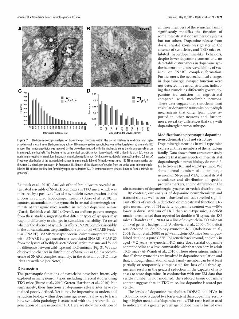
No gross ultrastructural changes of striatal dopaminergicaxons in TKO miceQuantitative electron-microscopic analyses of dopaminergicprofiles in the dorsal striatum of TKO and wild-type mice per-formed on sections immunostained for TH using two techniques(Fig. 7A,B) indicated the overall density of TH-positive profiles,the number of mitochondria per structure and synaptic inci-dence, were not different between two genotypes. Analysis of allTH-immunogold profiles, as well as the subgroup forming syn-
aptic specializations, showed trends toward reduction in someparameters in TKO mice, such as the cross-sectional area of TH-positive profiles (18.6% smaller in all profiles; 35.6% smaller insynapse-forming profiles), the number of synaptic vesicles perprofile (6.2 and 38.5% less), and also in the average distance of asynaptic vesicle from the plasma membrane (5.2 and 14.4%smaller) (Fig. 7D), but trends toward opposite outcomes in allversus synapse-forming structures for other parameters, for ex-ample, for the density of vesicles (9.5% increase across all versus14.3% decrease in synapse-forming) and the average intervesicledistance, an indication of clustering [24.8% decrease (Fig. 7C) vs3.0% increase]. In TH-immunogold profiles that formed syn-apses, the average distance of vesicles to the active zone wassmaller in the mutant animals (22.4% decreased) (Fig. 7D).However, differences in these or other studied parameters[data are available (see Notes)] were not statistically signifi-cant, suggesting that the ultrastructure of dopaminergic syn-apses in the striatum is not substantially affected by theabsence of synucleins.
Normal abundance of SNARE complexes in the dorsalstriatum of TKO miceRecent in vivo studies demonstrated that synucleins might regu-late neurotransmitter release by regulating synaptic SNARE com-plex assembly and/or distribution of SNARE proteins withinsynapses (Chandra et al., 2005; Burre et al., 2010; Garcia-
Figure 6. Behavior of wild-type and triple-synuclein-null mutant mice after pharmacological challenging of dopamineneurotransmission. The locomotor activity (A–C) or climbing behavior (D) of wild-type (WT) (white diamonds) and TKO (��/�
� �/�� �/�) (gray squares). A, Animals were injected with 4 mg/kg dAMPH after monitoring of their activity in novel environ-ment (nonanxiogenic activity camera with infrared beams) for 30 min and returned to the same camera for an additional 90 min.Statistically significant increase in the locomotor activity of mutant mice before treatment and decrease after treatment wasobserved. B, A graph showing locomotor activity of triple-synuclein-null mutant mice pretreated with 50 mg/kg L-DOPA 20 minbefore placing in the activity camera and consequent injecting dAMPH (black circles) overlays the same graphs as in A. Statisticallysignificant difference in the locomotor activity of wild-type and L-DOPA-pretreated mice (*) or naive and L-DOPA-pretreatedmutant mice ( #) is shown. C, The same protocol as described for A was used to assess locomotion of mice treated with 10 mg/kgcocaine. For all panels, statistically significant difference between two groups is shown for each 4 min interval (** ,##p � 0.01;*p � 0.05; Kolmogorov–Smirnov test). D, Scoring of climbing behavior of mice after injection with 4 mg/kg APO was performedas described in Materials and Methods.
7270 • J. Neurosci., May 18, 2011 • 31(20):7264 –7274 Anwar et al. • Nigrostriatal Defects in Triple-Synuclein-KO Mice
Reitbock et al., 2010). Analysis of total brain lysates revealed at-tenuated assembly of SNARE complexes in TKO mice, which wasmirrored by a positive effect of �-synuclein overexpression on thisprocess in cultured hippocampal neurons (Burre et al., 2010). Incontrast, accumulation of �-synuclein in striatal dopaminergic ter-minals of transgenic mice resulted in reduced dopamine release(Garcia-Reitbock et al., 2010). Overall, no uniform pattern emergesfrom these studies, suggesting that different types of synapses mayrespond differently to changes in synucleins availability. To revealwhether the absence of synucleins affects SNARE complex assemblyin the dorsal striatum, we quantified the amount of vSNARE (vesic-ular SNARE) VAMP2/synaptobrevin coimmunoprecipitatedwith tSNARE (target membrane-associated SNARE) SNAP-25from the lysates of freshly dissected dorsal striatum tissue and foundno difference between wild-type and TKO animals (Fig. 8). We alsoobserved no changes in distribution of SNAP-25 or CSP, a cochap-erone of SNARE complex assembly, in the striatum of TKO mice[data are available (see Notes)].
DiscussionThe presynaptic functions of synucleins have been intensivelyexplored in many neuron types, including in recent studies usingTKO mice (Burre et al., 2010; Greten-Harrison et al., 2010), butsurprisingly, their functions at dopamine release sites have re-mained poorly defined. Yet it may be imperative to understandsynuclein biology within dopaminergic neurons if we are to learnhow synuclein pathology is associated with the preferential de-generation of these neurons in PD. Here, we show that deletion of
all three members of the synuclein familysignificantly modifies the function ofsome mesostriatal dopaminergic systemsbut not others. Dopamine release fromdorsal striatal axons was greater in theabsence of synucleins, and TKO mice ex-hibited hyperdopamine-like behaviors,despite lower dopamine content and nodetectable disturbances in dopamine syn-thesis, neuron number, availability of ves-icles, or SNARE complex formation.Furthermore, the neurochemical changesin dopaminergic synapse function werenot detected in ventral striatum, indicat-ing that synucleins differently govern do-pamine transmission in nigrostriatalcompared with mesolimbic neurons.These data suggest that synucleins limitvesicular dopamine transmission throughmechanisms that differ from those re-ported in other neurons and, further-more, reveal key differences that vary withdopaminergic neuron subtype.
Modifications to presynaptic dopamineneurochemistry but not structureDopaminergic neurons in wild-type miceexpress all three members of the synucleinfamily. Data drawn from across our studyindicate that many aspects of mesostriataldopaminergic neuron biology do not dif-fer between TKO and wild-type mice. Weshow normal numbers of dopaminergicneurons in SNpc and VTA, normal striatalabundance and distribution of specificproteins markers, and no difference in the
ultrastructure of dopaminergic synapses or vesicle distribution.By contrast, our analysis of dopamine neurochemistry and
transmission as well as our behavioral analysis revealed signifi-cant effects of synuclein depletion on mesostriatal function. De-spite normal level of TH activity, dopamine content was �40%lower in dorsal striatum of TKO than wild-type mice, a deficitmuch more marked than reported for double-�/�-synuclein-KOmice (Chandra et al., 2004) or a line of �-synuclein-KO mice ona mixed genetic background (Abeliovich et al., 2000). No deficitwas detected in double-�/�-synuclein-KO (Robertson et al.,2004; Senior et al., 2008) or �/�-synuclein-KO mice (our unpub-lished data) on a pure C57BL/6J genetic background, and only inaged (�2 years) �-synuclein-KO mice does striatal dopaminecontent decline to a level comparable with that seen here in adultTKO mice (Al-Wandi et al., 2010). These observations indicatethat all three synucleins are involved in dopamine regulation andthat, although elimination of each family member can be at leastpartially or temporarily compensated for, loss of all three sy-nucleins results in the greatest reduction in the capacity of syn-apses to store dopamine. In conjunction with our EM data thatvesicle number is not modified, the reduced tissue dopaminecontent suggests that, in TKO mice, less dopamine is stored pervesicle.
The levels of dopamine metabolites DOPAC and HVA inTKO mice were reduced to a lesser extent than dopamine, result-ing in higher metabolite/dopamine ratios. This ratio is often usedto indicate that a greater percentage of dopamine is turned over
Figure 7. Electron-microscopic analyses of dopaminergic structures within the dorsal striatum in wild-type and triple-synuclein-null mutant mice. Electron micrographs of TH-immunoreactive synaptic boutons in the dorsolateral striatum of a TKOmouse. The immunoreactivity was revealed by the peroxidase method with diaminobenzidine as the chromogen (A) or theimmunogold method (B). The bouton forms symmetrical synaptic contact (arrowheads) with a dendritic shaft (d). Note thenonimmunoreactive terminals forming an asymmetrical synaptic contact (white arrowheads) with a spine. Scale bars, 0.5 �m. C,Frequency distribution of the intervesicle distances in immunogold-labeled TH-positive structures (150 TH-immunoreactive pro-files from 3 animals per genotype). D, Frequency distribution of the distances of vesicles from the active zone in immunogold-labeled TH-positive profiles that formed synaptic specializations (23 TH-immunoreactive synaptic boutons from 3 animals pergenotype).
Anwar et al. • Nigrostriatal Defects in Triple-Synuclein-KO Mice J. Neurosci., May 18, 2011 • 31(20):7264 –7274 • 7271

or, in other words, is released from vesicular stores with access toextracellular and cytosolic metabolic pathways. Indeed, directstudy of dopamine release using FCV revealed a striking elevationof the releasability of dopamine in the striatum in TKO mice.This increase was not caused by a reduction in dopamine uptakerate, an upregulation of nicotinic acetylcholine receptor func-tion, or by increased sensitivity of the presynaptic exocytotic ma-chinery to calcium or increased dopamine release probability [asdetermined by measurements of frequency sensitivity, whichshould be inversely correlated with release probability (Rice andCragg, 2004)]. Rather, the data suggest a generalized increase indopamine releasability per stimulus. Behavioral tests also sug-gested that TKO mice had increased rather than decreased levelsof striatal dopamine. Furthermore, the experiments with adopamine-releasing agent (amphetamine) and a receptor ligand(apomorphine) were in agreement with enhanced presynapticdopamine release despite reduced dopamine storage, and notwith any additional apparent upregulation of postsynaptic recep-tor number or efficacy.
Several mechanisms, which are not mutually exclusive, canunderlie enhanced releasability from presynaptic terminals withdiminished content of the neurotransmitter. For example, neu-rotransmitter releasability can be elevated if the recycling, readilyreleasable pool of these vesicles is increased at the expense of thereserve pool. Increased expression of �-synuclein in culturedhippocampal neurons reduces the size of the recycling pool and
can thus inhibit synaptic vesicle exocytosis (Nemani et al., 2010;Scott et al., 2010). However, striatal dopaminergic synapses aremorphologically and functionally distinct from these synapses:they lack spatially defined clusters of reserve and recycling vesi-cles. Our ultrastructural analyses indicated that the presynapticdistribution of synaptic vesicles in dorsal striatum is not modifiedin TKO mice, suggesting that synucleins do not regulate the over-all anatomical distribution of vesicles within dopaminergic ax-ons, unlike in other axon types, and that this does not explain achange in dopamine releasability in TKO mice.
Another possible explanation for enhanced neurotransmitterreleasability is increased rate of vesicle fusion. However, such anassumption is inconsistent with the recently obtained evidencethat in hippocampal neurons �-synuclein promotes activity-dependent assembly of SNARE complexes (Chandra et al., 2005;Burre et al., 2010), which is a crucial step in the vesicle fusioncycle. Moreover, we demonstrate here that, in dorsal striatum,the abundance of SNARE complexes is not different in TKO andwild-type mice. Therefore, it is unlikely that increased releasabil-ity of dopamine in the absence of synucleins is caused by modi-fied levels of SNARE complexes with consequent acceleration ofvesicle fusion.
It remains feasible that the presence or absence of synucleinsmight affect formation of a fusion pore and thus the fate of a fusedvesicle (i.e., kiss-and-run or full-collapse fusion with the presyn-aptic plasma membrane). A boost in dopamine release could thusarise from the release of a greater proportion of contents of eachvesicle or the number of dopamine quanta released per stimulus.Additional detailed studies are required to test this hypothesisand understand whether and why any of these mechanisms differin nigrostriatal compared with mesolimbic neurons. We cannotentirely exclude the possibility that changes to dopamine releasein the absence of synucleins reflect consequences of the develop-mental compensation of the nigrostriatal neurons for the absenceof synucleins.
These data together suggest that, in the absence of synucleins,dopamine axons release more vesicles (or more content of eachvesicle), each of which has a decreased dopamine load. The mostparsimonious explanation for reduced dopamine content of eachvesicle could be a downregulation in response to a gain in releas-ability: indeed content is lower when extracellular availability ofdopamine is increased in other models (e.g., DAT-KO mice)(Jones et al., 1998). However, differences in dopamine releasabil-ity have been reported without complementary reductions in do-pamine content in striatal terminals of �/�-synuclein double-KOmice (Senior et al., 2008). Therefore, it is feasible that multiplesynuclein-dependent mechanisms might differently upregulatereleasability on the one hand and disrupt dopamine storage onthe other (e.g., compromise VMAT2 function, which in turndownregulates dopamine content).
Regionally distinct effects of synuclein deletion: nigrostriatalversus mesolimbic dopamine synapsesIt is striking that synuclein depletion affects presynaptic dopa-mine neurochemistry only in dorsal and not ventral striatum.Thus, the roles of synucleins that we have described here in do-paminergic neurons may differ substantially between neurons ofdifferent neurotransmitter type (e.g., dopaminergic vs hippocampalglutamatergic neurons) and, moreover, may be specific to neuronsubtype even for a given transmitter. Furthermore, since dopami-nergic neurons innervating the dorsal striatum (nigrostriatal path-way) degenerate in preference to those innervating the ventralstriatum (mesolimbic pathway) in PD, our finding may offer signif-
Figure 8. Quantification of SNARE complexes in the dorsal striatum of wild-type and triple-synuclein-null mutant mice. Coimmunoprecipitation of VAMP2/synaptobrevin with SNAP-25was used to assess the abundance of SNARE complexes in the dorsal striatum of wild-type (WT)and TKO (� �/�� �/�� �/�) mice. The bar chart shows amount of VAMP2/synaptobrevin inSNAP-25 immunoprecipitates normalized to the amount of precipitated SNAP-25 and ex-pressed as percentage of mean amount WT samples (�SEM; results of 3 independent experi-ments; 3 WT and 3 TKO mice used in each of these experiments). A representative Western blotsshows results of analysis of two independent pairs of WT and TKO mice.
7272 • J. Neurosci., May 18, 2011 • 31(20):7264 –7274 Anwar et al. • Nigrostriatal Defects in Triple-Synuclein-KO Mice

icant new insight into the preferential degeneration seen in PD. Al-though striking, the finding that a mechanism regulating dopamineneurotransmission might differ in nigrostriatal versus mesolimbicneurons is not entirely unsurprising. A large body of work suggeststhat nigrostriatal neurons differ from mesolimbic neurons in manyfeatures [e.g., embryonic source and stage of ontogenetic develop-ment, neuroanatomy of projections and inputs, proteins expressionlevels including dopamine cell markers (DAT, VMAT2, D2 recep-tors), ion channels and other proteins, influence of neuromodula-tory receptors as well as the regulation of dopamine releaseprobability and their susceptibility to neurodegeneration (for re-view, see Korotkova et al., 2004; Bjorklund and Dunnett, 2007; Lissand Roeper, 2008)]. Synuclein function may be another key featurethat variably influences the function of nigrostriatal versus mesolim-bic neurons.
ConclusionsThese data significantly revise our understanding of synucleinfunction. They indicate that synucleins in some dopaminergicneurons limit synaptic neurotransmission through mechanismsthat differ from those reported in other neurons, which haveincluded vesicle pool redistribution or SNARE complex forma-tion. These data shed light on the distinct role of synucleins indopaminergic neurons and reveal type-specific differences be-tween dopaminergic neurons, which may ultimately offer insightinto the preferential degeneration of neurons subsets in PD.
NotesSupplemental Figures S1–S5 and supplemental Tables 1– 4 for this articleare available at https://docs.google.com/viewer?a�v&pid�explorer&chrome�true&srcid�0B-2G31EgcACfZjRjZDFjZTEtNGRlZi00ODI2LTg2NWUtOWYyYzQ4YzA2OGUx&hl�en. This material has not beenpeer reviewed.
ReferencesAbeliovich A, Schmitz Y, Farinas I, Choi-Lundberg D, Ho WH, Castillo PE,
Shinsky N, Verdugo JM, Armanini M, Ryan A, Hynes M, Phillips H,Sulzer D, Rosenthal A (2000) Mice lacking alpha-synuclein displayfunctional deficits in the nigrostriatal dopamine system. Neuron25:239 –252.
Al-Wandi A, Ninkina N, Millership S, Williamson SJ, Jones PA, Buchman VL(2010) Absence of alpha-synuclein affects dopamine metabolism andsynaptic markers in the striatum of aging mice. Neurobiol Aging31:796 – 804.
Bjorklund A, Dunnett SB (2007) Dopamine neuron systems in the brain: anupdate. Trends Neurosci 30:194 –202.
Buchman VL, Hunter HJ, Pinon LG, Thompson J, Privalova EM, NinkinaNN, Davies AM (1998) Persyn, a member of the synuclein family, has adistinct pattern of expression in the developing nervous system. J Neuro-sci 18:9335–9341.
Burre J, Sharma M, Tsetsenis T, Buchman V, Etherton MR, Sudhof TC(2010) Alpha-synuclein promotes SNARE-complex assembly in vivoand in vitro. Science 329:1663–1667.
Cabin DE, Shimazu K, Murphy D, Cole NB, Gottschalk W, McIlwain KL,Orrison B, Chen A, Ellis CE, Paylor R, Lu B, Nussbaum RL (2002) Syn-aptic vesicle depletion correlates with attenuated synaptic responses toprolonged repetitive stimulation in mice lacking �-synuclein. J Neurosci22:8797– 8807.
Carlsson A, Davis JN, Kehr W, Lindqvist M, Atack CV (1972) Simultaneousmeasurement of tyrosine and tryptophan hydroxylase activities in brainin vivo using an inhibitor of the aromatic amino acid decarboxylase.Naunyn Schmiedebergs Arch Pharmacol 275:153–168.
Chandra S, Fornai F, Kwon HB, Yazdani U, Atasoy D, Liu X, Hammer RE,Battaglia G, German DC, Castillo PE, Sudhof TC (2004) Double-knockout mice for alpha- and beta-synucleins: effect on synaptic func-tions. Proc Natl Acad Sci U S A 101:14966 –14971.
Chandra S, Gallardo G, Fernandez-Chacon R, Schluter OM, Sudhof TC
(2005) Alpha-synuclein cooperates with CSPalpha in preventing neuro-degeneration. Cell 123:383–396.
Cragg SJ (2003) Variable dopamine release probability and short-term plas-ticity between functional domains of the primate striatum. J Neurosci23:4378 – 4385.
Cragg SJ, Hille CJ, Greenfield SA (2000) Dopamine release and uptake dy-namics within nonhuman primate striatum in vitro. J Neurosci20:8209 – 8217.
Exley R, Clements MA, Hartung H, McIntosh JM, Cragg SJ (2008) Alpha6-containing nicotinic acetylcholine receptors dominate the nicotine controlof dopamine neurotransmission in nucleus accumbens. Neuropsychopharma-cology 33:2158–2166.
Fountaine TM, Venda LL, Warrick N, Christian HC, Brundin P, ChannonKM, Wade-Martins R (2008) The effect of alpha-synuclein knockdownon MPP� toxicity in models of human neurons. Eur J Neurosci28:2459 –2473.
Garcia-Reitbock P, Anichtchik O, Bellucci A, Iovino M, Ballini C, Fineberg E,Ghetti B, Della Corte L, Spano P, Tofaris GK, Goedert M, Spillantini MG(2010) SNARE protein redistribution and synaptic failure in a transgenicmouse model of Parkinson’s disease. Brain 133:2032–2044.
Giros B, Jaber M, Jones SR, Wightman RM, Caron MG (1996) Hyperloco-motion and indifference to cocaine and amphetamine in mice lacking thedopamine transporter. Nature 379:606 – 612.
Goedert M (2001) Alpha-synuclein and neurodegenerative diseases. NatRev Neurosci 2:492–501.
Greten-Harrison B, Polydoro M, Morimoto-Tomita M, Diao L, WilliamsAM, Nie EH, Makani S, Tian N, Castillo PE, Buchman VL, Chandra SS(2010) ���-Synuclein triple knockout mice reveal age-dependent neu-ronal dysfunction. Proc Natl Acad Sci U S A 107:19573–19578.
Jenco JM, Rawlingson A, Daniels B, Morris AJ (1998) Regulation of phos-pholipase D2: selective inhibition of mammalian phospholipase D isoen-zymes by alpha- and beta-synucleins. Biochemistry 37:4901– 4909.
Jones SR, Gainetdinov RR, Jaber M, Giros B, Wightman RM, Caron MG(1998) Profound neuronal plasticity in response to inactivation of thedopamine transporter. Proc Natl Acad Sci U S A 95:4029 – 4034.
Korotkova TM, Ponomarenko AA, Brown RE, Haas HL (2004) Functionaldiversity of ventral midbrain dopamine and GABAergic neurons. MolNeurobiol 29:243–259.
Larsen KE, Schmitz Y, Troyer MD, Mosharov E, Dietrich P, Quazi AZ, SavalleM, Nemani V, Chaudhry FA, Edwards RH, Stefanis L, Sulzer D (2006)�-Synuclein overexpression in PC12 and chromaffin cells impairs cate-cholamine release by interfering with a late step in exocytosis. J Neurosci26:11915–11922.
Larsson M, Broman J (2005) Different basal levels of CaMKII phosphory-lated at Thr286/287 at nociceptive and low-threshold primary afferentsynapses. Eur J Neurosci 21:2445–2458.
Lee FJ, Liu F, Pristupa ZB, Niznik HB (2001) Direct binding and functionalcoupling of alpha-synuclein to the dopamine transporters acceleratedopamine-induced apoptosis. FASEB J 15:916 –926.
Liss B, Roeper J (2008) Individual dopamine midbrain neurons: functionaldiversity and flexibility in health and disease. Brain Res Rev 58:314 –321.
Liu D, Jin L, Wang H, Zhao H, Zhao C, Duan C, Lu L, Wu B, Yu S, ChanP, Li Y, Yang H (2008) Silencing alpha-synuclein gene expressionenhances tyrosine hydroxylase activity in MN9D cells. Neurochem Res33:1401–1409.
Liu S, Ninan I, Antonova I, Battaglia F, Trinchese F, Narasanna A, Kolo-dilov N, Dauer W, Hawkins RD, Arancio O (2004) alpha-Synucleinproduces a long-lasting increase in neurotransmitter release. EMBO J23:4506 – 4516.
Mizuta I, Tsunoda T, Satake W, Nakabayashi Y, Watanabe M, Takeda A,Hasegawa K, Nakashima K, Yamamoto M, Hattori N, Murata M, Toda T(2008) Calbindin 1, fibroblast growth factor 20, and alpha-synuclein insporadic Parkinson’s disease. Hum Genet 124:89 –94.
Moss J, Bolam JP (2008) A dopaminergic axon lattice in the striatum andits relationship with cortical and thalamic terminals. J Neurosci28:11221–11230.
Nemani VM, Lu W, Berge V, Nakamura K, Onoa B, Lee MK, Chaudhry FA,Nicoll RA, Edwards RH (2010) Increased expression of alpha-synucleinreduces neurotransmitter release by inhibiting synaptic vesicle recluster-ing after endocytosis. Neuron 65:66 –79.
Ninkina N, Papachroni K, Robertson DC, Schmidt O, Delaney L, O’Neill F,Court F, Rosenthal A, Fleetwood-Walker SM, Davies AM, Buchman VL
Anwar et al. • Nigrostriatal Defects in Triple-Synuclein-KO Mice J. Neurosci., May 18, 2011 • 31(20):7264 –7274 • 7273

(2003) Neurons expressing the highest levels of gamma-synuclein areunaffected by targeted inactivation of the gene. Mol Cell Biol23:8233– 8245.
Ninkina N, Peters O, Millership S, Salem H, van der Putten H, Buchman VL(2009) Gamma-synucleinopathy: neurodegeneration associated withoverexpression of the mouse protein. Hum Mol Genet 18:1779 –1794.
Pankratz N, Wilk JB, Latourelle JC, DeStefano AL, Halter C, Pugh EW,Doheny KF, Gusella JF, Nichols WC, Foroud T, Myers RH (2009)Genomewide association study for susceptibility genes contributing tofamilial Parkinson disease. Hum Genet 124:593– 605.
Peng X, Tehranian R, Dietrich P, Stefanis L, Perez RG (2005) Alpha-synuclein activation of protein phosphatase 2A reduces tyrosine hydrox-ylase phosphorylation in dopaminergic cells. J Cell Sci 118:3523–3530.
Perez RG, Waymire JC, Lin E, Liu JJ, Guo F, Zigmond MJ (2002) A role for�-synuclein in the regulation of dopamine biosynthesis. J Neurosci22:3090 –3099.
Protais P, Costentin J, Schwartz JC (1976) Climbing behavior induced byapomorphine in mice: a simple test for the study of dopamine receptors instriatum. Psychopharmacology (Berl) 50:1– 6.
Rice ME, Cragg SJ (2004) Nicotine amplifies reward-related dopamine sig-nals in striatum. Nat Neurosci 7:583–584.
Robertson DC, Schmidt O, Ninkina N, Jones PA, Sharkey J, Buchman VL(2004) Developmental loss and resistance to MPTP toxicity of dopami-nergic neurones in substantia nigra pars compacta of gamma-synuclein,alpha-synuclein and double alpha/gamma-synuclein null mutant mice.J Neurochem 89:1126 –1136.
Satake W, Nakabayashi Y, Mizuta I, Hirota Y, Ito C, Kubo M, Kawaguchi T,Tsunoda T, Watanabe M, Takeda A, Tomiyama H, Nakashima K, Hase-gawa K, Obata F, Yoshikawa T, Kawakami H, Sakoda S, Yamamoto M,Hattori N, Murata M, et al. (2009) Genome-wide association studyidentifies common variants at four loci as genetic risk factors for Parkin-son’s disease. Nat Genet 41:1303–1307.
Scholz SW, Houlden H, Schulte C, Sharma M, Li A, Berg D, Melchers A,Paudel R, Gibbs JR, Simon-Sanchez J, Paisan-Ruiz C, Bras J, Ding J, ChenH, Traynor BJ, Arepalli S, Zonozi RR, Revesz T, Holton J, Wood N, et al.(2009) SNCA variants are associated with increased risk for multiplesystem atrophy. Ann Neurol 65:610 – 614.
Scott DA, Tabarean I, Tang Y, Cartier A, Masliah E, Roy S (2010) A patho-logic cascade leading to synaptic dysfunction in �-synuclein-inducedneurodegeneration. J Neurosci 30:8083– 8095.
Senior SL, Ninkina N, Deacon R, Bannerman D, Buchman VL, Cragg SJ,Wade-Martins R (2008) Increased striatal dopamine release andhyperdopaminergic-like behaviour in mice lacking both alpha-synucleinand gamma-synuclein. Eur J Neurosci 27:947–957.
Simon-Sanchez J, Schulte C, Bras JM, Sharma M, Gibbs JR, Berg D, Paisan-Ruiz C, Lichtner P, Scholz SW, Hernandez DG, Kruger R, Federoff M,Klein C, Goate A, Perlmutter J, Bonin M, Nalls MA, Illig T, Gieger C,Houlden H, et al. (2009) Genome-wide association study reveals geneticrisk underlying Parkinson’s disease. Nat Genet 41:1308 –1312.
Tehranian R, Montoya SE, Van Laar AD, Hastings TG, Perez RG (2006)Alpha-synuclein inhibits aromatic amino acid decarboxylase activity indopaminergic cells. J Neurochem 99:1188 –1196.
Trojanowski JQ, Lee VM (2002) Parkinson’s disease and related synucle-inopathies are a new class of nervous system amyloidoses. Neurotoxicol-ogy 23:457– 460.
Unger EL, Eve DJ, Perez XA, Reichenbach DK, Xu Y, Lee MK, Andrews AM(2006) Locomotor hyperactivity and alterations in dopamine neu-rotransmission are associated with overexpression of A53T mutant hu-man alpha-synuclein in mice. Neurobiol Dis 21:431– 443.
Venda LL, Cragg SJ, Buchman VL, Wade-Martins R (2010) alpha-Synucleinand dopamine at the crossroads of Parkinson’s disease. Trends Neurosci33:559 –568.
Wersinger C, Sidhu A (2003) Attenuation of dopamine transporter activityby alpha-synuclein. Neurosci Lett 340:189 –192.
Yavich L, Tanila H, Vepsalainen S, Jakala P (2004) Role of �-synuclein inpresynaptic dopamine recruitment. J Neurosci 24:11165–11170.
Yavich L, Oksman M, Tanila H, Kerokoski P, Hiltunen M, van Groen T,Puolivali J, Mannisto PT, García-Horsman A, MacDonald E, BeyreutherK, Hartmann T, Jakala P (2005) Locomotor activity and evoked dopa-mine release are reduced in mice overexpressing A30P-mutated humanalpha-synuclein. Neurobiol Dis 20:303–313.
Zhuang X, Oosting RS, Jones SR, Gainetdinov RR, Miller GW, Caron MG,Hen R (2001) Hyperactivity and impaired response habituation in hy-perdopaminergic mice. Proc Natl Acad Sci U S A 98:1982–1987.
7274 • J. Neurosci., May 18, 2011 • 31(20):7264 –7274 Anwar et al. • Nigrostriatal Defects in Triple-Synuclein-KO Mice