neurobiologyofdisease ... lrrk2inhibitionattenuatesmicroglialinflammatory responses...
TRANSCRIPT
Neurobiology of Disease
LRRK2 Inhibition Attenuates Microglial InflammatoryResponses
Mark S. Moehle,1,4,5* Philip J. Webber,1,4,5* Tonia Tse,1,4 Nour Sukar,1,4 David G. Standaert,1,4 Tara M. DeSilva,2,4
Rita M. Cowell,3,4 and Andrew B. West1,4
Departments of 1Neurology, 2Physical Medicine and Rehabilitation, and 3Psychiatry and Behavioral Neurobiology, 4Center for Neurodegeneration andExperimental Therapeutics, and 5Neuroscience Graduate Program, University of Alabama at Birmingham, Birmingham, Alabama 35294
Missense mutations in leucine-rich repeat kinase 2 (LRRK2) cause late-onset Parkinson’s disease (PD), and common genetic variation inLRRK2 modifies susceptibility to Crohn’s disease and leprosy. High levels of LRRK2 expression in peripheral monocytes and macro-phages suggest a role for LRRK2 in these cells, yet little is known about LRRK2 expression and function in immune cells of the brain. Here,we demonstrate a role for LRRK2 in mediating microglial proinflammatory responses and morphology. In a murine model of neuroin-flammation, we observe robust induction of LRRK2 in microglia. Experiments with toll-like receptor 4 (TLR4)-stimulated rat primarymicroglia show that inflammation increases LRRK2 activity and expression, while inhibition of LRRK2 kinase activity or knockdown ofprotein attenuates TNF� secretion and nitric oxide synthase (iNOS) induction. LRRK2 inhibition blocks TLR4 stimulated microglialprocess outgrowth and impairs ADP stimulated microglial chemotaxis. However, actin inhibitors that phenocopy inhibition of processoutgrowth and chemotaxis fail to modify TLR4 stimulation of TNF� secretion and inducible iNOS induction, suggesting that LRRK2 actsupstream of cytoskeleton control as a stress-responsive kinase. These data demonstrate LRRK2 in regulating responses in immune cellsof the brain and further implicate microglial involvement in late-onset PD.
IntroductionThe leucine-rich repeat kinase 2 (LRRK2) gene was discovered aspart of an evolutionarily conserved family of proteins marked byGTPase domains usually encoded together with kinase domains(Bosgraaf and Van Haastert, 2003). Missense mutations in boththe kinase and GTPase domain in LRRK2 cause late-onset Parkin-son’s disease (PD) with clinical and pathological phenotypes nearlyindistinguishable from idiopathic disease, possibly throughthe upregulation of LRRK2 kinase activity (Paisan-Ruíz et al.,2004; Zimprich et al., 2004; West et al., 2005). Disease penetranceof LRRK2 mutations in PD is incomplete as lifetime risk in clin-ical populations is estimated at �22–32%, suggesting strongmodifiers of LRRK2 disease (Goldwurm et al., 2007). A modify-ing role for the immune system in PD susceptibility is supportedby the association of the human leukocyte antigen (HLA) regionwith late-onset disease (Hamza et al., 2010), and pathologicalstudies of PD brains demonstrate strong microglial and T-cell
activation and infiltration in susceptible brain nuclei (McGeer etal., 1988). Genome-wide association studies also highlightLRRK2 in the modification of susceptibility to the chronic auto-immune Crohn’s disease and Mycobacterium leprae infection(Zhang et al., 2009; Umeno et al., 2011), raising the possibilitythat mutations in LRRK2 may modify immunogenic responsesin PD.
LRRK2 is expressed in many different cell types in mammals,but the intracellular function of LRRK2 is not clear. In the brain,LRRK2 is expressed in diverse neuronal subtypes and localizes tocytoskeletal structures and a variety of vesicular and membra-nous organelles (Biskup et al., 2006). In neurons, LRRK2 hasbeen described as a potent regulator of the cytoskeleton whereknockdown of protein enhances neurite outgrowth and mutant(overactive) LRRK2 expression inhibits outgrowth (MacLeod etal., 2006). LRRK2 may directly modify microtubule organizationand the actin cytoskeleton through phosphorylation of substrates(Gillardon, 2009; Parisiadou et al., 2009). LRRK2 may also playadditional kinase-dependent roles in the modification of synapticvesicle storage and mobilization, in addition to kinase-dependentroles in endocytosis, MAPK signaling, autophagy, and apoptosis(Alegre-Abarrategui et al., 2009; Gloeckner et al., 2009; Piccoli etal., 2011).
Particularly high LRRK2 expression has been discovered re-cently in macrophagic and monocytic cells, but not T-cells, lead-ing to speculation of a functional role for LRRK2 in the innateimmune system (Thevenet et al., 2011). A number of powerfultools, including highly specific rabbit monoclonal LRRK2 anti-bodies and potent and selective LRRK2 small-molecule kinaseinhibitors, have become available that allow for a careful dissec-
Received Nov. 7, 2011; accepted Dec. 6, 2011.Author contributions: M.S.M., P.J.W., T.M.D., R.M.C., and A.B.W. designed research; M.S.M., P.J.W., T.T., N.S.,
R.M.C., and A.B.W. performed research; T.M.D. and A.B.W. contributed unpublished reagents/analytic tools; M.S.M.,P.J.W., D.G.S., R.M.C., and A.B.W. analyzed data; D.G.S., R.M.C., and A.B.W. wrote the paper.
This work was supported by the Michael J. Fox Foundation for Parkinson’s Research, the American Parkinson’sDisease Association, National Institutes of Health Grant R01-NS064934, UAB Neuroscience Core Center GrantNS047466, and the benevolence of John A. and Ruth R. Jurenko. M.S.M. is supported by NIH/NINDS Grant T32NS061788. We thank Dario Alessi for L2in1 compound; Bassel Sawaya for purified human microglia cells; andHeather Melrose and Matthew Farrer for LRRK2 knock-out mice.
*M.S.M. and P.J.W. contributed equally to this work.Correspondence should be addressed to Andrew B. West, 1719 6th Avenue S, University of Alabama at Birming-
ham, Birmingham, AL 35226. E-mail: [email protected]:10.1523/JNEUROSCI.5601-11.2012
Copyright © 2012 the authors 0270-6474/12/321602-10$15.00/0
1602 • The Journal of Neuroscience, February 1, 2012 • 32(5):1602–1611
tion of LRRK2 function in cells of the immune system. Based onthe expression of LRRK2 in monocytes, we hypothesized a rolefor LRRK2 in the immune cells of the brain. Our results show thatLRRK2 is expressed in activated microglia and that LRRK2 mod-ulates proinflammatory responses in these cells. Alterations inLRRK2 function may modify inflammatory responses in neuro-degenerative and infectious diseases, potentially leading to dis-ease initiation or modification of progression.
Materials and MethodsImmunohistochemistry and immunofluorescence. Male 8 –12-week-oldWT or LRRK2 KO C57BL/6J mice (provided by Heather Melrose, MayoClinic, Jacksonville, FL) or Tg(TH-EGFP)DJ76Gsat were perfused withroom temperature (RT) PBS, pH 7.4, then 4% paraformaldehyde (PFA)in PBS, and brains removed and post-fixed in 4% PFA in PBS at 4°C for12 h with agitation, then embedded in 30% sucrose/PBS for 24 h at 4°C,then frozen in isopentane and sectioned at 40 �m width on a freezingmicrotome. Freshly cut sections were rinsed and immediately treatedwith 0.3% H2O2 in methanol for 30 min at RT with mild agitation,rinsed, and treated with 10 mM Na-Citrate, pH 6.0, 0.05% Tween, for 30min at 37°C. Sections were rinsed and blocked first in 3% nonfat milk inPBS with 0.3% Triton X-100 for 1 h RT and then in 10% normal goatserum in PBS with 0.3% Triton X-100 for 1 h. LRRK2 antibody solution[containing 0.2 �g/ml for DAB or 1 �g/ml for immunofluorescencerabbit monoclonal C-41 (Epitomics) 5% goat serum, 0.1% Triton X-100,and 0.01% sodium azide] was applied to sections for 24 h at 4°C withmild agitation. Sections were rinsed and Goat Anti-Rabbit:biotin (VectorLabs), Goat Anti-Rabbit DyLight 649 (Jackson Laboratories), Isolectin-B4:FITC or Isolectin-B4:Biotin (Sigma) was added (as indicated) for 24 hat 4°C. Sections for immunofluorescence were mounted with ProLongGold (Invitrogen) onto coverslips. Sections with biotinylated markerswere developed with the Vectastain Elite ABC kit and Impact DAB (Vec-tor Labs) according to manufacturer’s recommendations.
LPS injections. Five micrograms of LPS (15,000 endotoxin units,Sigma) was stereotactically injected in a 1 �l volume with a flow rate of0.2 �l/min using a NanoMite pump (Harvard Apparatus) fitted with a 32gauge fully beveled needle and gas-tight syringe (Hamilton), with a 5 minwait for needle withdrawal, in mice anesthetized with isoflurane. Coor-dinates were �3.4 anteroposterior (AP), �1.1 ML, and �3.9 DV forsubstantia nigra pars compacta (SNpc), and �0.4 AP, �1.5 ML, and�2.5 DV for striatum, with respect to bregma. For LRRK2 activity assays,FLAG-LRRK2 BAC mice (Jackson Laboratory strain 012466) were used,and FLAG-M2 resin (Sigma) was used according to manufacturer’s rec-ommendations to immunoprecipitate LRRK2 protein. Animal usage wasinstitutionally approved.
Western blotting, ELISA, and chemicals. Antibodies to LRRK2 (cloneC-41, Epitomics), �-actin, GFAP, MBP (Sigma), CD-68 (Serotec),MAP2 (Millipore), GAPDH, interferon regulatory factor-1, voltage-dependent anion channel (VDAC) (Santa Cruz Biotechnology), nitricoxide synthase (iNOS), Phospho-p38, and Ikk-� (Cell Signaling Tech-nology) were used according to manufacturer’s suggestions. Antibodiesto pT1503 were previously described and were combined with de-phosphopeptide at a concentration of 10 �g/ml during antibody incuba-tions (Webber et al., 2011). Rat TNF� ELISA assays were fromeBioscience. Sunitinib (LC Laboratories), L2In1 (provided by DarioAlessi, Medical Research Council, Protein Phosphorylation Unit,Dundee, United Kingdom), and cytochalasin D (Sigma) were dissolvedat a concentration of 10 mM in DMSO. DMSO controls represent DMSOconcentrations present at the highest amount in the experiment, and didnot exceed 0.04% in any experiment.
Quantitative RT-PCR. Total RNA was extracted with Trizol reagent(Invitrogen), and first strand cDNA was generated with Superscript III (In-vitrogen). qPCR was performed using Taqman assays Rn01455646_m1TATA-binding protein (TBP) and Rn00562055_m1 TNF� primer sets (In-vitrogen), and iQ Powermix (Bio-Rad). Thermocycling was performed on aBio-Rad CFX96 machine.
LRRK2 kinase assays. Kinase assays were performed as previously de-scribed (Sen et al., 2009). Recombinant purified human LRRK2 (Invit-
rogen) was combined into kinase buffer with LRRKtide substrate (EnzoBioscience), and activity was measured by scintillation counting of P-81Whatman phosphocellulose paper.
Lentiviral purification. Lentivirus preparation was performed as previ-ously described (Tomlinson, 2008). HEK293-FT cells were transfected withpLP1, pLP2, pVSV-G, and lentiviral expression vectors pLKO.1_LRRK2[plasmids TRCN0000022655 (shRNA-A) and TRCN0000022658 (shRNA-B), Open Biosystems] or pLKO.1_Noncoding (NC) shRNA (Addgene plas-mid 1864), or cFUGW (no RNAi) control. For determination of titer, RNAwas extracted and cDNA was synthesized using the SuperScript VILO cDNAsynthesis kit (Invitrogen). Real-time PCR was performed using primers thattarget the Rev response element (F-GCA GCA GGA AGC ACT ATG;R-CGC CTC AAT AGC CCT CAG C). Cycle threshold values obtained fromthe virus were compared with the plasmid standard curve to determine thenumber of copies of virus per microliter. EGFP epifluorescence was used toverify that �90% of cells were transduced for the entirety of the experiment.
Primary microglia cell cultures. Primary mixed glial cultures were iso-lated from the forebrains of 2-d-old Swiss Webster rats of either sex usinga differential detachment method. Forebrains were digested with HBSS(Invitrogen) containing 0.01% trypsin and 10 �g/ml DNase, and tritu-rated with DMEM (Invitrogen) containing 20% heat-inactivated fetalbovine serum (FBS; Hyclone) and 1% penicillin-streptomycin. The dis-sociated cell suspension was plated onto poly-D-lysine-coated flasks. Me-dia changes with DMEM containing 20% fetal bovine serum and 1%penicillin-streptomycin were performed every other day for 7 d. Micro-glia were separated by shaking the flasks for 1 h at 200 rpm. The resultingmicroglial cell suspension was removed and plated at a density of �1 �10 5 cells/cm 2 in DMEM supplemented with 10% FBS and 10 ng/mlgranulocyte macrophage-CSF (Peprotech). The purity of microglia wasverified by anti-rat CD-68 (Serotec) immunolabeling and Western blot.
Morphological assessment. Randomized captured phase contrast im-ages were derived from live cultures in a 37°C humidified chamber at 5%CO2 on a Carl Zeiss Cell Observer using Axiovision 4.7 Mark and Findcontroller. Before image collection, cells were incubated with 2.5 �M
propidium iodide and 10 �M Hoechst 33342 stain for 10 min. The resul-tant images were analyzed by an observer blinded to experimental iden-tity using ImageJ software to calculate process length in microglia cells.
Chemotaxis assay. A total of 90,000 primary microglia cells were addedto transwell plates (8 �m pore, 24-well inserts, Corning) immediatelyafter microglial removal from astrocyte beds and were allowed to adhereto the upper chamber for 6 h. The lower chamber was then supplementedwith 100 �M ADP (Sigma) to encourage migration through the mem-brane to the lower chamber, and experimental drug or DMSO was addedto both the upper and lower chambers. After 30 additional hours, mediawas removed and the total number of cells counted, after incubation with10 �M Hoechst 33342 for 10 min on a Carl Zeiss Cell Observer usingMark and Find software.
Statistics. Data from all experiments were analyzed with GraphPadPrism and InStat software.
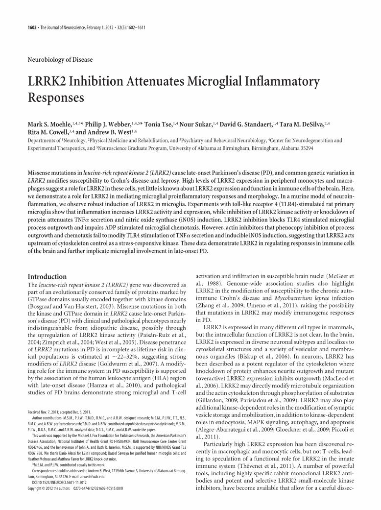
ResultsLRRK2 expression in toll-like receptor 4-activated microgliaHigh LRRK2 expression in peripheral mouse monocytes andmacrophages led us to examine whether LRRK2 may be ex-pressed in brain-resident macrophage cells (i.e., microglia). Wefirst applied recently characterized and highly specific rabbitmonoclonal antibodies directed against LRRK2 to normal mousebrain tissue and failed to detect any cells positive for LRRK2 withmorphology consistent with microglia, despite strong LRRK2immunoreactivity in several neuronal populations. Since LRRK2has been hypothesized as a stress-responsive kinase, we analyzedbrain tissue from mice subjected to an intracranial injection ofthe potent toll-like receptor 4 (TLR4) agonist lipopolysaccharide(LPS) for a period of 24 h. Although we observed no loss oftyrosine hydroxylase (TH)-positive cells at this time point, theintensity of TH expression was slightly diminished (Fig. 1A–C).Whereas only blood vessels were labeled by isolectin B4 in the
Moehle et al. • LRRK2 Activation in Neuroinflammation J. Neurosci., February 1, 2012 • 32(5):1602–1611 • 1603
contralateral side to LPS injection (Fig.1D), numerous small strongly stainedcells consistent with activated microgliawere detected in the LPS-treated SNpc(Fig. 1E,F). LRRK2 staining in the LPS-injected SNpc revealed a strong inductionof LRRK2 immunoreactivity in small cellswith a morphology and size consistentwith the activated microglia identified byisolectin B4 (Fig. 1H). This staining wasabolished in LRRK2 knock-out mice (Fig.1 I). LRRK2 protein was undetectable inwhite matter tracts in normal brain tissue,although many LRRK2-positive smallcells were found in the corpus callosum ofmice after an intrastriatal LPS injection(Fig. 1K). To rule out nonspecific orcross-reactive labeling, nonimmune rab-bit IgG was also applied at concentrationscomparable to LRRK2 antibody-treatedsections; negligible immunoreactivity wasobserved in these negative control sec-tions, and none were reminiscent of mi-croglia cells. Finally, staining in LRRK2KO mice revealed the LRRK2 monoclo-nal antibody to be specific for LRRK2(Fig. 1 I, L).
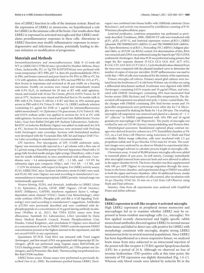
To colocalize LRRK2 with microglialmarkers in TLR4-activated microglia, atriple-staining protocol that uses a singlefluorescently labeled antibody was devel-oped and applied to mice LPS-treated ineither the SNpc or striatum (Fig. 2).Twenty-four hours postinjection, micro-glial cells rapidly accumulated and sur-rounded TH-positive neurons (Fig. 2A).Microglia could not be detected in the SNpcin animals that did not receive an LPS injec-tion (Fig. 2B). Similar to the SNpc, LRRK2colocalized to microglial cells present in thewhite matter tract post-striatal LPS injection(Fig. 2C). Resident microglia with restingmorphologies in noninjected animals failedto demonstrate immunoreactivity forLRRK2 (Fig. 2D). As in Figure 1, whole-rabbit IgG control stained sections con-firmed specificity of staining, and the LRRK2 antibodyproduces a single band of the correct size on Western blotanalysis (Fig. 2 E).
LRRK2 induction in TLR4-stimulatedcellsTLR4 activation stimulates signal transduction pathways responsi-ble for the upregulation of proinflammatory factors. To further ex-plore LRRK2 activity in response to TLR4 stimulation, micetransgenic for a FLAG-LRRK2 bacterial artificial chromosome insertwere injected (bilateral) with LPS in the SNpc, and the SNpc andimmediately surrounding regions were removed after 24 h. AfterLRRK2 immunoprecipitation from this tissue, LRRK2 was allowedto autophosphorylate for 15 min in the presence of 100 �M ATP, andprotein eluted and transferred to membranes and probed with therecently described autophosphorylation-specific antibody pT1503(Webber et al., 2011). LPS treatment significantly increased the pro-
portion of LRRK2 in an activated state as revealed by an en-hanced proportion of autophosphorylated LRRK2 (Fig. 2 B).To ensure the pT1503 antibody could not cross-react withnon-autophosphorylated LRRK2 protein, recombinant pro-tein harboring mutations in the 1503 autophosphoraytion siteor in the kinase domain (kinase dead, D1994A) was derivedfrom transiently transfected cells and evaluated by Westernblot (Fig. 2C). The pT1503 antibody could not detect signal inthe kinase dead or the T1503A mutant LRRK2 protein, sug-gesting a high degree of specificity for this antibody.
To address whether LRRK2 expression also becomes upregu-lated during TLR4 activation, primary microglia in culture weretreated with increasing concentrations of LPS for 12 h andLRRK2 protein levels determined by Western blot (Fig. 3E). Aconcentration of 100 ng/ml LPS was sufficient to increase levels ofLRRK2 protein obtained in SDS-solubilized cell lysates, whilehigher concentrations of LPS failed to further increase LRRK2
Figure 1. TLR4 stimulationPer triggers LRRK2 expression in microglia cells. Five micrograms of LPS (Escherichia coli 0111:B4)was unilaterally injected into the SNpc or striatum of 12-week-old male WT and LRRK2 KO C57BL/6J 12 mice. A–L, Immunohisto-chemistry for TH (A–C), isolectin B4 (marker for microglia and endothelial cells) (D–F ), or LRRK2 was performed on serial coronalsections spanning the SNpc and striatum (G–L). Arrowheads indicate LRRK2 immunoreactivity on cells in the SNpc with the sizeand location of TH-positive neurons on both the contralateral and ipsilateral injection sides. Arrows indicate intense LRRK2 stainingin numerous small cells observed exclusively on the ipsilateral side. WM, White matter; Str, striatum. No specific cellular staining inthese areas was observed when primary antibodies were replaced with species-matched whole IgG (data not shown). Scalebar: A–L (in A), 50 �m.
1604 • J. Neurosci., February 1, 2012 • 32(5):1602–1611 Moehle et al. • LRRK2 Activation in Neuroinflammation
induction. These primary microglia cultures were free from othercell types, and �99% of cells in culture were CD-68 positive (datanot shown). Quantification of LRRK2 protein levels across pri-mary microglia, astrocytes, hippocampal neurons, and oligoden-drocytes, all derived from postnatal day 2 rats, unexpectedlyrevealed LRRK2 expression in primary astrocytes despite the lackof expression we could observe in these cells in vivo. LRRK2 ex-pression in primary microglia cells is comparable to that of pri-mary neurons in culture (Fig. 3D).
We next determined whether LRRK2 expression is upregu-lated at the mRNA level but failed to detect any significant differ-ences after 12 h of induction with various doses of LPS, suggestingimportant post-transcriptional regulation of LRRK2 in microglia(Fig. 3G). LRRK2 distribution by immunofluorescence in pri-mary microglia is consistent with that of previous reports with
strong perinuclear staining and nuclear exclusion, both in non-LPS- and LPS-treated microglia (Fig. 3H). LRRK2 expression inhuman-derived cells of monocytic and microglia origin appearsto be conserved (Fig. 3I), with similar LRRK2 upregulation inTHP-1 cells treated with LPS. Quantitative PCR analysis forLRRK2 mRNA also revealed no significant changes in LRRK2levels in these cells, despite strong upregulation of LRRK2 proteinlevels (Fig. 3K; data not shown).
Inhibition of LRRK2 kinase activity attenuatesproinflammatory microglial signalingA post-transcriptional induction of LRRK2 and enhanced auto-phosphorylation in purified protein suggests possible involve-ment of LRRK2 during a proinflammatory response. We firstevaluated the inhibitory potential of the two most potent and
Figure 2. LRRK2 colocalizes with TLR4-stimulated microglia. TH-EGFP BAC mice were unilaterally injected with 5 �g of LPS (E. coli 0111:B4) into the SNpc or striatum. A, Using a triple-stainingprotocol (anti-rabbit IgG-Cy5 to detect LRRK2 antibody, ExtraAvidin-Cy3 to detect Isolectin-B4:biotin-positive cells, EGFP epifluorescence in TH-positive cells), LRRK2-labeled cells were observed inthe SNpc as either large (arrows) or small (arrowheads) cells that colocalized with EGFP (arrows) or isolectin (arrowheads). Scale bar, 20 �m. B, LRRK2 staining in the SNpc in control no-injectionmice. Scale bar, 20 �m. C, In striatal injected mice, LRRK2 was observed colocalized with most microglial cells in the white matter projection tract. Scale bar, 30 �m. D, LRRK2 staining could not bedetected in microglia with resting morphology. Scale bar, 10 �m. Overlap of green (LRRK2) and magenta (microglia) is white, and overlap of blue (TH-positive cells) and green (LRRK2) is cyan. E,Western blot for LRRK2 with 20 �g of total protein lysate loaded per well that was derived from LRRK2 KO or WT whole-brain tissue.
Moehle et al. • LRRK2 Activation in Neuroinflammation J. Neurosci., February 1, 2012 • 32(5):1602–1611 • 1605
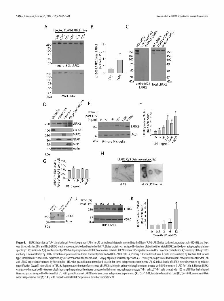
Figure 3. LRRK2 induction by TLR4 stimulation. A, Five micrograms of LPS or no LPS control was bilaterally injected into the SNpc of FLAG-LRRK2 mice (Jackson Laboratory strain 012466), the SNpcwas dissected after 24 h, and FLAG-LRRK2 was immunoprecipitated and treated with ATP. Eluted protein was analyzed by Western blot with either a total LRRK2 antibody- or autophosphorylation-specific pT1503 antibody. B, Quantification of pT1503-autophosphorylated LRRK2 normalized to total LRRK2 from four LPS-injected mice and four injection control mice. C, Specificity of the pT1503antibody is demonstrated by LRRK2 recombinant protein derived from transiently transfected HEK-293FT cells. D, Primary cultures derived from P2 rats were analyzed by Western blot for celltype-specific markers and LRRK2 expression. Lysates were normalized to actin, and�20 �g of protein was loaded per lane. E, F, Primary microglia treated with various concentrations of LPS for 12 hand LRRK2 expression evaluated by Western blot (E), with quantification normalized to actin for three independent experiments (F ). G, mRNA levels of LRRK2 were determined by relativequantification (��cT) normalized to TBP. H, Representative immunofluorescence of LRRK2 staining in primary microglia cultures treated with LPS or control (-LPS) for 12 h. I, Human LRRK2expression characterized by Western blot in human primary microglia cultures compared with human macrophage/monocyte THP-1 cells. J, THP-1 cells treated with 100 ng of LPS for the indicatedtime and lysates analyzed by Western blot (J ), with quantification of LRRK2 levels from three independent experiments (K ). *p � 0.01, two-tailed unpaired t test (B); *p�0.01, one-way ANOVAwith Tukey–Kramer test (B, F, K ), with respect to initial LRRK2 expression. Error bars indicate SEM.
1606 • J. Neurosci., February 1, 2012 • 32(5):1602–1611 Moehle et al. • LRRK2 Activation in Neuroinflammation
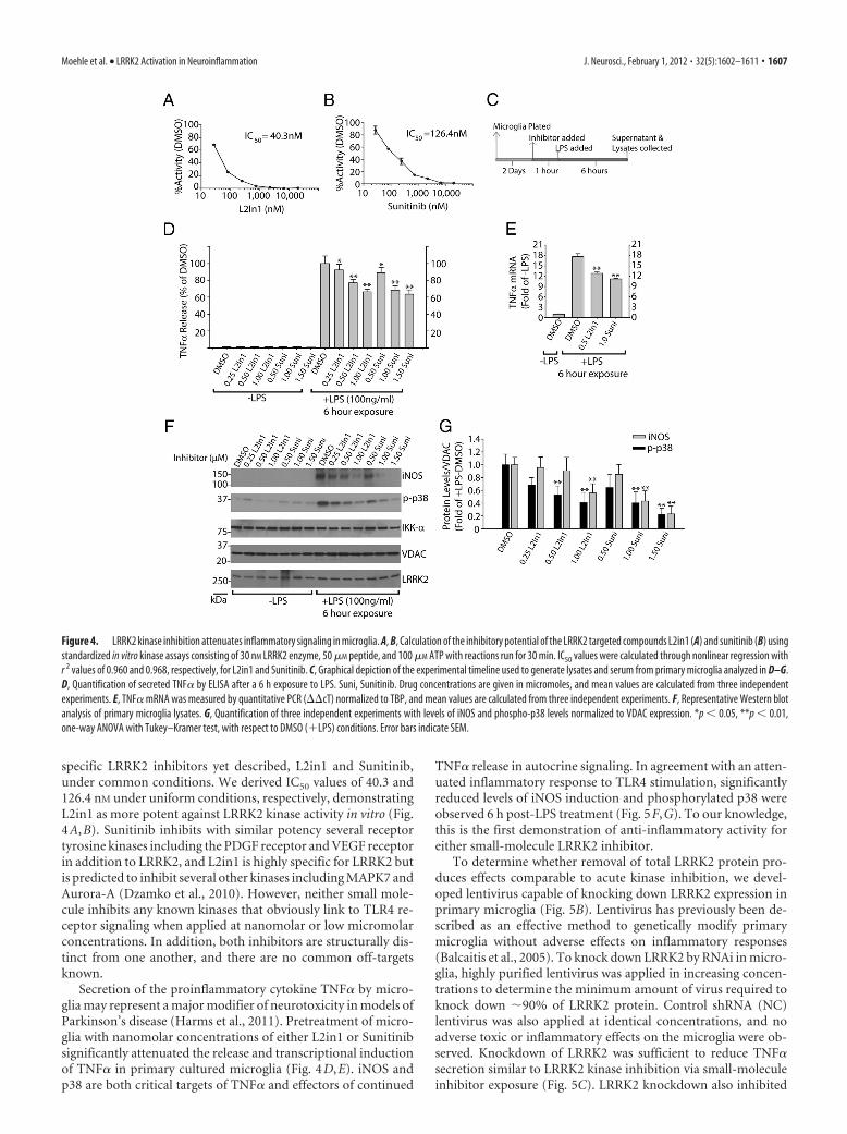
specific LRRK2 inhibitors yet described, L2in1 and Sunitinib,under common conditions. We derived IC50 values of 40.3 and126.4 nM under uniform conditions, respectively, demonstratingL2in1 as more potent against LRRK2 kinase activity in vitro (Fig.4A,B). Sunitinib inhibits with similar potency several receptortyrosine kinases including the PDGF receptor and VEGF receptorin addition to LRRK2, and L2in1 is highly specific for LRRK2 butis predicted to inhibit several other kinases including MAPK7 andAurora-A (Dzamko et al., 2010). However, neither small mole-cule inhibits any known kinases that obviously link to TLR4 re-ceptor signaling when applied at nanomolar or low micromolarconcentrations. In addition, both inhibitors are structurally dis-tinct from one another, and there are no common off-targetsknown.
Secretion of the proinflammatory cytokine TNF� by micro-glia may represent a major modifier of neurotoxicity in models ofParkinson’s disease (Harms et al., 2011). Pretreatment of micro-glia with nanomolar concentrations of either L2in1 or Sunitinibsignificantly attenuated the release and transcriptional inductionof TNF� in primary cultured microglia (Fig. 4D,E). iNOS andp38 are both critical targets of TNF� and effectors of continued
TNF� release in autocrine signaling. In agreement with an atten-uated inflammatory response to TLR4 stimulation, significantlyreduced levels of iNOS induction and phosphorylated p38 wereobserved 6 h post-LPS treatment (Fig. 5F,G). To our knowledge,this is the first demonstration of anti-inflammatory activity foreither small-molecule LRRK2 inhibitor.
To determine whether removal of total LRRK2 protein pro-duces effects comparable to acute kinase inhibition, we devel-oped lentivirus capable of knocking down LRRK2 expression inprimary microglia (Fig. 5B). Lentivirus has previously been de-scribed as an effective method to genetically modify primarymicroglia without adverse effects on inflammatory responses(Balcaitis et al., 2005). To knock down LRRK2 by RNAi in micro-glia, highly purified lentivirus was applied in increasing concen-trations to determine the minimum amount of virus required toknock down �90% of LRRK2 protein. Control shRNA (NC)lentivirus was also applied at identical concentrations, and noadverse toxic or inflammatory effects on the microglia were ob-served. Knockdown of LRRK2 was sufficient to reduce TNF�secretion similar to LRRK2 kinase inhibition via small-moleculeinhibitor exposure (Fig. 5C). LRRK2 knockdown also inhibited
Figure 4. LRRK2 kinase inhibition attenuates inflammatory signaling in microglia. A, B, Calculation of the inhibitory potential of the LRRK2 targeted compounds L2in1 (A) and sunitinib (B) usingstandardized in vitro kinase assays consisting of 30 nM LRRK2 enzyme, 50 �M peptide, and 100 �M ATP with reactions run for 30 min. IC50 values were calculated through nonlinear regression withr 2 values of 0.960 and 0.968, respectively, for L2in1 and Sunitinib. C, Graphical depiction of the experimental timeline used to generate lysates and serum from primary microglia analyzed in D–G.D, Quantification of secreted TNF� by ELISA after a 6 h exposure to LPS. Suni, Sunitinib. Drug concentrations are given in micromoles, and mean values are calculated from three independentexperiments. E, TNF� mRNA was measured by quantitative PCR (��cT) normalized to TBP, and mean values are calculated from three independent experiments. F, Representative Western blotanalysis of primary microglia lysates. G, Quantification of three independent experiments with levels of iNOS and phospho-p38 levels normalized to VDAC expression. *p � 0.05, **p � 0.01,one-way ANOVA with Tukey–Kramer test, with respect to DMSO (�LPS) conditions. Error bars indicate SEM.
Moehle et al. • LRRK2 Activation in Neuroinflammation J. Neurosci., February 1, 2012 • 32(5):1602–1611 • 1607
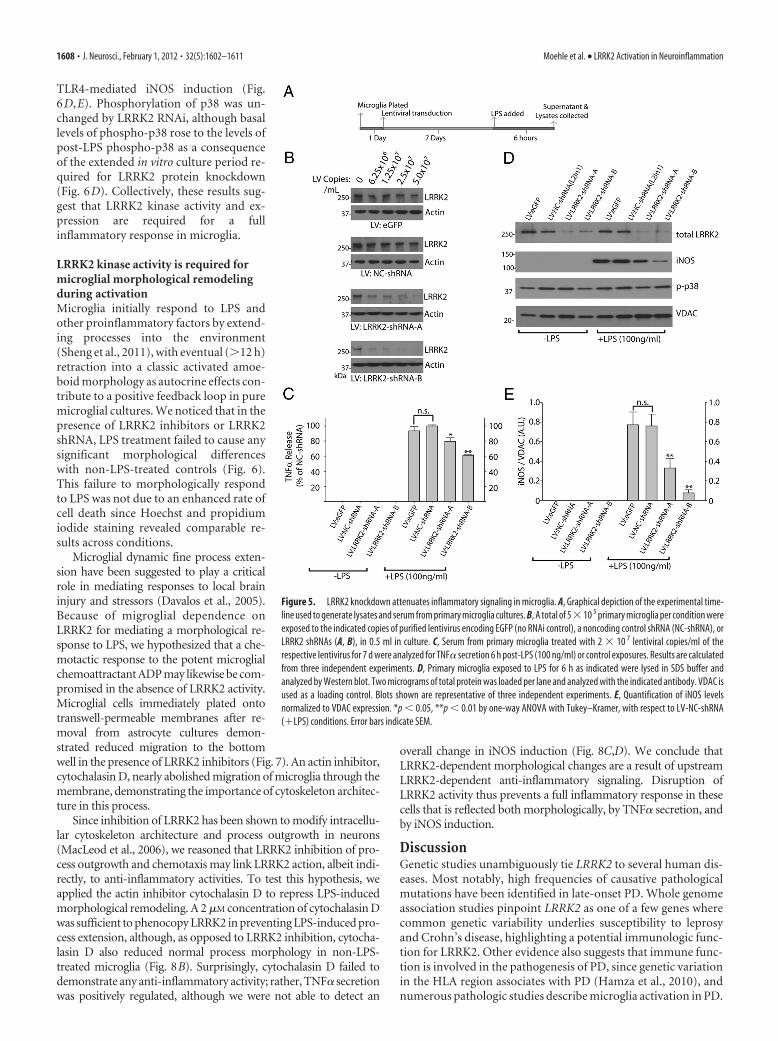
TLR4-mediated iNOS induction (Fig.6D,E). Phosphorylation of p38 was un-changed by LRRK2 RNAi, although basallevels of phospho-p38 rose to the levels ofpost-LPS phospho-p38 as a consequenceof the extended in vitro culture period re-quired for LRRK2 protein knockdown(Fig. 6D). Collectively, these results sug-gest that LRRK2 kinase activity and ex-pression are required for a fullinflammatory response in microglia.
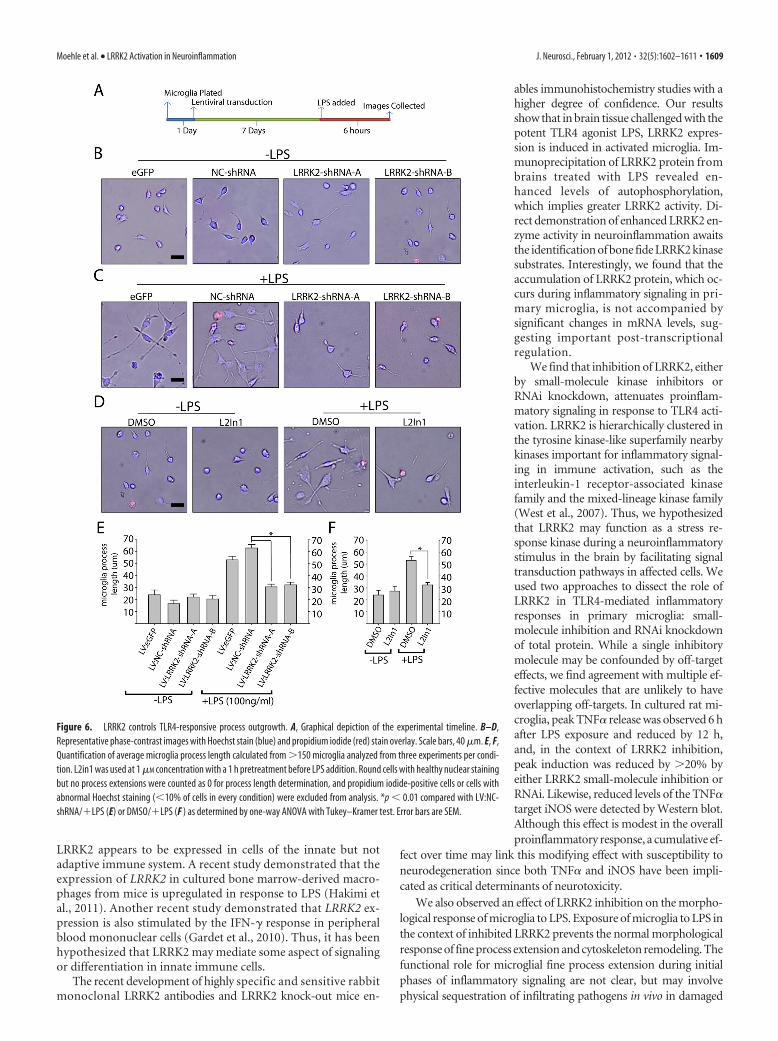
LRRK2 kinase activity is required formicroglial morphological remodelingduring activationMicroglia initially respond to LPS andother proinflammatory factors by extend-ing processes into the environment(Sheng et al., 2011), with eventual (�12 h)retraction into a classic activated amoe-boid morphology as autocrine effects con-tribute to a positive feedback loop in puremicroglial cultures. We noticed that in thepresence of LRRK2 inhibitors or LRRK2shRNA, LPS treatment failed to cause anysignificant morphological differenceswith non-LPS-treated controls (Fig. 6).This failure to morphologically respondto LPS was not due to an enhanced rate ofcell death since Hoechst and propidiumiodide staining revealed comparable re-sults across conditions.
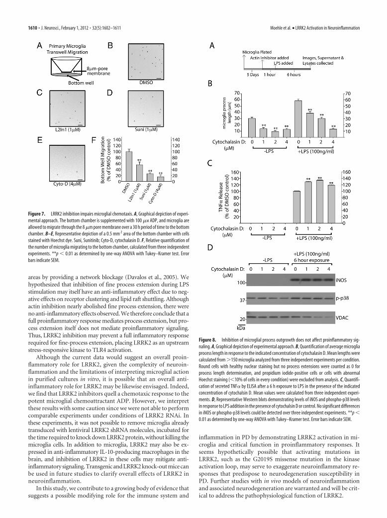
Microglial dynamic fine process exten-sion have been suggested to play a criticalrole in mediating responses to local braininjury and stressors (Davalos et al., 2005).Because of migroglial dependence onLRRK2 for mediating a morphological re-sponse to LPS, we hypothesized that a che-motactic response to the potent microglialchemoattractant ADP may likewise be com-promised in the absence of LRRK2 activity.Microglial cells immediately plated ontotranswell-permeable membranes after re-moval from astrocyte cultures demon-strated reduced migration to the bottomwell in the presence of LRRK2 inhibitors (Fig. 7). An actin inhibitor,cytochalasin D, nearly abolished migration of microglia through themembrane, demonstrating the importance of cytoskeleton architec-ture in this process.
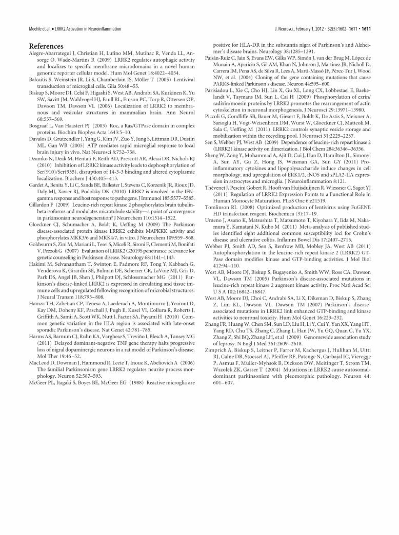
Since inhibition of LRRK2 has been shown to modify intracellu-lar cytoskeleton architecture and process outgrowth in neurons(MacLeod et al., 2006), we reasoned that LRRK2 inhibition of pro-cess outgrowth and chemotaxis may link LRRK2 action, albeit indi-rectly, to anti-inflammatory activities. To test this hypothesis, weapplied the actin inhibitor cytochalasin D to repress LPS-inducedmorphological remodeling. A 2 �M concentration of cytochalasin Dwas sufficient to phenocopy LRRK2 in preventing LPS-induced pro-cess extension, although, as opposed to LRRK2 inhibition, cytocha-lasin D also reduced normal process morphology in non-LPS-treated microglia (Fig. 8B). Surprisingly, cytochalasin D failed todemonstrate any anti-inflammatory activity; rather, TNF� secretionwas positively regulated, although we were not able to detect an
overall change in iNOS induction (Fig. 8C,D). We conclude thatLRRK2-dependent morphological changes are a result of upstreamLRRK2-dependent anti-inflammatory signaling. Disruption ofLRRK2 activity thus prevents a full inflammatory response in thesecells that is reflected both morphologically, by TNF� secretion, andby iNOS induction.
DiscussionGenetic studies unambiguously tie LRRK2 to several human dis-eases. Most notably, high frequencies of causative pathologicalmutations have been identified in late-onset PD. Whole genomeassociation studies pinpoint LRRK2 as one of a few genes wherecommon genetic variability underlies susceptibility to leprosyand Crohn’s disease, highlighting a potential immunologic func-tion for LRRK2. Other evidence also suggests that immune func-tion is involved in the pathogenesis of PD, since genetic variationin the HLA region associates with PD (Hamza et al., 2010), andnumerous pathologic studies describe microglia activation in PD.
Figure 5. LRRK2 knockdown attenuates inflammatory signaling in microglia. A, Graphical depiction of the experimental time-line used to generate lysates and serum from primary microglia cultures. B, A total of 5�10 5 primary microglia per condition wereexposed to the indicated copies of purified lentivirus encoding EGFP (no RNAi control), a noncoding control shRNA (NC-shRNA), orLRRK2 shRNAs (A, B), in 0.5 ml in culture. C, Serum from primary microglia treated with 2 � 10 7 lentiviral copies/ml of therespective lentivirus for 7 d were analyzed for TNF� secretion 6 h post-LPS (100 ng/ml) or control exposures. Results are calculatedfrom three independent experiments. D, Primary microglia exposed to LPS for 6 h as indicated were lysed in SDS buffer andanalyzed by Western blot. Two micrograms of total protein was loaded per lane and analyzed with the indicated antibody. VDAC isused as a loading control. Blots shown are representative of three independent experiments. E, Quantification of iNOS levelsnormalized to VDAC expression. *p � 0.05, **p � 0.01 by one-way ANOVA with Tukey–Kramer, with respect to LV-NC-shRNA(�LPS) conditions. Error bars indicate SEM.
1608 • J. Neurosci., February 1, 2012 • 32(5):1602–1611 Moehle et al. • LRRK2 Activation in Neuroinflammation
LRRK2 appears to be expressed in cells of the innate but notadaptive immune system. A recent study demonstrated that theexpression of LRRK2 in cultured bone marrow-derived macro-phages from mice is upregulated in response to LPS (Hakimi etal., 2011). Another recent study demonstrated that LRRK2 ex-pression is also stimulated by the IFN-� response in peripheralblood mononuclear cells (Gardet et al., 2010). Thus, it has beenhypothesized that LRRK2 may mediate some aspect of signalingor differentiation in innate immune cells.
The recent development of highly specific and sensitive rabbitmonoclonal LRRK2 antibodies and LRRK2 knock-out mice en-
ables immunohistochemistry studies with ahigher degree of confidence. Our resultsshow that in brain tissue challenged with thepotent TLR4 agonist LPS, LRRK2 expres-sion is induced in activated microglia. Im-munoprecipitation of LRRK2 protein frombrains treated with LPS revealed en-hanced levels of autophosphorylation,which implies greater LRRK2 activity. Di-rect demonstration of enhanced LRRK2 en-zyme activity in neuroinflammation awaitsthe identification of bone fide LRRK2 kinasesubstrates. Interestingly, we found that theaccumulation of LRRK2 protein, which oc-curs during inflammatory signaling in pri-mary microglia, is not accompanied bysignificant changes in mRNA levels, sug-gesting important post-transcriptionalregulation.
We find that inhibition of LRRK2, eitherby small-molecule kinase inhibitors orRNAi knockdown, attenuates proinflam-matory signaling in response to TLR4 acti-vation. LRRK2 is hierarchically clustered inthe tyrosine kinase-like superfamily nearbykinases important for inflammatory signal-ing in immune activation, such as theinterleukin-1 receptor-associated kinasefamily and the mixed-lineage kinase family(West et al., 2007). Thus, we hypothesizedthat LRRK2 may function as a stress re-sponse kinase during a neuroinflammatorystimulus in the brain by facilitating signaltransduction pathways in affected cells. Weused two approaches to dissect the role ofLRRK2 in TLR4-mediated inflammatoryresponses in primary microglia: small-molecule inhibition and RNAi knockdownof total protein. While a single inhibitorymolecule may be confounded by off-targeteffects, we find agreement with multiple ef-fective molecules that are unlikely to haveoverlapping off-targets. In cultured rat mi-croglia, peak TNF� release was observed 6 hafter LPS exposure and reduced by 12 h,and, in the context of LRRK2 inhibition,peak induction was reduced by �20% byeither LRRK2 small-molecule inhibition orRNAi. Likewise, reduced levels of the TNF�target iNOS were detected by Western blot.Although this effect is modest in the overallproinflammatory response, a cumulative ef-
fect over time may link this modifying effect with susceptibility toneurodegeneration since both TNF� and iNOS have been impli-cated as critical determinants of neurotoxicity.
We also observed an effect of LRRK2 inhibition on the morpho-logical response of microglia to LPS. Exposure of microglia to LPS inthe context of inhibited LRRK2 prevents the normal morphologicalresponse of fine process extension and cytoskeleton remodeling. Thefunctional role for microglial fine process extension during initialphases of inflammatory signaling are not clear, but may involvephysical sequestration of infiltrating pathogens in vivo in damaged
Figure 6. LRRK2 controls TLR4-responsive process outgrowth. A, Graphical depiction of the experimental timeline. B–D,Representative phase-contrast images with Hoechst stain (blue) and propidium iodide (red) stain overlay. Scale bars, 40 �m. E, F,Quantification of average microglia process length calculated from �150 microglia analyzed from three experiments per condi-tion. L2in1 was used at 1 �M concentration with a 1 h pretreatment before LPS addition. Round cells with healthy nuclear stainingbut no process extensions were counted as 0 for process length determination, and propidium iodide-positive cells or cells withabnormal Hoechst staining (�10% of cells in every condition) were excluded from analysis. *p � 0.01 compared with LV:NC-shRNA/�LPS (E) or DMSO/�LPS (F ) as determined by one-way ANOVA with Tukey–Kramer test. Error bars are SEM.
Moehle et al. • LRRK2 Activation in Neuroinflammation J. Neurosci., February 1, 2012 • 32(5):1602–1611 • 1609
areas by providing a network blockage (Davalos et al., 2005). Wehypothesized that inhibition of fine process extension during LPSstimulation may itself have an anti-inflammatory effect due to neg-ative effects on receptor clustering and lipid raft shuttling. Althoughactin inhibition nearly abolished fine process extension, there wereno anti-inflammatory effects observed. We therefore conclude that afull proinflammatory response mediates process extension, but pro-cess extension itself does not mediate proinflammatory signaling.Thus, LRRK2 inhibition may prevent a full inflammatory responserequired for fine-process extension, placing LRRK2 as an upstreamstress-responsive kinase to TLR4 activation.
Although the current data would suggest an overall proin-flammatory role for LRRK2, given the complexity of neuroin-flammation and the limitations of interpreting microglial actionin purified cultures in vitro, it is possible that an overall anti-inflammatory role for LRRK2 may be likewise envisaged. Indeed,we find that LRRK2 inhibitors quell a chemotaxic response to thepotent microglial chemoattractant ADP. However, we interpretthese results with some caution since we were not able to performcomparable experiments under conditions of LRRK2 RNAi. Inthese experiments, it was not possible to remove microglia alreadytransduced with lentiviral LRRK2 shRNA molecules, incubated forthe time required to knock down LRRK2 protein, without killing themicroglia cells. In addition to microglia, LRRK2 may also be ex-pressed in anti-inflammatory IL-10-producing macrophages in thebrain, and inhibition of LRRK2 in these cells may mitigate anti-inflammatory signaling. Transgenic and LRRK2 knock-out mice canbe used in future studies to clarify overall effects of LRRK2 inneuroinflammation.
In this study, we contribute to a growing body of evidence thatsuggests a possible modifying role for the immune system and
inflammation in PD by demonstrating LRRK2 activation in mi-croglia and critical function in proinflammatory responses. Itseems hypothetically possible that activating mutations inLRRK2, such as the G2019S missense mutation in the kinaseactivation loop, may serve to exaggerate neuroinflammatory re-sponses that predispose to neurodegeneration susceptibility inPD. Further studies with in vivo models of neuroinflammationand associated neurodegeneration are warranted and will be crit-ical to address the pathophysiological function of LRRK2.
Figure 7. LRRK2 inhibition impairs microglial chemotaxis. A, Graphical depiction of experi-mental approach. The bottom chamber is supplemented with 100 �M ADP, and microglia areallowed to migrate through the 8 �m pore membrane over a 30 h period of time to the bottomchamber. B–E, Representative depiction of a 0.5 mm 2 area of the bottom chamber with cellsstained with Hoechst dye. Suni, Sunitinib; Cyto-D, cytochalasin D. F, Relative quantification ofthe number of microglia migrating to the bottom chamber, calculated from three independentexperiments. **p � 0.01 as determined by one-way ANOVA with Tukey–Kramer test. Errorbars indicate SEM.
Figure 8. Inhibition of microglial process outgrowth does not affect proinflammatory sig-naling. A, Graphical depiction of experimental approach. B, Quantification of average microgliaprocess length in response to the indicated concentration of cytochalasin D. Mean lengths werecalculated from �150 microglia analyzed from three independent experiments per condition.Round cells with healthy nuclear staining but no process extensions were counted as 0 forprocess length determination, and propidium iodide-positive cells or cells with abnormalHoechst staining (�10% of cells in every condition) were excluded from analysis. C, Quantifi-cation of secreted TNF� by ELISA after a 6 h exposure to LPS in the presence of the indicatedconcentration of cytochalsin D. Mean values were calculated from three independent experi-ments. D, Representative Western blots demonstrating levels of iNOS and phospho-p38 levelsin response to LPS addition in the presence of cytochalsin D or control. No significant differencesin iNOS or phospho-p38 levels could be detected over three independent experiments. **p �0.01 as determined by one-way ANOVA with Tukey–Kramer test. Error bars indicate SEM.
1610 • J. Neurosci., February 1, 2012 • 32(5):1602–1611 Moehle et al. • LRRK2 Activation in Neuroinflammation
ReferencesAlegre-Abarrategui J, Christian H, Lufino MM, Mutihac R, Venda LL, An-
sorge O, Wade-Martins R (2009) LRRK2 regulates autophagic activityand localizes to specific membrane microdomains in a novel humangenomic reporter cellular model. Hum Mol Genet 18:4022– 4034.
Balcaitis S, Weinstein JR, Li S, Chamberlain JS, Moller T (2005) Lentiviraltransduction of microglial cells. Glia 50:48 –55.
Biskup S, Moore DJ, Celsi F, Higashi S, West AB, Andrabi SA, Kurkinen K, YuSW, Savitt JM, Waldvogel HJ, Faull RL, Emson PC, Torp R, Ottersen OP,Dawson TM, Dawson VL (2006) Localization of LRRK2 to membra-nous and vesicular structures in mammalian brain. Ann Neurol60:557–569.
Bosgraaf L, Van Haastert PJ (2003) Roc, a Ras/GTPase domain in complexproteins. Biochim Biophys Acta 1643:5–10.
Davalos D, Grutzendler J, Yang G, Kim JV, Zuo Y, Jung S, Littman DR, DustinML, Gan WB (2005) ATP mediates rapid microglial response to localbrain injury in vivo. Nat Neurosci 8:752–758.
Dzamko N, Deak M, Hentati F, Reith AD, Prescott AR, Alessi DR, Nichols RJ(2010) Inhibition of LRRK2 kinase activity leads to dephosphorylation ofSer(910)/Ser(935), disruption of 14-3-3 binding and altered cytoplasmiclocalization. Biochem J 430:405– 413.
Gardet A, Benita Y, Li C, Sands BE, Ballester I, Stevens C, Korzenik JR, Rioux JD,Daly MJ, Xavier RJ, Podolsky DK (2010) LRRK2 is involved in the IFN-gamma response and host response to pathogens. J Immunol 185:5577–5585.
Gillardon F (2009) Leucine-rich repeat kinase 2 phosphorylates brain tubulin-beta isoforms and modulates microtubule stability—a point of convergencein parkinsonian neurodegeneration? J Neurochem 110:1514–1522.
Gloeckner CJ, Schumacher A, Boldt K, Ueffing M (2009) The Parkinsondisease-associated protein kinase LRRK2 exhibits MAPKKK activity andphosphorylates MKK3/6 and MKK4/7, in vitro. J Neurochem 109:959–968.
Goldwurm S, Zini M, Mariani L, Tesei S, Miceli R, Sironi F, Clementi M, BonifatiV, Pezzoli G (2007) Evaluation of LRRK2 G2019S penetrance: relevance forgenetic counseling in Parkinson disease. Neurology 68:1141–1143.
Hakimi M, Selvanantham T, Swinton E, Padmore RF, Tong Y, Kabbach G,Venderova K, Girardin SE, Bulman DE, Scherzer CR, LaVoie MJ, Gris D,Park DS, Angel JB, Shen J, Philpott DJ, Schlossmacher MG (2011) Par-kinson’s disease-linked LRRK2 is expressed in circulating and tissue im-mune cells and upregulated following recognition of microbial structures.J Neural Transm 118:795– 808.
Hamza TH, Zabetian CP, Tenesa A, Laederach A, Montimurro J, Yearout D,Kay DM, Doheny KF, Paschall J, Pugh E, Kusel VI, Collura R, Roberts J,Griffith A, Samii A, Scott WK, Nutt J, Factor SA, Payami H (2010) Com-mon genetic variation in the HLA region is associated with late-onsetsporadic Parkinson’s disease. Nat Genet 42:781–785.
Harms AS, Barnum CJ, Ruhn KA, Varghese S, Trevino I, Blesch A, Tansey MG(2011) Delayed dominant-negative TNF gene therapy halts progressiveloss of nigral dopaminergic neurons in a rat model of Parkinson’s disease.Mol Ther 19:46 –52.
MacLeod D, Dowman J, Hammond R, Leete T, Inoue K, Abeliovich A (2006)The familial Parkinsonism gene LRRK2 regulates neurite process mor-phology. Neuron 52:587–593.
McGeer PL, Itagaki S, Boyes BE, McGeer EG (1988) Reactive microglia are
positive for HLA-DR in the substantia nigra of Parkinson’s and Alzhei-mer’s disease brains. Neurology 38:1285–1291.
Paisan-Ruíz C, Jain S, Evans EW, Gilks WP, Simon J, van der Brug M, Lopez deMunain A, Aparicio S, Gil AM, Khan N, Johnson J, Martinez JR, Nicholl D,Carrera IM, Pena AS, de Silva R, Lees A, Martí-Masso JF, Perez-Tur J, WoodNW, et al. (2004) Cloning of the gene containing mutations that causePARK8-linked Parkinson’s disease. Neuron 44:595–600.
Parisiadou L, Xie C, Cho HJ, Lin X, Gu XL, Long CX, Lobbestael E, Baeke-landt V, Taymans JM, Sun L, Cai H (2009) Phosphorylation of ezrin/radixin/moesin proteins by LRRK2 promotes the rearrangement of actincytoskeleton in neuronal morphogenesis. J Neurosci 29:13971–13980.
Piccoli G, Condliffe SB, Bauer M, Giesert F, Boldt K, De Astis S, Meixner A,Sarioglu H, Vogt-Weisenhorn DM, Wurst W, Gloeckner CJ, Matteoli M,Sala C, Ueffing M (2011) LRRK2 controls synaptic vesicle storage andmobilization within the recycling pool. J Neurosci 31:2225–2237.
Sen S, Webber PJ, West AB (2009) Dependence of leucine-rich repeat kinase 2(LRRK2) kinase activity on dimerization. J Biol Chem 284:36346–36356.
Sheng W, Zong Y, Mohammad A, Ajit D, Cui J, Han D, Hamilton JL, SimonyiA, Sun AY, Gu Z, Hong JS, Weisman GA, Sun GY (2011) Pro-inflammatory cytokines and lipopolysaccharide induce changes in cellmorphology, and upregulation of ERK1/2, iNOS and sPLA2-IIA expres-sion in astrocytes and microglia. J Neuroinflammation 8:121.
Thevenet J, Pescini Gobert R, Hooft van Huijsduijnen R, Wiessner C, Sagot YJ(2011) Regulation of LRRK2 Expression Points to a Functional Role inHuman Monocyte Maturation. PLoS One 6:e21519.
Tomlinson RL (2008) Optimized production of lentivirus using FuGENEHD transfection reagent. Biochemica (3):17–19.
Umeno J, Asano K, Matsushita T, Matsumoto T, Kiyohara Y, Iida M, Naka-mura Y, Kamatani N, Kubo M (2011) Meta-analysis of published stud-ies identified eight additional common susceptibility loci for Crohn’sdisease and ulcerative colitis. Inflamm Bowel Dis 17:2407–2715.
Webber PJ, Smith AD, Sen S, Renfrow MB, Mobley JA, West AB (2011)Autophosphorylation in the leucine-rich repeat kinase 2 (LRRK2) GT-Pase domain modifies kinase and GTP-binding activities. J Mol Biol412:94 –110.
West AB, Moore DJ, Biskup S, Bugayenko A, Smith WW, Ross CA, DawsonVL, Dawson TM (2005) Parkinson’s disease-associated mutations inleucine-rich repeat kinase 2 augment kinase activity. Proc Natl Acad SciU S A 102:16842–16847.
West AB, Moore DJ, Choi C, Andrabi SA, Li X, Dikeman D, Biskup S, ZhangZ, Lim KL, Dawson VL, Dawson TM (2007) Parkinson’s disease-associated mutations in LRRK2 link enhanced GTP-binding and kinaseactivities to neuronal toxicity. Hum Mol Genet 16:223–232.
Zhang FR, Huang W, Chen SM, Sun LD, Liu H, Li Y, Cui Y, Yan XX, Yang HT,Yang RD, Chu TS, Zhang C, Zhang L, Han JW, Yu GQ, Quan C, Yu YX,Zhang Z, Shi BQ, Zhang LH, et al (2009) Genomewide association studyof leprosy. N Engl J Med 361:2609 –2618.
Zimprich A, Biskup S, Leitner P, Farrer M, Kachergus J, Hulihan M, UittiRJ, Calne DB, Stoessel AJ, Pfeiffer RF, Patenge N, Carbajal IC, ViereggeP, Asmus F, Muller-Myhsok B, Dickson DW, Meitinger T, Strom TM,Wszolek ZK, Gasser T (2004) Mutations in LRRK2 cause autosomal-dominant parkinsonism with pleomorphic pathology. Neuron 44:601– 607.
Moehle et al. • LRRK2 Activation in Neuroinflammation J. Neurosci., February 1, 2012 • 32(5):1602–1611 • 1611