neurobiologyofdisease insulindysfunctioninduces invivo … ·...
TRANSCRIPT

Neurobiology of Disease
Insulin Dysfunction Induces In Vivo TauHyperphosphorylation through Distinct Mechanisms
Emmanuel Planel,1,4 Yoshitaka Tatebayashi,1,2 Tomohiro Miyasaka,1,3 Li Liu,4 Lili Wang,4 Mathieu Herman,4
W. Haung Yu,4 Jose A. Luchsinger,5 Brian Wadzinski,6 Karen E. Duff,4 and Akihiko Takashima1
1Laboratory for Alzheimer’s Disease, Brain Science Institute, The Institute of Physical and Chemical Research, Saitama 351-0198, Japan, 2DepressionProject, Mood Disorder Research Team, Tokyo Institute of Psychiatry, Tokyo 156-8585, Japan, 3Department of Neuropathology, Faculty of Medicine,University of Tokyo, Tokyo 113-0033, Japan, 4Department of Pathology, Taub Institute for Alzheimer’s Disease Research, Columbia University MedicalCenter, 5Columbia University, New York, New York 10032, and 6Vanderbilt University Medical Center, Nashville, Tennessee 37232
Hyperphosphorylated tau is the major component of paired helical filaments in neurofibrillary tangles found in Alzheimer’s disease (AD)brains, and tau hyperphosphorylation is thought to be a critical event in the pathogenesis of the disease. The large majority of AD cases islate onset and sporadic in origin, with aging as the most important risk factor. Insulin resistance, impaired glucose tolerance, anddiabetes mellitus (DM) are other common syndromes in the elderly also strongly age dependent, and there is evidence supporting a linkbetween insulin dysfunction and AD. To investigate the possibility that insulin dysfunction might promote tau pathology, we inducedinsulin deficiency and caused DM in mice with streptozotocin (STZ). A mild hyperphosphorylation of tau could be detected 10, 20, and30 d after STZ injection, and a massive hyperphosphorylation of tau was observed after 40 d. The robust hyperphosphorylation of tau waslocalized in the axons and neuropil, and prevented tau binding to microtubules. Neither mild nor massive tau phosphorylation inducedtau aggregation. Body temperature of the STZ-treated mice did not differ from control animals during 30 d, but dropped significantlythereafter. No change in �-amyloid (A�) precursor protein (APP), APP C-terminal fragments, or A� levels were observed in STZ-treatedmice; however, cellular protein phosphatase 2A activity was significantly decreased. Together, these data indicate that insulin dysfunc-tion induced abnormal tau hyperphosphorylation through two distinct mechanisms: one was consequent to hypothermia; the other wastemperature-independent, inherent to insulin depletion, and probably caused by inhibition of phosphatase activity.
Key words: Alzheimer’s disease; tau hyperphosphorylation; �-amyloid precursor protein; insulin deficiency; streptozotocin; diabetesmellitus; hypothermia; kinase; serine/threonine protein phosphatase; PP2A; GSK-3; cdk5; JNK; MAPK; CaMKII
IntroductionAlzheimer’s disease (AD) brains are characterized by extracellu-lar aggregates of the �-amyloid peptide (A�) (Glenner andWong, 1984), and intraneuronal neurofibrillary tangles (NFTs),composed of hyperphosphorylated tau protein assembled inpaired helical filaments (PHFs) (Grundke-Iqbal et al., 1986). Tauhyperphosphorylation can induce aggregation (Alonso et al.,2001; Sato et al., 2002), and is thought to induce NFTs and neu-rodegeneration in AD (Trojanowski and Lee, 1994). However,the causes of tau hyperphosphorylation in AD are not wellunderstood.
Only a small proportion of AD is attributable to genetic vari-ants, the large majority of cases is late onset and sporadic in
origin. Aging is considered to be the most important risk factorfor AD (Harman, 2002). Other common syndromes in the el-derly, diabetes mellitus (DM), impaired glucose tolerance, insu-lin resistance, and relative decrease of pancreas endocrine activityand insulin secretion, are also associated with aging (Lamberts etal., 1997). Moreover, insulin signaling seems to be involved in themodulation of lifespan and brain aging (Cole and Frautschy,2007).
There is increasing evidence to support a link between AD andinsulin dysfunction. Compared with age-matched controls, ADpatients show reduced CSF insulin (Craft et al., 1998), impairedinsulin-like signal transduction (Frolich et al., 1999), decreasedbrain insulin and insulin-like growth factor I expression andfunction (Rivera et al., 2005), and reduced glucose metabolism(Mosconi, 2005). From these observations, it was suggested thatbrain insulin signaling dysfunction and the possible consequentreduction in glucose metabolism are crucial in the genesis of AD(Meier-Ruge and Bertoni-Freddari, 1996; Salehi and Swaab,1999; Heininger, 2000; Hoyer, 2000, 2002; Gasparini et al., 2002;Watson and Craft, 2003). Insulin dysfunction is a feature of DM,and, although AD and DM were first found to be strongly asso-ciated or mutually exclusive in a series of conflicting epidemio-logical studies (Finch and Cohen, 1997), many longitudinal
Received March 15, 2007; revised Oct. 17, 2007; accepted Oct. 18, 2007.This work was supported by National Institutes of Health/New York University Alzheimer’s Disease Center Pilot
Project Grant AG008051 (E.P.) and National Institute of Neurological Disorders and Stroke Grant NS048447 (K.E.D.).We thank Drs. Peter Davies (Albert Einstein University, New York, NY), Yasuo Ihara (Tokyo University, Tokyo, Japan),Sonia Jung (Centocor R&D, Radnor, PA), and Paul Mathews (Nathan Kline Institute/New York University, Orange-burg, NY) for the generous gift of antibodies.
Correspondence should be addressed to Dr. Emmanuel Planel, Columbia University Medical Center, Departmentof Pathology, Taub Institute for Alzheimer’s Disease Research, Black Building #5-513, 650 West 168th Street, NewYork, NY 10032. E-mail: [email protected].
DOI:10.1523/JNEUROSCI.3949-07.2007Copyright © 2007 Society for Neuroscience 0270-6474/07/2713635-14$15.00/0
The Journal of Neuroscience, December 12, 2007 • 27(50):13635–13648 • 13635

population-based studies detected higher AD incidences rates indiabetic patients (Biessels et al., 2006). In addition, insulin can actas a neurotrophic and regulatory peptide in human brain, and iscapable of modulating tau phosphorylation in neuronal cultures(Hong and Lee, 1997; Lesort et al., 1999; Lesort and Johnson,2000). Despite all these data, very little is known on the effects ofinsulin dysfunction on tau phosphorylation in vivo.
To investigate the role of insulin dysfunction on tau phos-phorylation, we deprived the mice of insulin by injections ofstreptozotocin (STZ), which induces experimental DM by specif-ically destroying the pancreatic islets of Langerhans (Tjalve,1983). In contrast with many peripheral tissues, brain insulinbinding and insulin receptors levels are not upregulated duringexperimental DM (Pacold and Blackard, 1979; Sechi et al., 1992;Pezzino et al., 1996), which leads to lower brain insulin content(Figlewicz et al., 1983; Frank et al., 1986). Our data indicate thatinsulin dysfunction induced by STZ results in tau hyperphospho-rylation through two distinct mechanisms: one is consequent tohypothermia and induced by impaired glucose/energy metabo-lism; the other is temperature-independent, inherent to insulindeficiency, and induced by inhibition of Ser/Thr phosphatasesactivities.
Materials and MethodsAnimalsC57BL/6NJcl adult mice of either sex (Clea, Tokyo, Japan) ranging from3 to 6 months of age were used in these studies. Younger animals wereavoided, because juvenile rodents tend to develop hypothermia very rap-idly after injection of streptozotocin (Howarth et al., 2005). Mice weremaintained in a temperature-controlled room (�23°C) with a 12 h light/dark cycle, and experiments were performed during the light period.Animals were handled according to procedures approved by the Institu-tional Animal Care and Use Committee of the Institute of Physical andChemical Research. The body temperature of the animals was monitoredusing a rectal probe (Thermalert TH-5; Physitemp, Clifton, NJ).
Streptozotocin injections, insulin pumps implantation, plasmaglucose, and insulin quantificationBatches of four, nonfasted animals with food and water ad libitum wereinjected intraperitoneally with 200 mg/kg of STZ (Sigma, St. Louis, MO)freshly dissolved in 0.05 M citrate buffer, pH 4.5; control mice were in-jected with citrate buffer. STZ is used at various doses to induce experi-mental diabetes (Weber et al., 1984), usually after a period of fasting,which was avoided here because fasting can induce tau hyperphospho-rylation (Planel et al., 2001). The dose for our studies was specificallychosen to avoid induction of progressive diabetes mellitus (with normalserum insulin levels) that can occur in mice with lower doses. For exam-ple, one injection of 100 mg/kg can induce non-insulin-dependent DM,whereas one injection of 200 mg/kg induces insulin-dependent DM (Itoet al., 1999). Brains were collected 10, 20, 30, or 40 d after injection, andanalyzed as described below. To reverse the effect of STZ, 3 d after injec-tion, mice were implanted with subcutaneous osmotic pumps delivering0.75 U/d insulin (Humulin R; Eli Lilly, Indianapolis, IN) according to aprotocol modified from Cuthbertson et al. (1988). Pump implantationcompletely reversed diabetic symptoms in mice treated with a singleintraperitoneal injection of 250 mg/kg of STZ (Cuthbertson et al., 1988).Plasma glucose was determined using the mutarotase-glucose oxydasemethod according to the manufacturer’s instructions (Autokit Glucose;Wako Pure Chemical, Osaka, Japan). Plasma insulin was determinedusing a sandwich enzyme immunoassay according to the manufacturer’sinstructions (mouse insulin kit; Shibayagi, Gunma, Japan).
Analysis of tau solubilityMice were killed by cervical dislocation, brains were immediately re-moved, and tissues were dissected on ice. Hemispheres (hippocampusand neocortex) were quickly weighed, frozen on dry ice, and kept at�80°C. Tau solubility was analyzed by a modification of the protocol of
Greenberg and Davies (1990) (Noble et al., 2005). Briefly, frozen hemi-spheres were homogenized without thawing in 5� (v/w) RIPA buffer (50mM Tris-HCl, pH 7.4, 1% NP-40, 0.25% Na-deoxycholate, 150 mM NaCl,1 mM EDTA, 1 mM PMSF, 1 mM Na3VO4, 1 mM NaF, 1 �g/ml aprotinin,leupeptin, and pepstatin) with a mechanical microhomogenizer (Phy-scotron NS-310E; Microtek, Chiba, Japan), and centrifuged at 20,000 �g for 20 min at 4°C. An aliquot of the supernatant representing the totaltau fraction was kept for analysis. The heat-stable, aggregate-free fractionwas obtained by boiling another aliquot for 5 min and removing proteinaggregates by spinning at 20,000 � g for 20 min at 4°C. The rest of thesupernatant was adjusted to 1% Sarkosyl (N-lauroylsarcosine), incu-bated for 30 min at room temperature with constant shaking, and cen-trifuged at 100,000 � g for 1 h at 20°C. The pellet containing Sarkosyl-insoluble aggregated tau was resuspended and analyzed by SDS-PAGE.Tau in the Sarkosyl pellet has been shown by immunoelectron micros-copy to be filamentous (Noble et al., 2003), and it is synonymous withthat identified by immunohistochemistry in NFTs. All three fractionswere diluted in O� buffer [62.5 mM Tris-HCl, pH 6.8, 10% glycerol, 5%2-mercaptoethanol, 2.3% SDS, 1 mM EGTA, 1 mM EDTA, 1 mM PMSF, 1mM Na3VO4, 1 mM NaF, 10 �l/ml protease inhibitor mixture P8340(Sigma)], a modified O buffer (O’Farrell, 1975), boiled for 3 min, andkept at �20°C.
Microtubule binding assayThe microtubule (MT)-binding assay was performed as described previ-ously (Maas et al., 2000), with a few modifications. Hemispheres werehomogenized in 5� (w/v) BRB80 (Brinkley Reassembly Buffer; 80 mM
PIPES/KOH, pH 6.8, 1 mM EGTA, 1 mM MgCl2) with protease andphosphatase inhibitors. Homogenates were incubated on ice for 15 minand centrifuged at 20,000 � g for 20 min at 4°C. Supernatants wererespun at 100,000 � g for 1 h at 4°C. Equal amounts of protein lysate wereused in each binding assay. High-speed supernatants were adjusted to 1mM GTP and 10 �M taxol and incubated with taxol-stabilized microtu-bules (30 �M; Cytoskeleton, Denver, CO) in a final volume of 50 �l for 10min at 37°C. The mixtures were centrifuged through 100 �l of 30% (w/v)sucrose cushions in BRB80 containing 1 mM GTP and 10 �M taxol, at100,000 � g for 30 min at room temperature. The supernatant (MT-freefraction) was collected and diluted with O� buffer, and the pellet (MT-bound fraction) was resuspended in O� buffer.
Protein extractionMice were killed by cervical dislocation, brains were immediately re-moved, and tissues were dissected on ice. Hemispheres (hippocampusand neocortex) were quickly weighed, frozen on dry ice, and maintainedat �80°C until they were homogenized without thawing in 5� (v/w)RIPA buffer with a mechanical microhomogenizer. Samples were thencentrifuged for 20 min at 100,000 � g at 4°C, diluted in O� buffer, andboiled for 3 min. Depending on the antibody used, 7–14 �g of proteinwere analyzed as described previously (Planel et al., 2001).
AntibodiesThe anti-tau PHF-1 (phospho-Ser-396/404) monoclonal antibody was agenerous gift from Dr. Peter Davies (Albert Einstein University, NewYork, NY). Total tau was detected with either Tau T57120 (monoclonal;BD Transduction Laboratories, San Jose, CA) or Tau A0024 (polyclonal;DakoCytomation, Carpinteria, CA). AT8 and AT180 (Pierce Biotechnol-ogy, Rockford, IL) reacts with tau phosphorylated at Ser-202 and Thr-205 (Goedert et al., 1995), and tau phosphorylated at Thr-231 (Goedertet al., 1994), respectively. Tau-1 (Chemicon International, Temecula,CA) recognizes tau dephosphorylated at Ser-195, Ser-198, Ser-199, andSer-202 (Szendrei et al., 1993). Purified rabbit polyclonal anti-tau anti-bodies anti-tau pS199, pS262, pS356, pS400, and pS422 were purchasedfrom Biosource International (Camarillo, CA). Anti-microtubule-associated protein 2 (MAP2) antibody was kindly provided by Dr. YasuoIhara (Tokyo University, Tokyo, Japan). Changes in the expression andphosphorylation of tau kinases were investigated with the following an-tibodies: cyclin-dependent kinase 5 (cdk-5), p35C, calcium/calmodulin-dependent kinase II (CaMKII), phospho-CaMKII (Santa Cruz Biotech-nology, Santa Cruz, CA); glycogen synthase kinase 3� (GSK-3�) (BDTransduction Laboratories); anti-GSK-3�/� [pY219/pY216] (Biosource
13636 • J. Neurosci., December 12, 2007 • 27(50):13635–13648 Planel et al. • Insulin Dysfunction and Tau Hyperphosphorylation

International), phospho-GSK-3� (Ser9), stress-activated protein kinase(SAPK)/c-Jun N-terminal kinase (JNK), phospho-SAPK/JNK (T183/Y185) G9, p44/42 mitogen-activated protein kinase (MAPK), phospho-p44/42 MAPK (T202/Y204) (Cell Signaling Technology, Danvers, MA).Levels of protein phosphatases were analyzed with the following antibod-ies: protein phosphatase 1 (PP1) (E-9) and PP2A-A (H-300) from SantaCruz Biotechnology; PP2B (calcineurin A) from R&D Systems (Minne-apolis, MN); PP2A-C (7A6) from Upstate Biotechnology (Lake Placid,NY); PP2A B� and B� were described previously (Strack et al., 1998). ForA� precursor protein (APP) and A� analysis, the following antibodieswere used: C1/6.1 anti-APP full-length and C-terminal fragments (CTFs)(Mathews et al., 2002) (generous gift from Dr. Paul Mathews, NathanKline Institute/New York University, Orangeburg, NY); and JRF/cA�40and JRF/cA�42/26 (murine A�1– 40 and A�1– 42, respectively; generousgift from Dr. Sonia Jung, Centocor R&D, Radnor, PA).
Western blot analysis and ELISAMembrane blocking and antibody incubations were performed as de-scribed previously (Planel et al., 2001), with appropriate primary andsecondary antibody dilutions. Anti-mouse and anti-rabbit HRP-conjugated secondary antibodies were purchased from Jackson Immu-noResearch (West Grove, PA). Immunoreactive bands were visualizedand analyzed by enhanced chemiluminescence reagent (SuperSignalWest Pico or Femto; Pierce Biotechnology) using a Fujifilm LAS3000imaging system and the Image Gauge Mac OS X software. Serial dilutionsof brain extracts were loaded in gels to obtain calibration curves forreliable quantification. ELISAs for soluble murine A�40 and A�42 wereperformed as described previously, using diethyl acetate extracts fromfresh hemibrains (Burns et al., 2003; Schmidt et al., 2005).
ImmunohistochemistryTissue fixation was done according to the “cold Bouin’s method” (Planelet al., 2004). This procedure minimizes postmortem tau dephosphoryla-tion in mice brains (our unpublished observations). Briefly, mice werekilled by cervical dislocation, and the brain was quickly removed andimmersed in ice-cold Bouin’s solution (saturated picric acid, formalin,acetic acid at 15:5:1), for 24 h and then embedded in paraffin blocks.Eight- to 10-�m-thick sections were processed for immunohistochemi-cal analyses. Deparaffinized and hydrated sections were incubated inTarget Retrieval Solution (DakoCytomation) at 70°C for 25 min for en-hancement of the immunoreactivity, and then incubated in 10% normalgoat serum in PBS at 4°C for 2 h. The specimens were incubated inprimary antibodies diluted in 1% BSA in PBS containing 0.05% of Tween20 overnight at 4°C. Bound antibodies were visualized with Alexa 488-conjugated anti-mouse or Alexa 568-conjugated anti-rabbit IgG (In-vitrogen, Eugene, OR). Immunolabeled tissues were observed under anOlympus (Tokyo, Japan) BX50 microscope equipped with an OlympusDP50 digital CCD camera and the Studio Lite 1.0 software (Pixera Cor-poration, Tokyo, Japan).
Phosphatases activitiesThe effect of STZ injections on brain phosphatase activities was deter-mined using three different assays.
Endogenous tau dephosphorylation assay. This assay was performed asdescribed previously (Planel et al., 2007). Briefly, control or 10 d STZ-treated mice were killed by cervical dislocation, brains were immediatelyremoved, and one hemisphere (hippocampus and neocortex) was ho-mogenized in 5� (v/w) phosphatase sample buffer [50 mM Tris-HCl, pH7.5, 0.25 M sucrose, 10 mM 2-mercaptoethanol, 0.1 mM EDTA, 1 mM
benzamidine, 1 mM PMSF, 10 �l/ml protease inhibitor mixture P8340Sigma)]. The supernatant was diluted 10� in phosphatase assay buffer[50 mM Tris-HCl, pH 7.0, 0.1 mM EDTA, 5 mM DTT, 0.01% Brij 35, 1 mM
PMSF, 10 �l/ml protease inhibitor mixture P8340 (Sigma)], incubated at37°C, and sampled at different intervals. Endogenous PHF-1 phosphor-ylation levels were monitored by Western blotting.
Phosphatase assay with synthetic peptide. To verify the results from theabove phosphatase assay, we performed a PP2A assay using the Serine/Threonine Phosphatase Assay System from Promega (Madison, WI)(V2460). Briefly, mice were killed by cervical dislocation, brain was im-mediately removed, and one hemisphere (hippocampus and neocortex)
was homogenized in 5� (v/w) phosphatase sample buffer and immedi-ately processed as indicated by the manufacturer to remove particulatematter and endogenous free phosphate. For PP2A activity, 2.0 �l of theresulting solution was prepared in duplicate, and the release of phosphatefrom a chemically synthesized phosphopeptide was assessed over a pe-riod of 10 min in PP2A buffer at 37°C (calibration data not shown). Theamount of phosphate released was measured by the absorbance of themolybdate-malachite green-phosphate complex at 630 nm.
PP2A immunoprecipitation assay. We performed this assay with thePP2A Immunoprecipitation Phosphatase BioAssay kit from US Biologi-cal (Swampscott, MA), according to the manufacturer’s instructions.Briefly, brain hemispheres were homogenized in 40� (v/w) of 20 mM
imidazole-HCl, pH 7.0, 2 mM EDTA, 2 mM EGTA, 1 mM PMSF, 10 �l/mlprotease inhibitor mixture P8340 (Sigma), and centrifuged at 2000 � gfor 5 min at 4°C. PP2A catalytic subunit was immunoprecipitated fromthe supernatant with a monoclonal antibody and protein A-agarose for2 h at 4°C. The activity of the immunoprecipitated PP2A was assessed bythe release of phosphate from a chemically synthesized phosphopeptideover a period of 10 min at 30°C. The amount of phosphate released wasmeasured by the absorbance of the molybdate-malachite green-phosphate complex at 630 nm.
Statistical analysisExcept for Figure 10 A, in which a two-way ANOVA was performed,statistical analysis was performed by one-way ANOVA. Significant fac-tors from the ANOVA ( p � 0.05) were followed by Dunnett’s or Tukey–Kramer’s post hoc tests. Data are means � SD. The asterisks indicatesignificant differences versus control as follows: *p � 0.05, **p � 0.01,and ***p � 0.001.
ResultsEffect of diabetes on tau pathologyWe first investigated whether STZ treatment influences tau pa-thology by investigating tau phosphorylation and solubility inanimals sampled 10, 20, 30, and 40 d after one injection of STZ.There was a mild increase in tau phosphorylation at AT8 (pS202and pT205) and PHF-1 (pS396 and pS404) epitopes in the totaltau fraction at 10, 20, and 30 d (Fig. 1Aa,Ab), and a massivehyperphosphorylation after 40 d (Fig. 1Aa), whereas phospho-independent tau protein did not change (Fig. 1Ac). Note that theAT8 immunoblots are overexposed to reveal the mild hyperphos-phorylation during the first 30 d, and that the data at 40 d are notincluded in the statistical analysis because they are outside of thelinear range. Tau levels and phosphorylation in the heat-stablefraction were more variable, but displayed the same trend of mildtau phosphorylation during the first 30 d, followed by massivehyperphosphorylation 40 d after STZ injection (Fig. 1B). Exam-ination of the Sarkosyl-insoluble fraction did not reveal the pres-ence of aggregated tau in the STZ-treated nontransgenic mice(Fig. 1C, lanes 1–15). A transgenic hTau mouse hemibrain wasextracted at the same time and used as positive control (Fig. 1C,lane 16), because these mice develop tau pathology in the cortex(Andorfer et al., 2003). Thus, induction of diabetes in nontrans-genic mice leads to a biphasic phosphorylation of tau, with a mildhyperphosphorylation for the first 30 d and a massive one after40 d. However, aggregated tau was not observed in either phase.
Impact of diabetes on tau binding to microtubulesTau is a microtubule-associated protein whose function is to bindan stabilize MTs, and tau binding to MTs is regulated by phos-phorylation (Avila et al., 2004). We therefore investigated theeffect of diabetes on tau binding to MTs. Despite the mild tauhyperphosphorylation observed during the first 30 d after STZinjection, there was no change in tau binding to MTs (Fig. 2A).However, the massive tau hyperphosphorylation occurring at40 d impaired tau binding to MTs, as revealed by �40% less tau
Planel et al. • Insulin Dysfunction and Tau Hyperphosphorylation J. Neurosci., December 12, 2007 • 27(50):13635–13648 • 13637

in the MT-bound fraction (Fig. 2Ab).There was also �20% more tau in the MT-free fraction, but this increase did notreach significance. Equal loading of MTswas confirmed by examining the levels of�-tubulin in the different fractions (Fig.2B). Thus, only the massive hyperphos-phorylation observed at 40 d had a signif-icant effect on tau binding to MTs.
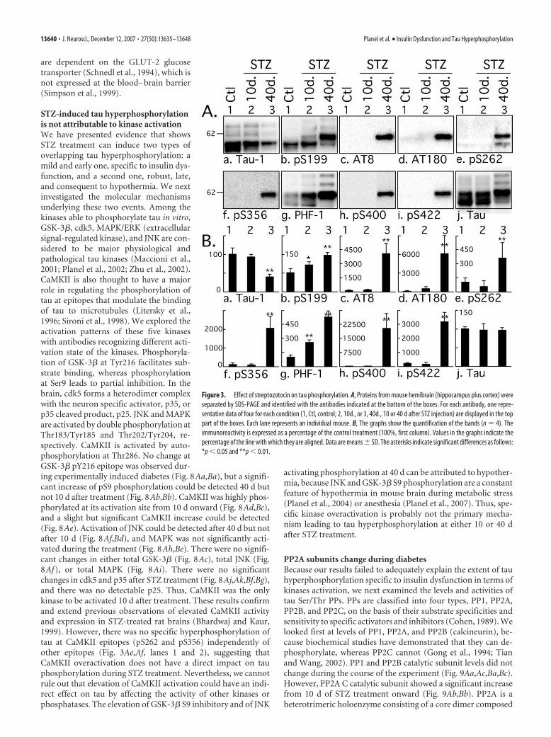
Streptozotocin treatment induces tauhyperphosphorylation atmultiple epitopesHaving shown that tau phosphorylation inresponse to STZ treatment was biphasic,with a mild hyperphosphorylation duringthe first 30 d and a massive one at 40 d, weconcentrated additional analysis on onerepresentative point of the first phase andone of the second phase: another set ofmice was treated with STZ and sampled at10 and 40 d after treatment. Tau phos-phorylation was investigated by semi-quantitative Western blot using an exten-sive panel of anti-tau antibodies. Anincrease of tau phosphorylation at thephospho-serine 199 (pS199) and PHF-1(pS396 and pS404) epitopes was observedafter 10 d, and at all the phospho-epitopesstudied after 40 d (Fig. 3). In fact, most ofthe epitopes investigated here were hyper-phosphorylated after 10 d, but this is notvisible in Figure 3 because the blots wereexposed to be in the linear range for quan-tification of the results at 40 d. The hyper-phosphorylation specific to STZ treatmentafter 10 d at Tau-1, AT8, and AT180epitopes can be seen in Figures 1, 6, and 7.Early phosphorylation at PS199 is particu-larly interesting because this epitope en-ables the visualization of early stages of taupathology, even when aggregates of tau arenot detectable by biochemical approaches(Maurage et al., 2003). Furthermore, de-tection of PS199 in the CSF can be used inthe antemortem diagnostic of AD (Itoh etal., 2001). AT8 (pS202 and pT205) andpS422 epitopes were also highly phosphor-ylated. AT8 is considered an early marker of tau dysfunction,whereas pS422 is characteristic of abnormal, AD-like tau phos-phorylation (Matsuo et al., 1994; Hasegawa et al., 1996; Bussiereet al., 1999). Thus, STZ-induced insulin dysfunction can rapidlylead to a biochemical pattern of tau phosphorylation similar tothat seen in early AD. The STZ-induced changes in tau phosphor-ylation appear to occur in two phases. First, a mild hyperphos-phorylation as observed at 10 d (up to 300% increase), followedby a massive hyperphosphorylation at 40 d (�15,000% increaseat some epitopes).
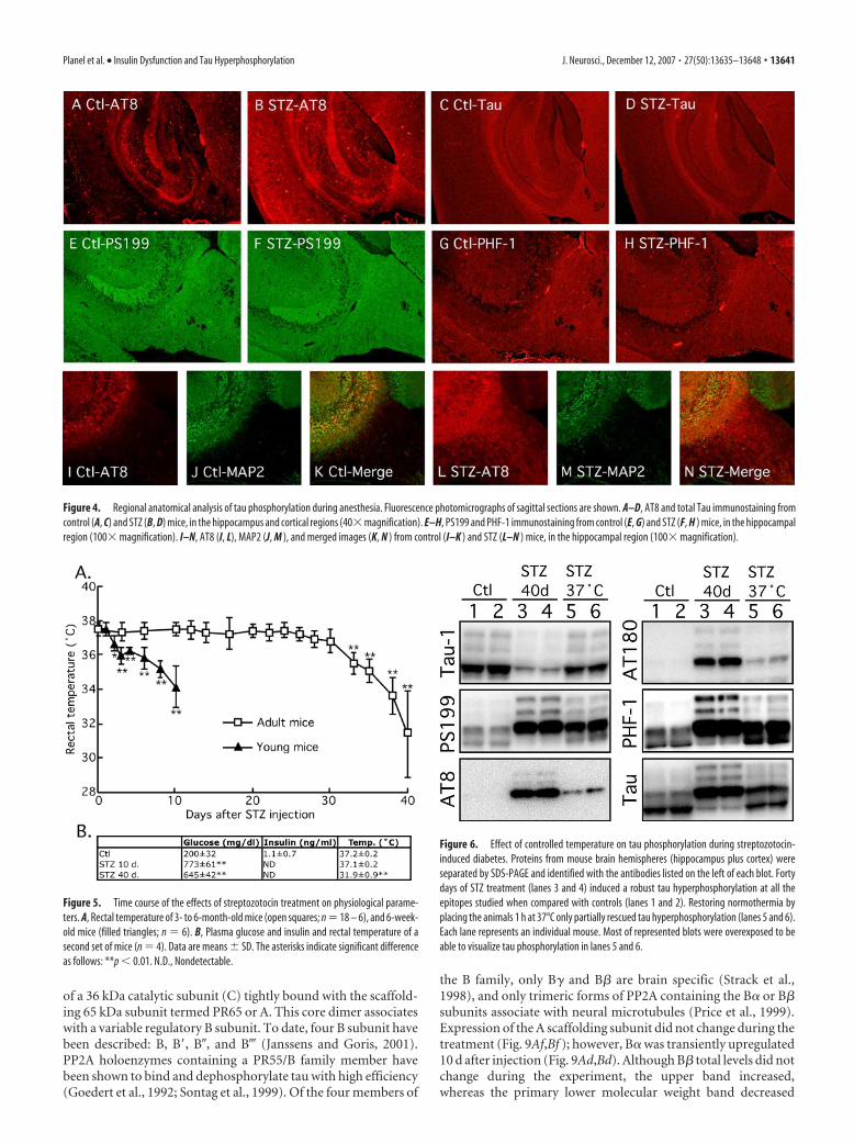
Hyperphosphorylated tau is limited to the neuropiland axonsWe then examined the gross anatomical pattern of tau phosphor-ylation. There were no evident differences in the pattern of tau
phosphorylation 10 d after STZ treatment (data not shown). At40 d, tau was hyperphosphorylated at AT8 in most anterior areas,such as the neocortex and hippocampus (Fig. 4A,B). The patternof total tau axonal (fimbria, alveus, and mossy fibers, for exam-ple) and neuropil staining was not altered after STZ treatment(Fig. 4C,D). Immunostaining for pS199 (Fig. 4E,F) and PHF-1(Fig. 4G,H) epitopes was not visibly altered in response to STZ.These results confirm our Western blot data, which revealed largechanges in tau phosphorylation at the AT8 epitope (Fig. 3Ac,Bc)in response to 40 d of STZ, but smaller changes in phosphoryla-tion at the PS199 and PHF-1 epitopes, which could already beobserved in control animals (Fig. 3Ab,Ag,Bb,Bg). In the hip-pocampus, hyperphosphorylated tau mainly localized in the neu-ropil and the axonal tracks of the fimbria, as demonstrated bydouble staining with AT8 and MAP2 antibodies (Fig. 4 I–N). In
Figure 1. Immunoblot analysis of tau solubility after streptozotocin treatment. Brain proteins (hippocampus and neocortex)from control mice and mice treated for 10, 20, 30, and 40 d were extracted according to a modified method of Greenberg andDavies (1990). Tau from total brain lysates (A), heat-stable soluble (HS) (B), and Sarkosyl-insoluble fractions (C) were evaluated byimmunoblot analysis with the following antibodies: AT8 (pS202/pT205), PHF-1 (pS396/pS404), and Tau (phospho-independent).The bar graphs represent the quantification of the immunoblot bands displayed above them. Each graph displays the immuno-reactivity expressed as a percentage of the control (100%). The numbers in the graphs indicate the percentage of the tick line withwhich they are aligned. Data represented are means � SD (n � 3 for each condition; 1 representative data displayed; each lanerepresents an individual mouse). The asterisks indicate significant differences from controls as follows: *p�0.05 and **p�0.01.
13638 • J. Neurosci., December 12, 2007 • 27(50):13635–13648 Planel et al. • Insulin Dysfunction and Tau Hyperphosphorylation

the normal adult brain, tau is an axonal protein, whereas MAP2 ispresent in the soma and dendrites, but not in the axons. In con-trast, tau is found in cell bodies and dendrites of affected neuronsin AD. In diseased brains, tau-positive dystrophic axons are wide-spread and can be easily detected in the alveus and fimbria of thehippocampus (Su et al., 1993). In fact, the earliest detectablehyperphosphorylated tau in AD is preferentially localized in neu-rites of vulnerable neurons before extending to the soma (Su etal., 1994) where NFTs are formed (Bancher et al., 1989; Braak etal., 1994; Trojanowski and Lee, 1994). Thus, insulin dysfunctionappears to induce axonal and neuropil tau hyperphosphoryla-tion, reminiscent of incipient AD, but without observable soma-todendritic relocalization.
Insulin dysfunction induces two types oftau hyperphosphorylationWe previously demonstrated that alterations of glucose metabo-lism can induce hypothermia leading to tau hyperphosphoryla-tion (Planel et al., 2004). Thus, we looked for a possible hypother-
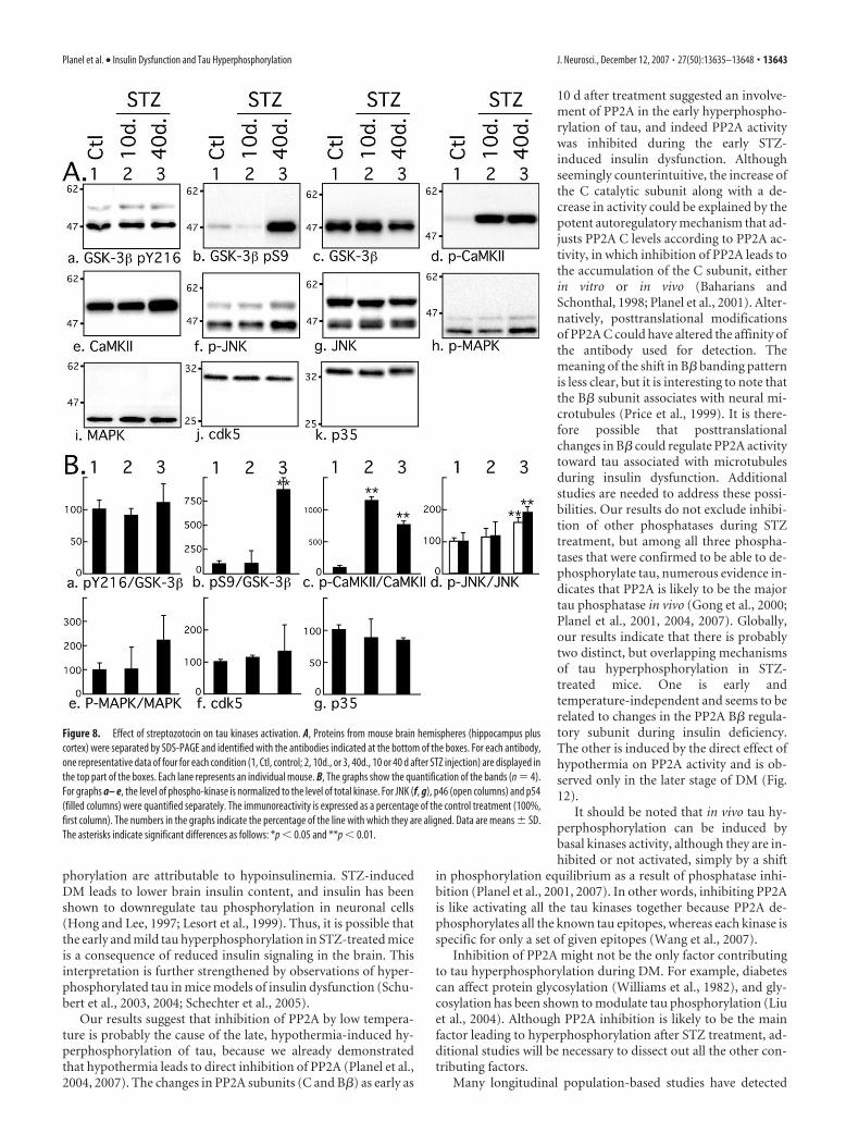
mic effect of STZ and its correlation with tauhyperphosphorylation. The temperature of the mice was�37.5 � 0.5°C for the first 26 d and began to drop nonsignifi-cantly thereafter: 37.0 � 0.5°C at 28 d, and 36.8 � 0.7°C at 30 d.However, after 30 d, the drop in temperature was significant andreached 31.4 � 2.5°C at 40 d (Fig. 5A). Hypothermia correlatedwith the massive phosphorylation of tau at 40 d (Figs. 1, 3), butnot with the mild tau phosphorylation observed while the ani-mals were still normothermic (Fig. 3, lanes 1 and 2, pS199 andPHF-1; Fig. 6, lanes 1– 4, all the phospho-epitopes). These tem-perature data were confirmed in the new batch of STZ-treatedmice, which displayed hypothermia at 40 d but not at 10 d (Fig.5B). STZ-treated mice displayed hyperglycemia from 10 d on-ward after injection, with insulin below detection levels (Fig. 5B).Thus, STZ treatment induced two types of tau hyperphosphory-lation: one is probably consequent to hypothermia induced byDM; the other is possibly temperature-independent and specificto hypoinsulinemia.
We also injected young mice (6 weeks of age) with 150 mg/kgSTZ, to confirm previously published data that young rodentstend to develop hypothermia very rapidly after injection of strep-tozotocin (Howarth et al., 2005). Two days after STZ injection,the animals began to be significantly hypothermic (Ctl, 37.6 �0.2°C; 2 d, 36.7 � 0.4°C; 3 d, 35.9 � 0.5°C), and by 10 d theyreached 34.2 � 1.2°C, a temperature attained 38 d after STZtreatment in the adults (Fig. 5). Hypothermia is a powerful reg-ulator of tau phosphorylation, and a 1°C drop in body tempera-ture can bring about a 100% increase of AT8 signal (Planel et al.,2007). Thus, young animals are not suitable to discriminate theeffect of hypoinsulinemia from hypothermia.
Prevention of hypothermia partially rescuestau hyperphosphorylationTo explore the role of temperature on tau phosphorylation in the40-d-treated mice, we placed the STZ-treated animals at in aventilated incubator at 37°C for 2 h before sampling. Animalsincubated at 37°C had rectal temperature equivalent to the con-trol group, did not became hyperthermic (data not shown). Tauhyperphosphorylation was partially, but not completely reversed,as can be seen in the overexposed blots of Figure 6. It is importantto note that, in other models of mouse hypothermia, placementof animals at 37°C completely rescues tau phosphorylation(Planel et al., 2004, 2007). Thus, 40 d after STZ treatment, tauhyperphosphorylation can only be partially rescued by restoringnormothermia, which suggest that the temperature-independent, hypoinsulinemia-specific mild hyperphosphoryla-tion observed at 10 d is still present after 40 d.
Insulin treatment prevents early tau hyperphosphorylationRecently, direct injections of STZ in the brain have been shown toincrease phospho-tau immunoreactivity (Lester-Coll et al., 2006;Grunblatt et al., 2007), raising the possibility that our observa-tions could be the consequence of infiltration of peripheral STZin the brain, rather than an effect of peripheral insulin deficit perse. To exclude this possibility, we implanted subcutaneous insu-lin pumps in mice 3 d after STZ injection, and analyzed the brains7 d later (for a total of 10 d STZ treatment). Our data show thatinsulin treatment totally blocked the mild tau hyperphosphory-lation of all the epitopes studied (Fig. 7). Thus, early and mild tauhyperphosphorylation in STZ-treated mice is a consequence ofhypoinsulinemia rather than a direct effect of STZ on the brain.This is consistent with the fact that peripheral STZ does not im-pact directly on the brain because its transport and cytotoxicity
Figure 2. Microtubule binding assay of tau after streptozotocin injection. Brain proteins(hippocampus and neocortex) from control mice and mice treated for 10, 20, 30, and 40 d wereextracted and incubated with taxol-stabilized microtubules. Tau and tubulin levels from MT-free and -bound fractions were evaluated by immunoblot analysis with the following antibod-ies: Tau (phospho-independent) (A); �-tubulin (B). The bar graphs represent the quantificationof the immunoblot bands displayed above them. Each graph displays the immunoreactivityexpressed as a percentage of the control (100%). The numbers in the graphs indicate thepercentage of the tick line with which they are aligned. Data represented are means � SD (n �3 for each condition; 1 representative data displayed; each lane represents an individualmouse). The asterisks indicate significant differences from controls as follows: **p � 0.01.
Planel et al. • Insulin Dysfunction and Tau Hyperphosphorylation J. Neurosci., December 12, 2007 • 27(50):13635–13648 • 13639
are dependent on the GLUT-2 glucosetransporter (Schnedl et al., 1994), which isnot expressed at the blood– brain barrier(Simpson et al., 1999).
STZ-induced tau hyperphosphorylationis not attributable to kinase activationWe have presented evidence that showsSTZ treatment can induce two types ofoverlapping tau hyperphosphorylation: amild and early one, specific to insulin dys-function, and a second one, robust, late,and consequent to hypothermia. We nextinvestigated the molecular mechanismsunderlying these two events. Among thekinases able to phosphorylate tau in vitro,GSK-3�, cdk5, MAPK/ERK (extracellularsignal-regulated kinase), and JNK are con-sidered to be major physiological andpathological tau kinases (Maccioni et al.,2001; Planel et al., 2002; Zhu et al., 2002).CaMKII is also thought to have a majorrole in regulating the phosphorylation oftau at epitopes that modulate the bindingof tau to microtubules (Litersky et al.,1996; Sironi et al., 1998). We explored theactivation patterns of these five kinaseswith antibodies recognizing different acti-vation state of the kinases. Phosphoryla-tion of GSK-3� at Tyr216 facilitates sub-strate binding, whereas phosphorylationat Ser9 leads to partial inhibition. In thebrain, cdk5 forms a heterodimer complexwith the neuron specific activator, p35, orp35 cleaved product, p25. JNK and MAPKare activated by double phosphorylation atThr183/Tyr185 and Thr202/Tyr204, re-spectively. CaMKII is activated by auto-phosphorylation at Thr286. No change atGSK-3� pY216 epitope was observed dur-ing experimentally induced diabetes (Fig. 8Aa,Ba), but a signifi-cant increase of pS9 phosphorylation could be detected 40 d butnot 10 d after treatment (Fig. 8Ab,Bb). CaMKII was highly phos-phorylated at its activation site from 10 d onward (Fig. 8Ad,Bc),and a slight but significant CaMKII increase could be detected(Fig. 8Ae). Activation of JNK could be detected after 40 d but notafter 10 d (Fig. 8Af,Bd), and MAPK was not significantly acti-vated during the treatment (Fig. 8Ah,Be). There were no signifi-cant changes in either total GSK-3� (Fig. 8Ac), total JNK (Fig.8Af), or total MAPK (Fig. 8Ai). There were no significantchanges in cdk5 and p35 after STZ treatment (Fig. 8Aj,Ak,Bf,Bg),and there was no detectable p25. Thus, CaMKII was the onlykinase to be activated 10 d after treatment. These results confirmand extend previous observations of elevated CaMKII activityand expression in STZ-treated rat brains (Bhardwaj and Kaur,1999). However, there was no specific hyperphosphorylation oftau at CaMKII epitopes (pS262 and pS356) independently ofother epitopes (Fig. 3Ae,Af, lanes 1 and 2), suggesting thatCaMKII overactivation does not have a direct impact on tauphosphorylation during STZ treatment. Nevertheless, we cannotrule out that elevation of CaMKII activation could have an indi-rect effect on tau by affecting the activity of other kinases orphosphatases. The elevation of GSK-3� S9 inhibitory and of JNK
activating phosphorylation at 40 d can be attributed to hypother-mia, because JNK and GSK-3� S9 phosphorylation are a constantfeature of hypothermia in mouse brain during metabolic stress(Planel et al., 2004) or anesthesia (Planel et al., 2007). Thus, spe-cific kinase overactivation is probably not the primary mecha-nism leading to tau hyperphosphorylation at either 10 or 40 dafter STZ treatment.
PP2A subunits change during diabetesBecause our results failed to adequately explain the extent of tauhyperphosphorylation specific to insulin dysfunction in terms ofkinases activation, we next examined the levels and activities oftau Ser/Thr PPs. PPs are classified into four types, PP1, PP2A,PP2B, and PP2C, on the basis of their substrate specificities andsensitivity to specific activators and inhibitors (Cohen, 1989). Welooked first at levels of PP1, PP2A, and PP2B (calcineurin), be-cause biochemical studies have demonstrated that they can de-phosphorylate, whereas PP2C cannot (Gong et al., 1994; Tianand Wang, 2002). PP1 and PP2B catalytic subunit levels did notchange during the course of the experiment (Fig. 9Aa,Ac,Ba,Bc).However, PP2A C catalytic subunit showed a significant increasefrom 10 d of STZ treatment onward (Fig. 9Ab,Bb). PP2A is aheterotrimeric holoenzyme consisting of a core dimer composed
Figure 3. Effect of streptozotocin on tau phosphorylation. A, Proteins from mouse hemibrain (hippocampus plus cortex) wereseparated by SDS-PAGE and identified with the antibodies indicated at the bottom of the boxes. For each antibody, one repre-sentative data of four for each condition (1, Ctl, control; 2, 10d., or 3, 40d., 10 or 40 d after STZ injection) are displayed in the toppart of the boxes. Each lane represents an individual mouse. B, The graphs show the quantification of the bands (n � 4). Theimmunoreactivity is expressed as a percentage of the control treatment (100%, first column). Values in the graphs indicate thepercentage of the line with which they are aligned. Data are means � SD. The asterisks indicate significant differences as follows:*p � 0.05 and **p � 0.01.
13640 • J. Neurosci., December 12, 2007 • 27(50):13635–13648 Planel et al. • Insulin Dysfunction and Tau Hyperphosphorylation
of a 36 kDa catalytic subunit (C) tightly bound with the scaffold-ing 65 kDa subunit termed PR65 or A. This core dimer associateswith a variable regulatory B subunit. To date, four B subunit havebeen described: B, B, B, and B� (Janssens and Goris, 2001).PP2A holoenzymes containing a PR55/B family member havebeen shown to bind and dephosphorylate tau with high efficiency(Goedert et al., 1992; Sontag et al., 1999). Of the four members of
the B family, only B� and B� are brain specific (Strack et al.,1998), and only trimeric forms of PP2A containing the B� or B�subunits associate with neural microtubules (Price et al., 1999).Expression of the A scaffolding subunit did not change during thetreatment (Fig. 9Af,Bf ); however, B� was transiently upregulated10 d after injection (Fig. 9Ad,Bd). Although B� total levels did notchange during the experiment, the upper band increased,whereas the primary lower molecular weight band decreased
Figure 4. Regional anatomical analysis of tau phosphorylation during anesthesia. Fluorescence photomicrographs of sagittal sections are shown. A–D, AT8 and total Tau immunostaining fromcontrol (A, C) and STZ (B, D) mice, in the hippocampus and cortical regions (40� magnification). E–H, PS199 and PHF-1 immunostaining from control (E, G) and STZ (F, H ) mice, in the hippocampalregion (100� magnification). I–N, AT8 (I, L), MAP2 (J, M ), and merged images (K, N ) from control (I–K ) and STZ (L–N ) mice, in the hippocampal region (100� magnification).
Figure 5. Time course of the effects of streptozotocin treatment on physiological parame-ters. A, Rectal temperature of 3- to 6-month-old mice (open squares; n � 18 – 6), and 6-week-old mice (filled triangles; n � 6). B, Plasma glucose and insulin and rectal temperature of asecond set of mice (n � 4). Data are means � SD. The asterisks indicate significant differenceas follows: **p � 0.01. N.D., Nondetectable.
Figure 6. Effect of controlled temperature on tau phosphorylation during streptozotocin-induced diabetes. Proteins from mouse brain hemispheres (hippocampus plus cortex) wereseparated by SDS-PAGE and identified with the antibodies listed on the left of each blot. Fortydays of STZ treatment (lanes 3 and 4) induced a robust tau hyperphosphorylation at all theepitopes studied when compared with controls (lanes 1 and 2). Restoring normothermia byplacing the animals 1 h at 37°C only partially rescued tau hyperphosphorylation (lanes 5 and 6).Each lane represents an individual mouse. Most of represented blots were overexposed to beable to visualize tau phosphorylation in lanes 5 and 6.
Planel et al. • Insulin Dysfunction and Tau Hyperphosphorylation J. Neurosci., December 12, 2007 • 27(50):13635–13648 • 13641

(Fig. 9Ae,Be). Thus only changes in the C and B� subunits ofPP2A inversely correlate with tau phosphorylation duringhypoinsulinemia.
PP2A is inhibited during STZ treatmentWe previously demonstrated that hypothermia induces tau hy-perphosphorylation by direct inhibition of PP2A (Planel et al.,2004). Because tau hyperphosphorylation 40 d after STZ injec-tion is mainly attributable to hypothermia (Fig. 5), we focused onexamining the mechanism of tau hyperphosphorylation at 10 d,when there is no overlapping inhibition of PP2A activity by tem-perature. To determine whether the changes in the subunits ofPP2A were associated with the inhibition of the enzyme, we per-formed a phosphatase assay using brain lysates and endogenousbrain tau as a substrate. Specifically, we monitored the dephos-phorylation of the PHF-1 epitope at 10 d after STZ injection. Wepreviously demonstrated that, in these conditions, PHF-1 de-phosphorylation is mainly attributable to endogenous PP2A ac-tivity (Planel et al., 2007). Brain extracts from 10-d-injected mice(Fig. 10A) (decay rate constant, 3.87 min; R 2 � 0.997) showed asignificant reduction ( p � 0.0001) in tau dephosphorylationcompared with controls (Fig. 10A) (decay rate constant, 6.48min; R 2 � 0.992), suggesting that PP2A is inhibited during STZ-induced diabetes. To verify this endogenous dephosphorylationassay, we performed the same experiment with a commerciallyavailable PP2A assay system, and included STZ-treated mice im-planted with insulin pumps, as described above. This assay re-vealed a �15% decrease in brain PP2A activity during STZ-induced diabetes, but no significant change in insulin-competentanimals (Fig. 10B). Lastly, we performed an immunoprecipita-tion (IP) assay of PP2A in the samples at 10, 20, 30, and 40 d afterSTZ injection. This IP assay had more variability than the twoprevious ones, but revealed a �30% inhibition of PP2A afterinduction of diabetes (Fig. 10C). The differences between the twoassays are probably attributable to the different protocols. Noadditional inhibition of PP2A at 40 d was detected by this assaybecause the inhibition of phosphatase by temperature at 40 dafter STZ treatment is caused by a direct effect of hypothermia on
enzyme activities in the brain, and is therefore not detectable byan assay performed at constant temperature for all the samples(Planel et al., 2004, 2007). Thus, PP2A inhibition occurs earlyduring STZ treatment, is attributable to hypoinsulinemia, andmight be responsible for the increased levels of hyperphosphory-lated tau observed 10 d after injection.
APP and A� are not affected by STZ treatmentPrevious data have shown that diet-induced insulin resistancepromotes A�1– 40 and A�1– 42 peptide generation in a transgenicmouse model of �-amyloidosis (Ho et al., 2004). In addition,other studies suggest that insulin can modulate A� levels (Gaspa-rini et al., 2002; Hoyer, 2004), which is known to influence tauphosphorylation (Blurton-Jones and Laferla, 2006). We there-fore examined the possibility of change in APP metabolism andA� levels in STZ-treated mice, because it could be a plausiblemechanism for the mild tau hyperphosphorylation observed. Nochange was observed in either APP, APP C-terminal fragments,or A� levels after 10 d of hypoinsulinemia (Fig. 11A–C), suggest-ing no association between A� and the mild and early tau hyper-phosphorylation after STZ injection.
DiscussionWe investigated the in vivo relationship between insulin dysfunc-tion and tau phosphorylation. Our results indicate that STZ-induced insulin deficiency rapidly leads to a biochemical andhistochemical pattern of tau phosphorylation similar to that seenin early AD, with hyperphosphorylation of tau at multipleepitopes seen in the neuropil and axonal tracks. Our data suggestthat tau hyperphosphorylation during diabetes is likely a result ofPP2A inhibition.
Hypothermia is a common outcome in both human (Neil etal., 1986; Scott et al., 1987) and experimental diabetes (Shalaby etal., 1989; Kilgour and Williams, 1996, 1998). In STZ-inducedDM, the occurrence of hypothermia has generally been associ-ated with numerous abnormalities such as decrease in nonshiv-ering thermogenesis (Seydoux et al., 1983), lack of shivering (Kil-gour and Williams, 1996), and the inability to use carbohydratesfor heat production (Smith and Davidson, 1982). These abnor-malities develop over time and, whereas at 10 –30 d after STZinjection the animals were still able to maintain normothermiaand had mild tau hyperphosphorylation, by 40 d they were fullyhypothermic and had massive tau phosphorylation. This massivehyperphosphorylation was mostly, but not completely, rescuedby returning the animals to normothermia, demonstrating that itis mainly attributable to hypothermia and not just correlative toit. However, insulin treatment of diabetic animals completelyrestored tau phosphorylation to control levels, demonstratingthat the mild hyperphosphorylation was attributable to the lackof insulin. In light of our previous results demonstrating thathypothermia induces tau hyperphosphorylation that is com-pletely reversible by normothermia (Planel et al., 2004, 2007), ourdata indicate that there are probably two distinct and overlappingtau hyperphosphorylation events during DM. One is mild, tem-perature independent, hypoinsulinemia dependent, and ob-served for the whole duration of diabetes. The other is massiveand consequent to hypothermia and overlaps the first type in thelater stage of STZ-induced DM. These results confirm and extendprevious studies reporting tau hyperphosphorylation after STZtreatment in mice (Zhao et al., 2003; Clodfelder-Miller et al.,2006).
Our data show that insulin treatment totally prevented hyper-phosphorylation, thus demonstrating that changes in tau phos-
Figure 7. Effect of insulin on tau phosphorylation during streptozotocin-induced diabetes.Proteins from mouse brain hemispheres (hippocampus plus cortex) were separated by SDS-PAGE and identified with the antibodies listed on the left of each blot. Ten days of STZ treatment(lanes 3 and 4) induced a slight hyperphosphorylation of tau at all the epitopes studied whencompared with controls (lanes 1 and 2). Implantation of subcutaneous pumps delivering 0.75U/d of insulin rescued tau phosphorylation to control levels (lanes 5 and 6). Each lane representsan individual mouse. INS, Insulin.
13642 • J. Neurosci., December 12, 2007 • 27(50):13635–13648 Planel et al. • Insulin Dysfunction and Tau Hyperphosphorylation
phorylation are attributable to hypoinsulinemia. STZ-inducedDM leads to lower brain insulin content, and insulin has beenshown to downregulate tau phosphorylation in neuronal cells(Hong and Lee, 1997; Lesort et al., 1999). Thus, it is possible thatthe early and mild tau hyperphosphorylation in STZ-treated miceis a consequence of reduced insulin signaling in the brain. Thisinterpretation is further strengthened by observations of hyper-phosphorylated tau in mice models of insulin dysfunction (Schu-bert et al., 2003, 2004; Schechter et al., 2005).
Our results suggest that inhibition of PP2A by low tempera-ture is probably the cause of the late, hypothermia-induced hy-perphosphorylation of tau, because we already demonstratedthat hypothermia leads to direct inhibition of PP2A (Planel et al.,2004, 2007). The changes in PP2A subunits (C and B�) as early as
10 d after treatment suggested an involve-ment of PP2A in the early hyperphospho-rylation of tau, and indeed PP2A activitywas inhibited during the early STZ-induced insulin dysfunction. Althoughseemingly counterintuitive, the increase ofthe C catalytic subunit along with a de-crease in activity could be explained by thepotent autoregulatory mechanism that ad-justs PP2A C levels according to PP2A ac-tivity, in which inhibition of PP2A leads tothe accumulation of the C subunit, eitherin vitro or in vivo (Baharians andSchonthal, 1998; Planel et al., 2001). Alter-natively, posttranslational modificationsof PP2A C could have altered the affinity ofthe antibody used for detection. Themeaning of the shift in B� banding patternis less clear, but it is interesting to note thatthe B� subunit associates with neural mi-crotubules (Price et al., 1999). It is there-fore possible that posttranslationalchanges in B� could regulate PP2A activitytoward tau associated with microtubulesduring insulin dysfunction. Additionalstudies are needed to address these possi-bilities. Our results do not exclude inhibi-tion of other phosphatases during STZtreatment, but among all three phospha-tases that were confirmed to be able to de-phosphorylate tau, numerous evidence in-dicates that PP2A is likely to be the majortau phosphatase in vivo (Gong et al., 2000;Planel et al., 2001, 2004, 2007). Globally,our results indicate that there is probablytwo distinct, but overlapping mechanismsof tau hyperphosphorylation in STZ-treated mice. One is early andtemperature-independent and seems to berelated to changes in the PP2A B� regula-tory subunit during insulin deficiency.The other is induced by the direct effect ofhypothermia on PP2A activity and is ob-served only in the later stage of DM (Fig.12).
It should be noted that in vivo tau hy-perphosphorylation can be induced bybasal kinases activity, although they are in-hibited or not activated, simply by a shift
in phosphorylation equilibrium as a result of phosphatase inhi-bition (Planel et al., 2001, 2007). In other words, inhibiting PP2Ais like activating all the tau kinases together because PP2A de-phosphorylates all the known tau epitopes, whereas each kinase isspecific for only a set of given epitopes (Wang et al., 2007).
Inhibition of PP2A might not be the only factor contributingto tau hyperphosphorylation during DM. For example, diabetescan affect protein glycosylation (Williams et al., 1982), and gly-cosylation has been shown to modulate tau phosphorylation (Liuet al., 2004). Although PP2A inhibition is likely to be the mainfactor leading to hyperphosphorylation after STZ treatment, ad-ditional studies will be necessary to dissect out all the other con-tributing factors.
Many longitudinal population-based studies have detected
Figure 8. Effect of streptozotocin on tau kinases activation. A, Proteins from mouse brain hemispheres (hippocampus pluscortex) were separated by SDS-PAGE and identified with the antibodies indicated at the bottom of the boxes. For each antibody,one representative data of four for each condition (1, Ctl, control; 2, 10d., or 3, 40d., 10 or 40 d after STZ injection) are displayed inthe top part of the boxes. Each lane represents an individual mouse. B, The graphs show the quantification of the bands (n � 4).For graphs a– e, the level of phospho-kinase is normalized to the level of total kinase. For JNK (f, g), p46 (open columns) and p54(filled columns) were quantified separately. The immunoreactivity is expressed as a percentage of the control treatment (100%,first column). The numbers in the graphs indicate the percentage of the line with which they are aligned. Data are means � SD.The asterisks indicate significant differences as follows: *p � 0.05 and **p � 0.01.
Planel et al. • Insulin Dysfunction and Tau Hyperphosphorylation J. Neurosci., December 12, 2007 • 27(50):13635–13648 • 13643

higher AD incidence rates in type 2 DM patients (Leibson et al.,1997; Ott et al., 1999; Peila et al., 2002; Arvanitakis et al., 2004;Luchsinger et al., 2005). Type 2 DM, the most common in adultsand in the elderly, is preceded in most individuals by insulinresistance and hyperinsulinemia to maintain glucose homeosta-sis (Festa et al., 2006). Over time, the pancreas becomes unable toproduce enough insulin to overcome the resistance; the �-cellsburnout and insulin production decreases dramatically, causingfull-blown diabetes (DeFronzo, 2004). Interestingly, at least twolongitudinal studies found that diabetic patients who requiredtreatment with insulin replacement, presumably those with thelowest pancreatic insulin production, had the highest risk of ADcompared with persons using oral medications and those withoutdiabetes (Ott et al., 1999; Luchsinger et al., 2001). Thus, althoughSTZ-induced diabetes is not a model of early type 2 DM, it ad-dresses mechanisms related to the low insulin levels that accom-pany type 2 DM in the end of its natural history. Here, we dem-
onstrated that STZ-induced insulin dysfunction led to AD-liketau hyperphosphorylation induced by inhibition of PP2A. In AD,decreased PP2A expression (Vogelsberg-Ragaglia et al., 2001)along with upregulation of its inhibitors (Tanimukai et al., 2005),results in overall inhibition of its activity (Gong et al., 1993,1995), which might be an important factor in the evolution of thepathology (Tian and Wang, 2002). Thus, during insulin dysfunc-tion, inhibition of PP2A and the consequent tau hyperphospho-
Figure 9. Effect of streptozotocin on phosphatases levels. A, Proteins from mouse brainhemispheres (hippocampus plus cortex) were separated by SDS-PAGE and identified with theantibodies indicated at the bottom of the boxes. PP1 C, PP2A C, and PP2B C detect the catalyticsubunits of PP1, PP2A, and PP2B, respectively. PP2A B� and B� detect B� and B� regulatorysubunits of PP2A, and PP2A A detects anchoring subunits. For each antibody, one representa-tive data of four for each condition (1, Ctl, control; 2, 10d., or 3, 40d., 10 or 40 d after STZinjection) are displayed in the top part of the boxes. Each lane represents an individual mouse.B, The graphs show the quantification of the bands (n � 4). The immunoreactivity is expressedas a percentage of the control treatment (100%, first column). The numbers in the graphsindicate the percentage of the line with which they are aligned. Data are means � SD. Theasterisks indicate significant differences as follows: *p � 0.05 and **p � 0.01. Figure 10. Effect of streptozotocin treatment on phosphatase activity. A, Phosphatase ac-
tivity was evaluated by endogenous tau dephosphorylation followed by Western blot analysis(n �3). Tau from control brains (solid squares, solid line) was dephosphorylated faster than taufrom animals treated with STZ after 10 d (solid circles, dotted line). B, To confirm this result,PP2A activity was evaluated with the PP2A Assay System from Promega. Similarly to the en-dogenous assay, PP2A activity was inhibited in 10 d STZ-treated mice (n � 3). Implantation ofsubcutaneous pumps delivering 0.75 U/d insulin in STZ-treated mice restored PP2A activity tocontrol levels. C, Immunoprecipitation assay of PP2A after STZ treatment. PP2A presented sim-ilar level of inhibition at 10, 20, 30, and 40 d after injection. Data are means � SD. The asterisksindicate significant differences as follows: *p � 0.05, **p � 0.01, and ***p � 0.001.
13644 • J. Neurosci., December 12, 2007 • 27(50):13635–13648 Planel et al. • Insulin Dysfunction and Tau Hyperphosphorylation

rylation might interact with biological or genetic susceptibilitiesto increase the vulnerability of the brain to insults associated withAD. PP2A inhibition may not be a direct consequence of insulindepletion; but the outcome of this (either directly or indirectly) istau hyperphosphorylation that is potentially of significance forAD, which is relevant regardless of the intermediate steps. It isunclear whether the high insulin levels that precede full-blowndiabetes has a role in the relationship observed between diabetesand AD, and this needs to be taken into account for future studies(Luchsinger and Mayeux, 2007).
In AD, the hyperphosphorylation of tau by the deregulation ofkinases and/or phosphatases has been proposed to lead to tauaggregation and prevent tau binding to MTs, thereby destabiliz-ing the MT network (Alonso et al., 1994; Feinstein and Wilson,2005; Mi and Johnson, 2006). Hyperphosphorylation of humantau induces MT disruption (Ebneth et al., 1999), as well as tauaggregation in vitro (Alonso et al., 2001; Sato et al., 2002). Here,despite sustained mild hyperphosphorylation of tau for 30 d andmassive late hyperphosphorylation, we could not detect aggre-gated tau in mice, and tau binding to MTs was affected only at40 d after STZ treatment. The differences in sequence and iso-form composition between human and mouse tau (Janke et al.,1999), cannot explain the lack of tau aggregates in nontransgenicmice, because rodent tau can form PHFs as readily as human tauafter being hyperphosphorylated in vitro (Chohan et al., 2005).Thus, absence of aggregates could be attributable to insufficientlevels of total tau phosphorylation in the STZ model, or to insuf-ficient levels of phosphorylation at epitopes promoting aggrega-tion. What remain to be determined is how much impact diabetescan have on tau pathogenesis. Examination of the development
of tau pathology in diabetic transgenic animal models expressingmutant tau that assemble at considerably lower levels of phos-phorylation than normal tau (Alonso et al., 2004), may be able toaddress this question.
ReferencesAlonso AC, Zaidi T, Grundke-Iqbal I, Iqbal K (1994) Role of abnormally
phosphorylated tau in the breakdown of microtubules in Alzheimer dis-ease. Proc Natl Acad Sci USA 91:5562–5566.
Alonso AC, Zaidi T, Novak M, Grundke-Iqbal I, Iqbal K (2001) Hyperphos-phorylation induces self-assembly of tau into tangles of paired helicalfilaments/straight filaments. Proc Natl Acad Sci USA 98:6923– 6928.
Alonso AC, Mederlyova A, Novak M, Grundke-Iqbal I, Iqbal K (2004) Pro-motion of hyperphosphorylation by frontotemporal dementia tau muta-tions. J Biol Chem 279:34873–34881.
Andorfer C, Kress Y, Espinoza M, de Silva R, Tucker KL, Barde YA, Duff K,Davies P (2003) Hyperphosphorylation and aggregation of tau in miceexpressing normal human tau isoforms. J Neurochem 86:582–590.
Arvanitakis Z, Wilson RS, Bienias JL, Evans DA, Bennett DA (2004) Diabe-tes mellitus and risk of Alzheimer disease and decline in cognitive func-tion. Arch Neurol 61:661– 666.
Avila J, Lucas JJ, Perez M, Hernandez F (2004) Role of tau protein in bothphysiological and pathological conditions. Physiol Rev 84:361–384.
Baharians Z, Schonthal AH (1998) Autoregulation of protein phosphatasetype 2A expression. J Biol Chem 273:19019 –19024.
Bancher C, Brunner C, Lassmann H, Budka H, Jellinger K, Wiche G, Seitel-berger F, Grundke-Iqbal I, Iqbal K, Wisniewski HM (1989) Accumula-tion of abnormally phosphorylated tau precedes the formation of neuro-fibrillary tangles in Alzheimer’s disease. Brain Res 477:90 –99.
Figure 11. Effect of streptozotocin on APP metabolism. Proteins from mouse brain hemi-spheres (hippocampus plus cortex) of control and 10 d STZ-treated mice were separated bySDS-PAGE and identified with C1/6.1 antibody, which recognize APP (A) and CTFs (B). C showsthe levels of A�1– 40 (open bars) and A�1– 42 (filled bars) for control and STZ 10 d, as detectedby ELISA (n � 4). Data are means � SD.
Figure 12. Putative mechanism of tau hyperphosphorylation during insulin dysfunction.Injection of STZ induces immediate decrease of pancreatic insulin secretion and results ininsulin-dependent diabetes mellitus. During the early phase of DM, when the adult mice are stillnormothermic, PP2A is inhibited, presumably through changes in the B� regulatory subunit,which leads to a mild hyperphosphorylation of tau specific to insulin deficiency, as demon-strated in Figures 7 and 10. In the later phase of DM, deficits in peripheral glucose/energymetabolism lead to hypothermia. This leads to a direct inhibition of PP2A activity by low tem-peratures, resulting in massive hyperphosphorylation of tau not specific to hypoinsulinemia.The early and mild tau hyperphosphorylation is still present at this stage, but is masked by theeffects of hypothermia, as demonstrated in Figure 6.
Planel et al. • Insulin Dysfunction and Tau Hyperphosphorylation J. Neurosci., December 12, 2007 • 27(50):13635–13648 • 13645

Bhardwaj SK, Kaur G (1999) Effect of diabetes on calcium/calmodulin de-pendent protein kinase-II from rat brain. Neurochem Int 35:329 –335.
Biessels GJ, Staekenborg S, Brunner E, Brayne C, Scheltens P (2006) Risk ofdementia in diabetes mellitus: a systematic review. Lancet Neurol5:64 –74.
Blurton-Jones M, Laferla FM (2006) Pathways by which Abeta facilitates taupathology. Curr Alzheimer Res 3:437– 448.
Braak E, Braak H, Mandelkow EM (1994) A sequence of cytoskeletonchanges related to the formation of neurofibrillary tangles and neuropilthreads. Acta Neuropathol 87:554 –567.
Burns M, Gaynor K, Olm V, Mercken M, LaFrancois J, Wang L, Mathews PM,Noble W, Matsuoka Y, Duff K (2003) Presenilin redistribution associ-ated with aberrant cholesterol transport enhances �-amyloid productionin vivo. J Neurosci 23:5645–5649.
Bussiere T, Hof PR, Mailliot C, Brown CD, Caillet-Boudin ML, Perl DP, BueeL, Delacourte A (1999) Phosphorylated serine422 on tau proteins is apathological epitope found in several diseases with neurofibrillary degen-eration. Acta Neuropathol (Berl) 97:221–230.
Chohan MO, Haque N, Alonso A, El-Akkad E, Grundke-Iqbal I, Grover A,Iqbal K (2005) Hyperphosphorylation-induced self assembly of murinetau: a comparison with human tau. J Neural Transm 112:1035–1047.
Clodfelder-Miller BJ, Zmijewska AA, Johnson GV, Jope RS (2006) Tau ishyperphosphorylated at multiple sites in mouse brain in vivo afterstreptozotocin-induced insulin deficiency. Diabetes 55:3320 –3325.
Cohen P (1989) The structure and regulation of protein phosphatases.Annu Rev Biochem 58:453–508.
Cole GM, Frautschy SA (2007) The role of insulin and neurotrophic factorsignaling in brain aging and Alzheimer’s Disease. Exp Gerontol 42:10 –21.
Craft S, Peskind E, Schwartz MW, Schellenberg GD, Raskind M, Porte Jr D(1998) Cerebrospinal fluid and plasma insulin levels in Alzheimer’s dis-ease: relationship to severity of dementia and apolipoprotein E genotype.Neurology 50:164 –168.
Cuthbertson RA, Koulmanda M, Mandel TE (1988) Detrimental effect ofchronic diabetes on growth and function of fetal islet isografts in mice.Transplantation 46:650 – 654.
DeFronzo RA (2004) Pathogenesis of type 2 diabetes mellitus. Med ClinNorth Am 88:787– 835, ix.
Ebneth A, Drewes G, Mandelkow EM, Mandelkow E (1999) Phosphoryla-tion of MAP2c and MAP4 by MARK kinases leads to the destabilization ofmicrotubules in cells. Cell Motil Cytoskeleton 44:209 –224.
Feinstein SC, Wilson L (2005) Inability of tau to properly regulate neuronalmicrotubule dynamics: a loss-of-function mechanism by which tau mightmediate neuronal cell death. Biochim Biophys Acta 1739:268 –279.
Festa A, Williams K, D’Agostino Jr R, Wagenknecht LE, Haffner SM (2006)The natural course of beta-cell function in nondiabetic and diabetic indi-viduals: the Insulin Resistance Atherosclerosis Study. Diabetes55:1114 –1120.
Figlewicz DP, Dorsa D, Ikeda H, Stein LJ, Baskin D, Woods SC (1983) Braininsulin binding and contents are altered by streptozotocin treatment inrats. Diabetes 32:134a.
Finch CE, Cohen DM (1997) Aging, metabolism, and Alzheimer disease:review and hypotheses. Exp Neurol 143:82–102.
Frank HJ, Pardridge WM, Jankovic-Vokes T, Vinters HV, Morris WL (1986)Insulin binding to the blood-brain barrier in the streptozotocin diabeticrat. J Neurochem 47:405– 411.
Frolich L, Blum-Degen D, Riederer P, Hoyer S (1999) A disturbance in theneuronal insulin receptor signal transduction in sporadic Alzheimer’sdisease. Ann NY Acad Sci 893:290 –293.
Gasparini L, Netzer WJ, Greengard P, Xu H (2002) Does insulin dysfunc-tion play a role in Alzheimer’s disease? Trends Pharmacol Sci 23:288 –293.
Glenner GG, Wong CW (1984) Alzheimer’s disease: initial report of thepurification and characterization of a novel cerebrovascular amyloid pro-tein. Biochem Biophys Res Commun 120:885– 890.
Goedert M, Cohen ES, Jakes R, Cohen P (1992) p42 MAP kinase phosphor-ylation sites in microtubule-associated protein tau are dephosphorylatedby protein phosphatase 2A1. Implications for Alzheimer’s disease. FEBSLett [Erratum (1992) 313:203] 312:95–99.
Goedert M, Jakes R, Crowther RA, Cohen P, Vanmechelen E, VandermeerenM, Cras P (1994) Epitope mapping of monoclonal antibodies to thepaired helical filaments of Alzheimer’s disease: identification of phos-phorylation sites in tau protein. Biochem J 301:871– 877.
Goedert M, Jakes R, Vanmechelen E (1995) Monoclonal antibody AT8
recognises tau protein phosphorylated at both serine 202 and threonine205. Neurosci Lett 189:167–169.
Gong CX, Singh TJ, Grundke-Iqbal I, Iqbal K (1993) Phosphoprotein phos-phatase activities in Alzheimer disease brain. J Neurochem 61:921–927.
Gong CX, Grundke-Iqbal I, Damuni Z, Iqbal K (1994) Dephosphorylationof microtubule-associated protein tau by protein phosphatase-1 and -2Cand its implication in Alzheimer disease. FEBS Lett 341:94 –98.
Gong CX, Shaikh S, Wang JZ, Zaidi T, Grundke-Iqbal I, Iqbal K (1995)Phosphatase activity toward abnormally phosphorylated tau: decrease inAlzheimer disease brain. J Neurochem 65:732–738.
Gong CX, Lidsky T, Wegiel J, Zuck L, Grundke-Iqbal I, Iqbal K (2000) Phos-phorylation of microtubule-associated protein tau is regulated by proteinphosphatase 2A in mammalian brain. Implications for neurofibrillarydegeneration in Alzheimer’s disease. J Biol Chem 275:5535–5544.
Greenberg SG, Davies P (1990) A preparation of Alzheimer paired helicalfilaments that displays distinct tau proteins by polyacrylamide gel electro-phoresis. Proc Natl Acad Sci USA 87:5827–5831.
Grunblatt E, Salkovic-Petrisic M, Osmanovic J, Riederer P, Hoyer S (2007)Brain insulin system dysfunction in streptozotocin intracerebroventricu-larly treated rats generates hyperphosphorylated tau protein. J Neuro-chem 101:757–770.
Grundke-Iqbal I, Iqbal K, Tung YC, Quinlan M, Wisniewski HM, Binder LI(1986) Abnormal phosphorylation of the microtubule-associated pro-tein tau (tau) in Alzheimer cytoskeletal pathology. Proc Natl Acad SciUSA 83:4913– 4917.
Harman D (2002) Alzheimer’s disease: role of aging in pathogenesis. AnnNY Acad Sci 959:384 –395; discussion 463– 465.
Hasegawa M, Jakes R, Crowther RA, Lee VM, Ihara Y, Goedert M (1996)Characterization of mAb AP422, a novel phosphorylation-dependentmonoclonal antibody against tau protein. FEBS Lett 384:25–30.
Heininger K (2000) A unifying hypothesis of Alzheimer’s disease. IV. Cau-sation and sequence of events. Rev Neurosci 11:213–328.
Ho L, Qin W, Pompl PN, Xiang Z, Wang J, Zhao Z, Peng Y, Cambareri G,Rocher A, Mobbs CV, Hof PR, Pasinetti GM (2004) Diet-induced insu-lin resistance promotes amyloidosis in a transgenic mouse model of Alz-heimer’s disease. FASEB J 18:902–904.
Hong M, Lee VM (1997) Insulin and insulin-like growth factor-1 regulatetau phosphorylation in cultured human neurons. J Biol Chem272:19547–19553.
Howarth FC, Jacobson M, Naseer O, Adeghate E (2005) Short-term effectsof streptozotocin-induced diabetes on the electrocardiogram, physicalactivity and body temperature in rats. Exp Physiol 90:237–245.
Hoyer S (2000) Brain glucose and energy metabolism abnormalities in spo-radic Alzheimer disease. Causes and consequences: an update. Exp Ger-ontol 35:1363–1372.
Hoyer S (2002) The aging brain. Changes in the neuronal insulin/insulinreceptor signal transduction cascade trigger late-onset sporadic Alzhei-mer disease (SAD). A mini-review. J Neural Transm 109:991–1002.
Hoyer S (2004) Glucose metabolism and insulin receptor signal transduc-tion in Alzheimer disease. Eur J Pharmacol 490:115–125.
Ito M, Kondo Y, Nakatani A, Naruse A (1999) New model of progressivenon-insulin-dependent diabetes mellitus in mice induced by streptozo-tocin. Biol Pharm Bull 22:988 –989.
Itoh N, Arai H, Urakami K, Ishiguro K, Ohno H, Hampel H, Buerger K,Wiltfang J, Otto M, Kretzschmar H, Moeller HJ, Imagawa M, Kohno H,Nakashima K, Kuzuhara S, Sasaki H, Imahori K (2001) Large-scale,multicenter study of cerebrospinal fluid tau protein phosphorylated atserine 199 for the antemortem diagnosis of Alzheimer’s disease. Ann Neu-rol 50:150 –156.
Janke C, Beck M, Stahl T, Holzer M, Brauer K, Bigl V, Arendt T (1999)Phylogenetic diversity of the expression of the microtubule-associatedprotein tau: implications for neurodegenerative disorders. Brain Res MolBrain Res 68:119 –128.
Janssens V, Goris J (2001) Protein phosphatase 2A: a highly regulated familyof serine/threonine phosphatases implicated in cell growth and signalling.Biochem J 353:417– 439.
Kilgour RD, Williams PA (1996) Effects of diabetes and food deprivation onshivering activity during progressive hypothermia in the rat. Comp Bio-chem Physiol A Physiol 114:159 –165.
Kilgour RD, Williams PA (1998) Diabetes affects blood pressure and heartrate responses during acute hypothermia. Acta Physiol Scand 162:27–32.
13646 • J. Neurosci., December 12, 2007 • 27(50):13635–13648 Planel et al. • Insulin Dysfunction and Tau Hyperphosphorylation

Lamberts SW, van den Beld AW, van der Lely AJ (1997) The endocrinologyof aging. Science 278:419 – 424.
Leibson CL, Rocca WA, Hanson VA, Cha R, Kokmen E, O’Brien PC, PalumboPJ (1997) Risk of dementia among persons with diabetes mellitus: apopulation-based cohort study. Am J Epidemiol 145:301–308.
Lesort M, Johnson GV (2000) Insulin-like growth factor-1 and insulin me-diate transient site-selective increases in tau phosphorylation in primarycortical neurons. Neuroscience 99:305–316.
Lesort M, Jope RS, Johnson GV (1999) Insulin transiently increases tauphosphorylation: involvement of glycogen synthase kinase-3beta and Fyntyrosine kinase. J Neurochem 72:576 –584.
Lester-Coll N, Rivera EJ, Soscia SJ, Doiron K, Wands JR, de la Monte SM(2006) Intracerebral streptozotocin model of type 3 diabetes: relevanceto sporadic Alzheimer’s disease. J Alzheimers Dis 9:13–33.
Litersky JM, Johnson GV, Jakes R, Goedert M, Lee M, Seubert P (1996) Tauprotein is phosphorylated by cyclic AMP-dependent protein kinase andcalcium/calmodulin-dependent protein kinase II within its microtubule-binding domains at Ser-262 and Ser-356. Biochem J 316:655– 660.
Liu F, Iqbal K, Grundke-Iqbal I, Hart GW, Gong CX (2004)O-GlcNAcylation regulates phosphorylation of tau: a mechanism in-volved in Alzheimer’s disease. Proc Natl Acad Sci USA 101:10804 –10809.
Luchsinger JA, Mayeux R (2007) Adiposity and Alzheimer’s disease. CurrAlzheimer Res 4:127–134.
Luchsinger JA, Tang MX, Stern Y, Shea S, Mayeux R (2001) Diabetes melli-tus and risk of Alzheimer’s disease and dementia with stroke in a multi-ethnic cohort. Am J Epidemiol 154:635– 641.
Luchsinger JA, Reitz C, Honig LS, Tang MX, Shea S, Mayeux R (2005) Ag-gregation of vascular risk factors and risk of incident Alzheimer disease.Neurology 65:545–551.
Maas T, Eidenmuller J, Brandt R (2000) Interaction of tau with the neuralmembrane cortex is regulated by phosphorylation at sites that are modi-fied in paired helical filaments. J Biol Chem 275:15733–15740.
Maccioni RB, Otth C, Concha II, Munoz JP (2001) The protein kinaseCdk5. Structural aspects, roles in neurogenesis and involvement in Alz-heimer’s pathology. Eur J Biochem 268:1518 –1527.
Mathews PM, Jiang Y, Schmidt SD, Grbovic OM, Mercken M, Nixon RA(2002) Calpain activity regulates the cell surface distribution of amyloidprecursor protein. Inhibition of clapains enhances endosomal generationof beta-cleaved C-terminal APP fragments. J Biol Chem277:36415–36424.
Matsuo ES, Shin RW, Billingsley ML, Van deVoorde A, O’Connor M, Tro-janowski JQ, Lee VM (1994) Biopsy-derived adult human brain tau isphosphorylated at many of the same sites as Alzheimer’s disease pairedhelical filament tau. Neuron 13:989 –1002.
Maurage CA, Sergeant N, Ruchoux MM, Hauw JJ, Delacourte A (2003)Phosphorylated serine 199 of microtubule-associated protein tau is a neu-ronal epitope abundantly expressed in youth and an early marker of taupathology. Acta Neuropathol (Berl) 105:89 –97.
Meier-Ruge W, Bertoni-Freddari C (1996) The significance of glucose turn-over in the brain in the pathogenetic mechanisms of Alzheimer’s disease.Rev Neurosci 7:1–19.
Mi K, Johnson GV (2006) The role of tau phosphorylation in the pathogen-esis of Alzheimer’s disease. Curr Alzheimer Res 3:449 – 463.
Mosconi L (2005) Brain glucose metabolism in the early and specific diag-nosis of Alzheimer’s disease. FDG-PET studies in MCI and AD. Eur J NuclMed Mol Imaging 32:486 –510.
Neil HA, Dawson JA, Baker JE (1986) Risk of hypothermia in elderly pa-tients with diabetes. Br Med J (Clin Res Ed) 293:416 – 418.
Noble W, Olm V, Takata K, Casey E, Mary O, Meyerson J, Gaynor K, LaFran-cois J, Wang L, Kondo T, Davies P, Burns M, Veeranna, Nixon R, DicksonD, Matsuoka Y, Ahlijanian M, Lau LF, Duff K (2003) Cdk5 is a key factorin tau aggregation and tangle formation in vivo. Neuron 38:555–565.
Noble W, Planel E, Zehr C, Olm V, Meyerson J, Suleman F, Gaynor K, WangL, LaFrancois J, Feinstein B, Burns M, Krishnamurthy P, Wen Y, Bhat R,Lewis J, Dickson D, Duff K (2005) Inhibition of glycogen synthasekinase-3 by lithium correlates with reduced tauopathy and degenerationin vivo. Proc Natl Acad Sci USA 102:6990 – 6995.
O’Farrell PH (1975) High resolution two-dimensional electrophoresis ofproteins. J Biol Chem 250:4007– 4021.
Ott A, Stolk RP, van Harskamp F, Pols HA, Hofman A, Breteler MM (1999)Diabetes mellitus and the risk of dementia: The Rotterdam Study. Neu-rology 53:1937–1942.
Pacold ST, Blackard WG (1979) Central nervous system insulin receptors innormal and diabetic rats. Endocrinology 105:1452–1457.
Peila R, Rodriguez BL, Launer LJ (2002) Type 2 diabetes, APOE gene, andthe risk for dementia and related pathologies: The Honolulu-Asia AgingStudy. Diabetes 51:1256 –1262.
Pezzino V, Costantino A, Russo P, Gullo D, Papa V (1996) Insulin receptorcontent in tissues of normal and diabetic rats measured by radioimmu-noassay. J Endocrinol Invest 19:593–597.
Planel E, Yasutake K, Fujita SC, Ishiguro K (2001) Inhibition of proteinphosphatase 2A overrides Tau protein kinase I/glycogen synthase kinase3beta and Cyclin-dependant kinase 5 inhibition and results in tau hyper-phosphorylation in the hippocampus of starved mouse. J Biol Chem276:34298 –34306.
Planel E, Sun X, Takashima A (2002) Role of GSK-3 beta in Alzheimer’sdisease pathology. Drug Development Res 56:491–510.
Planel E, Miyasaka T, Launey T, Chui DH, Tanemura K, Sato S, Murayama O,Ishiguro K, Tatebayashi Y, Takashima A (2004) Alterations in glucosemetabolism induce hypothermia leading to tau hyperphosphorylationthrough differential inhibition of kinase and phosphatase activities: im-plications for Alzheimer’s disease. J Neurosci 24:2401–2411.
Planel E, Richter KEG, Nolan CE, Finley JE, Liu L, Wen Y, Krishnamurthy P,Herman M, Wang L, Schachter JB, Nelson RB, Lau L-F, Duff KE (2007)Anesthesia leads to tau hyperphosphorylation through inhibition ofphosphatase activity by hypothermia. J Neurosci 27:3090 –3097.
Price NE, Wadzinski B, Mumby MC (1999) An anchoring factor targetsprotein phosphatase 2A to brain microtubules. Brain Res Mol Brain Res73:68 –77.
Rivera EJ, Goldin A, Fulmer N, Tavares R, Wands JR, de la Monte SM (2005)Insulin and insulin-like growth factor expression and function deterioratewith progression of Alzheimer’s disease: link to brain reductions in ace-tylcholine. J Alzheimers Dis 8:247–268.
Salehi A, Swaab DF (1999) Diminished neuronal metabolic activity in Alz-heimer’s disease. Review article. J Neural Transm 106:955–986.
Sato S, Tatebayashi Y, Akagi T, Chui DH, Murayama M, Miyasaka T, Planel E,Tanemura K, Sun X, Hashikawa T, Yoshioka K, Ishiguro K, Takashima A(2002) Aberrant tau phosphorylation by glycogen synthase kinase-3betaand JNK3 induces oligomeric tau fibrils in COS-7 cells. J Biol Chem277:42060 – 42065.
Schechter R, Beju D, Miller KE (2005) The effect of insulin deficiency on tauand neurofilament in the insulin knockout mouse. Biochem Biophys ResCommun 334:979 –986.
Schmidt SD, Nixon RA, Mathews PM (2005) ELISA method for measure-ment of amyloid-beta levels. Methods Mol Biol 299:279 –297.
Schnedl WJ, Ferber S, Johnson JH, Newgard CB (1994) STZ transport andcytotoxicity. Specific enhancement in GLUT2-expressing cells. Diabetes43:1326 –1333.
Schubert M, Brazil DP, Burks DJ, Kushner JA, Ye J, Flint CL, Farhang-FallahJ, Dikkes P, Warot XM, Rio C, Corfas G, White MF (2003) Insulin re-ceptor substrate-2 deficiency impairs brain growth and promotes tauphosphorylation. J Neurosci 23:7084 –7092.
Schubert M, Gautam D, Surjo D, Ueki K, Baudler S, Schubert D, Kondo T,Alber J, Galldiks N, Kustermann E, Arndt S, Jacobs AH, Krone W, KahnCR, Bruning JC (2004) Role for neuronal insulin resistance in neurode-generative diseases. Proc Natl Acad Sci USA 101:3100 –3105.
Scott AR, Bennett T, Macdonald IA (1987) Diabetes mellitus and thermo-regulation. Can J Physiol Pharmacol 65:1365–1376.
Sechi LA, Griffin CA, Grady EF, Grunfeld C, Kalinyak JE, Schambelan M(1992) Tissue-specific regulation of insulin receptor mRNA levels in ratswith STZ-induced diabetes mellitus. Diabetes 41:1113–1118.
Seydoux J, Chinet A, Schneider-Picard G, Bas S, Imesch E, Assimacopoulos-Jeannet F, Giacobino JP, Girardier L (1983) Brown adipose tissue me-tabolism in streptozotocin-diabetic rats. Endocrinology 113:604 – 610.
Shalaby TH, Yousef MK, Dupre RK (1989) Thermoregulatory responses ofdiabetic rats. Comp Biochem Physiol A 94:153–157.
Simpson IA, Appel NM, Hokari M, Oki J, Holman GD, Maher F, Koehler-Stec EM, Vannucci SJ, Smith QR (1999) Blood-brain barrier glucosetransporter: effects of hypo- and hyperglycemia revisited. J Neurochem72:238 –247.
Sironi JJ, Yen SH, Gondal JA, Wu Q, Grundke-Iqbal I, Iqbal K (1998) Ser-262 in human recombinant tau protein is a markedly more favorable sitefor phosphorylation by CaMKII than PKA or PhK. FEBS Lett436:471– 475.
Planel et al. • Insulin Dysfunction and Tau Hyperphosphorylation J. Neurosci., December 12, 2007 • 27(50):13635–13648 • 13647
Smith OL, Davidson SB (1982) Shivering thermogenesis and glucose uptakeby muscles of normal or diabetic rats. Am J Physiol 242:R109 –R115.
Sontag E, Nunbhakdi-Craig V, Lee G, Brandt R, Kamibayashi C, Kuret J,White III CL, Mumby MC, Bloom GS (1999) Molecular interactionsamong protein phosphatase 2A, tau, and microtubules. Implications forthe regulation of tau phosphorylation and the development of tauopa-thies. J Biol Chem 274:25490 –25498.
Strack S, Zaucha JA, Ebner FF, Colbran RJ, Wadzinski BE (1998) Brainprotein phosphatase 2A: developmental regulation and distinct cellularand subcellular localization by B subunits. J Comp Neurol 392:515–527.
Su JH, Cummings BJ, Cotman CW (1993) Identification and distribution ofaxonal dystrophic neurites in Alzheimer’s disease. Brain Res625:228 –237.
Su JH, Cummings BJ, Cotman CW (1994) Early phosphorylation of tau inAlzheimer’s disease occurs at Ser-202 and is preferentially located withinneurites. NeuroReport 5:2358 –2362.
Szendrei GI, Lee VM, Otvos Jr L (1993) Recognition of the minimal epitopeof monoclonal antibody Tau-1 depends upon the presence of a phosphategroup but not its location. J Neurosci Res 34:243–249.
Tanimukai H, Grundke-Iqbal I, Iqbal K (2005) Up-regulation of inhibitorsof protein phosphatase-2A in Alzheimer’s disease. Am J Pathol166:1761–1771.
Tian Q, Wang J (2002) Role of serine/threonine protein phosphatase in Alz-heimer’s disease. Neurosignals 11:262–269.
Tjalve H (1983) Streptozotocin: distribution, metabolism and mechanismsof action. Uppsala J Med Sci Suppl 39:145–157.
Trojanowski JQ, Lee VM (1994) Paired helical filament tau in Alzheimer’sdisease. The kinase connection. Am J Pathol 144:449 – 453.
Vogelsberg-Ragaglia V, Schuck T, Trojanowski JQ, Lee VM (2001) PP2AmRNA expression is quantitatively decreased in Alzheimer’s disease hip-pocampus. Exp Neurol 168:402– 412.
Wang JZ, Grundke-Iqbal I, Iqbal K (2007) Kinases and phosphatases andtau sites involved in Alzheimer neurofibrillary degeneration. Eur J Neu-rosci 25:59 – 68.
Watson GS, Craft S (2003) The role of insulin resistance in the patho-genesis of Alzheimer’s disease: implications for treatment. CNS Drugs17:27– 45.
Weber C, Pernis B, Ting W, Rosenkrantz K, Reemtsma K (1984) Murinestreptozotocin diabetes: influences of the major histocompatibility com-plex, genetic background and blood transfusion. Diabetologia 27[Suppl]:160 –162.
Williams SK, Howarth NL, Devenny JJ, Bitensky MW (1982) Structural andfunctional consequences of increased tubulin glycosylation in diabetesmellitus. Proc Natl Acad Sci USA 79:6546 – 6550.
Zhao YM, Pei JJ, Ji ZJ, Zhao ZW, Qian YY, Sheng SL (2003) Effect of amy-loid precursor protein 17mer peptide on microtubule structure and tauprotein hyperphosphorylation in hippocampal neurons of experimentaldiabetic mice. NeuroReport 14:61– 66.
Zhu X, Lee HG, Raina AK, Perry G, Smith MA (2002) The role of mitogen-activated protein kinase pathways in Alzheimer’s disease. Neurosignals11:270 –281.
13648 • J. Neurosci., December 12, 2007 • 27(50):13635–13648 Planel et al. • Insulin Dysfunction and Tau Hyperphosphorylation