neurobiologyofdisease ... · chronic pain patients (malinen et al., 2010; baliki et al., 2011;...
TRANSCRIPT

Neurobiology of Disease
Patients with Chronic Visceral Pain Show Sex-RelatedAlterations in Intrinsic Oscillations of the Resting Brain
Jui-Yang Hong,1,3,6,8 Lisa A. Kilpatrick,1,2,3,6 Jennifer Labus,1,2,3,5,6 Arpana Gupta,1,3,6 Zhiguo Jiang,9,10
Cody Ashe-McNalley,1,2,3,6 Jean Stains,1,3,6 Nuwanthi Heendeniya,1,3,6 Bahar Ebrat,1 Suzanne Smith,1,3,6
Kirsten Tillisch,1,2,3,6 Bruce Naliboff,1,2,3,4,6 and Emeran A. Mayer1,2,3,4,5,6,7
1Gail and Gerald Oppenheimer Family Center for Neurobiology of Stress, 2Pain and Interoception Imaging Network (PAIN), 3Department of Medicine,4Department of Psychiatry, 5Brain Research Institute, 6Division of Digestive Diseases, 7Ahmanson Lovelace Brain Mapping Center, David Geffen School ofMedicine, and 8Department of Bioengineering, University of California, Los Angeles, California 90095, 9Department of Biomedical Engineering, New JerseyInstitute of Technology, Newark, New Jersey 07102, and 10Human Performance and Engineering Laboratory, Kessler Foundation Research Center, WestOrange, New Jersey 07052
Abnormal responses of the brain to delivered and expected aversive gut stimuli have been implicated in the pathophysiology of irritablebowel syndrome (IBS), a visceral pain syndrome occurring more commonly in women. Task-free resting-state functional magneticresonance imaging (fMRI) can provide information about the dynamics of brain activity that may be involved in altered processingand/or modulation of visceral afferent signals. Fractional amplitude of low-frequency fluctuation is a measure of the power spectrumintensity of spontaneous brain oscillations. This approach was used here to identify differences in the resting-state activity of the humanbrain in IBS subjects compared with healthy controls (HCs) and to identify the role of sex-related differences. We found that both thefemale HCs and female IBS subjects had a frequency power distribution skewed toward high frequency to a greater extent in the amygdalaand hippocampus compared with male subjects. In addition, female IBS subjects had a frequency power distribution skewed toward highfrequency in the insula and toward low frequency in the sensorimotor cortex to a greater extent than male IBS subjects. Correlations wereobserved between resting-state blood oxygen level-dependent signal dynamics and some clinical symptom measures (e.g., abdominaldiscomfort). These findings provide the first insight into sex-related differences in IBS subjects compared with HCs using resting-statefMRI.
IntroductionIrritable bowel syndrome (IBS) is the most common chronicvisceral pain disorder and occurs with a greater prevalence inwomen (Mykletun et al., 2010; Chang, 2011; Fukudo and Ka-nazawa, 2011). Functional brain imaging studies have demon-strated greater blood oxygen level-dependent (BOLD) responsesin IBS subjects in regions of a homeostatic afferent, emotionalarousal, and cognitive modulatory networks (Wilder-Smith etal., 2004; Mayer and Bushnell, 2009; Tillisch et al., 2011). Sex-related differences in visceral perception, in the autonomic ner-vous system, and in brain responses to visceral stimuli or theirexpectation have been demonstrated (Mayer et al., 2001; Changand Heitkemper, 2002; Mayer et al., 2004; Mayer et al., 2005;
Mayer et al., 2006; Lieberman et al., 2007; Wang et al., 2007;Labus et al., 2008; van Marle et al., 2009). Similar to brain imagingstudies in rodents, these studies have shown greater engagementof cortical regions in males (prefrontal cortical subregions) andgreater engagement of emotional brain regions and circuits infemales (amygdala [AMYG], subgenual cingulate cortex) duringrectal distension and expectation of abdominal pain (Lawal et al.,2006; Naliboff et al., 2006).
Characterization of spontaneous intrinsic BOLD oscillationsin the brain (resting-state functional magnetic resonance imag-ing [fMRI]) using different analytic approaches, including frac-tional amplitude of low-frequency fluctuation (fALFF), ameasure of the power spectrum intensity of spontaneous brainfrequency oscillations, have been used to identify brain abnor-malities in psychiatric patients and in somatic pain conditions(Zang et al., 2007; Hoptman et al., 2010; Malinen et al., 2010;Davis and Moayedi, 2012; Farmer et al., 2012; Kwak et al., 2012;Wang et al., 2012). For example, patients with chronic spinal andlimb pain have been shown to exhibit greater high-frequency(HF; 0.12– 0.25 Hz) spectral power in the insula (INS) and ante-rior cingulate cortex (ACC) compared with healthy controls(HCs) (Malinen et al., 2010). Regional abnormalities in sponta-neous BOLD oscillations in the INS, medial prefrontal cortex,and posterior cingulate cortex have also been observed in chronicback pain patients (Baliki et al., 2011). These alterations in re-
Received Dec. 14, 2012; revised May 8, 2013; accepted June 5, 2013.Author contributions: L.A.K., J.L., K.T., B.N., and E.A.M. designed research; J.S., B.E., N.H., and S.S. performed
research; J.-Y.H., L.A.K., J.L., Z.J., C.A.-M., and B.E. analyzed data; J.-Y.H., L.A.K., J.L., A.G., K.T., and E.A.M. wrote thepaper.
This work was supported by the National Institutes of Health (Grants #R01 DK048351, #P50 DK064539, #U01DK082370, #P30 DK041301, #R03 DK084169, and #K01 DK085133). Pilot scanning was provided by the UCLAAhmanson-Lovelace Brain Mapping Center.
The authors declare no competing financial interests.Correspondence should be addressed to Emeran A. Mayer, MD, Oppenheimer Family Center for Neurobiology of
Stress, 10833 Le Conte Avenue, CHS 42-210 MC 737818, Los Angeles, CA 90095-7378. E-mail: [email protected]:10.1523/JNEUROSCI.5733-12.2013
Copyright © 2013 the authors 0270-6474/13/3311994-09$15.00/0
11994 • The Journal of Neuroscience, July 17, 2013 • 33(29):11994 –12002

gional frequency spectral power have been suggested to reflectabnormal intrinsic neuronal activities, functional connectivity,and spontaneous pain (Zou et al., 2008; Baliki et al., 2011).
In the present study, using fALFF, we aimed to identifydisease-related regional differences in intrinsic oscillatory dy-namics of BOLD signal as previously reported for other persistentpain conditions by comparing patients with IBS and HCs. Inaddition, we aimed to identify possible sex-related differencesand to determine whether the identified regions with altered os-cillatory dynamics are associated with IBS symptoms and behav-ioral characteristics. Based on previous published studies inchronic pain patients (Malinen et al., 2010; Baliki et al., 2011;Farmer et al., 2012), we aimed to test the following hypothesesusing a region of interest (ROI) approach: (1) regarding disease-related differences, there is a shift of regional frequency spectralpower toward HF in the interoceptive related regions in the pa-tient group; (2) sex-related differences in oscillatory dynamicsexist in emotional, somatosensory, and interoceptive regions;and (3) abnormal regional oscillatory dynamics are correlatedwith clinical symptoms.
Materials and MethodsSubjects. A total of 178 right-handed subjects were recruited throughthe University of California-Los Angeles (UCLA) Digestive DiseasesClinic and advertisements. The sample included 76 female HCs(mean age, 29.39 � 9.93 years), 42 male HCs (mean age, 35.95 �12.97 years), 29 male IBS subjects (mean age, 37.28 � 10.75 years),and 31 female IBS subjects (mean age, 30.65 � 10.71 years). Diagnosisof IBS was made by a gastroenterologist or nurse practitioner withexpertise in functional gastrointestinal disorders based on the ROMEII or ROME III symptom criteria during a clinical assessment (Dross-man, 2000; Drossman, 2006). The diagnostic criteria include recur-rent abdominal pain or discomfort associated with two or more of thefollowing: (1) pain/discomfort is relieved/improved by defecation,(2) the onset of pain/discomfort is related to a change in frequency ofstool, and (3) the onset of pain/discomfort is related to a change in theform (appearance) of stool. All procedures were approved by theUCLA Medical Institutional Review Board and all subjects providedinformed consent. Exclusion criteria comprised pregnancy, substanceabuse, abdominal surgery, tobacco dependence, and psychiatric ill-ness. In addition, IBS subjects with current regular use of analgesicdrugs (including narcotics, opioids, and �2-� ligands) were excluded.Use of medications such as antidepressants (low-dose tricyclic anti-depressants, selective serotonin uptake inhibitors, nonselective sero-tonin reuptake inhibitors) was only allowed if patients had been on astable dose for a minimum of 3 months. In our sample, only three IBSsubjects were on low-dose tricyclic antidepressants (one female on�75 mg/day) or selective serotonin uptake inhibitors (one female andone male). Questionnaires were completed before scanning to deter-mine IBS symptom type, severity, duration of symptoms, and abdom-inal sensation using the UCLA Bowel Symptom Questionnaire (BSQ;Chang et al., 2001), comorbid affective and mood disorders using theHospital Anxiety Depression (HAD) scale (Mykletun et al., 2001),IBS-related fears and anxiety using the Visceral Sensitivity Index(VSI; Labus et al., 2004; Labus et al., 2007), and the big five personalitytraits using the NEO Personality Inventory-Revised (NEO PI-R;Costa and McCrae, 1995).
Resting-state MRI data acquisition. All resting-state scans were col-lected with subjects having their eyes closed while in the scanner. Noise-cancelling headphones were used to help reduce the noise from thescanner. Images were acquired with echo planar sequence on Siemens 3Tesla Trio and Allegra scanners as follows: (1) Siemens 3 Tesla Trio usingthe following parameters: TE � 28 ms, TR � 2000 ms, scan duration �10 m 6 s, flip angle � 77 degrees, FOV � 220, slice thickness � 4.0 mm,40 slices were obtained with whole-brain coverage (27 female HCs, 28male HCs, 19 male IBS subjects, 25 female IBS subjects); (2) Siemens 3Tesla Trio using the following parameters: TE � 26 ms, TR � 2500 ms,
scan duration � 5 m 8 s, flip angle � 90 degrees, FOV � 200, slicethickness � 3.0 mm, 38 slices were obtained with whole-brain coverage(41 female HCs); (3) Siemens 3 Tesla Trio using the following parame-ters: TE � 28 ms, TR � 2000 ms, scan duration � 8 m 6 s, flip angle � 77degrees, FOV � 220, slice thickness � 4.0 mm, 40 slices were obtainedwith whole-brain coverage (8 female HCs, 6 female IBS subjects); and (4)Siemens 3 Tesla Allegra using the following parameters: TE � 28 ms,TR � 3000 ms, scan duration � 6 m 6 s, flip angle � 90 degrees, FOV �200, slice thickness � 3.0 mm, 38 slices were obtained with whole-braincoverage. (14 male HCs, 10 male IBS subjects).
Structural MRI. A standard T1-weighted magnetization-preparedrapid acquisition gradient echo (MP-RAGE) scan was obtained beforeresting scan session using the following parameters: TR � 2200 ms, TE �3.26 ms, slice thickness � 1 mm, 176 slices, 256 � 256 voxel matrices, and1.0 � 1.0 � 1.0 mm voxel size.
Resting-state MRI image preprocessing. All image processing anddata analysis were performed using Statistical Parametric Mapping 8(SPM8) software (Wellcome Department of Cognitive Neurology,London). Images were first imported from DICOM into NIFTI-1format followed by slice timing correction, spatial realignment, andmotion correction. A native-to-MNI transformation matrix was ob-tained by running the MP-RAGE scan through a segmentation pro-cedure. This matrix was then used to bring fMRI images, which werealigned to the MP-RAGE scan into MNI space. All scans were resa-mpled to a voxel size of 2 � 2 � 2 mm.
Between group analyses of normalized fALFF. We used fALFF, a power-density frequency spectrum approach applied to resting-state timecourse data, to identify region-specific abnormalities in resting-statebrain function (Zou et al., 2008). The examination of low-frequency (LF)fluctuations of BOLD signal using this type of approach has exhibited aclose relationship to spontaneous neuronal activity, adequate gray mat-ter–white matter differentiation, and the improved fALFF algorithm spe-cifically has been shown to reduce the confounding effects ofphysiological noise in the data compared with earlier frequency-basedmethods (Goldman et al., 2002; Mantini et al., 2007; Zou et al., 2008;Biswal et al., 2010). fALFF analysis was performed using in-house codeswritten in C��. Linear trends were removed before the time course datafrom each voxel was subjected to fast Fourier transformations into thefrequency domain (Zou et al., 2008). Similar to many previous studies(Wise et al., 2004; Duff et al., 2008; Zou et al., 2008; Baliki et al., 2011;Baria et al., 2011), we subdivided the BOLD frequency into three differ-ent bands. We pooled resting-state fMRI data with different TRs (2–3 s)from multiple studies for the purposes of comparing patterns of BOLDoscillation in a large sample size. To make comparisons possible acrossmultiple studies, we modified the fALFF by setting a cutoff frequency(0.16 Hz). The modified fALFF was calculated as the ratio of the squareroot of the oscillatory amplitude sum across the LF band (0.01– 0.05 Hz),the middle frequency (MF) band (0.05– 0.10 Hz), and the HF band(0.10 – 0.16 Hz) to the square root of the power across the entire fre-quency band (0 – 0.16 Hz). We normalized the fALFF for each voxel,transformed it to a Z-score by subtracting the global mean fALFF value,and then dividing by the SD within a whole brain mask (provided inMRIcro, http://www.mccauslandcenter.sc.edu/mricro/mricron/). An 8mm isotropic Gaussian kernel was then used to smooth normalizedfALFF maps (Zou et al., 2008).
Between-group analyses of normalized fALFF. Group differences infALFF in the different frequency bands were tested using linear con-trast analyses on estimates from a general linear model implementedin SPM8. Using the flexible factorial specification, normalized fALFFmaps were entered as dependent variables and group (male HC, fe-male HC, male IBS, and female IBS) and frequency band (LF, MF, andHF) were entered as factors. The main effect and interactions betweengroup and frequency band were entered as effects in the model. Sub-ject was also included as an effect in the model (Glascher and Gitel-man, 2008). Because chronic pain has been associated with shifts inBOLD frequency power distribution (Malinen et al., 2010; Farmer etal., 2012), we specified interaction contrasts (i.e., differences of dif-ferences) to localize regions that exhibited changes/shifts in fre-quency power distribution due to group; for example: female HCs
Hong et al. • Resting-State Bold Oscillations in IBS J. Neurosci., July 17, 2013 • 33(29):11994 –12002 • 11995

(HF vs LF) � female IBS (HF vs LF). As asensitivity analysis, we entered subject age(in years), depression score, and factors forscanning protocols as nuisance covariates,but this did not change the results; for exam-ple, contrast maps for female IBS (HF vsLF) � male IBS (HF vs LF) showed 95.99%overlap. Therefore, we do not report resultsfor the model controlling for these potentialnuisance covariates.
Based on the findings from previous studies(Labus et al., 2008; Malinen et al., 2010; Apkar-ian et al., 2011; Baliki et al., 2011; Tillisch et al.,2011; Van Oudenhove, 2011), we investigatedthe resting-state signal in specific ROIs, includ-ing the sensorimotor cortex, subregions of INS(including anterior INS [aINS]; mid INS[mINS]; posterior INS [pINS]), and affectiveregions (AMYG and HIPP). ROIs were con-structed in the WFU PickAtlas tool using theaal human brain atlas with a two-dimensionaldilation factor of one (Maldjian et al., 2003;Maldjian et al., 2004). ROI analysis was appliedby thresholding whole-brain fALFF activationmaps at an uncorrected significance thresholdof p � 0.001, using the small volume correctionprocedure in SPM8, and considering results assignificant at a cluster threshold of p � 0.05corrected for FWE correction. To better quan-tify the differences between groups, we ex-tracted normalized fALFF values (Z-scores)averaged over the significant voxels withineach ROI by MarsBaR (http://marsbar.sourceforge.net) and plotted them using SPSSversion 19 software.
Independent sample t tests were conducted to examine bowel symp-tom severity (BSQ) between male and female IBS subjects. ANOVA wasperformed to examine differences in non-BSQ clinical and behavioralcharacteristics, including visceral sensitivity (VSI), anxiety and depres-sion (HAD), and personality traits (NEO). Significant group effectswere further examined using linear contrasts using false-discovery rate(FDR) for the four comparisons at 5% (Benjamini and Hochberg, 2000;Benjamini et al., 2006). Associations between clinical characteristics(BSQ, symptom duration, VSI, and HAD) and frequency oscillationshifts were conducted in SPSS by correlating clinical scores for each of thevariables of interest with the frequency shifts between averaged Z-scoreof different fALFF maps for each voxel within the significant ROIs andcorrecting for multiple comparisons by FDR.
ResultsClinical characteristicsSubjects’ clinical data are summarized in Table 1. Female IBSsubjects tended to have greater IBS-related symptom scores com-pared with male IBS subjects (p � 0.05). Although within normalclinical ranges, IBS subjects had higher anxiety and depressionsymptom scores on the HAD than HCs, with male IBS patientshaving significantly higher scores than male HCs for anxiety (ad-justed p (q) � 0.006), and female IBS having significantly higheranxiety (adjusted p (q) � 0.006) and depression (adjusted p (q) �0.002) scores compared with female HCs. In addition, male andfemale IBS subjects had significantly higher VSI scores than male and
Figure 1. Altered BOLD frequency power distribution of INS subregions in male subjects. Significant differences of HF versus MFpower distribution in male HCs compared with male IBS subjects are shown in red ( p � 0.05, FWE corrected). For display purposesonly, all statistical results ( p � 0.001 uncorrected) were overlapped on a MRIcron ch2better template.
Table 1. Subject clinical and behavioral characteristics
HC males IBS males HC females IBS females
F pn Mean SD n Mean SD n Mean SD n Mean SD
Neuroticisma 37 47.03 10.40 29 52.72 14.78 72 43.60 10.14 31 49.06 9.52 5.259 0.002Anxietyb 42 3.26 2.60 29 5.69 4.45 76 3.04 2.436 31 5.03 3.67 7.123 0.000Depressionc 42 1.64 1.56 29 2.79 3.40 76 1.09 1.73 31 2.42 2.50 5.475 0.001Overall Symptomsc 24 9.71 4.24 31 9.97 4.38 0.058 0.826Abd Paind 25 8.92 4.70 31 9.23 4.90 0.410 0.814Abd Discomforte 25 8.80 5.66 31 11.58 5.15 0.668 0.060Durationf 24 12.83 9.77 30 11.30 8.75 0.039 0.546Symptom related worriesg 41 3.41 5.85 29 32.14 17.39 72 3.29 5.77 31 34.94 14.83 107.76 0.000
F is the main effect of group from ANOVA and t test for four and two group comparisons, respectively. Statistical significance was set at p � 0.05.aNEO personality inventory (Costa and McCrae, 1995).bHAD (Mykletun et al., 2001); BSQ (Chang et al., 2001).cBSQ overall symptoms in the past week (0 –20).dBSQ abdominal pain in the past week (0 –20).eBSQ discomfort in the past week (0 –20).fBSQ duration in years, derived from onset of symptoms.gVSI (Labus et al., 2004; Labus et al., 2007).
11996 • J. Neurosci., July 17, 2013 • 33(29):11994 –12002 Hong et al. • Resting-State Bold Oscillations in IBS

female HCs, respectively (males: adjusted p (q) � 0.001; females:adjusted p (q) � 0.001).
IBS-related differences in frequency power distributionMale HCs showed greater HF versus MF and LF power distribu-tion in the left aINS, bilateral mINS, and left pINS compared withmale IBS subjects (Fig. 1 and Table 2). These results indicate thatmale HCs had a frequency power distribution skewed toward HFto a greater extent than male IBS subjects in the left aINS, bilateralmINS, and left pINS (Fig. 4a).
Female IBS patients showed greater HF and MF versus LFpower distribution in the left AMYG, right HIPP, and aINScompared with female HCs (Fig. 2a and Table 3). ROI analysisalso revealed that female IBS subjects had greater frequencydistribution of LF versus MF and HF power in the sensorimo-tor regions (precentral, postcentral, and paracentral cortexand supplementary motor area) compared with female HCs(Fig. 2b and Table 3). These results indicate that female IBShad a frequency power distribution skewed toward HF in theleft AMYG, right HIPP, and aINS and toward LF in the senso-rimotor regions to a greater extent compared with female HCs(Fig. 4b).
There were no significant differences between IBS subjectsand HCs when males and females were combined in each group.
Sex-related differences in frequency power distributionHCsMale subjects showed greater LF versus HF power distribution insensorimotor cortical regions (bilateral precentral and right post-
central cortex) compared with female subjects (Table 4). Femalesubjects had larger HF and MF versus LF power distribution inAMYG and HIPP, compared with males (Table 4). These resultsindicate that male HCs had a frequency power distributionskewed toward LF in the sensorimotor cortex to a greater extentthan female HCs, whereas female HCs had a distribution skewedtoward HF in affective regions to a greater extent than in maleHCs (Fig. 4c).
IBS groupFemale compared with male IBS subjects had greater HF versusLF power distribution in all INS subregions (aINS, mINS, andpINS) and several affective regions (AMYG and HIPP) (Fig. 3aand Table 5). In addition, female subjects showed significantlygreater LF versus HF power distribution in the left precentral,postcentral cortex, and supplementary motor area (Fig. 3b andTable 5). These results indicate that female IBS subjects had afrequency power distribution skewed toward LF in sensorimotorregions, as well as toward HF in the interoceptive and emo-tional arousal regions to a greater extent than in male IBSsubjects (Fig. 4d).
Correlations of BOLD signal dynamics with clinical andbehavioral characteristicsCorrelation analyses were performed separately in male and fe-male subjects between clinical symptom scores and the frequencypower distributions from regions displaying sex-specific IBS-related alterations. In female IBS subjects, IBS symptom-related
Table 2. Regions showing altered frequency power distribution in male IBS patients compared with male HCs
ROI Hemisphere Cluster size (voxels) p-value T Z-score x y z
Male HCs versus male IBS (amplitude of HF vs MF)aINS L 23 0.024 3.48 3.45 �34 14 �14mINS L 18 0.022 3.55 3.52 �40 �4 0pINS L 92 0.008 4.02 3.97 �40 �8 0mINS R 26 0.017 3.9 3.85 40 �8 0
Male HCs versus male IBS (amplitude of HF vs LF)pINS L 22 0.032 4.02 3.97 �38 �10 6
ROI analysis was applied by thresholding whole-brain fALFF activation maps at an uncorrected significance threshold of p � 0.001 and using the small volume correction procedure in SPM8. Results were considered significant at clusterthreshold of p � 0.05 corrected for voxel wise error rate. MNI coordinates (x,y,z) for peak voxels show significance.
Figure 2. Altered BOLD frequency power distribution in female IBS compared with female HCs. a, Significant differences of MF versus LF power distribution in female IBS compared with femaleHCs are shown in red ( p � 0.05, FWE corrected). b, ROIs with significant differences between LF versus HF power distribution in female IBS compared with female HCs are shown in red ( p � 0.05,FWE corrected).
Hong et al. • Resting-State Bold Oscillations in IBS J. Neurosci., July 17, 2013 • 33(29):11994 –12002 • 11997
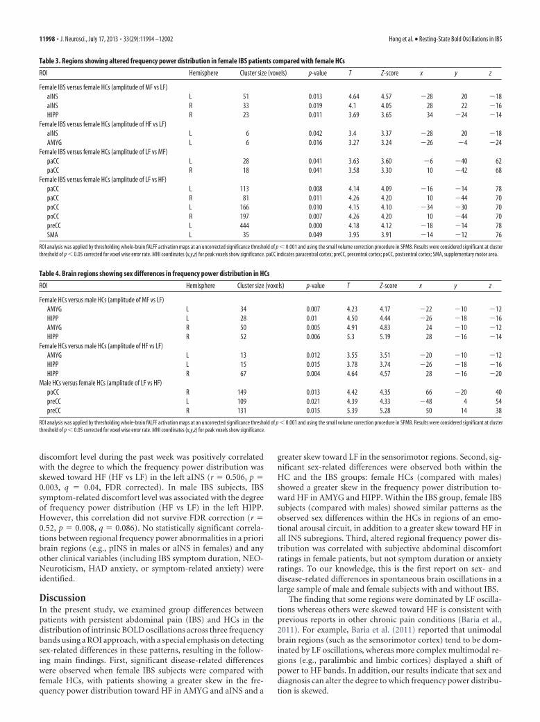
discomfort level during the past week was positively correlatedwith the degree to which the frequency power distribution wasskewed toward HF (HF vs LF) in the left aINS (r � 0.506, p �0.003, q � 0.04, FDR corrected). In male IBS subjects, IBSsymptom-related discomfort level was associated with the degreeof frequency power distribution (HF vs LF) in the left HIPP.However, this correlation did not survive FDR correction (r �0.52, p � 0.008, q � 0.086). No statistically significant correla-tions between regional frequency power abnormalities in a prioribrain regions (e.g., pINS in males or aINS in females) and anyother clinical variables (including IBS symptom duration, NEO-Neuroticism, HAD anxiety, or symptom-related anxiety) wereidentified.
DiscussionIn the present study, we examined group differences betweenpatients with persistent abdominal pain (IBS) and HCs in thedistribution of intrinsic BOLD oscillations across three frequencybands using a ROI approach, with a special emphasis on detectingsex-related differences in these patterns, resulting in the follow-ing main findings. First, significant disease-related differenceswere observed when female IBS subjects were compared withfemale HCs, with patients showing a greater skew in the fre-quency power distribution toward HF in AMYG and aINS and a
greater skew toward LF in the sensorimotor regions. Second, sig-nificant sex-related differences were observed both within theHC and the IBS groups: female HCs (compared with males)showed a greater skew in the frequency power distribution to-ward HF in AMYG and HIPP. Within the IBS group, female IBSsubjects (compared with males) showed similar patterns as theobserved sex differences within the HCs in regions of an emo-tional arousal circuit, in addition to a greater skew toward HF inall INS subregions. Third, altered regional frequency power dis-tribution was correlated with subjective abdominal discomfortratings in female patients, but not symptom duration or anxietyratings. To our knowledge, this is the first report on sex- anddisease-related differences in spontaneous brain oscillations in alarge sample of male and female subjects with and without IBS.
The finding that some regions were dominated by LF oscilla-tions whereas others were skewed toward HF is consistent withprevious reports in other chronic pain conditions (Baria et al.,2011). For example, Baria et al. (2011) reported that unimodalbrain regions (such as the sensorimotor cortex) tend to be dom-inated by LF oscillations, whereas more complex multimodal re-gions (e.g., paralimbic and limbic cortices) displayed a shift ofpower to HF bands. In addition, our results indicate that sex anddiagnosis can alter the degree to which frequency power distribu-tion is skewed.
Table 3. Regions showing altered frequency power distribution in female IBS patients compared with female HCs
ROI Hemisphere Cluster size (voxels) p-value T Z-score x y z
Female IBS versus female HCs (amplitude of MF vs LF)aINS L 51 0.013 4.64 4.57 �28 20 �18aINS R 33 0.019 4.1 4.05 28 22 �16HIPP R 23 0.011 3.69 3.65 34 �24 �14
Female IBS versus female HCs (amplitude of HF vs LF)aINS L 6 0.042 3.4 3.37 �28 20 �18AMYG L 6 0.016 3.27 3.24 �26 �4 �24
Female IBS versus female HCs (amplitude of LF vs MF)paCC L 28 0.041 3.63 3.60 �6 �40 62paCC R 18 0.041 3.58 3.30 10 �42 68
Female IBS versus female HCs (amplitude of LF vs HF)paCC L 113 0.008 4.14 4.09 �16 �14 78paCC R 81 0.011 4.26 4.20 10 �44 70poCC L 166 0.010 4.15 4.10 �34 �30 70poCC R 197 0.007 4.26 4.20 10 �44 70preCC L 444 0.000 4.18 4.12 �18 �14 78SMA L 35 0.049 3.95 3.91 �14 �12 76
ROI analysis was applied by thresholding whole-brain fALFF activation maps at an uncorrected significance threshold of p � 0.001 and using the small volume correction procedure in SPM8. Results were considered significant at clusterthreshold of p � 0.05 corrected for voxel wise error rate. MNI coordinates (x,y,z) for peak voxels show significance. paCC indicates paracentral cortex; preCC, precentral cortex; poCC, postcentral cortex; SMA, supplementary motor area.
Table 4. Brain regions showing sex differences in frequency power distribution in HCs
ROI Hemisphere Cluster size (voxels) p-value T Z-score x y z
Female HCs versus male HCs (amplitude of MF vs LF)AMYG L 34 0.007 4.23 4.17 �22 �10 �12HIPP L 28 0.01 4.50 4.44 �26 �18 �16AMYG R 50 0.005 4.91 4.83 24 �10 �12HIPP R 52 0.006 5.3 5.19 28 �16 �14
Female HCs versus male HCs (amplitude of HF vs LF)AMYG L 13 0.012 3.55 3.51 �20 �10 �12HIPP L 15 0.015 3.78 3.74 �26 �18 �16HIPP R 67 0.004 4.64 4.57 28 �16 �20
Male HCs versus female HCs (amplitude of LF vs HF)poCC R 149 0.013 4.42 4.35 66 �20 40preCC L 109 0.021 4.39 4.33 �48 4 54preCC R 131 0.015 5.39 5.28 50 14 38
ROI analysis was applied by thresholding whole-brain fALFF activation maps at an uncorrected significance threshold of p � 0.001 and using the small volume correction procedure in SPM8. Results were considered significant at clusterthreshold of p � 0.05 corrected for voxel wise error rate. MNI coordinates (x,y,z) for peak voxels show significance.
11998 • J. Neurosci., July 17, 2013 • 33(29):11994 –12002 Hong et al. • Resting-State Bold Oscillations in IBS

Sex-specific alterations in oscillatory dynamics between IBSand HCsIn contrast to previous reports from other chronic pain condi-tions, no disease-related differences in regional oscillation fre-quencies were identified when male and female subjects werecombined, despite the significantly larger sample size comparedwith previous reports. Even though these earlier studies in pa-tients with different persistent pain conditions have providedevidence for abnormal resting-state oscillatory dynamics (Mali-nen et al., 2010; Baliki et al., 2011), the majority of studies werelimited by small sample size, sometimes heterogeneous pa-tient populations, and by the combination of male and femalesubjects.
When female and male subjects were compared separately,robust differences between IBS subjects and HCs were observed.Female IBS subjects showed oscillatory alterations in affect re-lated brain regions (aINS and AMYG) and sensorimotor regionscompared with female HCs. In contrast, male HCs showed alter-ations in viscerosensory regions (mainly in mINS and pINS)compared with male IBS subjects. Similar to previous observa-tions (Malinen et al., 2010; Napadow et al., 2010; Baliki et al.,2011; Ichesco et al., 2012), we observed disease-related alterations
in oscillatory dynamics in the INS, even though the affected INSsubregions differed between males and females. Although pINSand mINS receive primary interoceptive and somatosensory in-puts, the aINS is considered a multimodal association cortex re-ceiving interoceptive and affective inputs (Craig, 2009). One mayspeculate that an upregulation of emotional arousal/salience cir-cuits involving the aINS and AMYG contributes more to symp-toms in female patients, whereas an increased engagement ofnetworks related to sensorimotor integration and interoceptiveprocessing may play a greater role in male patients.
Sex-related differences in HCsThe present study found significant sex-related differences asso-ciated with spontaneous BOLD fluctuation patterns in the HCgroup. Female subjects showed different oscillation dynamicscompared with males in affect-related regions and in sensorimo-tor regions. The pattern of the observed sex-related difference inintrinsic BOLD oscillations are similar to those reported fromstudies on brain responses to emotional stimuli (Kilpatrick et al.,2006; Dickie and Armony, 2008; Stevens and Hamann, 2012) andpain stimuli (Derbyshire et al., 2002; Moulton et al., 2006) inhealthy subjects. For example, in response to negative emotional
Figure 3. Altered regional BOLD oscillation distribution in female IBS subjects compared with male IBS subjects. a, Significant differences of HF versus LF power distribution in female IBS subjectscompared with male IBS subjects are shown in red ( p �0.05, FWE corrected). b, ROIs with significant differences between LF versus HF power distribution in female IBS subjects compared with maleIBS subjects are shown in red ( p � 0.05, FWE corrected).
Table 5. Brain regions showing sex differences in frequency power distribution in IBS
ROI Hemisphere Cluster size (voxels) p-value T Z-score x y z
Female IBS versus male IBS (amplitude of HF vs LF)aINS L 52 0.013 3.89 3.84 �32 20 �16pINS L 53 0.016 3.47 3.44 �36 �6 �12aINS R 44 0.015 4.54 4.48 30 22 �16mINS R 82 0.005 4.00 3.95 36 6 �12pINS R 115 0.005 4.36 4.30 38 �4 �14AMYG L 27 0.008 3.44 3.41 �30 �2 �18HIPP L 46 0.007 4.02 3.98 �26 �26 �10
Female IBS versus male IBS (amplitude of LF vs HF)poCC L 287 0.002 4.56 4.50 �34 �16 40preCC L 544 0.000 4.93 4.84 �18 �20 68SMA L 67 0.026 4.51 4.44 �14 �14 72
ROI analysis was applied by thresholding whole-brain fALFF activation maps at an uncorrected significance threshold of p � 0.001 and using the small volume correction procedure in SPM8. Results were considered significant at clusterthreshold of p � 0.05 corrected for voxel wise error rate. MNI coordinates (x,y,z) for peak voxels show significance.
Hong et al. • Resting-State Bold Oscillations in IBS J. Neurosci., July 17, 2013 • 33(29):11994 –12002 • 11999

stimuli, female subjects had greater activation in HIPP andAMYG compared with male subjects (Stevens and Hamann,2012). In response to noxious heat, male HCs had greater activa-tions in somatosensory and viscerosensory regions, as well as theprefrontal and parietal regions compared with female HCs (Der-byshire et al., 2002; Moulton et al., 2006). The similarity betweensex differences in resting brain measures and task-based mea-sures is consistent with previous studies demonstrating a rela-tionship between rest and task-evoked responses (Mennes et al.,2011), even though the precise relationship between task-basedBOLD signals and resting-state frequency fluctuations remains tobe determined (Mennes et al., 2011). In summary, our findingssuggest a fundamental difference in the central processing of af-fective and nociceptive stimuli in the healthy male and femalebrain, which can be detected in the absence of any stimulus.
Sex-related differences in IBS subjectsSimilar to the healthy group, female IBS subjects (compared withmales) had greater oscillatory dynamics in affect-related regions(AMYG and HIPP) and in all INS subregions, which were notobserved in HCs. The current findings suggest some similaritieswith previously published sex-related differences observed inevoked brain responses in IBS subjects (Naliboff et al., 2003; La-bus et al., 2008). Abnormal power spectral density shifts from LFtoward HF, especially at 0.16 Hz in the INS, had been reportedpreviously in patients with chronic musculoskeletal pain (Mali-nen et al., 2010). Our study also showed that IBS subjects hadsimilar aberrant frequency power distributions in all the INS sub-regions, with female IBS subjects showing greater fluctuationshifts from LF to HF. Even though the reason behind the greaterfrequency dynamics in female IBS subjects remains unclear, onecan speculate that increased activity in affective regions (e.g.,AMYG and HIPP) contributed to this sex-related difference infrequency power distributions of the anterior aspects of the INS.
Correlation of frequency dynamics withclinical/behavioral parametersIn female patients, a significantly positive correlation betweenaberrant frequency power distribution in the left aINS and thesubjective report of abdominal discomfort was observed, consis-tent with previous reports on correlations of the aINS with sub-jective ratings of visceral stimuli (Dunckley et al., 2005; Lowenet al., 2013). In view of the close bidirectional interactions ofaINS with the AMYG, it is conceivable that tonically increasedinput to the aINS from emotional arousal circuits plays a roleboth in the generation of subjective symptoms and in the ob-served abnormalities in intrinsic oscillations. The lack of a signif-icant correlation between symptom duration and alteredoscillatory dynamics makes it less likely that the observed find-ings are a consequence of longstanding alteration in gut functionand visceral afferent signaling, and one could speculate that it repre-sents a primary, possibly genetically/epigenetically determined ab-normality of central sensory processing. Alternatively, the lack ofsignificant correlations with clinical symptoms, including duration,anxiety, or NEO-Neuroticism, may be related to sample size or to thefact that such complex clinical variables are more correlated withalterations in brain networks than with activity in individual regions.
Possible pathophysiological implicationsThe findings of this study are consistent with a dysregulation of atleast two resting-state networks involving affective and sensorybrain regions. Evidence has been provided for the existence oftwo distinct and anticorrelated intrinsic resting-state networksassociated with different INS subregions that function as hubs.One network is centered in the ventral aspect of the aINS withconnections to the ACC, AMYG, and temporal lobe regions, isprimarily related to attentional and affective regions, and mayplay a role in salience detection and other emotional aspects. Theother network links the mINS and pINS to sensorimotor, premo-
Figure 4. Comparison of frequency power in represented ROIs across groups. Graphs show normalized mean Z-score of frequency power distribution for selected ROIs. a, Male HCs showed greaterfrequency power distributions skewed from MF toward HF in left INS subregions. b, Female IBS showed greater frequency power distributions skewed toward HF in left AMYG and aINS, and towardLF in left precentral cortex compared with female HCs. c, Female HCs had greater frequency power shifts from LF toward higher frequencies in right AMYG and right HIPP compared with Male HCs.Male HCs had a greater skew from HF toward LF in right precentral cortex compared with female HCs. d, Female IBS subjects showed greater frequency power oscillation skewed toward HF in rightpINS, right HIPP, and left AMYG and toward LF power in left precentral cortex compared with male IBS. *Significant difference, p � 0.05, FWE corrected.
12000 • J. Neurosci., July 17, 2013 • 33(29):11994 –12002 Hong et al. • Resting-State Bold Oscillations in IBS

tor, MCC, and pACC, indicating a role in sensorimotor integra-tion (Cauda et al., 2011). The fact that previous work has showngood correspondence of networks derived from resting-statestudies and from task-relevant brain activation studies (De Lucaet al., 2005; Vincent et al., 2006; Fox and Raichle, 2007; Smith etal., 2009; Baria et al., 2011) is consistent with our observation thatdisease- and sex-related findings in the current resting-statestudy are similar to previously published activation studies inIBS.
ConclusionsTo our knowledge, this is the first large scale resting-state fMRIanalysis performed in IBS subjects identifying disease-related os-cillation frequencies that are sex specific. The findings are consis-tent with sex-specific dysregulations in INS centric networksengaged in emotional arousal/salience detection and in sensori-motor processing (Cauda et al., 2011; Cauda et al., 2012), con-firming previous findings obtained in studies using evoked brainresponses to nociceptive stimuli and their expectation. The lackof identified correlations between these frequency abnormalitiesand symptom duration, together with the observation that sev-eral of the observed sex-related differences in IBS were also ob-served in the HCs, suggests that some of these alterations may beprimary and not a consequence of longstanding symptoms.
ReferencesApkarian AV, Hashmi JA, Baliki MN (2011) Pain and the brain: specificity
and plasticity of the brain in clinical chronic pain. Pain 152:S49 –S64.CrossRef Medline
Baliki MN, Baria AT, Apkarian AV (2011) The cortical rhythms of chronicback pain. J Neurosci 31:13981–13990. CrossRef Medline
Baria AT, Baliki MN, Parrish T, Apkarian AV (2011) Anatomical and func-tional assemblies of brain BOLD oscillations. J Neurosci 31:7910 –7919.CrossRef Medline
Benjamini Y, Hochberg Y (2000) On the adaptive control of the false dis-covery rate in multiple testing with independent statistics. Journal ofEducational and Behavioral Statistics 25:60 – 83. CrossRef
Benjamini Y, Krieger AM, Yekutieli D (2006) Adaptive linear step-up pro-cedures that control the false discovery rate. Biometrika 93:491–507.CrossRef
Biswal BB, Mennes M, Zuo XN, Gohel S, Kelly C, Smith SM, Beckmann CF,Adelstein JS, Buckner RL, Colcombe S, Dogonowski AM, Ernst M, Fair D,Hampson M, Hoptman MJ, Hyde JS, Kiviniemi VJ, Kotter R, Li SJ, LinCP, et al. (2010) Toward discovery science of human brain function.Proc Natl Acad Sci U S A 107:4734 – 4739. CrossRef Medline
Cauda F, D’Agata F, Sacco K, Duca S, Geminiani G, Vercelli A (2011) Func-tional connectivity of the insula in the resting brain. Neuroimage 55:8 –23.CrossRef Medline
Cauda F, Costa T, Torta DM, Sacco K, D’Agata F, Duca S, Geminiani G, FoxPT, Vercelli A (2012) Meta-analytic clustering of the INS cortex: char-acterizing the meta-analytic connectivity of the insula when involved inactive tasks. Neuroimage 62:343–355. CrossRef Medline
Chang L (2011) The role of stress on physiologic responses and clinicalsymptoms in irritable bowel syndrome. Gastroenterology 140:761–765.CrossRef Medline
Chang L, Heitkemper MM (2002) Gender differences in irritable bowel syn-drome. Gastroenterology 123:1686 –1701. CrossRef Medline
Chang L, Lee OY, Naliboff B, Schmulson M, Mayer EA (2001) Sensation ofbloating and visible abdominal distension in patients with irritable bowelsyndrome. Am J Gastroenterol 96:3341–3347. CrossRef Medline
Costa PT Jr, McCrae RR (1995) Domains and facets: hierarchical personal-ity assessment using the revised NEO personality inventory. J Pers Assess64:21–50. CrossRef Medline
Craig AD (2009) How do you feel–now? The anterior insula and humanawareness. Nat Rev Neurosci 10:59 –70. CrossRef Medline
Davis KD, Moayedi M (2012) Central mechanisms of pain revealed throughfunctional and structural MRI. J Neuroimmune Pharmacol 8:518 –534.CrossRef Medline
De Luca M, Smith S, De Stefano N, Federico A, Matthews PM (2005) Blood
oxygenation level dependent contrast resting state networks are relevantto functional activity in the neocortical sensorimotor system. Exp BrainRes 167:587–594. CrossRef Medline
Derbyshire SW, Nichols TE, Firestone L, Townsend DW, Jones AK (2002)Gender differences in patterns of cerebral activation during equal experi-ence of painful laser stimulation. J Pain 3:401– 411. CrossRef Medline
Dickie EW, Armony JL (2008) Amygdala responses to unattended fearfulfaces: Interaction between sex and trait anxiety. Psychiatry Res 162:51–57.CrossRef Medline
Drossman DA (2000) Rome II: the functional gastrointestinal disorders: di-agnosis, pathophysiology, and treatment: a multinational consensus, Ed2. McLean, VA: Degnon Associates.
Drossman DA (2006) The functional gastrointestinal disorders and theRome III process. Gastroenterology 130:1377–1390. CrossRef Medline
Duff EP, Johnston LA, Xiong J, Fox PT, Mareels I, Egan GF (2008) Thepower of spectral density analysis for mapping endogenous BOLD signalfluctuations. Hum Brain Mapp 29:778 –790. CrossRef Medline
Dunckley P, Wise RG, Aziz Q, Painter D, Brooks J, Tracey I, Chang L (2005)Cortical processing of visceral and somatic stimulation: differentiatingpain intensity from unpleasantness. Neuroscience 133:533–542. CrossRefMedline
Farmer MA, Baliki MN, Apkarian AV (2012) A dynamic network perspec-tive of chronic pain. Neurosci Lett 520:197–203. CrossRef Medline
Fox MD, Raichle ME (2007) Spontaneous fluctuations in brain activity ob-served with functional magnetic resonance imaging. Nat Rev Neurosci8:700 –711. CrossRef Medline
Fukudo S, Kanazawa M (2011) Gene, environment, and brain-gut interac-tions in irritable bowel syndrome. J Gastroenterol Hepatol 26:110 –115.CrossRef Medline
Glascher J, Gitelman D (2008) Contrast weights in flexible factorial designwith multiple groups of subjects. Retrieved May 10, 2010, fromhttp://www.sbirc.ed.ac.uk/cyril/fMRI.html.
Goldman RI, Stern JM, Engel J Jr, Cohen MS (2002) Simultaneous EEG andfMRI of the alpha rhythm. Neuroreport 13:2487–2492. CrossRef Medline
Hoptman MJ, Zuo XN, Butler PD, Javitt DC, D’Angelo D, Mauro CJ, MilhamMP (2010) Amplitude of low-frequency oscillations in schizophrenia: aresting state fMRI study. Schizophr Res 117:13–20. CrossRef Medline
Ichesco E, Quintero A, Clauw DJ, Peltier S, Sundgren PM, Gerstner GE,Schmidt-Wilcke T (2012) Altered functional connectivity between theinsula and the cingulate cortex in patients with temporomandibular dis-order: a pilot study. Headache 52:441– 454. CrossRef Medline
Kilpatrick LA, Zald DH, Pardo JV, Cahill LF (2006) Sex-related differencesin amygdala functional connectivity during resting conditions. Neuroim-age 30:452– 461. CrossRef Medline
Kwak Y, Peltier SJ, Bohnen NI, Muller ML, Dayalu P, Seidler RD (2012)L-DOPA changes spontaneous low-frequency BOLD signal oscillations inParkinson’s disease: a resting state fMRI study. Front Syst Neurosci 6:52.CrossRef Medline
Labus JS, Bolus R, Chang L, Wiklund I, Naesdal J, Mayer EA, Naliboff BD(2004) The Visceral Sensitivity Index: development and validation of agastrointestinal symptom-specific anxiety scale. Aliment Pharmacol Ther20:89 –97. CrossRef Medline
Labus JS, Mayer EA, Chang L, Bolus R, Naliboff BD (2007) The central roleof gastrointestinal-specific anxiety in irritable bowel syndrome: furthervalidation of the visceral sensitivity index. Psychosom Med 69:89 –98.CrossRef Medline
Labus JS, Naliboff BN, Fallon J, Berman SM, Suyenobu B, Bueller JA, Man-delkern M, Mayer EA (2008) Sex differences in brain activity duringaversive visceral stimulation and its expectation in patients with chronicabdominal pain: a network analysis. Neuroimage 41:1032–1043. CrossRefMedline
Lawal A, Kern M, Sidhu H, Hofmann C, Shaker R (2006) Novel evidence forhypersensitivity of visceral sensory neural circuitry in irritable bowel syn-drome patients. Gastroenterology 130:26 –33. CrossRef Medline
Lieberman MD, Eisenberger NI, Crockett MJ, Tom SM, Pfeifer JH, Way BM(2007) Putting feelings into words: affect labeling disrupts amygdala ac-tivity in response to affective stimuli. Psychol Sci 18:421– 428. CrossRefMedline
Lowen MB, Mayer EA, Sjoberg M, Tillisch K, Naliboff B, Labus J, Lundberg P,Strom M, Engstrom M, Walter SA (2013) Effect of hypnotherapy andeducational intervention on brain response to visceral stimulus in theirritable bowel syndrome. Aliment Pharmacol Ther.
Hong et al. • Resting-State Bold Oscillations in IBS J. Neurosci., July 17, 2013 • 33(29):11994 –12002 • 12001

Maldjian JA, Laurienti PJ, Kraft RA, Burdette JH (2003) An automatedmethod for neuroanatomic and cytoarchitectonic atlas-based interroga-tion of fMRI data sets. Neuroimage 19:1233–1239. CrossRef Medline
Maldjian JA, Laurienti PJ, Burdette JH (2004) Precentral gyrus discrepancyin electronic versions of the Talairach atlas. Neuroimage 21:450 – 455.CrossRef Medline
Malinen S, Vartiainen N, Hlushchuk Y, Koskinen M, Ramkumar P, Forss N,Kalso E, Hari R (2010) Aberrant temporal and spatial brain activity dur-ing rest in patients with chronic pain. Proc Natl Acad Sci U S A 107:6493–6497. CrossRef Medline
Mantini D, Perrucci MG, Del Gratta C, Romani GL, Corbetta M (2007)Electrophysiological signatures of resting state networks in the humanbrain. Proc Natl Acad Sci U S A 104:13170 –13175. CrossRef Medline
Mayer EA, Naliboff BD, Chang L, Coutinho SV (2001) V. Stress and irritablebowel syndrome. Am J Physiol Gastrointest Liver Physiol 280:G519–G524.Medline
Mayer EA, Berman S, Chang L, Naliboff BD (2004) Sex-based differences ingastrointestinal pain. Eur J Pain 8:451– 463. CrossRef Medline
Mayer EA, Berman S, Suyenobu B, Labus J, Mandelkern MA, Naliboff BD,Chang L (2005) Differences in brain responses to visceral pain betweenpatients with irritable bowel syndrome and ulcerative colitis. Pain 115:398 – 409. CrossRef Medline
Mayer EA, Naliboff BD, Craig AD (2006) Neuroimaging of the brain-gutaxis: from basic understanding to treatment of functional GI disorders.Gastroenterology 131:1925–1942. CrossRef Medline
Mayer EA, Bushnell MC; International Association for the Study of Pain.(2009) Functional pain syndromes: presentation and pathophysiology.Seattle: IASP.
Mennes M, Zuo XN, Kelly C, Di Martino A, Zang YF, Biswal B, CastellanosFX, Milham MP (2011) Linking inter-individual differences in neuralactivation and behavior to intrinsic brain dynamics. Neuroimage 54:2950 –2959. CrossRef Medline
Moulton EA, Keaser ML, Gullapalli RP, Maitra R, Greenspan JD (2006) Sexdifferences in the cerebral BOLD signal response to painful heat stimuli.Am J Physiol Regul Integr Comp Physiol 291:R257–R267. CrossRefMedline
Mykletun A, Stordal E, Dahl AA (2001) Hospital Anxiety and Depression(HAD) scale: factor structure, item analyses and internal consistency in alarge population. Br J Psychiatry 179:540 –544. CrossRef Medline
Mykletun A, Jacka F, Williams L, Pasco J, Henry M, Nicholson GC, KotowiczMA, Berk M (2010) Prevalence of mood and anxiety disorder in selfreported irritable bowel syndrome (IBS). An epidemiological populationbased study of women. BMC Gastroenterol 10:88. CrossRef Medline
Naliboff BD, Berman S, Chang L, Derbyshire SW, Suyenobu B, Vogt BA,Mandelkern M, Mayer EA (2003) Sex-related differences in IBS pa-tients: central processing of visceral stimuli. Gastroenterology 124:1738 –1747. CrossRef Medline
Naliboff BD, Berman S, Suyenobu B, Labus JS, Chang L, Stains J, MandelkernMA, Mayer EA (2006) Longitudinal change in perceptual and brain ac-
tivation response to visceral stimuli in irritable bowel syndrome patients.Gastroenterology 131:352–365. CrossRef Medline
Napadow V, LaCount L, Park K, As-Sanie S, Clauw DJ, Harris RE (2010)Intrinsic brain connectivity in fibromyalgia is associated with chronicpain intensity. Arthritis Rheum 62:2545–2555. CrossRef Medline
Smith SM, Fox PT, Miller KL, Glahn DC, Fox PM, Mackay CE, Filippini N,Watkins KE, Toro R, Laird AR, Beckmann CF (2009) Correspondenceof the brain’s functional architecture during activation and rest. Proc NatlAcad Sci U S A 106:13040 –13045. CrossRef Medline
Stevens JS, Hamann S (2012) Sex differences in brain activation to emo-tional stimuli: a meta-analysis of neuroimaging studies. Neuropsycholo-gia 50:1578 –1593. CrossRef Medline
Tillisch K, Mayer EA, Labus JS (2011) Quantitative meta-analysis identifiesbrain regions activated during rectal distension in irritable bowel syn-drome. Gastroenterology 140:91–100. CrossRef Medline
van Marle HJ, Hermans EJ, Qin S, Fernandez G (2009) From specificity tosensitivity: how acute stress affects amygdala processing of biologicallysalient stimuli. Biolo Psychiatry 66:649 – 655. CrossRef Medline
Van Oudenhove L (2011) Understanding gut-brain interactions in gastro-intestinal pain by neuroimaging: lessons from somatic pain studies. Neu-rogastroenterol Motil 23:292–302. CrossRef Medline
Vincent JL, Snyder AZ, Fox MD, Shannon BJ, Andrews JR, Raichle ME,Buckner RL (2006) Coherent spontaneous activity identifies a hippo-campal-parietal memory network. J Neurophysiol 96:3517–3531.CrossRef Medline
Wang J, Korczykowski M, Rao H, Fan Y, Pluta J, Gur RC, McEwen BS, DetreJA (2007) Gender difference in neural response to psychological stress.Social cognitive and affective neuroscience 2:227–239. CrossRef Medline
Wang L, Dai W, Su Y, Wang G, Tan Y, Jin Z, Zeng Y, Yu X, Chen W, Wang X,Si T (2012) Amplitude of low-frequency oscillations in first-episode,treatment-naive patients with major depressive disorder: a resting-statefunctional MRI study. PLoS One 7:e48658. CrossRef Medline
Wilder-Smith CH, Schindler D, Lovblad K, Redmond SM, Nirkko A (2004)Brain functional magnetic resonance imaging of rectal pain and activa-tion of endogenous inhibitory mechanisms in irritable bowel syndromepatient subgroups and healthy controls. Gut 53:1595–1601. CrossRefMedline
Wise RG, Ide K, Poulin MJ, Tracey I (2004) Resting fluctuations in arterialcarbon dioxide induce significant low frequency variations in BOLD sig-nal. Neuroimage 21:1652–1664. CrossRef Medline
Zang YF, He Y, Zhu CZ, Cao QJ, Sui MQ, Liang M, Tian LX, Jiang TZ, WangYF (2007) Altered baseline brain activity in children with ADHD re-vealed by resting-state functional MRI. Brain Dev 29:83–91. CrossRefMedline
Zou QH, Zhu CZ, Yang Y, Zuo XN, Long XY, Cao QJ, Wang YF, Zang YF(2008) An improved approach to detection of amplitude of low-frequency fluctuation (ALFF) for resting-state fMRI: fractional ALFF.J Neurosci Methods 172:137–141. CrossRef Medline
12002 • J. Neurosci., July 17, 2013 • 33(29):11994 –12002 Hong et al. • Resting-State Bold Oscillations in IBS