nervous system: neurons - napa valley college 9 - neurons a… · title: microsoft powerpoint -...
TRANSCRIPT

10/16/2013
1
Nervous System:Neurons
Biology 105
Lecture 9
Chapter 7
Outline
I. Nervous system overview and function
II. Central and peripheral nervous systems
III. Nervous system cells
IV. Myelinated neurons
V Nerve signal transmission
Copyright © 2009 Pearson Education, Inc.
V. Nerve signal transmission
VI. Neuronal synapse
Nervous Tissues
Nervous tissue functions to conduct messages throughout the body.
When a nerve cell is stimulated, an electrical signal quickly travels through the nerve cell to
Copyright © 2009 Pearson Education, Inc.
the nerve ending, triggering events.

10/16/2013
2
Nervous System
Includes nerve tissue and sensory organs
Nervous system functions to:
Sense the environment – receives information from both outside and inside
Copyright © 2009 Pearson Education, Inc.
the body.
Process the information it receives.
Respond to information – sends out orders.
Two Parts of the Nervous System
1. Central Nervous System (CNS)
Brain and spinal cord
2. Peripheral Nervous System (PNS)
N ti t id b i d i
Copyright © 2009 Pearson Education, Inc.
Nervous tissue outside brain and spine
Sensory organs
Central Nervous System
Peripheral
Copyright © 2009 Pearson Education, Inc.
10/16/2013
3
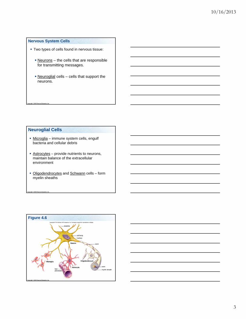
Nervous System Cells
Two types of cells found in nervous tissue:
Neurons – the cells that are responsible for transmitting messages.
Copyright © 2009 Pearson Education, Inc.
Neuroglial cells – cells that support the neurons.
Neuroglial Cells
Microglia – immune system cells, engulf bacteria and cellular debris
Astrocytes – provide nutrients to neurons, maintain balance of the extracellular
Copyright © 2009 Pearson Education, Inc.
environment
Oligodendrocytes and Schwann cells – form myelin sheaths
Figure 4.6
Copyright © 2009 Pearson Education, Inc.

10/16/2013
4
Parts of a Neuron
Cell body – the main body of the cell. Contains the nucleus.
Dendrites – many projections from the cell body that carry messages to the cell body.
Copyright © 2009 Pearson Education, Inc.
y y g y
Axons – one large projection that carries messages away from the cell body.
Dendrites receive information from other neurons or from the environment.
The cell body controlsthe cell’s metabolic activities.
Axon endings release chemicals called neurotransmitters that affect the activity of nearby neurons or an effector (muscle or gland).
Nucleus
Neuron!Neuron!
Copyright © 2009 Pearson Education, Inc. Figure 7.2
The cell bodyintegrates input from other neurons.
An axon conducts the nerve impulse away from the cell body.
Receiving portion of neuron
Sending portion of neuron
Cell body
Axon endings
DIRECTION OF NEURONAL SIGNAL
Copyright © 2009 Pearson Education, Inc.
10/16/2013
5
Neurons of the Peripheral Nervous System
Neurons in the PNS are either carrying messages to or from the CNS.
Afferent = sensory neurons = neurons carrying messages TO the CNS.
Copyright © 2009 Pearson Education, Inc.
Efferent = motor neurons = neurons carrying messages FROM the CNS.
Interneurons in the Central Nervous System
Located between sensory and motor neurons within the CNS.
Interneurons integrate and interpret sensory signals.
Copyright © 2009 Pearson Education, Inc.
g
Copyright © 2009 Pearson Education, Inc.

10/16/2013
6
Sensory Neurons
The afferent or sensory neuron cell bodies are located in dorsal root ganglion.
Motor Neurons
Th ff t t ll b di
Copyright © 2009 Pearson Education, Inc.
The efferent or motor neuron cell bodies are located in the gray matter of the spinal cord. Their axons leave the CNS and go to the
skeletal muscles.
Neurons of the Nervous System
Copyright © 2009 Pearson Education, Inc.
The cell bodies of these neurons are located in the dorsal root ganglion:
50%50%1. Motor
2. Sensory
Copyright © 2009 Pearson Education, Inc.
Moto
r
Sen
sory
10/16/2013
7
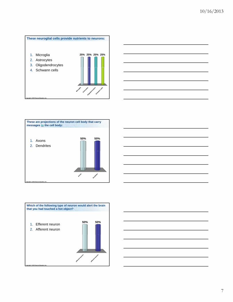
These neuroglial cells provide nutrients to neurons:
25% 25%25%25%1. Microglia
2. Astrocytes
3. Oligodendrocytes
Copyright © 2009 Pearson Education, Inc.
Mic
roglia
Ast
rocy
tes
Olig
oden
rocy
tes
Sch
wann c
ells
4. Schwann cells
These are projections of the neuron cell body that carry messages to the cell body:
50%50%1. Axons
2. Dendrites
Copyright © 2009 Pearson Education, Inc.
Axo
ns
Den
drite
s
Which of the following type of neuron would alert the brain that you had touched a hot object?
50%50%1. Efferent neuron
2. Afferent neuron
Copyright © 2009 Pearson Education, Inc.
effe
rent n
euro
n
affe
rent n
euro
n

10/16/2013
8
What type of neuron is the arrow pointing to?
50%50%
1. Sensory
2. Motor
Copyright © 2009 Pearson Education, Inc.
Sen
sory
Moto
r
Myelinated Neurons
Some neuroglial cells form a substance called myelin that is rich in lipid and contains proteins. These neuroglial cells wrap themselves around
neuronal axons.
The neurons that have axons covered with these ne roglial cells are called m elinated ne rons
Copyright © 2009 Pearson Education, Inc.
neuroglial cells are called myelinated neurons.
Myelinated neurons are able to carry messages faster than non-myelinated neurons. Myelin is an excellent electrical insulator!
Functions of Myelin Sheaths
1. The main benefit of myelin sheaths is that myelinated neurons are able to carry messages faster than non-myelinatedneurons.
Copyright © 2009 Pearson Education, Inc.
2. Myelin sheaths from Schwann cells also help regenerate injured PNS neuronal axons.

10/16/2013
9
Two Types of Cells Myelinate Neurons
Schwann cells are found in the PNS.
Oligodendrocytes are found in the CNS.
Schwann cells and oligodendrocytes both wrap around neuronal axons
Copyright © 2009 Pearson Education, Inc.
around neuronal axons.
Nodes of Ranvier are spaces on the axon between the glial cells.
Myelinated Neurons
Cellbody
Dendrites
Nucleus
In saltatory conduction, the nerve impulses jump from one node of Ranvier to the next.
Copyright © 2009 Pearson Education, Inc. Figure 7.3a
(a)Myelin sheath
Node of Ranvier
Schwann cell
Myelin Sheath
Copyright © 2009 Pearson Education, Inc. Figure 7.3b

10/16/2013
10
Myelin Sheath
Copyright © 2009 Pearson Education, Inc. Figure 7.3c
Multiple Sclerosis (MS)
Caused by the destruction of the myelin sheath that surrounds axons found in the CNS.
Can result in paralysis and loss of sensation,
Copyright © 2009 Pearson Education, Inc.
p y ,including loss of vision.
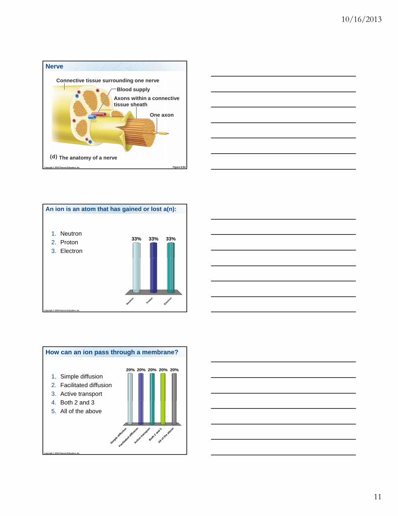
Nerve
A nerve contains many neuronal axons bundled together.
These bundles contain:
Axons
Copyright © 2009 Pearson Education, Inc.
Axons
Blood vessels
Connective tissue
10/16/2013
11
Nerve
Blood supply
Axons within a connectivetissue sheath
One axon
Connective tissue surrounding one nerve
Copyright © 2009 Pearson Education, Inc. Figure 8.9d
(d) The anatomy of a nerve
One axon
An ion is an atom that has gained or lost a(n):
33% 33%33%1. Neutron
2. Proton
3. Electron
Copyright © 2009 Pearson Education, Inc.
Neu
tron
Pro
ton
Ele
ctro
n
How can an ion pass through a membrane?
20% 20% 20%20%20%
1. Simple diffusion
2. Facilitated diffusion
3. Active transport
4 B th 2 d 3
Copyright © 2009 Pearson Education, Inc.
Sim
ple diff
usion
Fac
ilita
ted
diffus
ion
Act
ive tra
nspo
rt
Bot
h 2 and
3
All of t
he ab
ove
4. Both 2 and 3
5. All of the above

10/16/2013
12
The Nerve Impulse is an Electrochemical Signal
A nerve impulse, or action potential, involves sodium ions (Na+) and potassium ions (K+) that cross the cell membrane through ion channels.
Each ion channel is designed to allow only
Copyright © 2009 Pearson Education, Inc.
g ycertain ions to pass through.
Action Potential
Cross section
Axon membrane
Copyright © 2009 Pearson Education, Inc. Figure 7.4
Extracellular fluid
Neuron plasma membrane
Cytoplasm
Sodium-potassium pumpThe sodium-potassium pump uses cellular energy (ATP) to pump sodium ions out of the cell and potassium ions intothe cell
Continually open ion channels “Gated” ion channels Sodium-potassium pump
Ion channelsIon channels can be open continuously or opened and closed by a molecular gate
The difference in charge between the inside and outside of the neuron is the membrane potential.
Membrane Potential
Copyright © 2009 Pearson Education, Inc.

10/16/2013
13
A neuron that is not conducting a message is said to be “resting”.
When a neuron is resting, there is more sodium (Na+) outside the neuron cell and more potassium (K+) inside the cell.
Resting Membrane Potential
Copyright © 2009 Pearson Education, Inc.
potassium (K ) inside the cell.
The inside of the cell has a negative charge compared to the outside the cell.
Resting Membrane Potential
Copyright © 2009 Pearson Education, Inc.
The Nerve Impulse
Copyright © 2009 Pearson Education, Inc. Figure 7.5 (1 of 4)

10/16/2013
14
Sodium-Potassium Pump
To maintain this resting membrane potential the neuron pumps Na+ out of the cell and K+ into the cell.
The transport proteins (= Na+/K+ pumps) move
Copyright © 2009 Pearson Education, Inc.
3 Na+ ions out for every 2 K+ ions into the cell.
This is Active Transport and requires ATP!
Action Potential
Action Potential – an electrochemical signal conducted along an axon. An action potential is a wave of depolarization
followed by repolarization.
Copyright © 2009 Pearson Education, Inc.
Depolarization is caused by sodium ions entering the axon.
Repolarization is caused by potassium ions leaving the axon.
Steps of an Action Potential
1. The axon is depolarized when voltage-gated sodium ion channels open and Na+ comes rushing in. This causes the inside of the neuron to
become positively charged.
Copyright © 2009 Pearson Education, Inc.

10/16/2013
15
Action Potential
Copyright © 2009 Pearson Education, Inc. Figure 7.5 (2 of 4)
Steps of an Action Potential
2. The axon is repolarized when voltage-gated potassium ion channels open and allow K+ to leave the axon. This returns the membrane potential to
negative on the inside of the neuron.
Copyright © 2009 Pearson Education, Inc.
3. The action potential travels down the axon.
Action Potential
Copyright © 2009 Pearson Education, Inc. Figure 7.5 (3 of 4)

10/16/2013
16
Action Potential
After the action potential, the sodium-potassium pump restores the original conditions by pumping sodium (Na+) out of the cell and potassium (K+) back into the cell.
Copyright © 2009 Pearson Education, Inc.
The Nerve Impulse
Copyright © 2009 Pearson Education, Inc. Figure 7.5 (4 of 4)
The Nerve Impulse
Copyright © 2009 Pearson Education, Inc. Figure 7.6
10/16/2013
17
Action Potentials
An action potential is an all-or-nothing response: If there is not enough stimulation, the ion channels
will not open and there will not be an action potential.
The level of the action potential is always the same.
An action potential always moves in the same
Copyright © 2009 Pearson Education, Inc.
An action potential always moves in the same direction down the axon.
The sodium channels are inactivated for awhile after the action potential passes = refractory period.
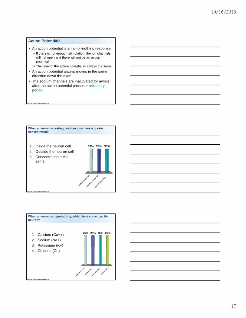
When a neuron is resting, sodium ions have a greater concentration:
33% 33%33%1. Inside the neuron cell
2. Outside the neuron cell
3. Concentration is the
Copyright © 2009 Pearson Education, Inc.
insi
de th
e neu
ron c
ell
outs
ide
the
neuro
n cel
l
conce
ntra
tion is
the
...
same
When a neuron is depolarizing, which ions come into the neuron?
25% 25%25%25%1. Calcium (Ca++)
2. Sodium (Na+)
3. Potassium (K+)
Copyright © 2009 Pearson Education, Inc.
Cal
cium
(Ca+
+)
Sodiu
m (N
a+)
Pota
ssiu
m (K
+)
Chlo
rine
(Cl-)
( )
4. Chlorine (Cl-)

10/16/2013
18
When a neuron is depolarizing, the inside of the neuron cell becomes:
50%50%1. Positively charged
2. Negatively charged
Copyright © 2009 Pearson Education, Inc.
Posi
tivel
y ch
arged
Neg
ativ
ely
char
ged
Neuronal Synapse
How are messages passed from one neuron to the next, or from a neuron to a muscle?
The junction between two neurons or between a neuron and a muscle is called a
Copyright © 2009 Pearson Education, Inc.
synapse.
Components of the Synapse
1. Presynaptic neuron is the transmitting neuron. It has synaptic vesicles that contain
neurotransmitters.
Copyright © 2009 Pearson Education, Inc.
2. Postsynaptic neuron is the receiving neuron or the muscle.
3. And the gap in between them = synaptic cleft.

10/16/2013
19
Synaptic Transmission
Nucleus
Impulse
Axon
Dendrites
Cellbody
Impulse
Step 1: The impulse reaches the axon ending of the
Copyright © 2009 Pearson Education, Inc. Figure 7.8 (1 of 3)
Synapticknob
Synaptic cleft
Synaptic vesicle
Membrane of postsynaptic neuron
the axon ending of the presynaptic membrane.
Step 2: Synaptic vesicles release neurotransmitter into the synaptic cleft.
Synaptic Transmission
Neurotransmitter
Step 3: Neurotransmitterdiffuses across synaptic cleft.
Synapticvesicle
Copyright © 2009 Pearson Education, Inc. Figure 7.8 (2 of 3)
Receptor (of sodium ion channel) on postsynaptic membrane
Synaptic Transmission
Step 5: Sodium ion channels open.
Step 4: Neurotransmitter molecules bind to receptors on the postsynaptic neuron.
Copyright © 2009 Pearson Education, Inc. Figure 7.8 (3 of 3)
open.
Step 6: Sodium ions enter the postsynaptic neuron, causing depolarization and possible action potential.

10/16/2013
20
1. The action potential gets to the end of the presynaptic axon.
2. The action potential triggers Ca2+ to enter the presynaptic axon terminal.
Transmission Across Synaptic Cleft
Copyright © 2009 Pearson Education, Inc.
p y p
3. The Ca2+ triggers synaptic vesicles located at the axon terminal to merge with the neural membrane.
4. The synaptic vesicles release the neurotransmitters into the synaptic cleft.
5. These neurotransmitters travel across the synaptic cleft to the postsynaptic
Transmission Across Synaptic Cleft
Copyright © 2009 Pearson Education, Inc.
y p p y pneuron (or the muscle).
6. Neurotransmitter binds to receptors on the postsynaptic neuron (or muscle).
Transmission Across Synaptic Cleft
7. These receptors are ligand-gated sodium ion channels which allow Na+ to enter the postsynaptic neuron (or muscle) and triggers an action potential in the postsynaptic neuron (or muscle
)
Copyright © 2009 Pearson Education, Inc.
contraction).
8. Once the neurotransmitters are released, they need to be destroyed or contained quickly or they will continue to stimulate the nerve.
10/16/2013
21
Neurotransmitters
Acetylcholine Acts in both the PNS and the CNS as a
neurotransmitter.
Causes voluntary muscles to contract.
Acetylcholinesterase: an enzyme that breaks d t l h li i th ti l ft
Copyright © 2009 Pearson Education, Inc.
down excess acetylcholine in the synaptic cleft.
Myasthenia gravis is an autoimmune disease that attacks the acetylcholine receptors, resulting in reduced muscle strength.
Important Concepts
Read Chapter 7
What are the functions of nervous system?
What are the two types of cells in nervous tissue? (Neuroglial cells and neurons!)
Copyright © 2009 Pearson Education, Inc.
What are the three types of neuroglial cells and what are their functions?
What are the two main divisions of the nervous system (CNS, PNS) and where are they each found?

10/16/2013
22
Important Concepts
What are the parts and functions of a neuron?
What are the three types of neurons (sensory, interneuron, and motor neuron) and their functions, and where are they located?
Copyright © 2009 Pearson Education, Inc.
Where are the cell bodies located for motor and sensory neuronal cells?
What are Schwann cells and oligodendrocytes, and what are their functions?
Where are Schwann cells and oligodendrocytesfound?
Important Concepts
Copyright © 2009 Pearson Education, Inc.
What is the cause and what are the effects of multiple sclerosis?
What are the parts of a nerve?
How do ions pass through membranes?
What is the function of the sodium-potassium pump?
Important Concepts
Copyright © 2009 Pearson Education, Inc.
What are the steps of signal transmission through the nervous system, starting with the resting stage of one neuron and ending with the next neuron or muscle being stimulated?

10/16/2013
23
Which ions enter and exit the neuron during the depolarization and repolarization steps of an action potential? What is the relative charge of the inside versus
the outside of the neuron during these events, and what is the correct order of these events?
Important Concepts
Copyright © 2009 Pearson Education, Inc.
and what is the correct order of these events?
What are the components of a synapse?
What is the function of neurotransmitters? How do they work and where do they work?
What is acetylcholine, where is it found, what effect does it have, and how is acetylcholine removed from the synaptic cleft?
What is the cause and effect of Myasthenia gravis?
Important Concepts
Copyright © 2009 Pearson Education, Inc.
gravis?
Definitions
Afferent neuron, efferent neuron, dendrite, axon, sensory neuron, interneuron, motor neuron, myelin, myelin sheath, myelinatedneuron, Schwann cell, oligodendrocytes, nodes of Ranvier, nerve, ion, ion channel, ligand-gated ion channel, voltage-gated ion channel, action
t ti l l i ti d l i ti
Copyright © 2009 Pearson Education, Inc.
potential, repolarization, depolarization, membrane potential, resting potential, sodium-potassium pump, refractory period, synapse, synaptic cleft, synaptic vesicle, neurotransmitter, acetylcholinesterase, presynaptic neuron, postsynaptic neuron, stimulate, inhibit