nervous control of liver metabolism and …control.pdfnervous control of liver metabolism and...
TRANSCRIPT

U n i v e r s i t ä t P o t s d a m
Andreas, Gardemann ; Gerhard, Puschel ; Kurt, Jungermann
Nervous control of liver metabolism andhemodynamics
first published in:European Journal of Biochemistry 207 (1992), 2, S. 399-411
Postprint published at the Institutional Repository of the Potsdam University:In: Postprints der Universitat PotsdamMathematisch-Naturwissenschaftliche Reihe ; 164http://opus.kobv.de/ubp/volltexte/2011/5134/http://nbn-resolving.de/urn:nbn:de:kobv:517-opus-51346
Postprints der Universitat PotsdamMathematisch-Naturwissenschaftliche Reihe ; 164

Review
Nervous control of liver metabolism and hemodynamics Andreas GARDEMANN, Gerd P. PUSCHEL and Kurt JUNGERMANN
Institut fur Biochemie, Georg-August-Universitat Gottingen, Federal Republic of Germany
(Received March 3,1992) - EJB 92 0292
Besides being a center of intermediary metabolism, a center of defense and a control center for the hormonal system, the liver acts as the glucose reservoir of the organism and, moreover, as an important blood reservoir, by taking up or releasing glucose and blood. The many diverse hepatic functions are controlled by the substrate concentrations in blood, the circulating hormone levels, the biomatrix and the autonomic hepatic nerves. In this review, the present knowledge on the metabolic and hemodynamic effects, the mechanism of action and the function of the hepatic nerves, as studied in the isolated perfused liver, are summarized.
Effects of hepatic nerve stimulation
In perfused rat liver, activation of the sympathetic liver nervesincreases glucose and lactate release, urate and allantoin formation, decreases ketogenesis, urea release, ammonia uptake, xenobiotics conjugation, bile flow and bile acid secretion as well as oxygen utilization, and causes an overflow of noradrenaline into the hepatic vein. Furthermore, sympathetic stimulation decreases the flow and elicits an intrahepatic redistribution as well as a mobilization of blood by the closing of sinusoids. Activation of parasympathetic nerves enhances glucose utilization and causes re-opening of previously closed sinusoids. The actions of hepatic nerves are modulated by the hormones glucagon, insulin, adrenaline, noradrenaline, vasopressin and angiotensin.
Extrahepatocellular signal chain of sympathetic nerve stimulation
The release of noradrenaline from the nerve endings and all effects of electrical nerve stimulation are strictly dependent on the presence of extracellular calcium. Sympathetic nerves regulate the hepatic metabolism of carbohydrates and ketone bodies and the conjugation of xenobiotics directly via interaction with non-parenchymal and parenchymal liver cells. However, sympathetic nerves regulate metabolism of nitrogenous compounds indirectly via reduction of blood flow. Studies with adrenergic and prostanoid agonists and antagonists support the following mechanism. Noradrenaline, released from the nerve endings via al-adrenergic receptors, directly interacts with periportal hepatocytes and also stimulates prostanoid formation in nearby non-parenchymal liver cells. Prostanoids, in turn, modulate metabolism in parenchymal cells. About 50% of the metabolic nerve effects in rat liver are mediated via signal propagation through gap junctions. By this mechanism, rat liver apparently compensates the scarce innervation of only a few parenchymal and non-parenchymal cells in the proximal periportal zone.
Intrahepatocellular signal chain of sympathetic nerve stimulation
Sympathetic nerve stimulation of the perfused rat liver causes an increase in the activity of glycogen phosphorylase and a decrease in the activity of glycogen synthase, but leaves the activity of pyruvate kinase unaltered ; fructose 2,6-bisphosphate and CAMP are only slightly enhanced. Various studies strongly support the notion that the intracellular signal chain is propagated by production of inositol 1,4,5-trisphosphate and an increase in cytosolic calcium.
In conclusion, hepatic nerves, together with the hormonal system, are directly involved in the regulation of liver metabolism and hemodynamics.
le-5-carboxamide sodium salt; TXA2, thromboxane A2 analog U46619.
Enzymes. Calmodulin-dependent myosin light-chain kinase (EC 2.7.1.37); cyclooxygenase (EC 1.14.99.1); glycogen phosphorylase
Fax: + 49-5 51-39 5960 (EC 2.4.1 .I); glycogen phosphorylase kinase (EC 2.7.1.38); glycogen Abbreviations. BM 13505, 4-[2-(4-~hlorobenzenesulfonamide)- synthase (EC 2.4.1 . l l ) ; phospholipase Az (EC 3.1.1.4); pyruvate
kinase (EC 2.7.1.40); 5’-lipoxygenase (EC 1.13.1 1.34); protein kinase C (EC 2.7.1.37); tyrosine aminotransferase (EC 2.7.1.38).
Correspondence to A. Gardemann, Institut fur Biochemie, Georg- August-Universitat Gottingen, Humboldtallee 23, W-3400 Gottin- gen, Federal Republic of Germany
ethyl] benzeneaceticacid; CGP 35949 B, N-{3-[3-(4-acetyl-3-hydroxy - 2-propylphenoxy)propoxy]-4-chlorine-6-methyl-phenyl}-l H-tetrazo-

400
Table 1. Functions of the liver.
Main function Subfunction Purpose
Center of metabolism energy supply of the body glucose uptake and release ketone body production urea production amino acid uptake and release lipid processing
biosynthesis and biodegradation
Center of defense
Control station of the hormonal system
plasma protein synthesis and degradation bile formation
xenobiotic metabolism phagocytosis and biochemical elimination of foreign macromolecules and macromolecular aggregates elimination of tumor cells
inactivation and elimination of hormones and me- diators synthesis and release of (pro)hormones and mediators
Blood reservoir active blood storage passive blood storage
Fig. 1. Schematic drawing of liver tissue. The parenchyma consists of liver cell plates formed by adjoining hepatocytes (H). Blood flow through the sinusoids (S) formed by the fenestrated endothelial cells (E) which are sorrounded by perisinusoidal or fat storing cells (F). Kupffer cells (K) and pit cells (P) bulge into the sinusoidal lumen from the endothelial cells. The space between the endothelial lining and the liver cell plates is the space of Disse (SD), in which the ectoskeleton of the liver composed of collagen, fibronectin, different glycoproteins and proteoglycans are located. BC, bile canaliculi. Taken from Sasse, D., Spornitz, U. M. & Maly, I. P. (1992) Liver Architrcfure. Enzyme, in the press.
The liver is the center of intermediary metabolism, a center of defense, a control center for the hormonal system and a blood reservoir (Table 1). The many diverse hepatic functions are provided by parenchymal and non-parenchymal cells (Fig. 1) and are regulated by substrate concentrations in blood, circulating hormone levels, autonomic hepatic nerves and the biomatrix. Moreover, the parenchymal cells can be under the control of the non-parenchymal cells and vice versa. Finally, the zonal heterogeneity of the parenchymal cells [l, 21 and the heterogeneity of the non-parenchymal cell popu- lation [3, 41, comprising endothelial cells, resident macro- phages (Kupffer cells), perisinusoidal cells (lipocytes, fat storing cells, Ito cells) and large granular lymphocytes (pit
cells) (Fig. 1) are important factors contributing to the various liver functions and their regulation.
It is the aim of this review first to give a short introduction into the anatomy of hepatic innervation and a brief overview on in vivo studies of hepatic nerve functions, then to summarize the present knowledge about the metabolic and hemodynamic effects, the mechanism of action and the func- tion of the hepatic nerves as studied in the isolated perfused liver.
Anatomy of hepatic innervation
This section provides only a short overview of hepatic innervation. Additional and more detailed information is avaiable in other publications [5 - 111.
Extrinsic inner vat ion
Nerves innervating the liver include afferent and efferent, sympathetic, parasympathetic and peptidergic components (Fig. 2). The hepatic Sympathetic fibers are derived from the splanchnic nerves, the parasympathetic fibers from the vagus nerves. Spianchnic, vagal and sometimes also phrenic nerves enter the liver primarily with the hepatic artery, portal vein and bile duct. Some fibers are not associated with the hepatic vasculature and enter the liver via the lesser omentum or the hepatic vein. Sympathetic and parasympathetic nerves form two separate, but communicating, plexuses; the anterior plexus around the hepatic artery is composed of fibers orig- inating from the celiac ganglion and anterior vagus, the pos- terior plexus around the portal vein and bile duct is formed by fibers from the celiac ganglion and right vagus.
The sensory afferent nerves were estimated to represent up to 90% of the fibers of the abdominal vagus and about 50% of sympathetic fibers in the splanchnic nerve [l l] . Afferent vagal fibers project primarily to the left nodose ganglion, then to the medial portion of.the left solitary nucleus [12]. Afferent splanchnic fibers also project to the left solitary nucleus [13]. Efferent sympathetic nerves originate in the ventromedial hypothalamus and Parasympathetic fibers in the lateral hypo- thalamus [14] (Fig. 2).

40 3
L Hypo2alamus lateralis
.-fN IL Nervus vaaus sinister
Artena hepatica 1 -/Aorta \ T V e n a partae
Fig. 2. Scheme of efferent liver innervation and sites of neurostimulation. (I) Stimulation of the arterial plexus at the common hepatic artery. (11) Stimulation of the portal plexus at the mesenteric vein. (111) Joint stimulation at the hepatic artery and portal vein in the liver hilus. L, liver; P, pancreas; A, adrenals; Ins, insulin; Ggn, glucagon; A, adrenaline; NA. noradrenaline; GC, ganglion coeliacum; Th7(10), seventh(tenth) thoracai segment of the splanchnic nerve.
Intrinsic innervation
Many intrinsic nerves of the liver are associated with the arterial and portovenous vasculature-forming plexuses in the adventitia and terminating on smooth muscle cells [15, 161. Nerve endings have also been demonstrated on Kupffer cells, fat-storing cells and the hepatocytes [ I l , 171. Whereas the density of the vascular innervation does not vary considerably [18], the extent and distribution of intralobular innervation exhibit remarkable interspecies variations [15, 18, 191. In the guinea pig and tree shrew, nearly all hepatocytes possess nerve contacts [18], whereas in the rat only a few periportal hepatocytes are innervated [18, 191. The extent of the intrinsic innervation of the parenchymal cells seems to be inversely correlated to the number of signal-propagating gap junctions, since in livers of the tree shrew and guinea pig only a low density [I 51, but in rat liver [I91 a high density of gap junctions has been demonstrated. Studies of primate liver [15] suggest that intralobular innervation is primarily, if not exclusively, monoaminergic. So far, a significant cholinergic innervation of the lobule has only been demonstrated in the guinea pig liver [I 1,201. Besides an adrenergic and a cholinergic innervation a peptidergic innervation has also been found in livers of various species. Vasoactive intestinal peptide could be demonstrated in the hilar region, substance P in the periportal area and neurotensin in the parenchymal region [21,22]. Neuropeptide
Y is apparently co-localized with noradrenaline in hepatic nerve terminals [23, 241 and was identified together with sub- stance P and calcitonin-gene-related peptide in nerve fibers around lymphatic capillaries in the interconnective tissue of rat liver [25]. The functions of the petidergic innervation of the liver have not been studied so Par.
In vivo studies on the role of hepatic nerves
Many studies have been performed in living animals in order to assess the physiological role of the hepatic nerves. It is, however, far beyond the scope of this review to discuss these experiments in detail. They will be considered only briefly to provide a useful background to the studies with isolated perfused livers, reviewed in the next sections. More detailed reviews presenting the in-vivo data have been published re- cently [7, 11, 26-28].
Acute metabolic and hemodynamic regulation by eJferent nerves
Efferent sympathetic hepatic nerves work in the frequency range 2 -20 Hz [29, 301, the efferent parasympathetic fibers in the range 5 - 15 Hz [31]. It is not known whether a change in the frequency range of sympathetic nerves can alter the ratio in which colocalized substances in nerve endings like neuropeptide Y [23, 241 can be released. The discharge rates of the nerves are regulated by the blood-glucose concentration via the central nervous system [ l l ] ; the frequency of parasympathetic nerves can be increased in vivo by taste stimu- lation [32].
Metabolism. Efferent sympathetic fibers, connected to the liver, have been stimulated either in the ventromedial hypo- thalamus [33, 341, preganglionically at the subdiaphragmatic splanchnic nerve [35 -401 or postganglionically at the liver hilus [41-461. Stimulation resulted in an increase in liver glycogen phosphorylase activity in rabbits [35] and an increase in blood glucose levels in anaesthetized dogs [40, 42, 441, cats [40, 41, 43, 451, rabbits [35, 39, 471, sheep [40], pig [40] and men [46], a decrease in hepatic oxygen consumption in cats [48] and an overflow of noradrenaline into the hepatic vein in dogs [42].
Efferent parasympathetic fibers connected to the liver have been stimulated either in the lateral hypothalamus [49] or preganglionically at the cervical left vagus nerve [41, 501. Stimulation increased hepatic glycogen synthesis in rabbits [50, 511 and reduced hepatic glucose output in chemically sympathectomized cats [52].
Hemodynamics. Stimulation of the sympathetic splanchnic nerves reduced hepatic blood flow in cats and dogs [48, 53, 541 and lowered the blood volume in the hepatic capacitance vessels, thereby replenishing the systemic circulation. These effects were antagonized by circulating adenosine and glucagon 155,561. In cats, bile secretion was reduced by stimu- lation of the splanchnic nerve [57]. Stimulation of the hepatic branch of the vagus nerve opened previously closed sinusoids in the rat [58, 591.
Chronic regulation by efferent nerves
There is also evidence that liver nerves are involved, not only in short-term, but also in long-term metabolic regulation. Local removal of sympathetic fibers decreased hepatic choles- terol synthesis, whereas destruction of the parasympathetic fibers increased hepatic cholesterol synthesis in rats [60]. Selec-

402
Table 2. Effects of stimulation of the arterial and portal nerve plexus on metabolism, perfusion flow, bile flow and noradrenaline overflow in perfused rat liver. Rat livers were perfused via the portal vein in the orthograde (0) or retrograde (R) direction with a Krebs-Henseleit bicarbonate buffer with (ERY) or without (KH) 30% (by vol.) erythrocytes containing 5 mM glucose, 2 mM lactate and 0.2 mM pyruvate (GLP) and, where indicated, additionally with 0.2 mM NH3 plus 2 mM ornithin (GLPN) or 0.75 mM oleate (GLPO). When indicated, I0 pM sodium-nitroprussiate (NP) or 20 pM nifedipin (NI) were infused before the start of nerve stimulation. The arterial and portal plexuses were jointly stimulated in the liver hilus (20 Hz, 2 ms, 20 V) for 5 min. Positive values indicate release or reduction of uptake; negative values indicate uptake or reduction of output. Values are means k SEM of 3 - 5 experiments. Data are from [74,77,79 - 84,971. Values for metabolism are in pmol. min-' . g-I , for hemodynamics in ml . min-' . g- I , for bile-acid secretion in nmol. min-' . g-', for signal propagation in pmol . min- ' - g- ' and for bile flow in P I . min-' . g-'.
Function Buffer Substrates Perfusion Values before Alterations by nerve stimulation nerve stimulation
Metabolism Glucose release
Lactate uptake/release
Oxygen uptake
Urea uptake
Glutamine uptake
Ammonia uptake
Ketone body production
Urate formation
p-Nitrophenol conjugation
Hemodynamics Portal flow
Bile acid secretion Bile flow
Signal propagation Noradrenaline overflow
GLP GLP GLP GLP
GLP GLP GLP GLP
GLP
GLPN
GLPN
GLPN
GLPO
GLP
GLP
GLP GLP GLP GLP
GLP GLP
GLP GLP GLP GLP
0 R 0, NP 0, NI
0 R 0, NP 0, NI
0
0
0
0
0
0
0
0 R 0, NP 0, NI
0
0
0 R 0, NP 0, NI
0.60 f 0.2 0.50 f 0.2 0.60 f 0.2 0.20 f 0.1
-0.40 k 0.2 -0.40 i 0.2 -0.30 f 0.1
0.10 _+ 0.1
-3.00 5 0.2
0.32 f 0.03
0.25 f 0.01
-0.40 f 0.06
0.47 f 0.07
0.02 5 0.002
0.20 * 0.02
2.0 +- 0.1 1.9 k 0.2 2.1 k 0.02 4.0 f 0.2
1.1 f 0.15 0.8 k 0.05
4.8 k 2.0 6.2 f 1.9 3.5 2.5 2.0 k 0.5
2.40 f 0.4 1.80 _+ 0.2 2.10 f 0.3 1.80 f 0.1
1.30 f 0.3 1.00 * 0.2 1.20 _+ 0.2 1.00 f 0.1
0.90 f 0.2
-0.14 f 0.02
-0.10 f 0.01
0.19 f 0.02
-0.16 k 0.04
0.12 k 0.01
-0.11 f 0.02
-0.7 * 0.1 -0.6 f 0.1 -0.2 f 0.1 -0.2 f 0.2
-0.3 f 0.09 -0.15 k 0.05
27.1 f 7.8 34.3 _+ 9.1 22.5 i 3.6 50.0 _+ 3.5
tive hepatic vagotomy abolished the day/night rhythm of tyro- sine aminotransferase [61] and reduced the cytochrome P450 content 1621 of the liver. Likewise, carbon tetrachloride-in- duced hepatic injury was aggravated by simultaneous stimu- lation of the ventromedial hypothalamus, causing an increased hepatic sympathicotonus [34]. Sympathetic denervation re- sulted in a hyperplasid of fiver fibroblasts [63] that could be antagonized by infusion of adenosine. Denervated livers showed a compensatory hypersensitivity to glucagon [64] and noradrenaline [65].
Communication with other organs via afferent nerves
The importance of afferent hepatic nerves in the regulation of glucose homeostasis has been studied extensively by the group of Niijima [25, 26, 66-69] and by others [70-721. Glucose infusion into the portal vein decreased the discharge rate of hepatic vagal afferents [66 - 681, subsequently reduced the activity of the hepatic branch of efferent splanchnic nerve
fibers [69] and the level of circulating catecholamines, in- creased the discharge rate of the efferent pancreatic branch of the vagus nerve [69] and thereby finally caused systemic hypoglycemia [70]. The stimulation of insulin secretion by intraperitoneal administration of glucose was abolished [71] and the stimulation of arginine was enhanced [72] by hepatic vagotomy. Electrical stimulation of the afferent branch of the hepatic vagus nerve decreased insulin secretion [69] in anesthetized rats. All these data provide evidence for a direct neural control of pancreatic and adrenal functions by glucose sensors in the liver.
Effects of hepatic nerves in isolated perfused liver
For studies on the importance of liver nerves in the regu- lation of hepatic metabolism and hemodynamics, rat livers were perfused either via the portal vein only with a pressure of about 2 kPa 1731, or simultaneously via both the hepatic artery with a high pressure of about 13 kPa (20-40% of total

403
0 LO 50 6030 LO 50 60'30 LO 50 60 Time (min)
Fig. 3. Effects of separate and simultaneous stimulation of the arterial and portal nerve plexuses on glucose and lactate balance and on arterial, portal and total flow in the isolated perfused rat liver. Livers were perfused via the hepatic artery and portal vein with a Krebs-Henseleit bicarbonate buffer containing 30% (by vol.) erythrocytes, 5 mM glu- cose, 2 mM lactate and 0.2 mM pyruvate. Hepatic nerves were stimu- lated (7.5 Hz, 2 ms, 20 V) separately at the arterial (Fig. 2, site I) and portal (Fig. 2, site 11) plexus and jointly at both plexuses in liver hilus (Fig. 2, site 111). Substrate balance is given by {[(concentration in he- patic vein (pmol. mI- ') x flow (ml . min-' . g-')I - [concentration in portal vein (pmol. ml- I) x portal flow (ml . min- . g- ')] + [con- centration in hepatic artery ( p o l . m1-I) x arterial flow (ml. min-* . g-')I}. A positive value indicates release, a negative value uptake. Values are means 5 SEM of seven experiments for glucose and lactate balance and three experiments for hemodynamics and noradrenaline overflow. More detailed informations are given in 1751.
flow) and the portal vein with a low pressure of about 2 kPa (80 - 60% of total flow) [74].
Metabolic effects of sympathetic nerves
Although sympathetic, parasympathetic and other nerve fibers are activated by perivascular nerve stimulation, sym- pathetic actions clearly predominated [73 - 751. Thus, in portally perfused rat livers, perivascular stimulation of the arterial and portal plexus increased glucose output and shifted lactate uptake to output 1731, enhanced urate and allantoin formation [76] as well as glutathione efflux [77], decreased ketone body production [78], urea and glutamine formation as well as ammonia uptake [79], reduced p-nitrophenol conju- gation [80], bile flow and bile acid secretion [Sl] and lowered oxygen uptake [82- 841 (Table 2). In portally perfused mouse liver, stimulation of the hepatic nerves also increased glucose release [85].
In hepatic-artery-plus-portal-vein-perfused rat liver, single separate stimulation of only the arterial plexus at the common
h W i t h E r y t h r o c y t e s I B Without E r y t h r o c y t e s 1
Fig. 4. Modulation by hormones of the nerve-stimulation-dependent alterations in glucose balance and perfusion flow in the isolated perfused liver. Perfusion of the liver via the portal vein with joint perivascular nerve stimulation (Fig. 2, site 111). (A) The organ was perfused with the standard erythrocyte-containing medium (see Fig. 3 ) ; nerves were stimulated with 20 Hz, 2 ms, 20 V for 5 min. When indicated, insulin (INS) and glucagon (GGN) were infused at 100 and 1 nM concen- tration, respectively, before the start of nerve stimulation. (B) The organ was perfused with standard medium (see Fig. 3) without eryth- rocytes; nerves were stimulated with 10 Hz, 0.5 ms, 12 V for 2 min. When indicated, adrenaline (ADR), noradrenaline (NOR), vas- opressin (VAS) and angiotensin (ANG) were infused at 40, 100, 0.1 and 0.2 nM concentration, respectively, before the start of nerve stimulation. Values are means & SEM of 3-5 experiments each. Student's t-test for unpaired values; * P < 0.05 for nerve stimulation versus nerve stimulation in the presence of hormone. Data are from [87 - 891.
hepatic artery caused 2 - 3-fold greater metabolic changes in the glucose and lactate balance than stimulation of the portal plexus at the mesenteric vein (Fig. 3 ) [74]. The effects after single separate stimulation were additive to those after simul- taneous separate stimulation, which in turn were the same as after joint stimulation of both plexuses in the liver hilus. The sympathetic actions on glucose and lactate balance, both i n portal-vein-perfused [86] and in hepatic-artery-plus-portal- vein-perfused [74] livers, were half-maximal at a frequency of around 5 Hz, which is within the physiological range of frequencies of up to 20 Hz [29].
Interactions between the hormonal and nervous systems were studied in portal-vein-perfused liver (Fig. 4). In the pres- ence of glucagon levels which enhanced glucose output and lactate uptake only submaximally, nerve stimulation altered the glucose and lactate balance to a similar extent as in con- trols [87]. Insulin, which under euglycemic conditions had no effect by itself, clearly reduced the nerve stimulation-depen- dent increase in glucose output without modifying the switch of lactate uptake to release [87,88]. Noradrenaline, adrenaline [89], vasopressin and angiotensin (Miura, H., Gardemann, A. , Rosa J. & Jungermann, K., unpublished results), which themselves caused about half-maximal alterations, inhibited the nerve stimulation-induced increase in glucose and lactate output with descending effectivity. Since these substances did not influence noradrenaline overflow, the inhibitory effects were apparently caused by a postsynaptic and not by a presyn- aptic mechanism.

404
NA
Hernodynamic effects of sympathetic nerves
In portal-vein-perfused livers, nerve stimulation reduced portal flow [73]. In hepatic-artery-plus-portal-vein-perfused livers, stimulation of the arterial plexus caused a drastic de- crease in arterial flow and also in portal flow; conversely, stirnulation of the portal plexus led to a less pronounced decrease in portal flow and again a decrease in arterial flow (Fig. 3) [74]. The sum of the hemodynamic effects of single separate stimulations was greater than the effects of simul- taneous separate or joint stimulation. Complex hemodynamic interactions between the hepatic artery and portal vein have also been observed in vivo [54, 901.
Sympathetic-nerve stimulation not only altered perfusion flow, but also the intrahepatic distribution of the perfusate; infusion of trypan blue 20 s after onset of nerve stimulation resulted in a heterogeneous staining of the liver both at the surface and in cross-sections, while it led to a homogeneous distribution in non-stimulated controls [82, 841. A redistri- bution of perfusion flow upon nerve stimulation was also detected in vivo in rats using X-ray angiography [91] and in dogs using heat-conduction measurements [92]; it was, how- ever, not observed in a study using radiolabelled microspheres [531.
A deeper insight into the function of the microcirculatory changes was provided by the observation in the perfused rat liver that, in periportal areas at the liver surface, the oxygen tension decreased immediately after stimulation of the arterial and portal plexuses, while, in contrast, in the perivenous zone it first increased sharply, then fell to low values [82 - 841. The transient rise in the perivenous pOz was probably caused by oxygen-rich perfusate, which was squeezed out by the blood reservoirs of the liver from the periportal zone through the perivenous zone. A nerve-stimulation-dependent constriction of the sinusoids had been observed before in morphological studies with rat liver in vivo [93].
The hemodynamic changes occurred within the physio- logical frequency range up to 20 Hz [29] with half-maximal effects around 1.5 Hz in portal-vein-perfused rat liver [86] and around 6 Hz in hepatic-artery-plus-portal-vein-perfused [74] rat liver. The latter value is similar to or higher than the corresponding values for glucose release. Thus, in the hepatic- artery-plus-portal-vein-perfused and portal-vein-perfused liver, hepatic flow was less sensitive to hepatic nerve stimu- lation than glucose output. These results are in line with earlier studies in vivo [41,43,45, 53, 941. Since hepatic flow changes, accompanied by an intrahepatic redistribution of flow, can be regarded to mirror the blood-reservoir function of the liver [86], these findings apparently show that acting as a blood reservoir is a liver function of lower priority than acting as a glucose store.
Metabolic effects of parasympathetic nerves
In portal-vein-perfused rat liver, parasympathetic stimu- lation (perivascular stimulation in the presence of the a- blocker phentolamine and the 8-blocker propranolol) had no influence on basal glucose metabolism either under euglycemic or under hyperglycemic conditions [95]. However, in the pres- ence of insulin which under hyperglycemic conditions caused a small enhancement of glucose utilization, parasympathetic stimulation led to a further increase in glucose uptake. In the presence of glucagon, which also stimulated glucose release under hyperglycemic conditions, parasympathetic stimulation antagonized the glucagon-mediated glucose output. This
P re s in u s o id al 1 S i”sups;ta;edar space
NA NA NA NA T7T
Noradrenaline H N 1 1 Noradrenaline
Direct innervation
Release of transmittei at each hepatocyte
Release of transmitter only at a few hepatocytes, signal propagation via gap junctions
Release of transmitter at a f e w non-parenchymal
cells to trigger release of eicosanoids , signal propagation via gap iunctions
Indirect innervation
Noradrenaline overflow from hepatic vasculature
Metabolic effects caused
by hemodynamic changes
Fig. 5. Possible extrahepatocellular mechanisms of the action of sym- pathetic hepatic nerves on metabolism. HN, hepatic nerve; NA, noradrenaline; SM, smooth muscle; H, hepatocyte; G, gap junction; NPC, non-parenchymal liver cell; EI, eicosanoids.
nerve effect could be mimicked by acetylcholine and was pre- vented by the parasympathetic antagonist atropine. Also, in- sulin could antagonize the glucagon-stimulated glucose re- lease; parasympathetic-nerve stimulation in the presence of insulin was even more effective than either stimulus alone [95].
Hemodynamic effects of parasympathetic nerves
In the liver perfused via the portal vein, parasympathetic nerve stimulation did not alter the perfusion flow [95]. In in- vivo studies it was observed that vagal stimulation opened sinusoids which had previously been closed [58 , 591. It was speculated that parasympathetic nerves may not change the overall liver flow, but cause an extensive redistribution of hepatic blood [54].
MECHANISM OF ACTION OF SYMPATHETIC HEPATIC NERVES
Signal propagation from the nerve cells to the effector cells
Noradrenaline release
The primary event following electrical stimulation of the hepatic sympathetic nerves is the release of noradrenaline from the varicosities at the nerve terminals. Noradrenaline is eliminated from its site of action by re-uptake into the nerve endings, degradation by the liver cells and diffusion into the vascular bed. The difference between noradrenaline release re- uptake plus degradation can be measured as noradrenaline overflow into the hepatic vein, which reaches a maximum about 0.5 - 1.5 min after the onset of stimulation [74, 861 (Fig. 3). The release of noradrenaline from the nerve endings and thus the noradrenaline overflow and all effects of electri- cal-nerve stimulation are strictly dependent on the presence of extracellular calcium [73, 96, 971.

405
Table 3. Nerve effects remaining in perfused rat liver in the presence of gap junction inhibitors or after partial hepatectomy. pph, post two thirds partial hepatectomy. The values in control experiments were set equal to 100%. Values are means f SEM of 3-5 experiments. Significance in Student’s t-test for unpaired values. Data are from [103, 1041. pmA, 4B-phorbol 12-myristate 13-acetate.
Parameters Gap junction inhibitor Regenerating liver -
72 h pph 11 dpph Carbenoxolone PMA 48 h pph
%
Glucose output 5 5 * 4b 21 f 9a,b Flow reduction 72 k 31 62 24 Noradrenaline overflow 130 f 32 85 f 40
23 l b 23 3b 157 & 20 123 f 23 103 f 4 103 k 12
94 f 8 84 f 18 54 f 9
in control experiments the noradrenaline-dependent glucose output was lowered to 70%. P< 0.01.
Metabolism
Noradrenaline action. The metabolic effects of sympathetic hepatic nerve stimulation are mediated via LX receptors. The increase in glucose, lactate and uric acid output can be inhibit- ed by :he a-receptor blockers phentolamine or prazosin almost completely both in monovascular and bivascular per- fusions [73 - 761. A study of monovascular perfusion using the subtype-specific a,-receptor, a2-receptor, pl-receptor and p2-receptor blockers prazosin, yohimbine, metoprolol and butoxamine provided evidence only for the involvement of al- adrenergic receptors in the signal chain of events, whereas a2, PI and p2 receptors seemed to play no role [75]. Yet, in liver perfused simultaneously via the hepatic artery and the portal vein, propranolol attenuated the nerve stimulation-dependent increase in glucose and lactate output, indicating that recep- tors, which were accessible only in the bivascular perfusion, had a permissive role in the regulation of carbohydrate metab- olism by the sympathetic hepatic nerves [74]. This permissive action could not be attributed to a modulation by presynaptic
receptors of noradrenaline and cotransmitter release from the nerve endings [75].
Direct regulation of liver metabolism by hepatic nerves. The metabolic effects of hepatic sympathetic nerves could be mediated either directly by contact of the nerve endings with the hepatocytes or indirectly by the overflow of noradrenaline from the vasculature or by a hypoxia due to the flow reduction (Fig. 5). These two indirect mechanisms could be excluded. Noradrenaline not taken up into the nerve endings at the smooth muscles of the vasculature and carried to the hepatocytes with the blood stream, should not reach the hepatocytes in perfusions with retrograde direction from the hepatic vein to the portal vein. Yet, metabolic alterations after nerve stimulation in retrograde perfusions were almost equal to those in orthograde perfusions [97]. In addition, circulating noradrenaline had no effect on glucose or lactate output in concentrations that could be expected to result from release of noradrenaline from the nerve endings into the circulation. The hypothesis that the metabolic effects of nerve stimulation were a mere consequence of an impaired sinusoidal circulation resulting in hepatocellular hypoxia could be dismissed, since the nerve-stimulation-elicited increase in glucose and lactate output remained nearly unaffected when the flow reduction was prevented by sodium nitroprusside [73, 971 or nifedipine [98]. Similarly, the nerve-stimulation-dependent formation of ketone bodies [78,88], allantoin and uric acid [76], conjugation of xenobiotics [80] and secretion of bile [81] were independent of flow changes in contrast to the changes in urea, glutamine
and ammonia balance [79] (not shown). Thus, hepatic sym- pathetic nerves seemed to regulate the metabolism of carbo- hydrates and ketone bodies or the conjugation of xenobiotics directly and the metabolism of nitrogenous compounds in- directly.
Involvement of gap junctions. The question was addressed as to whether every single hepatocyte is reached by the nerve endings or only some periportally located cells. In the latter case, the signal could be further propagated via gap junctions between hepatocytes. Large interspecies differences were found both on the basis of anatomical [15, 18, 191 and bio- chemical studies [99]. Thus, the liver parenchyma of guinea pig and tree shrew is densely innervated and has few gap junctions, whereas the parenchyma of the rat is scarcely in- nervated at the periportal borders of the acinus, but is rich in gap junctions [19]. Electrical stimulation of the hepatic nerves in these three species elicited about equal increases in glucose output, however, in guinea pig and three shrew livers, the noradrenaline content and its overflow were 6 - 7-fold higher than in rat liver, in line with the denser innervation [99]. Thus, signal propagation via gap junctions might be of outstanding importance in rat liver. This was demonstrated by two approaches. (1) Rat liver regenerating after partial hepa- tectomy was found to contain a clearly reduced number of gap junctions [loo, 1011. After 67% partial hepatectomy, the liver remnant reached 59% of the original liver mdSS after 48 h, and 75% of the original liver mass after 72 h [102], whereas gap junctions had not been redeveloped [loo, 1011. Stimulation of hepatic nerves in perfused rat liver resulted in a flow reduction and a noradrenaline overflow similar to that observed in control livers, indicating that the nerves were functioning [lo31 (Table 3). Nevertheless, the stimulation-de- pendent increase in glucose output was reduced by 80%. Eleven days after partial hepatectomy, the gap junctions were re-expressed and the nerve-stimulation-mediated increase in glucose output reached control levels again. (2) Gap junctions were inhibited by the glycerrhetinic acid derivative car- benoxolone or low concentrations of the phorbol ester 4p- phorbol 12-myristate 13-acetate. Again, the nerve-stimu- lation-dependent increase in glucose output was inhibited by 50% and 80%, respectively [1041, whereas noradrenaline over- flow was not significantly affected (Table 3). Both studies together provide strong evidence that about 50% of the meta- bolic nerve effects in rat liver are mediated via signal propa- gation through gap junctions (see below and Fig. 8).
Involvement of eicosanoids. Nerve endings are not only in contact with the parenchymal liver cells (hepatocytes), but also with non-parenchymal liver cells, especially
406
$ 1 'r 'r BPB I BM 13.505 CGP35949B
'r 'I Prazosin
PG?
BPB
b
Fig. 6. Inhibition by the al-receptor blocker prazosin, the calcium-chan- nel blocker nifedipine and eicosanoid synthesis and receptor blockers of nerve-stimulation-dependent or prostaglandin F,=-dependent alterations in glucose and lactate balance and perfusion flow in perfused rat liver. Rat livers were perfused via the portal vein with a Krebs-Henseleit buffer containing 5 mM glucose, 2 mM lactate and 0.2 mM pyruvate. Perivascular nerves were stimulated (20 Hz, 2 ms, 20 V) or prostaglan- din Fz. (PGFZ,, 10 pM) was infused for 5 min. Where indicated, the following inhibitors were infused before and during the stimulation: Nifedipine (NIFE, 25 pM), bromophenacylbromide (BPB, 20 pM) the thromboxane receptor antagonist BM 13505 (20 pM) the leukotriene receptor antagonist CGP 35949 B (20 pM) and the c(,-
receptor antagonist prazosin (10 nM). Alteration are shown as areas under the curves. Value are means f SEM of three experiments each. Sfafistics: Student's t-test for unpaired values, a P < 0.01, P < 0.005. Data are from [117, 118, 1211, Iwai, M. & Jungermann, K. (unpublished results).
perisinusoidal cells (It0 or fat-storing cells) [lo]. Only non- parenchymal liver cells can form eicosanoids (prostaglandins, thromboxanes and leukotrienes) from arachidonate, which is released from phospholipids by the action of phospholipase A2 and converted to prostaglandins and thromboxanes via the cyclooxygenase pathway and to leukotrienes via the 5'- lipoxygenase pathway [ 1051. Prostanoid production and re- lease is elicited in non-parenchymal liver cells by a variety of physiological, pathophysiological and chemical stimuli, which also increase glucose and lactate output and vascular resis- tance in perfused rat liver. Among these stimuli are extra- cellular nucleotides [106], nucleosides [107], zymosan [108, 1091, endotoxin [I 101, aggregated immunoglobulins [ l l l J, anaphylatoxins [ 1 12, 11 31, phorbol esters [ 1141 and calcium ionophores [106]. This led to the hypothesis, that eicosanoids might also play a role in the signal chain after nerve stimu- lation. Noradrenaline and/or other cotransmitters released from the nerve endings could stimulate prostanoid formation in nearby non-parenchymal liver cells. Prostanoids, in turn, might modulate metabolism in parenchymal cells (Fig. 5).
Indeed, the nerve-stimulation-dependent metabolic alter- ations could be inhibited in rat [115] and guinea pig [99] livers
Time (min)
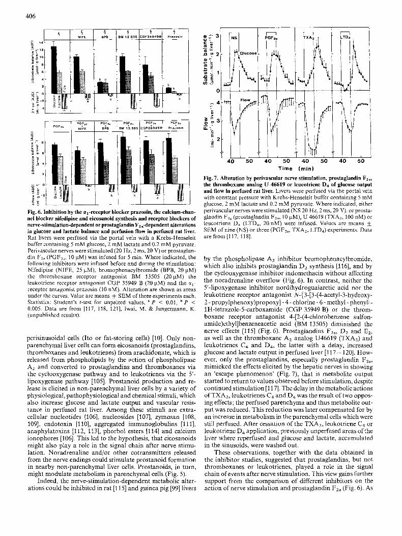
Fig. 7. Alteration by perivascular nerve stimulation, prostaglandin Fz,, the thromboxane analog U 46619 or leucotriene D4 of glucose output and flow in perfused rat liver. Livers were perfused via the portal vein with constant pressure with Krebs-Henseleit buffer containing 5 mM glucose, 2 mM lactate and 0.2 mM pyruvate. Where indicated, either perivascular nerves were stimulated (NS 20 Hz, 2 ms, 20 V), or prosta- glandin Fz. (prostaglandin FZo, 10 pM), U 46619 (TXA2, 100 nM) or leucotriene D, (LTD,, 20 nM) were infused. Values are means f SEM of nine (NS) or three (PGF2,, TXA2, LTD,) experiments. Data are from [I 17, 1 181.
by the phospholipase A2 inhibitor brornophenacylbromide, which also inhibits prostaglandin Dz synthesis [116], and by the cyclooxygenase inhibitor indomethacin without affecting the noradrenaline overflow (Fig. 6). In contrast, neither the 5'-lipoxygenase inhibitor nordihydroguaiaretic acid nor the leukotriene receptor antagonist N-(3-[3-(4-acetyl-3-hydroxy- 2 - propy1phenoxy)propoxyJ - 4 - chlorine - 6 - methyl - phenyl} - 1 H-tetrazole-Scarboxamide (CGP 35949 B) or the throm- boxane receptor antagonist 4-[2-(4-chlorobenzene sulfon- amide)ethyl]benzeneacetic acid (BM 13505) diminished the nerve effects [115] (Fig. 6). Prostaglandins FZo, Dz and E2, as well as the thromboxane A, analog U46619 (TXA2) and leukotrienes C4 and D4, the latter with a delay, increased glucose and lactate output in perfused liver [117- 1201. How- ever, only the prostaglandins, especially prostaglandin FZa, mimicked the effects elicited by the hepatic nerves in showing an 'escape phenomenon' (Fig. 7), that is metabolite output started to return to values observed before stimulation, despite continued stimulation [117]. The delay in the metabolic actions of TXAz, leukotrienes C4 and D4 was the result of two oppos- ing effects; the perfused parenchyma and thus metabolite out- put was reduced. This reduction was later compensated for by an increase in metabolism in the parenchymal cells which were still perfused. After cessation of the TXA2, leukotriene C4 or leukotriene D4 application, previously unperfused areas of the liver where reperfused and glucose and lactate, accumulated in the sinusoids, were washed out.
These observations, together with the data obtained in the inhibitor studies, suggested that prostaglandins, but not thromboxanes or leukotrienes, played a role in the signal chain of events after nerve stimulation. This view gains further support from the comparison of different inhibitors on the action of nerve stimulation and prostaglandin Fza (Fig. 6). As

407
with nerve stimulation, nordihydroguaiaretic acid, leukotriene and thromboxane receptor antagonists did not attenuate the prostaglandin F2,-elicited increase in metabolite output, nor did prazosin, bromophenacylbromide or nifedipine (see be-
The participation of prostaglandins in a signal chain of events from the nerve cell to the hepatocyte depends upon the existence of hepatocellular prostaglandin receptors. Evidence for the existence of such a receptor has accumulated. Prosta- glandin F2. [121], E2 [I221 and D, [123] stimulated glycogen phosphorylase activity or glucose release in hepatocyte sus- pensions, whereas the TXAz was inactive [121]. As in liver perfusions, half-maximal stimulation of glycogenolysis was observed in the concentration range 0.5 - 5 pM for all three prostaglandins (Piischel, G. P., Schroder, A. and Jungermann, K., unpublished results). The prostaglandins increased inosi- to1 1,4,5-trisphosphate but not CAMP [121, 1221. A common low affinity binding site with a Kd of about 2 pM could be demonstrated in hepatocyte plasma membranes besides a prostaglandin F,,-specific high-affinity binding site, for which a biological function is currently not evident (Neuschafer- Rube, F., Piischel, G. P. and Jungermann, K., unpublished results). In other studies with hepatocyte suspensions, prosta- glandin D2 stimulated glycogenolysis half-maximally at con- centrations of 20 nM and a prostaglandin D2-specific binding site with a Kd in the same concentration range was described
low).
[123].
Hemodynamics
Noradrenaline action. The nerve-stimulation-mediated flow reduction was inhibited almost completely by the a- receptor antagonist phentolamine [73], and the al-receptor- specific antagonist prazosin (Fig. 6) [74], but not by u2-recep- tor, P,-receptor and fi2-receptor blockers [75] and hence was mediated via u1 receptors. This could point to a direct contact of the nerve endings with the vascular smooth muscle cells regulating the flow.
involvement of eicosanoids. Flow reduction was also in- hibited by the phospholipase A,/prostaglandin D2-synthesis- inhibitor bromophenacylbromide (Fig. 6 ) and by the cy- clooxygenase inhibitor indomethacine [99, 11 51. Thus, the possibility can be envisaged that the nervous regulation of liver hemodynamics was at least partly mediated via stimulation of eicosanoid formation in non-parenchymal liver cells. As with the metabolic effects of nerve stimulation, neither nor- dihydroguaiaretic acid nor leukotriene or thromboxane recep- tor antagonists [118] interfered with flow reduction to a signifi- cant extent (Fig. 6) , indicating that only prostaglandins, but not thromboxanes or leukotrienes, participated in flow regu- lation by the sympathetic hepatic nerves. Prostaglandins, es- pecially prostaglandin F2,, mimicked the nerve-stimulation- mediated flow reduction in kinetics and extent [117]. Bromophenacylbromide and, in contrast to nerve stimulation, also thromboxane-receptor and leukotriene-receptor antag- onists (Iwai, M. and Jungermann, K., unpublished results), attenuated the prostaglandin F,,-elicited flow reduction slightly but significantly (Fig. 6). This might indicate that the flow effects of circulating prostaglandin F,,, in contrast to those of prostaglandin F,, released locally after nerve stimu- lation, could in part act via stimulation of leukotriene and thromboxane production in other non-parenchymal liver cells. Prazosin did not alter the prostaglandin F2,-mediated flow reduction, but the calcium channel blocker nifedipine abolished it [98, 11 81 (see below).
PP
P"
Hypothesis
PP
P V
4 Flow Glucose t Fig.8. Model for the signal propagation from nerve endings to the effector cells in rat liver. NE, nerve ending; NPC, non-parenchymal cell; PC, parenchymal cell; NA,,, synaptically released noradrenaline; PGSd, prostaglandins, locally released into the space of Disse; ctl- R, cc,-receptor; P-R, prostaglandin receptor; GJ, gap junction; pp, periportal zone; pv, perivenous zone. The model is expiained in the text.
Hypothetical signal chain from nerve endings to the effector cells
The model. Since the metabolic and hemodynamic effects of hepatic nerve stimulation were inhibited by the ul-receptor blocker prazosin and by bromophenacylbromide or in- domethacin, and since the actions of prostaglandin FZiT are unaffected by prazosin, noradrenaline and the u1 receptor must preceed prostaglandin and the prostaglandin receptor in the signal chain. Noradrenaline released from periportally located nerve endings might act partly via a1 receptors on nearby hepatocytes or partly via a, receptors on non- parenchymal liver cells, which in turn can release prostaglan- dins. Noradrenaline and prostaglandins would then stimulate glycogenolysis in neighbouring hepatocytes. The signal might be further propagated to more distant hepatocytes via gap junctions (Fig. 8), which have been shown to be permeable to inositol1,4,5-trisphosphate and Ca2+. In freshly isolated pairs or clusters of hepatocytes, signal propagation was mediated by the transjunctional flux of these intracellular messengers [ 1241. Similarly, noradrenaline released from nerve endings

408
might act partly via a1 receptors on smooth muscle cells of the hepatic blood vessels and partly via al receptors on non- parenchymal liver cells, which in turn could release prosta- glandins. Again, noradrenaline and prostaglandin would then cause a constriction of the sinusoids (Fig. 8).
Caveats to the model. The studies with inhibitors of eicosanoid synthesis do not prove the model, since inhibition might be caused by yet unknown unspecific side effects of these drugs. The model could be further supported by a prostanoid overflow into the hepatic vein, as has been shown after in- fusion of nucleotides, nucleosides, zymosan, endotoxin, aggre- gated immunoglobulins, anaphylatoxins, phorbol esters and calcium ionophores (see above) that stimulate glucose output and reduce flow in perfused livers. Such an overflow has not yet been demonstrated in a convincing manner after nerve stimulation. The overflow of prostaglandin Dz and prosta- glandin E2 [lo61 was at the edge of significance and with concentrations of 2.5 nM and 0.1 nM far below the range in which physiological responses to prostaglandins have been observed. One has, however, to keep in mind that the overflow is only the difference between prostaglandin formation and degradation during the passage through the sinusoids. In fact, prostagbandins are very efficiently degraded by the liver. Thus, 90 - 95% of prostaglandin Ez was extracted during a single passage through the sinusoids in rat liver perfused with defibrinated blood [125] and more than 95% of prostaglandin Fza immunoreactivity was eliminated during one passage through livers perfused with a Krebs-Henseleit buffer (Puschel, G. P., Hulsmann, M. and Jungermann, K., unpub- lished results). Taking into account that nerve action, in con- trast to the circulating agents mentioned above, probably stimulates only a few periportally located non-parenchymal liver cells to release prostaglandins, a small or even lacking prostaglandin overflow does not argue against the partici- pation of prostaglandins in the signal chain of events. In order to be a link in the signal chain of events from the nerve to the effector cell, non-parenchymal liver cells should possess a receptors and should react to stimulation by a agonists with prostaglandin formation. This has not yet been shown. Studies tempting to elucidate this are under way.
Signal chain within the effector cell
Metabolism
Following stimulation of the hepatic nerves, the activity of glycogen phosphorylase increased, that of glycogen synthase decreased and that of glycolytic pyruvate kinase remained unaffected. The level of the activators of glycogenolysis (CAMP) and glycolysis (fructose-2,6-bisphosphate), increased slightly [96]. The slight increase in CAMP could not account for the increase in glycogen phosphorylase activity. Therefore, it was assumed that liver nerves enhanced hepatic glucose output via an increase in the cytosolic calcium concentration (Fig. 9). A transient release of Ca2+ into the perfusate, fol- lowed by a prolonged reuptake of Ca2+ after nerve stimu- lation favoured this view [77]. There was no evidence for entry of extracellular calcium via nifedipine-sensitive calcium channels [98].
The possible involvement of intracellular calcium in the 'metabolic' signal chain of events was tested in 'calcium deprivation/replenishment' experiments [96]. The idea was that intracellular calcium pools should be emptied during a prolonged calcium-free perfusion and, thus, processes de- pending on intracellular calcium should be inhibited. After re-
NA A VP
I 1 I k , IP3
f i Glycogen GlP- G6P- Glucose
Fig. 9. Scheme of the mechanism of inhibition by noradrenaline, adrenaline and vasopressin of the nerve stimulation-dependent and noradrenaline-dependent alterations of metabolism and hemodynamics in rat liver. A, adrenaline; NA, noradrenaline; VP, vasopressin; a,R, a1 receptor; VPR, vasopressin receptor; GP ,,z'', putative a,-receptor- coupled guanosine-nucleotide-binding regulatory protein; GP ,,V", putative vasopressin-receptor-coupled guanosine-nucleotide-binding regulatory protein; PKC ,,a'', PKC ,,V" and PLC ,,a", PLC ,,V", putative agonist-specific protein kinase C and phospholipase C; PIP2, phosphatidylinositol4,5-bisphosphate; DAG, 1,2-diacylglycerol; IP3, inositol 1,4,5-trisphosphate; GIP (G6P), glucose 1(6)-phosphate; GgP(K) glycogen phosphorylase (kinase).
addition of calcium to the extracellular medium, processes depending on intracellular calcium should still be inhibited or at least delayed, since replenishment of intracellular calcium pools is a relatively slow process, lasting 2 - 3 min. Processes depending on extracellular calcium, however, should be pos- sible immediately after adding calcium to the medium. In calcium-free perfusions, nerve-stimulation-dependent meta- bolic alterations occurred with normal kinetics when calcium was added 3 min prior to stimulation; they were, however, delayed when calcium was added only 2 min after the onset of stimulation. This indicated that calcium release from intra- cellular pools was involved in the signal chain [96] (Fig. 9).
The second messenger causing a release of calcium from intracellular pools is generally thought to be inositol 1,4,5- trisphosphate, which, together with diacylglycerol, is released from phosphatidyl inositol4,S-bisphosphate after stimulation of a1 receptors. Both noradrenaline [126,127] and prostaglan- dins [121, 1221 have been shown to increase inositol 1,4,5- trisphosphate in hepatocytes. In perfused livers, however, a significant increase in inositol 1,4,5-trisphosphate could not be demonstrated after nerve stimulation, but only after in- fusion of noradrenaline [128]. This result would fit the hy- pothesis that, during nerve action, only a few periportal hepatocytes are directly stimulated by noradrenaline or pros- taglandins, leading to a small local increase in inositol 1,4,5- trisphosphate which is not measurable in whole liver homogenates, and that the signal is then propagated to the majority of hepatocytes via gap junctions (Fig. 8).
The metabolic nerve actions were inhibited by so-called calcium-releasing hormones such as adrenaline, noradrenaline [89], vasopressin and angiotensin (Miura, H., Gardemann, A., Rosa, J. and Jungermann, K., unpublished results) (Fig. 9), which increase inositol 1,4,5-trisphosphate and diacylglycerol, and by high concentrations of the phorbol ester 4P-phorbol 12-myristate 13-acetate. Diacylglycerol and 4P-phorbol 12- myristate 13-acetate would activate protein kinase C, which is known to inhibit the al-receptor-mediated hydrolysis of phosphatidyl inositol 4,5-bisphosphate to inositol 1,4,5-

Blood Glucose
Fig. 10. The nervous humoral network for the maintenance of the hepatic glucostat and blood reservoir function. A, adrenals; P, pancreas; HT, hypothalamus; (N)Adr, (n0r)adrenaline; Ggn, glucagon; Ins, insulin; $ , nervous activity. The sympathetic, adrenergic and glucagon system are shown with filled arrows, the parasympathetic and insulin system with open arrows. Uninterrupted lines, activation; broken lines, inhi- bition.
trisphosphate [129]. These findings would corroborate the participation of inositol 1,4,5-trisphosphate in the nerve-sig- nal chain of reactions.
Hemodynamics. The hemodynamic nerve effects were in- hibited by the calcium-channel blocker nifedipine (Fig. 6) [98]. In the ‘calcium deprivation/replenishment’ experiments, the nerve-stimulation-mediated flow reduction was restored im- mediately after readdition of calcium [96]. Thus, the hemodynamic nerve effects were dependent on the entry of extracellular calcium into the smooth muscle cells. Within the cell, Ca2 + would activate the calmodulin-dependent myosin light chain kinase and thus cause constriction. The attenuation of the flow effects by sodium nitroprusside [73], which de- creases the sensitivity of the system to calmodulin by increas- ing cGMP, would also be in line with this mechanism.
Functions of sympathetic and parasympathetic nerve actions
The liver is both the central organ for the maintenance of the energy supply, mainly by operating as a glucostat and also an active and passive blood reservoir 16, 71 (Table 1). A network of afferent and efferent, sympathetic and Parasympathetic nerves is important for the regulation of the glucose and blood reservoir function of the liver. The network senses the blood glucose concentration and blood pressure [25, 26, 66-72] and influences hepatic metabolism and hemodynamics directly via liver nerves and indirectly via mod- ifying the hormonal secretion rates of other organs, such as the pancreas and adrenal medulla (Fig. 10).
Activation of the sympathetic nervous system signals that the organism requires glucose and blood. The total amount of glucose and part of the required blood are provided by the liver in which some tissue areas release blood to the circulation by being squeezed out and temporarily restricted from receiv- ing further blood supply and in which other tissue areas re- main accessible mainly for the increased release of systemically
409
required glucose. Major signals for an increase in glucose output are declining glucose levels, increased portovenous concentrations of glucagon, enhanced systemic levels of cat- echolamines and an increased tone of the sympathetic liver nerves. Major signals for release of blood are elevated concen- trations of circulating catecholamines and again an increased tone of the sympathetic hepatic nerves. Thus, the sympathetic nervous system, activated in part via afferent hepatic nerve fibers [25, 26, 66-72], appears to control the glucose and the blood-reservoir functions mainly directly via sympathetic liver nerves and also indirectly by activating the release of glucagon from the pancreas and of adrenaline and noradrenaline from the adrenal medulla (Fig. 10).
After a normal meal, the liver takes up a substantial part of the absorbed glucose. Major signals for the activation of glucose uptake are an increased glucose concentration gradi- ent between the portal vein and the hepatic artery [130], an increased portovenous level of insulin and an enhanced tone of the parasympathetic liver nerves. The parasympathetic ner- vous system enhances hepatic glucose uptake indirectly by stimulation of insulin release and reduction of the activity of the hepatic branch of efferent splanchnic nerve fibers [69] and of the level of circulating catecholamines as well as directly via the hepatic nerves antagonistically to glucagon and syner- gistically with insulin (Fig. 10) [95].
REFERENCES 1 . 2. 3.
4.
5.
6. 7. 8. 9.
30.
11.
12.
13.
14. 15. 16. 17.
18.
19.
20.
21.
Jungermann, K. & Katz, N. (1989) Physiol. Rev. 69, 708-764. Jungermann, K. (1988) Semin. Liver Dis. 8, 329-341. Wisse, E., Geerts, B., Bouwens, L, Van Bossuyt, K., Vanderkerken, K. & Van Goethem, V. (1989) in Cells of the hepatic sinusoid 1988, an attempt to review the 4th International Kupffer cell symposium in cells of the hepatic sinusoids (Wisse, E., Knook, D. L. & Decker, K., eds), pp. 1-7, Kupffer Cell Foundation, Leiden, Netherlands.
Bouwens, L, De Bleser, P. Vanderkerken, K. Geerts, B. & Wisse, E. (1 992) Enzyme, in the press.
Sawchenko, P. E. & Friedman, M. I. (1979) Am. J . Physiol. 236, R5 - R20.
Lautt, W. W. (1980) Can. J . Physiol. Pharmacol. 58, 105-123. Lautt, W. W. (1983) Prog. Neurobiol. 21, 323-348. Sutherland, S. D. (1965) Rev. Int. Hepatol. 15, 569-578. Prechtl, J. C. & Powley, T. L. (1987) Anat. Embryol. 176,115-
126. Bioulac-Sage, P., Lafon, M. E., Saric, J. & Balabaud, C. (19901
J . Hepatol. 10, 105 - 112. Friedman, M. I. ( 1988) in Hepatic nervefunction in the liver (Arias, I. M., Jacoby, W. B., Popper, H., Schachter, D. & Shafritz, D. A., eds) pp. 949-959, Raven Press Ltd., New York.
Carobi, C., Della Torre, G. & Magni, F. (1985) Neurosci. Lett.
Rogers, R. C., Kahrilas, P. J. & Hermann, G. E. (1984)J. Auton.
Shimazu, T. (1981) Diahetologiu 20, 343-356. Forssmann, W. G. & Ito, S. (1977) J . Cell. Biol. 74, 299-313. Tsuneki, K . & Ichihara, K. (1981) Arch. Histol. JPN. 44, 1 - 13. Moghimzadeh, E., Nobin, A. & Rosengren, E. (1983) Cell Tissue
Rex 230, 605-613. Metz, W. & Forssmann, W. G. (1980) in Communications ofliver cells (Popper, H., Bianchi, L., Gudat, F. & Reutter, W., eds) pp. 121 - 127, MTP Press, Lancaster.
Reilly, F. D., McCuskey, P. A. & McCuskey, R. S. (1978) Anat. Rec. 191, 55-67.
Ungvary, G. & Donath, T. (1975) Exp. Mol. Puthol. 22, 29- 34.
Schubert, W., Metz, J. & Forssmann, W. G. (1984) Verh. Anat. Ges. 78, 461 -462.
62,255 -260.
Nerv. Syst. 11, 223 - 225.

410
22. Forssmann, W. G. (1986) Z. Gastroenterol. Suppl. 21,190-191. 23. Burt, A. D., Tiniakos, D., MacSween, R. N., Griffiths, M. R.,
Wisse, E. & Polak, J. M. (1989) Hepatology 9,839-845. 24. Lundberg, J. M., Terenius, L. & Hokfell, T. (1982) Acta Physiol.
Scand. 116,477-480. 25. Ito, Y., Magari, S. & Sakanaka, M. (1990) Arch. Histol. Cytol.
26. Niijima, A. (1989) Prog. Neurobiol. 33, 135 - 147. 27. Niijima, A. (1989) J. Nutr. 119, 833-840. 28. Shimazu, T. (1983) Adv. Metab. Disord. 10, 355-384. 29. Niijima, A. (1979) in Integrativefunctions of the autonomic ner-
vous system (Brooks, C. McC., Koizumi, K. & Sato, A., eds), pp. 68 -83, Elsevier, Amsterdam.
30. Cohen, M. I. & Gootman, P. M. (1970) Am. J. Physiol. 218, 1092-1101.
31. Niijima, A. (1985) J . Physiol. 364, 105-112. 32. Niijima, A. (1991) Brain Res. Bull. 26, 165-167. 33. Shimazu, T. (1981) Diabetologia 20, 343-356. 34. Iwai, M. & Shimazu, T. (1988) Life Sci. 42, 1833-1840. 35. Shimazu, T. & Usami, M. (1982) J. Physiol. {Lond.) 329,231 -
36. Shimazu, T. & Amakawa, A. (1968) Biochim. Biophys. Acta 165,
37. Shimazu, T. & Amakawa, A. (1968) Biochim. Biophys. Acta 165,
38. Proost, C., Carton, H. & DeWulf, H. (1979) Biochem.
39. Shimazu, T. & Amakawa, A. (1968) Biochim. Biophys. Acta 165,
40. Edwards, A. V. (1971) J . Physiol. (Lond.) 213,741 -753. 41. Jarhult, J., Andersson, P. O., Holst, J., Moghimzadeh, E. &
Nobin, A. (1 980) Acta Physiol. Scand. 110, 5 - 11. 42. Garceau, D., Yamaguchi, N., Goyer, R. & Guitard, F. (1984)
Can. J. Physiol. Pharmacol. 62, 1086- 1091. 43. Edwards, A. V. (1972) J. Physiol. (Lond.) 220, 697-710. 44. Edwards, A. V. & Silver, M. (1972) J . Physiol. (Lond.) 223,
45. Lautt, W. W. & Wong, C. (1978) Can. J . Physiol. Pharmacol.
46. Nobin, A., Falck, B., Ingemanson, S., Jarhuld, J. & Rosengren,
47. Shimazu, T. & Amakawa, A. (1975) Biochim. Biophys. Acta 385,
48. Lautt, W. W. (1977) Am. J . Physiol. 232, H652--656. 49. Yoshimatsu, H., Niijima, A., Oomura, Y. & Katafuchi, T. (1988)
50. Shimazu, T. & Fujimoto, T. (1971) Biochim. Biophys. Acta 252,
51. Shimazu, T. (1971) Biochim. Biophys. Acta 252,28-38. 52. Lautt, W. W. & Wong, C. (1978) Can. J. Physiol. Pharmacol.
53. Greenway, C. V. & Oshiro, G. (1972) J . Physiol. (Lond.) 227,
54. Richardson, P. D. I. & Withrington, P. G. (1982) Annu. Rev.
55. Lautt, W. W. & Legare, D. J. (1986) Can. J. Physiol. Pharmacol.
56. Lautt, W. W., Schafer, J. & Legare, D. J. (1991) Can. J. Physiol.
57. Friman, S., Radberg, G. & Svanvik, J. (1990) Acta Physiol.
58. Koo, A. & Liang, I. Y. S. (1979) Am. J. Physiol. 236, E728-
59. Koo, A. & Liang, I. Y. S. (1979) J. Physiol. 295, 191 - 199. 60. Shanygina, K. I., Fomina, M. P., Parfenova, N. S. &
61. Magni, F. &Viola, M. P. (1985) J. Auton. Nerv. Syst. 13, 245-
62. Ashirmetov, A. K. H. & Krakovskii, M. E. (1990) Fiziol. Zh.
63. Albino Teixeira. A.. Matias. A.. da Silva. P. Soares. Sarmento.
53 Suppl., 199 - 208.
42.
324 - 334.
335 -348.
Pharmacol. 28, 2187 -2191.
349 - 356.
571 - 593.
56,1022-1028.
E. (1 977) Acta Physiol. Scand. Suppl. 452, 103 - 106.
242 - 256.
Brain Res. Bull. 21, 239-44.
18-27.
56,679 - 682.
487 - 501.
Physiol. 44, 57 - 69.
64,449 - 454.
Pharmacol. 69,43 - 48.
Scand. 140,287 - 293.
E732.
Kalashnikova, N. M. (1981) Vopr. Med. Khim. 27, 505-509.
254.
(Kiev) 36,43-46.
64. Lindfeldt, J., Holmin, T. & Ahren, B. (1987) J . Auton. Nerv.
65. Hogestatt, E. D., Hammarstrom, L. E., Andersson, K. E. &
66. Niijima, A. (1982) J . Physiol. (Lond.) 332, 315-323. 67. Niijima, A. (1983) J. Auton. Nerv. Syst. 9,207-220. 68. Niijima, A., Fukuda, A,, Taguchi, T. & Okuda, J. (1983) Am. J.
69. Niijima, A. (1984) J. Auton. Nerv. Syst. 10, 279-285. 70. Donovan, C. M., Halter, J. B. & Bergman, R. N. (1991) Diabetes
71. Lee, K. C. &Miller, R. E. (1985) Endocrinology 117,307-314. 72. Tanaka, K., Inoue, S., Nagase, H. & Takamura, Y. (1990) Endo-
73. Hartmann, H., Beckh, K. & Jungermann, K. (1982) Eur. J .
74. Gardemann, A., Strulik, H. & Jungermann, K. (1987) Am. J .
75. Ulken, V., Piischel, G. P. & Jungermann, K. (1991) Biol. Chem.
76. Piischel, G. P., Nath, A. & Jungermann, K. (1987) FEBS Lett.
77. Haussinger, D., Stehle, T., Gerok, W. & Sies, H. (1987) Eur. J .
78. Beuers, U., Beckh, K. & Jungermann, K. (1986) Eur. J . Biochem.
79. Ball&, C. & Jungermann, K. (1986) Eur. J . Biochem. 158, 13-
80. Beuers, U., Pogonka, R., Esterline, R., Ji., S. & Jungermann, K.
81. Beckh, K. & Arnold, R. (1991) Am. J. Physiol. 261, G775-
82. Ji, S., Beckh, K. & Jungermann, K. (1984) FEBS Lett. 167,
83. Beckh, K., Hartmann, H., Jungermann, K. & Scholz, R. (1984)
84. Beckh, K., Otto, R., Ji, S. & Jungerrnann, K. (1985) Biol. Chem.
85. Seydoux, J., Brunsmann, M., Jeanrenaud, B. & Girardier, L.
86. Sannemann, J., Beckh, K. & Jungermann, K. (1986) Biol. Chem.
87. Beckh, K., Hartmann, H. & Jungermann, K. (1982) FEBS Lett.
88. Kiister, J., Beuers, U. & Jungermann, K. (1989) Biol. Chem. Hoppe Seyler 370, 1035-1044.
89. Miura, H., Gardemann, A., Rosa, J. & Jungermann, K. (1992) Hepatology, in the press.
90. Withrington, P. G. & Richardson, P. D. I. (1986) in Regulation of hepatic metabolism (Thurman, R. G., Kauffman, F. C. & Jungermann, K., eds) pp. 27 - 53, Plenum, New York.
91. Daniel, P. M. & Pritchard, P. P. M. (1951) J. Physiol. 114, 538- 548.
92. Ungvary, G. & Varga, B. (1971) Acta Physiol. Acad. Sci. Hung.
93. Reilly, F. D., McCuskey, R. S. & Cilento, E. V. (1981)
94. Greenway, C. V., Lawson, A. E. & Mellander, S. (1967) J.
95. Gardemann, A. & Jungermann, K. (1986) Hoppe Seyler Biol.
96. Balle, C., Beuers, U. Engelhardt R. & Jungerrnann, K. (1987)
97. Beckh, K., Beuers, U., Engelhardt, R. & K. Jungermann (1987)
98. Athari, A. & Jungermann, K. (1990) Biochem. Int. 20, 13-23. 99. Beckh, K., Fuchs, E., Balle, C. & Jungermann K. (1990) Biol.
100. Sugiyama, Y. & Ohta, H. (1990) Arch. Histol. Cytol. 53, 71-
101. Mivashita. T.. Takeda. A.. Iwai. M. & Shimazu, T. (1991) Eur.
Syst. 19, 211 -217.
Holmin, T. (1986) Acta Physiol. Scand. 128, 309-315.
Physiol. 244, R611-614.
40, 155-158.
crinology 127, 2017-2023.
Biochem. 123, 521 - 527.
Physiol. 253, E238- E245.
Hoppe Seyler 372,401 -409.
219,145 - 150.
Biochem. 163, 197-203.
158, 19-24.
18.
(1986) Toxicol. Lett. 34, 247-252.
G780.
117-122.
Pfiigers Arch. 401, 104- 106.
Hoppe Seyler 366,671 -678.
(1979) Am. J. Physiol. 236, G323-G327.
Hoppe Seyler 367,401 -409.
146,69 - 14.
40,347 - 356.
Microvasc. Res. 21, 103- 109.
Physiol. 192, 21 -41.
Chem. 367, 559 - 566.
Eur. J. Biochem. 170, 193- 199.
Biol. Chem. Hoppe Seyler 368, 379 - 386.
Chem. Hoppe Seyler 371,153- 158.
80. , , , ,
A. & Azevedo, I. (1990) J . Auton. Pharmacol. 10, 181 -189. J..Biochem. 196,37 -42.

41 1
102. Brinkmann, A., Katz, N., Sasse, D. & Jungermann, K. (1978)
103. Iwai, M., Miyashita, T. & Shimazu, T. (1991) Eur. J . Biochem.
104. Seseke, F., Gardemann, A. & Jungermann, K. (1992) FEBS
105. Decker, K. (1985) Semin. Liver Dis. 5 , 175-190. 106. Tran Thi, D. A., Haussinger, D., Gyufko, K. & Decker, K.
(1988) Biol. Chem. Hoppe Seyler 369, 65 - 68. 107. Vom Dahl, S., Wettstein, M., Gerok, W. & Haussinger, D. (1990)
Biochem. J. 270, 39-44. 108. Dieter, P., Altin, P. G., Decker, K. & Bygrave, F. L. (1987) Eur.
J . Biochem. 165,455 - 460. 109. Dieter, P., Altin, J. G. & Bygrave, F. L. (1987) FEBS Lett. 213,
174-178. 110. Casteleijn, E., Kuiper, J., Van Rooij, H. C., Kamps, J. A., Koster,
J. F. &Van Berkel, T. J. (1988) J . Biol. Chem. 263,6953-6955. 111. Buxton, D. B., Fisher, R. A., Briseno, D. L., Hanahan, D. J. &
Olson, M. S. (1987) Biochem. J . 243,493-498. 112. Puschel, G. P., Oppermann, M., Muschol, W., Gotze, 0. &
Hoppe-Seyler’s 2. Physiol. Chem. 359, 1561 - 1571.
200,69 - 74.
Lett., in the press.
116. Dieter, P., Schulze-Specking, A. &Decker, K. (1989) Biol. Chem.
117. Iwai, M., Gardemann, A., Piischel, G. P. & Jungermann, K.
118. Iwai, M. & Jungermann, K. (1989) Eur. J. Biochem. 180,273-
119. Haussinger, D., Stehle, T., Tran Thi, T. A,, Decker, K. & Gerok,
120. Altin, J. G. & Bygrave, F. L. (1988) Biochem. J . 249,677-685. 121. Athari, A. & Jungermann, K. (1989) Biochem. Biophys. Res.
126,
Hoppe Seyler 370,543 - 547.
(1988) Eur. J . Biochem. I75,45-50.
281.
W. (1987) Biol. Chem. Hoppe Seyler 368,1509-1513.
Commun. 163.1235 - 1242. 122. Mine, T., Kojima, I. & Ogata, E. (1990) Endocrinolog
123. Kuiper, J., Zijlstra, F. J., Kamps, J. A. &Van Berkel, T. J. Biochem. J. 262, 195-201.
124. Saez, J. C., Connor, J. A., Spray, D. C. & Bennett, M (1989) Proc. Natl Acad. Sci. USA 86,2708-2712.
125. Dawson, W. (1970) Brit. J . Pharmacol. 39,585. 126. Exton, J. H. (1987) Kidney Znt. Suppl. 23, S68-S81. 127. Exton, J. H. (1987) Diabetes Metab. Rev. 3, 163-183.
2831 - 2836. 1989)
v. L.
Jungermann, K. (1989) FEBS Lett. 2#3,83-87.
(1991) Eur. J . Biochem. 196,525 - 530.
J . Hepatol. 6, 151 - 157.
128. Piischel, G. P. & Jungermann, K. (1988) Eur. J . Biochem. 175,
129. Van de Werve, G. & Jeanrenaud, B. (1987) Diabetes Met. Rev.
130. Gardemann, A,, Strulik, H. & Jungermann, K. (1986) FEBS
113. Muschol, W., Piischel, G. P., Hulsmann, M. & Jungermann, K.
114. Tran Thi, T. A,, Gyufko, K., Haussinger, D. &Decker, K. (1988)
115. Iwai, M. & Jungermann, K. (1987) FEBS Lett. 221,155- 160.
187-191.
3, 47 - 78.
Lett. 202, 255 -259.