nervegrowthfactor(ngf)regulatesactivityofnuclear ... ·...
TRANSCRIPT
Nerve Growth Factor (NGF) Regulates Activity of NuclearFactor of Activated T-cells (NFAT) in Neurons via thePhosphatidylinositol 3-Kinase (PI3K)-Akt-Glycogen SynthaseKinase 3� (GSK3�) Pathway*
Received for publication, June 4, 2014, and in revised form, September 14, 2014 Published, JBC Papers in Press, September 17, 2014, DOI 10.1074/jbc.M114.587188
Man-Su Kim‡§1, Leonid P. Shutov‡, Aswini Gnanasekaran‡, Zhihong Lin‡, Jacob E. Rysted‡, Jason D. Ulrich‡,and Yuriy M. Usachev‡2
From the ‡Department of Pharmacology, University of Iowa Carver College of Medicine, Iowa City, Iowa 52242 and the §College ofPharmacy, Inje University, Gimhae 621-749, Korea
Background: Neurotrophins regulate transcription factor NFAT and NFAT-mediated neuronal functions, but the under-lying mechanisms are poorly defined.Results: NGF facilitated depolarization-induced NFAT activation in sensory neurons, which depended on PI3K, Akt, andGSK3� but not on PLC.Conclusion: NGF-dependent facilitation of NFAT activation is mediated by the PI3K-Akt-GSK3� pathway.Significance: This novel mechanism may represent an important component of NFAT-dependent gene regulation in neurons.
The Ca2�/calcineurin-dependent transcription factor nuclearfactor of activated T-cells (NFAT) plays an important role inregulating many neuronal functions, including excitability,axonal growth, synaptogenesis, and neuronal survival. NFATcan be activated by action potential firing or depolarization thatleads to Ca2�/calcineurin-dependent dephosphorylation ofNFAT and its translocation to the nucleus. Recent data suggestthat NFAT and NFAT-dependent functions in neurons can alsobe potently regulated by NGF and other neurotrophins. How-ever, the mechanisms of NFAT regulation by neurotrophins arenot well understood. Here, we show that in dorsal root ganglionsensory neurons, NGF markedly facilitates NFAT-mediatedgene expression induced by mild depolarization. The effects ofNGF were not associated with changes in [Ca2�]i and were inde-pendent of phospholipase C activity. Instead, the facilitatoryeffect of NGF depended on activation of the PI3K/Akt pathwaydownstream of the TrkA receptor and on inhibition of glycogensynthase kinase 3� (GSK3�), a protein kinase known to phos-phorylate NFAT and promote its nuclear export. Knockdown orknockout of NFATc3 eliminated this facilitatory effect. Simul-taneous monitoring of EGFP-NFATc3 nuclear translocationand [Ca2�]i changes in dorsal root ganglion neurons indicatedthat NGF slowed the rate of NFATc3 nuclear export but did notaffect its nuclear import rate. Collectively, our data suggest thatNGF facilitates depolarization-induced NFAT activation bystimulating PI3K/Akt signaling, inactivating GSK3�, andthereby slowing NFATc3 export from the nucleus. We propose
that NFAT serves as an integrator of neurotrophin action anddepolarization-driven calcium signaling to regulate neuronalgene expression.
Activity-dependent regulation of gene expression plays acrucial role in sculpting neural circuits during development andin controlling neuronal plasticity in adulthood (1, 2). Amongthe various Ca2�-dependent transcription factors, nuclear fac-tor of activated T-cells (NFAT)3 has emerged as an importantcomponent of excitation-transcription coupling in neurons(3– 8). Numerous neuronal functions are controlled by NFAT,including excitability, axonal growth, synaptic plasticity, neu-ronal development, and survival (4, 6, 8 –13). Moreover, recentstudies have linked aberrant NFAT activation to pain sensitiza-tion (14 –16) and the neurotoxicity associated with Alzheimerdisease, ischemia, and traumatic brain injury (17–20).
NFAT activation is regulated by reversible phosphorylation(21, 22). Ca2� entering neurons via voltage- or ligand-gatedCa2� channels activates the Ca2�- and calmodulin-dependentprotein phosphatase calcineurin (CaN), which induces CaN-mediated dephosphorylation of NFAT at multiple serine resi-dues within its regulatory domain (21–24). This, in turn,unmasks the nuclear localization signal of NFAT and facilitatesits import to the nucleus, enabling it to initiate transcription(21, 22). Several protein kinases, such as glycogen synthase kinase3� (GSK3�), casein kinase 1, and dual specificity tyrosine phos-
* This work was supported, in whole or in part, by National Institutes of HealthGrants NS072432 and NS087068. This work was also supported by theFraternal Order of Eagles Diabetes Research Center.
1 To whom correspondence may be addressed: College of Pharmacy, InjeUniversity, Inje-ro 197, Gimhae, Gyeongnam 621-749, Korea. Tel.: 82-55-320-3887; Fax: 82-55-320-3940; E-mail: [email protected].
2 To whom correspondence may be addressed: Dept. of Pharmacology,University of Iowa Carver College of Medicine, 2-450 BSB, 51 Newton Rd.,Iowa City, IA 52242. Tel.: 319-335-9388; Fax: 319-335-8930; E-mail: [email protected].
3 The abbreviations used are: NFAT, nuclear factor of activated T-cells;ANOVA, analysis of variance; BDNF, brain-derived neurotrophic factor;CaN, calcineurin; DRG, dorsal root ganglion/ganglia; EGFP, enhanced GFP;GSK3�, glycogen synthase kinase 3�; IP3, inositol 1,4,5-trisphosphate;mTOR, mammalian target of rapamycin; NFAT-luc, NFAT luciferase report-er; PLC, phospholipase C; TK-luc, R. reniformis luciferase reporter controlledby HSV-TK promoter; caPI3K, constitutively active PI3K�; CAPS, 3-(cyclo-hexylamino)propanesulfonic acid; K�10, K�15, K�20, and K�90, 10, 15, 20,and 90 mM KCl, respectively.
THE JOURNAL OF BIOLOGICAL CHEMISTRY VOL. 289, NO. 45, pp. 31349 –31360, November 7, 2014© 2014 by The American Society for Biochemistry and Molecular Biology, Inc. Published in the U.S.A.
NOVEMBER 7, 2014 • VOLUME 289 • NUMBER 45 JOURNAL OF BIOLOGICAL CHEMISTRY 31349
by guest on August 9, 2019
http://ww
w.jbc.org/
Dow
nloaded from

phorylation-regulated kinase 1, phosphorylate nuclear NFAT,which facilitates its binding by the nuclear exportin Crm1 and,consequently, its deactivation and nuclear export (3, 10, 22, 23,25–27). Thus, NFAT-mediated transcriptional responses aredetermined by the balance between the activities of the Ca2�/CaN pathways driving nuclear import of NFAT and the NFATkinases stimulating NFAT export from the nucleus.
Recent studies have demonstrated that not only electricalactivity and intracellular Ca2�, but also neurotrophins, in par-ticular nerve growth factor (NGF), potently regulate NFATfunction in neurons (4, 15, 28). For example, NGF and brain-derived neurotrophic factor (BDNF) stimulate NFAT-depen-dent expression of inositol 1,4,5-trisphosphate receptor 1,BDNF, cyclooxygenase-2, and plasminogen activation inhibi-tor-1 in peripheral and central neurons (15, 28, 29). BDNF-de-pendent survival of adult hippocampal neurons and the forma-tion of spatial memory have also been reported to requireNFAT activation (13). Furthermore, NFAT is essential forNGF-dependent axonal growth, and deletion of NFAT iso-forms NFATc2, NFATc3, and NFATc4 disrupts neurite out-growth (4). Despite the growing evidence that NFAT proteinsare important effectors of neurotrophin signaling, the mecha-nisms of NFAT regulation by neurotrophins are not well under-stood. It is also unclear whether and how electrical activity andneurotrophin signaling interact to regulate NFAT activity.
Here, by using genetic and pharmacological tools, we dem-onstrate that NGF facilitates depolarization-induced activationof NFAT in dorsal root ganglion (DRG) sensory neurons.Although Ca2� is a critical regulator of NFAT, NGF had nosignificant effect on the intracellular Ca2� concentration([Ca2�]i) in DRG neurons, and the potentiating actions of NGFwere independent of phospholipase C (PLC) activity. Instead,the NGF potentiation of NFAT activation required the PI3K-Akt signaling pathway and inhibition of the NFAT kinase,GSK3�. Furthermore, the silencing or deletion of specificallythe NFATc3 isoform abolished the potentiating effect of NGFon NFAT-mediated transcription, implicating NFATc3 as a keyNGF effector in sensory neurons.
EXPERIMENTAL PROCEDURES
DRG Cell Cultures and Transfection—Cultured DRG neu-rons were prepared as described previously (7, 30). Briefly, new-born (postnatal day 1–2) Sprague-Dawley rats or adult (2– 4-month-old) wild-type and NFATc3 knock-out mice (BALB/cbackground) were sacrificed, and DRG were isolated from cer-vical, thoracic, and lumbar segments. Suspensions of DRGneurons were plated onto 25-mm glass coverslips precoatedwith poly-L-ornithine and laminin. Approximately 30 min afterthe cells were plated, Dulbecco’s modified Eagle’s medium(DMEM) supplemented with 5% heat-inactivated horse serum,5% fetal bovine serum, and penicillin (100 units/ml)/streptomy-cin (100 g/ml) was added to the plates (hereafter referred to ascomplete DMEM). The DRG neurons were maintained in cul-ture in a 10% CO2 incubator at 37 °C and were used within 2–3days. All surgical protocols were approved by the University ofIowa Institutional Animal Care and Use Committee. Rats werepurchased from Charles River Laboratories (Wilmington, MA),
and NFATc3 knock-out mice were generously provided by Dr.Santana (University of Washington, Seattle, WA) (31, 32).
Prior to plating, DRG neurons were transfected using theAmaxa nucleofection system (Program G-013; Lonza, Switzer-land) as described previously (7, 30, 33). Typically, five rat pupsor three mice were required to obtain a sufficient number ofcells for performing a single transfection. EGFP-NFATc3 wasgenerated by ligating NFATc3 (gift of Dr. Iino, Tokyo Metro-politan Institute of Medical Science) (34) into pEGFP-C1(Clontech). A constitutively active form of the catalytic subunitPI3K� (caPI3K; p110 mutant (35)) was a gift from Dr. StevenGreen (University of Iowa). The shRNA constructs targetingNFATc3, NFATc4, and GSK3� were kindly provided by Dr.Michal Hetman (University of Kentucky) (12). We previouslyvalidated all of these constructs (33). A constitutively activeform of GSK3� (GSK3� S9A) (36) was purchased from Add-gene (plasmid 14754; Cambridge, MA).
NFAT Reporter Assays—NFAT reporter expression wasassessed using a Dual-Luciferase assay as described previously(7, 33). In brief, DRG neurons were co-transfected with theNFAT-luciferase (NFAT-luc) reporter plasmid (which encodesfirefly luciferase under the control of three copies of the NFAT-binding motif; pNFAT-TA-luciferase; Clontech) and the Renillareniformis luciferase (TK-luc) reporter (which encodes Renillaluciferase under the control of constitutively active HSV-TKpromoter; pRL-TK; Promega, Madison, WI). In some experi-ments, the investigated signaling pathways were modulated byco-transfecting GSK3� S9A or caPI3K with the luciferasereporter constructs. Initially, transfected cells were cultured incomplete DMEM containing 25 ng/ml NGF (50 ng/ml in thecase of mouse DRG cultures). Approximately 20 h later, theculture medium was replaced with fresh complete DMEM sup-plemented with B27 (Invitrogen) and ITS-A (Invitrogen) butdevoid of NGF. Twenty-four hours later, the cultured DRGneurons were stimulated with 20 mM KCl medium (15 mM KClfor mouse cultures), which was prepared by mixing completeDMEM with 150 mM KCl stock solution. The L-type Ca2�
channel agonist BayK8644 (1 �M) was added to the 20 mM (15mM) KCl medium to stabilize [Ca2�]i at the elevated levels forthe duration of stimulation (7). In some cultured DRG plates,the KCl medium was supplemented with 25 ng/ml NGF (50ng/ml for mouse DRG cultures). The cultured DRG neuronswere stimulated with 20 mM (15 mM) KCl medium for either 6or 12 h. In the case of the 6-h KCl stimulation protocol, cellswere cultured in complete DMEM supplemented with B27 andITS-A for an additional 6 h in the presence or absence of 25ng/ml NGF (50 ng/ml NGF for mouse DRG cultures). All chem-ical inhibitors were applied at least 30 min prior to stimulationwith 20 mM KCl. Twelve hours after the beginning of KCl stim-ulation, cells were lysed, and Dual-Luciferase assays were per-formed according to the manufacturer’s protocol (DLRTM
Assay System, Promega) using a Sirius luminometer (Berthold,Spain). NFAT-mediated transcription was quantified by nor-malizing the expression of NFAT-luciferase to that of constitu-tively active TK-luc (NFAT-luc/TK-luc). This approach mini-mizes variations in transfection efficiency and neuronalviability among various DRG cultures and culture conditionsused in this study.
Mechanisms of NFAT Regulation by NGF in Neurons
31350 JOURNAL OF BIOLOGICAL CHEMISTRY VOLUME 289 • NUMBER 45 • NOVEMBER 7, 2014
by guest on August 9, 2019
http://ww
w.jbc.org/
Dow
nloaded from
Western Blotting—Control and NGF-treated (25 ng/ml) DRGcultures were lysed in the presence of protease inhibitors(HaltTM protease inhibitor mixture kit, Pierce) and phospha-tase inhibitors (HaltTM phosphatase inhibitor mixture, Pierce).Loading volumes for SDS-polyacrylamide gels were calculatedbased on BCA assays (Thermo Scientific, Rockford, IL) mea-suring the protein concentration in lysate. Lysates were mixedwith SDS-PAGE sample buffer and boiled at 90 °C for 10 min tocompletely denature the proteins. Samples were loaded onto8 –20% gradient SDS-polyacrylamide gels (Bio-Rad) and run atconstant voltage (100 V) for 2–3 h in a chamber that containedrunning buffer (3.0 g of Tris-HCl, 14.3 g of glycine, and 1 g ofSDS dissolved in 1000 ml of H2O). Proteins in SDS-polyacryl-amide gels were transferred to PVDF membranes (Millipore,Billerica, MA) using transfer buffer (1.5 g of Tris-HCl, 7.2 g ofglycine, and 150 ml of MeOH dissolved in 1000 ml of H2O).Then the membrane was incubated with a blocking solutioncomposed of 5% skim milk in TBS (20 mM Tris-HCl, pH 7.5, and150 mM NaCl) for 1 h and then washed briefly with TBS priorto incubation with rabbit polyclonal anti-Ser(P)-9 GSK3�(1:10,000; catalog no. ab30619, Abcam, Cambridge, MA) dis-solved in 5% skim milk in TBS-T (0.05% Tween 20 in TBS) for2 h. The membrane was then washed with TBS-T and incu-bated with HRP-conjugated bovine anti-rabbit IgG (1:5000)dissolved in TBS-T for 1 h and then thoroughly washed withTBS-T. Phosphorylated GSK3� (Ser(P)-9 GSK3�) was visual-ized using the ECL Plus detection kit (GE Healthcare). TotalGSK3� was measured using the same membrane, following a30-min incubation in stripping buffer (0.7% �-mercaptoetha-nol, 2% SDS, and 62.7 mM Tris-HCl, pH 6.8) at 50 °C, twowashes with TBS-T, confirmation that there was no residualsignal from Ser(P)-9 GSK3� antibody, and immunoblottingusing the steps described above but using rabbit monoclonalanti-GSK3� (1:5000 –20,000, catalog no. 27C10, Cell Signaling,Danvers, MA) as the primary antibody. Similar procedureswere used for Western blotting of tropomyosin-related kinaseA (TrkA) and Tyr-490-phosphorylated TrkA, except for thefollowing modifications. After running SDS-PAGE, proteinswere transferred onto nitro-pure supported nitrocellulosemembrane (Fisher) using CAPS buffer. The membranes wereblocked with 5% BSA in TBS-T for 1 h. Total TrkA and phos-pho-TrkA (Tyr-490) were detected using anti-TrkA (1:500; cat-alog no. sc-118, Santa Cruz Biotechnology, Inc.) and anti-phos-pho-TrkA (Tyr-490) (1:500; catalog no. 9141, Cell Signaling)antibodies, respectively.
[Ca2�]i Imaging and EGFP-NFATc3 Nuclear Export/ImportAssays—Cultured rat DRG neurons were prepared and treatedas described under “NFAT Reporter Assays” with the exceptionthat cells were transfected with EGFP-NFATc3. The [Ca2�]ichanges and EGFP-NFATc3 movement were simultaneouslyrecorded as described previously (7, 33). In brief, DRG neuronswere loaded with the ratiometric Ca2� indicator dye Fura-2/AM (2 �M) for 30 min. The cells were then placed in a flow-through perfusion chamber that was mounted on an invertedIX-71 microscope (Olympus, Japan) and perfused with stan-dard extracellular HEPES buffered Hanks’ salt solution (HHbuffer) composed of 140 mM NaCl, 5 mM KCl, 1.3 mM CaCl2, 0.4mM MgSO4, 0.5 mM MgCl2, 0.4 mM KH2PO4, 0.6 mM NaHPO4,
3 mM NaHCO3, 10 mM glucose, 10 mM HEPES, pH 7.4, withNaOH (310 mosM/kg with sucrose). For the nuclear exportassay, the cells were perfused with 15 mM KCl medium (mixtureof HH buffer and 150 mM KCl stock solution with added 1 �M
BayK8644) for 40 – 60 min to allow EGFP-NFATc3 to translo-cate into the nucleus, and then cells were returned to HHbuffer. [Ca2�]i changes and EGFP-NFATc3 movement werecontinuously recorded by alternately exciting fluorescence at340 nm (12-nm bandpass), 380 nm (12-nm bandpass), and 475nm (12-nm bandpass) using a Polychrome IV monochromator(TILL Photonics, Munich, Germany) and focusing on the cellsvia a �40 oil immersion objective (numerical aperture � 1.35,Olympus). Fluorescence emission was collected at 530 nm(50-nm bandpass) using an IMAGO charge-coupled devicecamera (640 � 480 pixels; TILL Photonics). A 2 � 2 binningwas used for acquisition (1 pixel �500 nm). Series of 340, 380,and 475 nm images were acquired at 0.05 Hz. [Ca2�] was cal-culated by converting the fluorescence ratio (R � F340/F380)using the formula, [Ca2�] � Kd�(R � Rmin)/(Rmax � R). A dis-sociation constant (Kd) value of 275 nM was used, as provided byShuttleworth and Thompson (37). Rmin, Rmax, and � were cal-culated by applying 10 �M ionomycin in either Ca2�-free buffer(1 mM EGTA) or HH buffer (1.3 mM Ca2�) and were found to beas follows: Rmin � 0.22, Rmax � 2.75, and � � 5.9. [Ca2�]i datawere analyzed using TILLvisION version 4.0.12 software (TILLPhotonics). Background-corrected nuclear EGFP fluorescencewas plotted as a function of time, and the trace was fitted with amonoexponential decay function using pCLAMP version 9.0software (Axon Instruments, Sunnyvale, CA); the time constant(�) was used for quantifying nuclear export of EGFP-NFATc3.For the EGFP-NFATc3 nuclear import assay, cells were stimu-lated with 10 mM KCl � 1 �M BayK8644 for 15–30 min, with[Ca2�]i and nuclear EGFP fluorescence recorded simultane-ously. Nuclear import of NFATc3 was quantified by calculatingthe average rate (slope) of EGFP-NFATc3 translocation intothe nucleus during the first 5 min of translocation.
For the experiments shown in Figs. 2 (A and B) and 3,untransfected cultured rat DRG neurons were prepared,treated, and loaded with Fura-2/AM as described above. Fluo-rescent images (�ex � 340 and 380 nm) were collected at 0.05 or0.5 Hz using a �20 objective (numerical aperture � 0.75, Olym-pus, Japan) and a 2 � 2 binning set for an IMAGO charge-coupled device camera at room temperature. For the experi-ments shown in Fig. 2C, the cells were stimulated with 20 mM
KCl for 6 h in a 10% CO2 incubator at 37 °C, loaded with Fura-2/AM, and placed in a temperature-controlled flow-throughchamber (T � 37 °C). Experimentally determined calibrationconstants for �20 objective were Rmin � 0.21, Rmax � 3.45, and� � 6.97, which were used for the conversion to [Ca2�]i. Thedissociation constants (Kd) of Fura-2 used for the conversionwere 275 nM (experiments performed at room temperature)and 225 nM (experiments performed at 37 °C) (37).
Analysis of EGFP-NFATc3 Nuclear Translocation—Culturedrat DRG neurons were prepared, treated, and stimulated asdescribed under “NFAT Reporter Assays” with the exceptionthat they were transfected with EGFP-NFATc3. DRG neuronswere stimulated with 20 mM KCl for various times (0 min, 20min, 1 h, 3 h, and 6 h), fixed with 4% paraformaldehyde in
Mechanisms of NFAT Regulation by NGF in Neurons
NOVEMBER 7, 2014 • VOLUME 289 • NUMBER 45 JOURNAL OF BIOLOGICAL CHEMISTRY 31351
by guest on August 9, 2019
http://ww
w.jbc.org/
Dow
nloaded from

phosphate-buffered saline (PBS) for 15 min, and then washedthree times with PBS. They were counterstained with DAPI(Invitrogen) to mark the nucleus and mounted on glass slidesusing Fluoromount-G (Southern Biotechnology Associates,Birmingham, AL). The distribution of EGFP-NFATc3 was ana-lyzed using a �60 oil immersion objective (numerical aper-ture � 1.4) on an Olympus BX61 microscope equipped with theFluoview 300 laser-scanning confocal imaging system asdescribed previously (33). Data were acquired and analyzed by ablinded experimenter using the Fluoview software (Olympus).Average EGFP fluorescence intensity values were obtained forregions of interest in both the nucleus (overlap with DAPIstaining) and the cytosol. Background fluorescence was cor-rected for using a region of interest devoid of cells.
Reagents—The TrkA inhibitor GSK-Trk (catalog no. 648450),K252A, Akt inhibitor IV (catalog no. 124011), wortmannin, andLY294002 were obtained from Calbiochem/EMD Millipore.U73122 was purchased from R&D Systems/Tocris (Minneapo-lis, MN). Purified mouse nerve growth factor 2.5s was fromAbD Serotec (Raleigh, NC). Pronase E and collagenase A werefrom Roche Applied Science. All other reagents were pur-chased from Sigma.
Statistical Analysis—Data were analyzed by using Student’s ttest for comparing two groups and by using one-way analysis ofvariance (ANOVA) for comparing more than two groups, fol-lowed by Bonferroni’s post hoc test. The Kruskal-Wallis testwas used to compare kinetics of NFAT nuclear export (Fig. 7).The statistical tests were performed using GraphPad Prism ver-sion 5.0 software (San Diego, CA). All data are expressed asmean � S.E.
RESULTS
NGF Facilitates Depolarization-induced NFAT-dependentGene Expression in DRG Neurons—NGF regulates NFAT activ-ity in neurons, which is essential for axonal growth, neuronalsurvival, and pain signaling (4, 15). However, the mechanismsunderlying the NGF-dependent regulation of NFAT are notwell understood. To address this question, we examined theeffects of NGF on NFAT-mediated transcription in DRG neu-rons using an NFAT-luciferase expression reporter (Dual-Lu-ciferase assay) as described previously (7, 33) (also see “Experi-mental Procedures”). For this assay, the NFAT-mediatedexpression of firefly luciferase (NFAT-luc) was normalized tothe expression of Renilla luciferase driven by the constitutivelyactive TK-HSV promoter (TK-luc) and quantified as NFAT-luc/TK-luc. The experimental timeline is shown in Fig. 1A.DRG cultures were initially maintained in the presence of 25ng/ml NGF to maximize transfection efficiency and cell viabil-ity. Prior to the beginning of the experiment (t � 0 h; timeline inFig. 1A), the DRG cultures were deprived of NGF for 24 h. In theabsence of depolarization, a 12-h treatment with either 25 or100 ng/ml NGF did not affect the NFAT-dependent luciferaseexpression in DRG neurons (Fig. 1, B and C). However, milddepolarization using 20 mM KCl (K�20) increased NFAT-de-pendent luciferase expression, and this effect of depolarizationwas strongly potentiated by the addition of 25 ng/ml of NGF(Fig. 1B). We also found that the effect of NGF was somewhatstronger for a 6-h K�20 stimulation (2.9 � 0.3-fold increase in
luciferase expression) than for a 12-h stimulation protocol(2.4 � 0.3-fold increase). An increase in NGF concentrationfrom 25 to 100 ng/ml did not significantly change the magni-tude of the NGF effect (Fig. 1C). Thus, in further experimentsinvestigating the mechanisms by which NGF regulates NFATactivity in DRG neurons, we used 6-h K�20 stimulation in com-bination with the 25 ng/ml NGF treatment protocol. Collec-tively, the described data suggest that NGF facilitates NFAT-mediated transcription induced by depolarization in sensoryneurons.
NGF Does Not Affect [Ca2�]i, and the Facilitatory Effect ofNGF on NFAT Activation Is Independent of PLC in SensoryNeurons—Previous studies suggested that another neurotro-phin, BDNF, activates NFAT in central neurons via TrkB-dependent stimulation of phospholipase C (PLC), inositol1,4,5-trisphosphate (IP3) synthesis, Ca2� mobilization fromintracellular IP3-sensitive Ca2� stores, and resulting CaN acti-vation (28). To test the possibility that NGF acts via a similarsignaling pathway in DRG neurons, we first examined theeffects of NGF on [Ca2�]i in DRG neurons. DRG cultures weredeprived of NGF for 24 h prior to recordings. Treatment with25 ng/ml NGF did not produce any changes in [Ca2�]i in DRGneurons (Fig. 2, A and B; n � 64). All of the tested neuronsdemonstrated a normal [Ca2�]i response to depolarizationevoked by 90 mM KCl (K�90; Fig. 2, A and B), consistent with
FIGURE 1. NGF facilitates depolarization-induced NFAT-dependent tran-scription in DRG neurons. A, timeline describing the experimental protocols.NFAT-luc (NFAT reporter; firefly luciferase) and TK-luc (constitutively activeRenilla luciferase reporter) constructs were co-transfected into cultured DRGneurons. Cultures were deprived of NGF for 24 h prior to initiation of treat-ments (t � 0 h). Cultures were left untreated or were treated with NGF (whitebar) in the absence or presence of K�20 applied either for 6 h (gray bar) or 12 h(black bar) to induce depolarization. The K�20 solution was supplementedwith the L-type Ca2� channel agonist BayK8644 (1 �M) to stabilize [Ca2�]ielevation for the duration of treatments (6 and 12 h, respectively) (7). Cellswere lysed 12 h after the beginning of stimulation, and NFAT-dependenttranscription was quantified by Dual-Luciferase assay (NFAT-luc/TK-luc) asdescribed previously (7, 33) (also see “Experimental Procedures”). B, quantifi-cation of the effects of NGF on NFAT-dependent transcription in DRG neuronsinduced by K�20 stimulation for 6 or 12 h compared with that in unstimulatedcells (rest). Treatment with 25 ng/ml NGF (black bars) significantly increasedK�20-induced NFAT-luciferase expression compared with that in untreatedcells (white bars). ***, p � 0.001; one-way ANOVA with Bonferroni’s post hoctest (n � 9 – 42 experiments). C, quantification of the effects of NGF at variousconcentrations (0, 25 and 100 ng/ml) on NFAT-dependent transcription in theabsence (left) or presence (right) of K�20 (6-h stimulation). ***, p � 0.001; N.S.,not significant; one-way ANOVA with Bonferroni’s post hoc test (n � 6 – 42experiments). All data are presented as mean � S.E. (error bars).
Mechanisms of NFAT Regulation by NGF in Neurons
31352 JOURNAL OF BIOLOGICAL CHEMISTRY VOLUME 289 • NUMBER 45 • NOVEMBER 7, 2014
by guest on August 9, 2019
http://ww
w.jbc.org/
Dow
nloaded from

Ca2� signaling in these cells being undisturbed (7, 38). Brady-kinin and ATP are known to evoke Ca2� release from IP3-sen-sitive Ca2� stores in DRG neurons in a manner dependent onPLC activation (14, 39 – 42). Thus, as a positive control for nor-mally functioning IP3-sensitive stores in our system, we testedthe effects of bradykinin (300 nM) and ATP (100 �M) on [Ca2�]isignaling in DRG neurons. We found that both agonists wereable to induce Ca2� release from intracellular Ca2� stores insubsets of DRG neurons and that these effects were blocked bythe PLC inhibitor U73122 (1 �M; Fig. 3).
Next, we examined whether K�20-induced [Ca2�]i eleva-tions were affected by NGF under the same conditions as thoseused in the Dual-Luciferase experiments. We found that bothNGF-treated and untreated DRG neurons showed similar[Ca2�]i levels in the presence of 20 mM KCl (Fig. 2C). Thus, thefacilitatory effect of NGF on NFAT activation is probably inde-pendent of any alteration in Ca2� handling in DRG neurons.
To further examine the possibility that NGF recruits thePLC-IP3 signaling to regulate NFAT activation, we performedadditional experiments monitoring NFAT-luciferase expres-sion in DRG neurons. NGF produces its biological effects viatwo receptors, the high-affinity TrkA receptor and the low-affinity p75 receptor (43, 44). We found that NGF treatmentsignificantly increased phosphorylation of TrkA at Tyr-490,which is consistent with NGF-induced TrkA activation in oursystem (43, 45, 46) (Fig. 4B). We also showed that both a potentTrkA inhibitor, GSK-Trk (47, 48), and a potent inhibitor oftyrosine kinase commonly used to study TrkA signaling, K252A(49 –51), blocked the potentiating effect of NGF on K�20-in-
duced NFAT-luciferase expression in DRG neurons (Fig. 4C).However, the PLC inhibitor U73122 (1 �M) did not affect NGF-dependent facilitation of NFAT-luciferase expression (Fig. 4D).The experiments shown in Fig. 3 and our previous work dem-onstrate the effectiveness of this inhibitor in DRG neurons (14,41). Collectively, these data suggest that NGF potentiatesNFAT activation via a TrkA-dependent but PLC-independentpathway in sensory neurons.
NGF Recruits the PI3K-Akt-GSK3� Signaling PathwayDownstream of the TrkA Receptor to Facilitate NFAT-depen-dent Transcription—Another common signaling pathway initi-ated by NGF binding to the TrkA receptor in sensory neuronsinvolves the activation of PI3K and Akt (also known as proteinkinase B), followed by Akt-dependent phosphorylation andinactivation of GSK3� (43, 44, 52, 53). Notably, GSK3� is a wellestablished negative regulator of NFAT that phosphorylates SPmotifs in NFAT, thereby promoting its export from the nucleus(3, 25). Therefore, we hypothesized that NGF facilitates depo-larization-induced activation of NFAT through PI3K-Akt sig-naling, inactivation of GSK3�, and inhibition of the nuclearexport of NFAT.
To test this hypothesis, we first examined the effects of twostructurally distinct inhibitors of PI3K, wortmannin andLY294002. As shown in Fig. 4E, applying either wortmannin orLY294002 significantly reduced the effect of NGF. Conversely,overexpressing caPI3K (35) mimicked the potentiating effect ofNGF (Fig. 4E). Finally, the effect of NGF was blocked by the Aktinhibitor, Akt IV, in a concentration-dependent manner (Fig.4F). Akt IV was reported to inhibit Akt activity indirectly byblocking a protein kinase upstream of Akt and downstream ofPI3K (54).
Next, we examined the role of GSK3� in NGF-dependentfacilitation of NFAT activation. Transfecting DRG neuronswith a constitutively active form of GSK3� (GSK3� S9A; phos-phorylation-deficient form) (36), abolished the facilitatoryeffect of NGF on NFAT-luciferase expression (Fig. 5B). We per-formed reciprocal experiments in which we knocked downGSK3� expression using an shRNA construct that we previ-ously showed reduced GSK3� expression by over 90% (33).Knockdown of GSK3� markedly enhanced NFAT activation inDRG neurons that had not been treated with NGF (Fig. 5B).Notably, this dramatic increase in NFAT activity was notobserved in the absence of K�20 stimulation, consistent withthe inability of NGF to enhance NFAT activation in the absenceof depolarization (Fig. 1, B and C). In complementary experi-ments, we found that NGF treatment (25 ng/ml for 6 h) led to asignificant increase in the phosphorylation of GSK3� at Ser-9(Fig. 5C), which is known to inhibit GSK3� (55). Interestingly,the enhancement of NFAT activation by GSK3� knockdownwas much greater than that resulting from NGF treatment (Fig.5B). This difference may be explained by the fact that only�60% of postnatal DRG neurons express TrkA (56, 57),whereas GSK3� knockdown would affect the majority of cellsco-transfected with luciferase reporter constructs. Overall, thedescribed data are consistent with the hypothesis that NGFfacilitates depolarization-induced NFAT activation by stimu-lating the PI3K-Akt pathway and inhibiting GSK3�.
FIGURE 2. NGF does not affect [Ca2�]i in DRG neurons. Cultured DRG neu-rons were prepared using the protocol described in Fig. 1A. A, representativetraces of [Ca2�]i from Fura-2/AM-loaded DRG neurons during treatment with25 ng/ml NGF (gray bar). Cell viability and responsiveness to depolarizationwere confirmed at the end of the experiment by applying 90 mM KCl (30 s) tothe cells. Ca2� imaging was performed at room temperature (n � 64 cells, 4independent experiments). Each trace represents an individual cell. B, [Ca2�]iin cells is indicated by pseudocolor images taken at rest (left), during the NGFtreatment (middle) and at the peak of K�90-induced depolarization (right) forthe same experiment as described in A. C, comparison of the effects of pro-longed (6-h) K�20 depolarization on [Ca2�]i in the absence (�NGF; white) orpresence (�NGF; black) of 25 ng/ml NGF. The K�20 solutions were supple-mented with 1 �M BayK8644. [Ca2�]i was measured at the end of the 6-htreatment, in cells loaded with Fura-2/AM. All procedures were conducted at37 °C. p 0.05, Student’s t test; n � 99 cells (�NGF) and 121 cells (�NGF); 3independent experiments/condition. All data are presented as mean � S.E.(error bars).
Mechanisms of NFAT Regulation by NGF in Neurons
NOVEMBER 7, 2014 • VOLUME 289 • NUMBER 45 JOURNAL OF BIOLOGICAL CHEMISTRY 31353
by guest on August 9, 2019
http://ww
w.jbc.org/
Dow
nloaded from

NFATc3 Is Required for the Potentiating Effect of NGF onNFAT-dependent Transcription in Sensory Neurons—We andothers previously identified NFATc3 and NFATc4 as the mainNFAT isoforms that are expressed and functional in DRG neu-rons and showed that depolarization-induced NFAT-depen-dent gene expression is strongly dependent on the NFATc3isoform in both DRG and hippocampal neurons (7, 15, 16, 33).We tested whether NFATc3 or NFATc4 mediated NGF-de-pendent enhancement of NFAT transcriptional response. Thisquestion was addressed by using previously validated shRNAplasmids to knock down NFATc3 and NFATc4 in DRG neu-rons (33). Knocking down NFATc3 expression in cultured ratDRG neurons resulted in an almost complete elimination of theNGF effect, whereas knockdown of NFATc4 had virtually noeffect (Fig. 6B). To further validate the role of NFATc3 in theNGF effect, we examined the expression of NFAT-luciferase inDRG neurons prepared from NFATc3 knock-out (KO) (58) andwild-type adult mice (Fig. 6C). As in the case of DRG neuronsfrom neonatal rats, NGF treatment of DRG neurons obtainedfrom adult mice produced a marked enhancement of depolar-
ization-induced NFAT-luciferase expression but did not pro-duce an increase of NFAT activity in the absence of depolariza-tion (Fig. 6C). Notably, the NGF effect was abolished in DRGneurons from NFATc3 KO mice. Knocking out NFATc3also markedly reduced depolarization-induced expression ofNFAT-luciferase, consistent with our previous report demon-strating the key role of NFATc3 in DRG neurons (33). Collec-tively, these results suggest that NFATc3 is the main isoformthat is responsible for the potentiating effect of NGF on theNFAT-mediated gene expression in DRG neurons.
NGF Slows the Nuclear Export but Does Not Affect theNuclear Import of NFATc3 in DRG Neurons—The nucleartranslocation and retention of NFAT upon its activation aredetermined by the relative rates of nuclear NFAT import andexport (21, 22). Having established the importance of NFATc3for the potentiating effect of NGF on NFAT-mediated tran-scription, we next tested whether NGF inhibits the nuclearexport of NFATc3, facilitates the nuclear import of NFATc3, oraffects both processes. To address this question, we used simul-taneous imaging of EGFP-tagged NFATc3 dynamics and
FIGURE 3. The PLC inhibitor U73122 blocks bradykinin- and ATP-induced Ca2� mobilization in DRG neurons. [Ca2�]i measurements were carried out inneonatal rat DRG neurons using Fura-2/AM as described under “Experimental Procedures.” [Ca2�]i increases were induced by either 300 nM bradykinin (A–C; 1min; orange bars) or 100 �M ATP (D–F; 1 min; yellow bars). To isolate Ca2� release from intracellular Ca2� stores, cells were perfused with a Ca2�-free extracellularbuffer (supplied with 0.1 mM EGTA) during the applications of bradykinin and ATP. Shown are representative [Ca2�]i traces in response to repeated applicationsof bradykinin (A and B) and in response to ATP (D and E). The [Ca2�]i recordings were simultaneously made from several different DRG neurons, and each colortrace corresponds to an individual DRG neuron. Approximately 27% (n � 122) and 62% (n � 74) of tested DRG neurons responded to bradykinin and ATP,respectively, which is consistent with the previous reports (14, 39 – 41). Depolarization stimuli using 50 mM KCl (K�50, 15 s; black bar) were applied to ensurerefilling of the intracellular Ca2� stores prior to the second application of an agonist. With this approach, the amplitudes of the second bradykinin (ATP)-induced [Ca2�]i responses were 90% of the first ones (A, C, D, and F). Treating cells for 20 min with the PLC inhibitor U73122 (1 �M) prior to the second agonistapplication markedly reduced the amplitudes of the second [Ca2�]i responses. C and F, effects of U73122 (B and E) and vehicle/control (A and D) on the ratio ofthe second [Ca2�]i response amplitude to the first one (A2/A1) for bradykinin (C; n � 22 neurons for vehicle/control, and n � 11 neurons for U73122) and ATP(F; n � 29 neurons for vehicle/control, and n � 17 neurons for U73122). ***, p � 0.001, Student’s t test. Error bars, S.E.
Mechanisms of NFAT Regulation by NGF in Neurons
31354 JOURNAL OF BIOLOGICAL CHEMISTRY VOLUME 289 • NUMBER 45 • NOVEMBER 7, 2014
by guest on August 9, 2019
http://ww
w.jbc.org/
Dow
nloaded from

[Ca2�]i changes in DRG neurons, as described previously (7,33). This method allows monitoring nuclear translocation ofEGFP-NFATc3 in neurons in real time while controlling for theeffects of NGF on Ca2� signaling. EGFP-NFATc3-transfectedand Fura-2-loaded DRG neurons were depolarized using 15 mM
KCl (K�15) for 40 – 60 min, which led to an increase in [Ca2�]i
(Fig. 7A, black trace) and EGFP-NFATc3 translocation to thenucleus (Fig. 7A, green trace and EGFP-NFATc3 images). Oncethe nuclear localization of EGFP-NFATc3 reached a steadystate, the extracellular solution was changed to normal HHbuffer ([K�] � 5 mM), resulting in a rapid decrease in [Ca2�]iand initiating export of EGFP-NFATc3 from the nucleus (Fig.7A). The kinetics of NFATc3 export could be well approxi-mated by a monoexponential function, with � of �10 min (Fig.7). In the absence of NGF, EGFP-NFATc3 rapidly returned tothe cytoplasm upon termination of K�15-evoked depolariza-tion (Fig. 7, B and C). Treating cells with 25 ng/ml NGF orknocking down GSK3� resulted in a significant slowing ofEGFP-NFATc3 export from the nucleus, whereas treatmentwith the PI3K inhibitor wortmannin blocked the NGF effect(Fig. 7, B and C).
Because Ca2�/CaN-dependent NFAT nuclear import is con-stantly opposed by NFAT kinases, it is possible that NGF-me-diated inhibition of GSK3� enhances the nuclear import ofNFAT. This possibility was tested by mildly depolarizing cellsusing 10 mM KCl (K�10). We reasoned that submaximal acti-vation of Ca2�/CaN by mild depolarization would be optimalfor detecting any contribution that suppression of GSK3�might make to the nuclear import of NFATc3. As in the case ofthe NFATc3 nuclear export experiments, the translocation ofEGFP-NFATc3 was simultaneously monitored with [Ca2�]i inresponse to depolarization (Fig. 8A). The nuclear import ofEGFP-NFATc3 was quantified as the initial rate of increase ofEGFP fluorescence intensity in the nucleus. We found that nei-
FIGURE 4. Facilitation of NFAT-dependent transcription by NGF requiresactivation of the PI3K-Akt signaling pathway. A, timeline describing theexperimental protocol. Horizontal bars, treatments with 25 ng/ml NGF andK�20. BayK8644 (1 �M) was added to K�20 solution. All inhibitors wereapplied 30 min prior to stimulation with NGF/K�20. Dual-Luciferase assayswere performed to quantify NFAT-dependent transcription (NFAT-luc/TK-luc). caPI3K is a construct that expresses a constitutively active form of PI3K; itwas co-transfected with NFAT-luciferase and TK-luciferase in the experimentsshown in E. B, Western blot analysis of TrkA phosphorylation at Tyr-490. DRGneurons were deprived of NGF for 24 h and subsequently treated with eithervehicle control (�) or 25 ng/ml NGF (�) for 90 min (n � 5 experiments). Ashorter time (compared with the 6 h commonly used) was chosen becauseTrkA phosphorylation is expected to be an early event in the studied signal-ing. C, effects of the TrkA inhibitor GSK-Trk (2 �M; n � 11 experiments) andtyrosine kinase inhibitor K252A (1–2 �M, n � 11 experiments) on NGF-depen-dent potentiation of NFAT-luciferase expression. D, the PLC inhibitor U73122(1 �M; n � 5 experiments) had no effect on NGF-dependent potentiation ofNFAT-luciferase expression. E, effects of the PI3K inhibitors wortmannin(wortm; 100 nM, n � 15 experiments) and LY294002 (LY; 25 �M, n � 15 exper-iments) as well as those of caPI3K (n � 6 experiments) on NGF-dependentpotentiation of NFAT-luciferase expression in the presence or absence ofK�20 stimulation. Both inhibitors blocked the NGF effects in DRG neurons.Conversely, caPI3K mimicked the NGF effect in the presence but not in theabsence of K�20 stimulation. F, effects of the Akt inhibitor, Akt IV (0.5, 1, or 2�M) on NGF-dependent potentiation of NFAT-luciferase expression. Akt IVblocked the NGF effect in a concentration-dependent manner (n � 3– 8experiments). *, p � 0.05; **, p � 0.01; ***, p � 0.001. Student’s t test (B) andone-way ANOVA with Bonferroni’s post hoc test (C-F) were used. N.S., notsignificant. All data are presented as mean � S.E. (error bars).
FIGURE 5. The facilitatory effect of NGF on NFAT-mediated transcriptionstrongly depends on GSK3� activity in DRG neurons. A, timeline describ-ing the experimental protocols. The GSK3� shRNA or GSK3� S9A constructwas co-transfected with NFAT-luciferase and TK-luciferase into DRG neurons.Horizontal bars, treatments using 25 ng/ml NGF and K�20. The L-type Ca2�
channel agonist BayK8644 (1 �M) was added to K�20 solution to stabilize[Ca2�]i elevation. B, expression of constitutively active GSK3� (GSK3� S9A;n � 9 experiments) blocked the facilitatory effects of NGF on NFAT-luciferaseexpression, whereas GSK3� knockdown mimicked the effect of NGF (n � 5experiments). *, p � 0.05; ***, p � 0.001, one-way ANOVA with Bonferroni’spost hoc test; mean � S.E. (error bars). C, effects of NGF (25 ng/ml) on GSK3�phosphorylation at Ser-9, as determined by Western blotting. Cultured DRGneurons either remained untreated (�NGF; white bar) or were treated with 25ng/ml NGF (�NGF; black bar) for 6 h and subsequently processed for Westernblotting. Values were quantified as the ratio of Ser(P)-9 GSK3�/total GSK3�(n � 3 experiments); *, p � 0.05, Student’s t test.
Mechanisms of NFAT Regulation by NGF in Neurons
NOVEMBER 7, 2014 • VOLUME 289 • NUMBER 45 JOURNAL OF BIOLOGICAL CHEMISTRY 31355
by guest on August 9, 2019
http://ww
w.jbc.org/
Dow
nloaded from

ther NGF treatment nor GSK3� knockdown produced adetectable change in the rate of EGFP-NFATc3 import into thenuclei of DRG neurons (Fig. 8, B and C). Collectively, our find-ings suggest that NGF markedly slows the rate of NFATc3export from the nuclei of DRG neurons without significantlyaffecting the rate of NFATc3 nuclear import.
To better understand how the slowing of nuclear export ofNFATc3 affects its overall nuclear-cytosolic distribution dur-ing prolonged depolarization, we examined the translocation ofEGFP-NFATc3 under conditions similar to those used in theNFAT-luciferase experiments (Figs. 1 and 4 – 6). Specifically,EGFP-NFATc3-transfected DRG neurons deprived of NGF for24 h were depolarized using K�20 in the presence or absence ofNGF, fixed at various time points, and analyzed for the distri-bution of EGFP-NFATc3 as described previously (33) (see also“Experimental Procedures”). K�20-evoked depolarization pro-duced a rapid and robust nuclear translocation of NFATc3 thatwas similar between the NGF-treated and untreated cells at 20min and 1 h after the beginning of depolarization (Fig. 9A).Notably, in the absence of NGF, NFATc3 reached its maximalnuclear translocation at 1 h and was then steadily exportedfrom the nucleus to the cytosol despite the continuing depolar-ization. In contrast, DRG neurons treated with NGF not onlyretained NFATc3 in the nucleus but also showed that NFATc3continued to slowly accumulate in the nucleus throughout theperiod of depolarization (Fig. 9). These observations are con-sistent with the prominent potentiating effects of NGF ondepolarization-induced expression of NFAT-luciferase and
highlight the importance of NGF signaling for retaining acti-vated NFATc3 in the nucleus, probably via the suppression ofits nuclear export.
DISCUSSION
Electrical activity and neurotrophins are important regula-tors of gene expression in the nervous system and are respon-sible for long term structural and functional changes in neuronsduring development, synaptic plasticity, and adaptation toenvironmental conditions (1, 43, 44, 59). The work presentedhere identifies mechanisms of interaction between electricallydriven Ca2� signaling and neurotrophin signaling in regulatingthe transcription factor NFAT. Based on our results, we pro-pose a model in which Ca2�/CaN-dependent nuclear import ofNFAT is driven by electrical activity/depolarization in neurons(Fig. 10). The parallel NGF-induced activation of the TrkA-PI3K-Akt signaling pathway inhibits GSK3�, the protein kinasethat rephosphorylates NFAT and promotes its export from thenucleus (3, 25, 60), thereby prolonging retention of activatedNFAT in the nucleus. Thus, depolarization-driven Ca2� signal-ing and NGF signaling act in concert to stimulate NFAT-de-pendent gene expression by concurrently inducing the nuclearimport of NFAT and inhibiting its nuclear export (Fig. 10).
The finding that NGF potentiates NFAT activation by atten-uating NFAT kinase activity is novel, with previous studies hav-ing focused on the importance of a neurotrophin-inducedincrease in [Ca2�]i for NFAT activation (15, 28). Indeed, Grothet al. (15, 28) showed that another major neurotrophin, BDNF,activates NFAT-mediated transcription in hippocampal andspinal cord neurons and that these effects can be abolished byinhibiting either PLC or the endoplasmic reticulum Ca2�-ATPase. Based on these findings, the authors proposed thatBDNF activates NFAT in neurons via PLC- and IP3-dependentCa2� release from intracellular Ca2� stores, although the directeffects of BDNF on [Ca2�]i were not tested in these studies (15,28). Ozaki et al. (61) recently reported that DRG neurons cul-tured for 4 –14 days in the presence of NGF can subsequentlyproduce spontaneous [Ca2�]i transients when recorded in theabsence of NGF. However, the amplitudes of these [Ca2�]i fluc-tuations were rather small (�50 nM) and below the [Ca2�]ilevels required to activate NFAT in DRG neurons (200 –300nM) (7). In contrast, we and others observed no evidence of[Ca2�]i fluctuations under resting conditions in the majority ofDRG neurons cultured either with or without NGF (7, 30, 41,62– 69). One possible explanation for these differences is thatthe study by Ozaki et al. (61) used cells that had been main-tained in culture for an extended period prior to the experimen-tation (4 –14 days) and also a relatively high concentration ofNGF (100 ng/ml).
Overall, our data argue against the possibility that the PLC-IP3-Ca2� pathway downstream of TrkA contributed to theNGF-dependent enhancement of NFAT activation in DRGneurons described here. First, NGF had no detectable effect on[Ca2�]i in DRG neurons, either under resting conditions orduring depolarization (Fig. 2). Second, the potentiating effect ofNGF on NFAT activity was not sensitive to the potent PLCinhibitor U73122 (Fig. 4D). Note that in our hands, U73122 iscapable of effectively blocking bradykinin- and ATP-evoked
FIGURE 6. The facilitatory effect of NGF on NFAT-mediated gene expres-sion in DRG neurons relies on the NFATc3 isoform. A, timeline describingthe experimental protocols. For NFATc3 and NFATc4 knockdown experi-ments, the corresponding shRNA constructs were co-transfected with NFAT-luciferase and TK-luciferase. K�15 and K�20 solutions were supplementedwith the L-type Ca2� channel agonist BayK8644 (1 �M) for stabilizing [Ca2�]iat elevated levels (7). B, effects of NFATc3 and NFATc4 knockdown on NGF-dependent potentiation of NFAT-luciferase expression in DRG neurons.Knocking down NFATc3 (c3, red; n � 7 experiments) but not NFATc4 (c4, blue;n � 6 experiments) significantly reduced the effect of NGF (25 ng/ml) ondepolarization (K�20)-induced NFAT-luciferase expression in DRG neurons.C, effects of NGF (50 ng/ml) on depolarization (K�15)-induced expression ofNFAT-luciferase in DRG neurons from adult WT and NFATc3 KO mice. NGFtreatment significantly enhanced K�15-induced NFAT-luciferase expressionin DRG neurons from WT (black; n � 7 experiments) but not from NFATc3 KOmice (red; n � 4 experiments). ***, p � 0.001; N.S., not significant, one-wayANOVA with Bonferroni’s post hoc test. All data are presented as mean � S.E.(error bars).
Mechanisms of NFAT Regulation by NGF in Neurons
31356 JOURNAL OF BIOLOGICAL CHEMISTRY VOLUME 289 • NUMBER 45 • NOVEMBER 7, 2014
by guest on August 9, 2019
http://ww
w.jbc.org/
Dow
nloaded from
Ca2� release from the intracellular Ca2� stores (Fig. 3) as well asbradykinin-induced NFAT activation in DRG neurons (14, 41).Third, the facilitation of NFAT activity by NGF required con-comitant mild depolarization of neurons to induce nuclearimport of NFAT (7). Indeed, in DRG neurons from both ratsand mice, treatment with NGF alone had no effect on NFAT-mediated transcription (Figs. 1 and 6); this further arguesagainst the involvement of an NGF-induced [Ca2�]i increase inthe described phenomena. Instead, NGF-induced potentiationof the NFAT response in DRG neurons was blocked by inhibi-tors of PI3K and Akt (Fig. 4) as well as by constitutively activeGSK3� (Fig. 5), suggesting recruitment of the PI3K-Akt-GSK3� pathway downstream of TrkA. Moreover, we foundthat the NGF effect was mimicked by the introduction of con-stitutively active PI3K (Fig. 4) and by GSK3� knockdown (Fig.5). It was reported that insulin-like growth factor inducedpotentiation of L-type Ca2� channels via Akt in central neurons(70). However, it is unlikely that a similar mechanism playedrole in our system because depolarization-induced [Ca2�]i ele-vations were not affected by NGF (Fig. 2C). Interestingly, instudies using cortical neurons that lack endogenous TrkA asa model to recapitulate TrkA-NFAT signaling by transfectingthem with wild-type TrkA or signaling-deficient TrkA
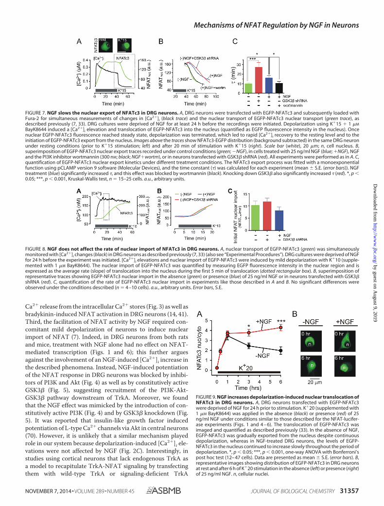
FIGURE 7. NGF slows the nuclear export of NFATc3 in DRG neurons. A, DRG neurons were transfected with EGFP-NFATc3 and subsequently loaded withFura-2 for simultaneous measurements of changes in [Ca2�]i (black trace) and the nuclear transport of EGFP-NFATc3 nuclear transport (green trace), asdescribed previously (7, 33). DRG cultures were deprived of NGF for at least 24 h before the recordings were initiated. Depolarization using K�15 � 1 �M
BayK8644 induced a [Ca2�]i elevation and translocation of EGFP-NFATc3 into the nucleus (quantified as EGFP fluorescence intensity in the nucleus). Oncenuclear EGFP-NFATc3 fluorescence reached steady state, depolarization was terminated, which led to rapid [Ca2�]i recovery to the resting level and to theinitiation of EGFP-NFATc3 export from the nucleus. Images above the traces show NFATc3-EGFP distribution (background subtracted) in the same DRG neuronunder resting conditions (prior to K�15 stimulation; left) and after 20 min of stimulation with K�15 (right). Scale bar (white), 20 �m; n, cell nucleus. B,superimposition of EGFP-NFATc3 nuclear export traces recorded under control conditions (green; �NGF), in cells treated with 25 ng/ml NGF (blue; �NGF), NGFand the PI3K inhibitor wortmannin (300 nM; black; NGF�wortm), or in neurons transfected with GSK3� shRNA (red). All experiments were performed as in A. C,quantification of EGFP-NFATc3 nuclear export kinetics under different treatment conditions. The NFATc3 export process was fitted with a monoexponentialfunction using pCLAMP version 9 software (Molecular Devices), and the time constant (�) was calculated for each experiment (mean � S.E. (error bars)). NGFtreatment (blue) significantly increased �, and this effect was blocked by wortmannin (black). Knocking down GSK3� also significantly increased � (red). *, p �0.05; ***, p � 0.001, Kruskal-Wallis test, n � 15–25 cells. a.u., arbitrary units.
FIGURE 8. NGF does not affect the rate of nuclear import of NFATc3 in DRG neurons. A, nuclear transport of EGFP-NFATc3 (green) was simultaneouslymonitored with [Ca2�]i changes (black) in DRG neurons as described previously (7, 33) (also see “Experimental Procedures”). DRG cultures were deprived of NGFfor 24 h before the experiment was initiated. [Ca2�]i elevations and nuclear import of EGFP-NFATc3 were induced by mild depolarization with K�10 (supple-mented with 1 �M BayK8644). The nuclear import of EGFP-NFATc3 was quantified by measuring EGFP fluorescence intensity in the nuclear region and isexpressed as the average rate (slope) of translocation into the nucleus during the first 5 min of translocation (dotted rectangular box). B, superimposition ofrepresentative traces showing EGFP-NFATc3 nuclear import in the absence (green) or presence (blue) of 25 ng/ml NGF or in neurons transfected with GSK3�shRNA (red). C, quantification of the rate of EGFP-NFATc3 nuclear import in experiments like those described in A and B. No significant differences wereobserved under the conditions described (n � 4 –10 cells). a.u., arbitrary units. Error bars, S.E.
FIGURE 9. NGF increases depolarization-induced nuclear translocation ofNFATc3 in DRG neurons. A, DRG neurons transfected with EGFP-NFATc3were deprived of NGF for 24 h prior to stimulation. K�20 (supplemented with1 �M BayK8644) was applied in the absence (black) or presence (red) of 25ng/ml NGF under conditions similar to those described for the NFAT-lucifer-ase experiments (Figs. 1 and 4 – 6). The translocation of EGFP-NFATc3 wasimaged and quantified as described previously (33). In the absence of NGF,EGFP-NFATc3 was gradually exported from the nucleus despite continuousdepolarization, whereas in NGF-treated DRG neurons, the levels of EGFP-NFATc3 in the nucleus continued to increase slowly throughout the period ofdepolarization. *, p � 0.05; ***, p � 0.001, one-way ANOVA with Bonferroni’spost hoc test (12– 47 cells). Data are presented as mean � S.E. (error bars). B,representative images showing distribution of EGFP-NFATc3 in DRG neuronsat rest and after 6 h of K�20 stimulation in the absence (left) or presence (right)of 25 ng/ml NGF. n, cellular nuclei.
Mechanisms of NFAT Regulation by NGF in Neurons
NOVEMBER 7, 2014 • VOLUME 289 • NUMBER 45 JOURNAL OF BIOLOGICAL CHEMISTRY 31357
by guest on August 9, 2019
http://ww
w.jbc.org/
Dow
nloaded from

mutants, both the PI3K and PLC signaling pathways were foundto be required for NGF/TrkA-dependent activation of NFAT(4). Collectively, our findings and work by others suggest that atleast two distinct pathways, the PI3K-Akt-GSK3� and/or PLC-IP3-Ca2� signaling cascades, can mediate neurotrophin-depen-dent regulation of NFAT in neurons and that their relative con-tribution depends on the type of neurotrophin involved, thetype of neurons, and the status of neuronal activity.
GSK3� is one of the first NFAT kinases to have been identi-fied and is known to regulate nuclear export of all of the canon-ical NFAT isoforms (NFATc1– c4) (3, 10, 25, 27, 71). Also,recent work has demonstrated that GSK3� suppresses NFAT-mediated gene expression in neurons (3, 72). However, itremained unclear whether endogenous neuromodulators affectGSK3�-NFAT signaling. Here, we demonstrate that neuronalGSK3�-NFAT signaling is modulated by NGF, resulting in thepotentiation of NFAT transcriptional activity. Notably, GSK3�knockdown resulted in a stronger enhancement of NFAT acti-vation (Fig. 5B) and a more prominent slowing of nuclearexport of NFATc3 (Fig. 7C) than did NGF treatment. Thiscould potentially be explained by the fact that only �60% (orfewer) of postnatal DRG neurons express the NGF receptorTrkA (56) and thus are likely to be affected by NGF. A relativelymodest but significant effect of NGF on GSK3� phosphoryla-tion at Ser-9 (Fig. 5C), which is known to inhibit GSK3� (55,73), is in agreement with this estimation. GSK3� is not the onlyNFAT kinase regulated by the PI3K-Akt signaling. For example,mammalian target of rapamycin (mTOR) kinase is known tophosphorylate and inhibit NFATc4 (74), and mTOR kinase isactivated by Akt signaling (75). However, its involvement wouldin theory result in NGF-dependent inhibition rather than theenhancement of NFAT activity described here (Figs. 1 and4 – 6) and thus is unlikely to account for the observed outcomes.
Our knockdown and knockout approaches revealed that theNFAT isoform NFATc3 is absolutely crucial for NGF-inducedpotentiation of NFAT-mediated gene expression in DRG neu-rons (Fig. 6). These findings are in good agreement with ourprevious molecular and functional data indicating thatNFATc3 is the predominant NFAT isoform in DRG neurons (7,
33). However, given that the expression and roles of specificNFAT isoforms vary among different types of neurons (4, 8, 12,13, 28), it is likely that other NFAT isoforms are also regulatedby neurotrophins. For example, NFATc4 is targeted by BDNFin hippocampal neurons (12, 13, 28). By monitoring the nucleartranslocation of EGFP-NFATc3 in DRG neurons in real time,we found that NGF and GSK3� inhibited nuclear export ofNFATc3 but had no detectable effect on the rate of nuclearimport of NFATc3 (Figs. 7 and 8). The latter is consistent withour previous finding that the rate of depolarization-inducednuclear import of NFATc3 is not affected by GSK3� knock-down in DRG neurons (33). Our data also further support theview that GSK3� is the major NFAT export kinase in neurons(3, 25, 60).
In summary, we have identified a novel mechanism of NFATactivation by NGF and suggest that neurotrophins act in con-cert with depolarization-driven Ca2� signaling to regulateNFATc3-dependent gene expression in sensory neurons.Although the functional significance of this mechanismremains to be determined, the new model proposed in thisstudy (Fig. 10) will guide further research directed at betterunderstanding of how electrical activity and neurotrophic fac-tors cooperate in long term regulation of neuronal excitabilityand synaptic function and how they orchestrate the wiring ofneuronal networks.
Acknowledgments—We thank Dr. Fernando Santana for providingNFATc3 KO mice; Dr. Michal Hetman for the NFATc3-shRNA,NFATc4-shRNA, and GSK3�-shRNA plasmids; and Dr. Steven Greenfor the caPI3K plasmid.
REFERENCES1. Deisseroth, K., Mermelstein, P. G., Xia, H., and Tsien, R. W. (2003) Sig-
naling from synapse to nucleus: the logic behind the mechanisms. Curr.Opin. Neurobiol. 13, 354 –365
2. Cohen, S., and Greenberg, M. E. (2008) Communication between the syn-apse and the nucleus in neuronal development, plasticity, and disease.Annu. Rev. Cell Dev. Biol. 24, 183–209
3. Graef, I. A., Mermelstein, P. G., Stankunas, K., Neilson, J. R., Deisseroth,K., Tsien, R. W., and Crabtree, G. R. (1999) L-type calcium channels andGSK-3 regulate the activity of NF-ATc4 in hippocampal neurons. Nature401, 703–708
4. Graef, I. A., Wang, F., Charron, F., Chen, L., Neilson, J., Tessier-Lavigne,M., and Crabtree, G. R. (2003) Neurotrophins and netrins require cal-cineurin/NFAT signaling to stimulate outgrowth of embryonic axons.Cell 113, 657– 670
5. Oliveria, S. F., Dell’Acqua, M. L., and Sather, W. A. (2007) AKAP79/150anchoring of calcineurin controls neuronal L-type Ca2� channel activityand nuclear signaling. Neuron 55, 261–275
6. Nguyen, T., and Di Giovanni, S. (2008) NFAT signaling in neural devel-opment and axon growth. Int. J. Dev. Neurosci. 26, 141–145
7. Kim, M. S., and Usachev, Y. M. (2009) Mitochondrial Ca2� cycling facili-tates activation of the transcription factor NFAT in sensory neurons.J. Neurosci. 29, 12101–12114
8. Zhang, J., and Shapiro, M. S. (2012) Activity-dependent TranscriptionalRegulation of M-type (Kv7) K� channels by AKAP79/150-mediatedNFAT actions. Neuron 76, 1133–1146
9. Benedito, A. B., Lehtinen, M., Massol, R., Lopes, U. G., Kirchhausen, T.,Rao, A., and Bonni, A. (2005) The transcription factor NFAT3 mediatesneuronal survival. J. Biol. Chem. 280, 2818 –2825
10. Arron, J. R., Winslow, M. M., Polleri, A., Chang, C. P., Wu, H., Gao, X.,Neilson, J. R., Chen, L., Heit, J. J., Kim, S. K., Yamasaki, N., Miyakawa, T.,
FIGURE 10. Model of concerted regulation of NFATc3 by electrical activityand NGF in sensory neurons. Electrical stimulation (depolarization in thisstudy) induces Ca2� influx into the cell via voltage-gated Ca2� channels,which leads to CaN-dependent dephosphorylation of NFATc3 and initiationof its import into the nucleus. NGF facilitates NFATc3 activation and retentionin the nucleus by stimulating the PI3K-Akt signaling pathway, which leads tophosphorylation-dependent inhibition of GSK3� and thereby to the slowingof NFATc3 export from the nucleus.
Mechanisms of NFAT Regulation by NGF in Neurons
31358 JOURNAL OF BIOLOGICAL CHEMISTRY VOLUME 289 • NUMBER 45 • NOVEMBER 7, 2014
by guest on August 9, 2019
http://ww
w.jbc.org/
Dow
nloaded from

Francke, U., Graef, I. A., and Crabtree, G. R. (2006) NFAT dysregulation byincreased dosage of DSCR1 and DYRK1A on chromosome 21. Nature441, 595– 600
11. Schwartz, N., Schohl, A., and Ruthazer, E. S. (2009) Neural activity regu-lates synaptic properties and dendritic structure in vivo through calcineu-rin/NFAT signaling. Neuron 62, 655– 669
12. Vashishta, A., Habas, A., Pruunsild, P., Zheng, J. J., Timmusk, T., andHetman, M. (2009) Nuclear factor of activated T-cells isoform c4(NFATc4/NFAT3) as a mediator of antiapoptotic transcription in NMDAreceptor-stimulated cortical neurons. J. Neurosci. 29, 15331–15340
13. Quadrato, G., Benevento, M., Alber, S., Jacob, C., Floriddia, E. M., Nguyen,T., Elnaggar, M. Y., Pedroarena, C. M., Molkentin, J. D., and Di Giovanni,S. (2012) Nuclear factor of activated T cells (NFATc4) is required forBDNF-dependent survival of adult-born neurons and spatial memoryformation in the hippocampus. Proc. Natl. Acad. Sci. U.S.A. 109,E1499 –E1508
14. Jackson, J. G., Usachev, Y. M., and Thayer, S. A. (2007) Bradykinin-in-duced nuclear factor of activated T-cells-dependent transcription in ratdorsal root ganglion neurons. Mol. Pharmacol. 72, 303–310
15. Groth, R. D., Coicou, L. G., Mermelstein, P. G., and Seybold, V. S. (2007)Neurotrophin activation of NFAT-dependent transcription contributesto the regulation of pro-nociceptive genes. J. Neurochem. 102, 1162–1174
16. Cai, Y. Q., Chen, S. R., and Pan, H. L. (2013) Upregulation of nuclear factorof activated T-cells by nerve injury contributes to development of neuro-pathic pain. J. Pharmacol. Exp. Ther. 345, 161–168
17. Shioda, N., Han, F., Moriguchi, S., and Fukunaga, K. (2007) Constitutivelyactive calcineurin mediates delayed neuronal death through Fas-ligandexpression via activation of NFAT and FKHR transcriptional activities inmouse brain ischemia. J. Neurochem. 102, 1506 –1517
18. Abdul, H. M., Sama, M. A., Furman, J. L., Mathis, D. M., Beckett, T. L.,Weidner, A. M., Patel, E. S., Baig, I., Murphy, M. P., LeVine, H., 3rd,Kraner, S. D., and Norris, C. M. (2009) Cognitive decline in Alzheimer’sdisease is associated with selective changes in calcineurin/NFAT signal-ing. J. Neurosci. 29, 12957–12969
19. Hudry, E., Wu, H. Y., Arbel-Ornath, M., Hashimoto, T., Matsouaka, R.,Fan, Z., Spires-Jones, T. L., Betensky, R. A., Bacskai, B. J., and Hyman, B. T.(2012) Inhibition of the NFAT pathway alleviates amyloid � neurotoxicityin a mouse model of Alzheimer’s disease. J. Neurosci. 32, 3176 –3192
20. Yan, H. Q., Shin, S. S., Ma, X., Li, Y., and Dixon, C. E. (2014) Differentialeffect of traumatic brain injury on the nuclear factor of activated T cells C3and C4 isoforms in the rat hippocampus. Brain Res. 1548, 63–72
21. Crabtree, G. R., and Olson, E. N. (2002) NFAT signaling: choreographingthe social lives of cells. Cell 109, S67–S79
22. Hogan, P. G., Chen, L., Nardone, J., and Rao, A. (2003) Transcriptionalregulation by calcium, calcineurin, and NFAT. Genes Dev. 17, 2205–2232
23. Okamura, H., Aramburu, J., Garcıa-Rodrıguez, C., Viola, J. P., Raghavan,A., Tahiliani, M., Zhang, X., Qin, J., Hogan, P. G., and Rao, A. (2000)Concerted dephosphorylation of the transcription factor NFAT1 inducesa conformational switch that regulates transcriptional activity. Mol. Cell 6,539 –550
24. Li, H., Pink, M. D., Murphy, J. G., Stein, A., Dell’Acqua, M. L., and Hogan,P. G. (2012) Balanced interactions of calcineurin with AKAP79 regulateCa2�-calcineurin-NFAT signaling. Nat. Struct. Mol. Biol. 19, 337–345
25. Beals, C. R., Sheridan, C. M., Turck, C. W., Gardner, P., and Crabtree, G. R.(1997) Nuclear export of NF-ATc enhanced by glycogen synthase ki-nase-3. Science 275, 1930 –1934
26. Zhu, J., and McKeon, F. (1999) NF-AT activation requires suppression ofCrm1-dependent export by calcineurin. Nature 398, 256 –260
27. Gwack, Y., Sharma, S., Nardone, J., Tanasa, B., Iuga, A., Srikanth, S., Oka-mura, H., Bolton, D., Feske, S., Hogan, P. G., and Rao, A. (2006) A genome-wide Drosophila RNAi screen identifies DYRK-family kinases as regula-tors of NFAT. Nature 441, 646 – 650
28. Groth, R. D., and Mermelstein, P. G. (2003) Brain-derived neurotrophicfactor activation of NFAT (nuclear factor of activated T-cells)-dependenttranscription: a role for the transcription factor NFATc4 in neurotrophin-mediated gene expression. J. Neurosci. 23, 8125– 8134
29. Stefos, G. C., Soppa, U., Dierssen, M., and Becker, W. (2013) NGF upregu-lates the plasminogen activation inhibitor-1 in neurons via the calcineu-
rin/NFAT pathway and the Down syndrome-related proteins DYRK1Aand RCAN1 attenuate this effect. PLoS One 8, e67470
30. Schnizler, K., Shutov, L. P., Van Kanegan, M. J., Merrill, M. A., Nichols, B.,McKnight, G. S., Strack, S., Hell, J. W., and Usachev, Y. M. (2008) Proteinkinase A anchoring via AKAP150 is essential for TRPV1 modulation byforskolin and prostaglandin E2 in mouse sensory neurons. J. Neurosci. 28,4904 – 4917
31. Rossow, C. F., Minami, E., Chase, E. G., Murry, C. E., and Santana, L. F.(2004) NFATc3-induced reductions in voltage-gated K� currents aftermyocardial infarction. Circ. Res. 94, 1340 –1350
32. Nieves-Cintron, M., Amberg, G. C., Nichols, C. B., Molkentin, J. D., andSantana, L. F. (2007) Activation of NFATc3 down-regulates the beta1subunit of large conductance, calcium-activated K� channels in arterialsmooth muscle and contributes to hypertension. J. Biol. Chem. 282,3231–3240
33. Ulrich, J. D., Kim, M. S., Houlihan, P. R., Shutov, L. P., Mohapatra, D. P.,Strack, S., and Usachev, Y. M. (2012) Distinct activation properties of thenuclear factor of activated T-cells (NFAT) isoforms NFATc3 andNFATc4 in neurons. J. Biol. Chem. 287, 37594 –37609
34. Tomida, T., Hirose, K., Takizawa, A., Shibasaki, F., and Iino, M. (2003)NFAT functions as a working memory of Ca2� signals in decoding Ca2�
oscillation. EMBO J. 22, 3825–383235. Hu, Q., Klippel, A., Muslin, A. J., Fantl, W. J., and Williams, L. T. (1995)
Ras-dependent induction of cellular responses by constitutively activephosphatidylinositol-3 kinase. Science 268, 100 –102
36. Hur, E. M., and Zhou, F. Q. (2010) GSK3 signalling in neural development.Nat. Rev. Neurosci. 11, 539 –551
37. Shuttleworth, T. J., and Thompson, J. L. (1991) Effect of temperature onreceptor-activated changes in [Ca2�]i and their determination using fluo-rescent probes. J. Biol. Chem. 266, 1410 –1414
38. Thayer, S. A., and Miller, R. J. (1990) Regulation of the intracellular freecalcium concentration in single rat dorsal root ganglion neurones in vitro.J. Physiol. 425, 85–115
39. Thayer, S. A., Perney, T. M., and Miller, R. J. (1988) Regulation of calciumhomeostasis in sensory neurons by bradykinin. J. Neurosci. 8, 4089 – 4097
40. Svichar, N., Shmigol, A., Verkhratsky, A., and Kostyuk, P. (1997) ATPinduces Ca2� release from IP3-sensitive Ca2� stores exclusively in largeDRG neurones. Neuroreport 8, 1555–1559
41. Usachev, Y. M., DeMarco, S. J., Campbell, C., Strehler, E. E., and Thayer,S. A. (2002) Bradykinin and ATP accelerate Ca2� efflux from rat sensoryneurons via protein kinase C and the plasma membrane Ca2� pump iso-form 4. Neuron 33, 113–122
42. Verkhratsky, A. (2005) Physiology and pathophysiology of the calciumstore in the endoplasmic reticulum of neurons. Physiol. Rev. 85, 201–279
43. Patapoutian, A., and Reichardt, L. F. (2001) Trk receptors: mediators ofneurotrophin action. Curr. Opin. Neurobiol. 11, 272–280
44. Chao, M. V. (2003) Neurotrophins and their receptors: a convergencepoint for many signalling pathways. Nat. Rev. Neurosci. 4, 299 –309
45. Stephens, R. M., Loeb, D. M., Copeland, T. D., Pawson, T., Greene, L. A.,and Kaplan, D. R. (1994) Trk receptors use redundant signal transductionpathways involving SHC and PLC-�1 to mediate NGF responses. Neuron12, 691–705
46. Marsh, H. N., Dubreuil, C. I., Quevedo, C., Lee, A., Majdan, M., Walsh,G. S., Hausdorff, S., Said, F. A., Zoueva, O., Kozlowski, M., Siminovitch, K.,Neel, B. G., Miller, F. D., and Kaplan, D. R. (2003) SHP-1 negatively regu-lates neuronal survival by functioning as a TrkA phosphatase. J. Cell Biol.163, 999 –1010
47. Wood, E. R., Kuyper, L., Petrov, K. G., Hunter, R. N., 3rd, Harris, P. A., andLackey, K. (2004) Discovery and in vitro evaluation of potent TrkA kinaseinhibitors: oxindole and aza-oxindoles. Bioorg. Med. Chem. Lett. 14,953–957
48. Martin, K. J., Shpiro, N., Traynor, R., Elliott, M., and Arthur, J. S. (2011)Comparison of the specificity of Trk inhibitors in recombinant and neu-ronal assays. Neuropharmacology 61, 148 –155
49. Knusel, B., and Hefti, F. (1992) K-252 compounds: modulators of neu-rotrophin signal transduction. J. Neurochem. 59, 1987–1996
50. Watson, J. J., Allen, S. J., and Dawbarn, D. (2008) Targeting nerve growthfactor in pain: what is the therapeutic potential? BioDrugs 22, 349 –359
Mechanisms of NFAT Regulation by NGF in Neurons
NOVEMBER 7, 2014 • VOLUME 289 • NUMBER 45 JOURNAL OF BIOLOGICAL CHEMISTRY 31359
by guest on August 9, 2019
http://ww
w.jbc.org/
Dow
nloaded from

51. Eibl, J. K., Strasser, B. C., and Ross, G. M. (2012) Structural, biological, andpharmacological strategies for the inhibition of nerve growth factor. Neu-rochem. Int. 61, 1266 –1275
52. Zhou, F. Q., Zhou, J., Dedhar, S., Wu, Y. H., and Snider, W. D. (2004)NGF-induced axon growth is mediated by localized inactivation ofGSK-3� and functions of the microtubule plus end binding protein APC.Neuron 42, 897–912
53. Jones, D. M., Tucker, B. A., Rahimtula, M., and Mearow, K. M. (2003) Thesynergistic effects of NGF and IGF-1 on neurite growth in adult sensoryneurons: convergence on the PI 3-kinase signaling pathway. J. Neurochem.86, 1116 –1128
54. Kau, T. R., Schroeder, F., Ramaswamy, S., Wojciechowski, C. L., Zhao, J. J.,Roberts, T. M., Clardy, J., Sellers, W. R., and Silver, P. A. (2003) A chemicalgenetic screen identifies inhibitors of regulated nuclear export of a Fork-head transcription factor in PTEN-deficient tumor cells. Cancer Cell 4,463– 476
55. Stambolic, V., and Woodgett, J. R. (1994) Mitogen inactivation of glycogensynthase kinase-3� in intact cells via serine 9 phosphorylation. Biochem. J.303, 701–704
56. Molliver, D. C., Wright, D. E., Leitner, M. L., Parsadanian, A. S., Doster, K.,Wen, D., Yan, Q., and Snider, W. D. (1997) Ib4-binding Drg neuronsswitch from Ngf to Gdnf dependence in early postnatal life. Neuron 19,849 – 861
57. Meyer, R. A., Ringhamp, M., Campbell, J. N., and Raja, S. N. (2006) inTextbook of Pain (McMahon, S. B., and Koltzenburg, M., eds) pp 3–34,Elsevier, Amsterdam
58. Wilkins, B. J., De Windt, L. J., Bueno, O. F., Braz, J. C., Glascock, B. J.,Kimball, T. F., and Molkentin, J. D. (2002) Targeted disruption ofNFATc3, but not NFATc4, reveals an intrinsic defect in calcineurin-me-diated cardiac hypertrophic growth. Mol. Cell Biol. 22, 7603–7613
59. West, A. E., Griffith, E. C., and Greenberg, M. E. (2002) Regulation oftranscription factors by neuronal activity. Nat. Rev. Neurosci. 3, 921–931
60. Muller, M. R., and Rao, A. (2010) NFAT, immunity and cancer: a tran-scription factor comes of age. Nat. Rev. Immunol. 10, 645– 656
61. Ozaki, Y., Kitamura, N., Tsutsumi, A., Dayanithi, G., and Shibuya, I. (2009)NGF-induced hyperexcitability causes spontaneous fluctuations of intra-cellular Ca2� in rat nociceptive dorsal root ganglion neurons. Cell Cal-cium 45, 209 –215
62. Usachev, Y. M., and Thayer, S. A. (1999) Ca2� influx in resting rat sensoryneurones that regulates and is regulated by ryanodine-sensitive Ca2�
stores. J. Physiol. 519, 115–13063. Kress, M., and Distler, C. (2004) Differences in calcium signalling in rat
peripheral sensory neurons. Neurosci. Lett. 354, 127–13064. Linhart, O., Obreja, O., and Kress, M. (2003) The inflammatory mediators
serotonin, prostaglandin E2 and bradykinin evoke calcium influx in ratsensory neurons. Neuroscience 118, 69 –74
65. Stucky, C. L., Thayer, S. A., and Seybold, V. S. (1996) Prostaglandin E2increases the proportion of neonatal rat dorsal root ganglion neurons thatrespond to bradykinin. Neuroscience 74, 1111–1123
66. Khasabova, I. A., Simone, D. A., and Seybold, V. S. (2002) Cannabinoidsattenuate depolarization-dependent Ca2� influx in intermediate-size pri-mary afferent neurons of adult rats. Neuroscience 115, 613– 625
67. Khasabova, I. A., Harding-Rose, C., Simone, D. A., and Seybold, V. S.(2004) Differential effects of CB1 and opioid agonists on two populationsof adult rat dorsal root ganglion neurons. J. Neurosci. 24, 1744 –1753
68. Oh, S. B., Tran, P. B., Gillard, S. E., Hurley, R. W., Hammond, D. L., andMiller, R. J. (2001) Chemokines and glycoprotein120 produce pain hyper-sensitivity by directly exciting primary nociceptive neurons. J. Neurosci.21, 5027–5035
69. Malin, S. A., Molliver, D. C., Koerber, H. R., Cornuet, P., Frye, R., Albers,K. M., and Davis, B. M. (2006) Glial cell line-derived neurotrophic factorfamily members sensitize nociceptors in vitro and produce thermal hyper-algesia in vivo. J. Neurosci. 26, 8588 – 8599
70. Blair, L. A., Bence-Hanulec, K. K., Mehta, S., Franke, T., Kaplan, D., andMarshall, J. (1999) Akt-dependent potentiation of L channels by insulin-like growth factor-1 is required for neuronal survival. J. Neurosci. 19,1940 –1951
71. van der Velden, J. L., Schols, A. M., Willems, J., Kelders, M. C., and Langen,R. C. (2008) Glycogen synthase kinase 3 suppresses myogenic differentia-tion through negative regulation of NFATc3. J. Biol. Chem. 283, 358 –366
72. Gomez-Sintes, R., and Lucas, J. J. (2010) NFAT/Fas signaling mediates theneuronal apoptosis and motor side effects of GSK-3 inhibition in a mousemodel of lithium therapy. J. Clin. Invest. 120, 2432–2445
73. Fang, X., Yu, S. X., Lu, Y., Bast, R. C., Jr., Woodgett, J. R., and Mills, G. B.(2000) Phosphorylation and inactivation of glycogen synthase kinase 3 byprotein kinase A. Proc. Natl. Acad. Sci. U.S.A. 97, 11960 –11965
74. Yang, T. T., Yu, R. Y., Agadir, A., Gao, G. J., Campos-Gonzalez, R.,Tournier, C., and Chow, C. W. (2008) Integration of protein kinasesmTOR and extracellular signal-regulated kinase 5 in regulating nucleocy-toplasmic localization of NFATc4. Mol. Cell Biol. 28, 3489 –3501
75. Sugden, P. H., Fuller, S. J., Weiss, S. C., and Clerk, A. (2008) Glycogensynthase kinase 3 (GSK3) in the heart: a point of integration in hyper-trophic signalling and a therapeutic target? A critical analysis. Br. J. Phar-macol. 153, S137–S153
Mechanisms of NFAT Regulation by NGF in Neurons
31360 JOURNAL OF BIOLOGICAL CHEMISTRY VOLUME 289 • NUMBER 45 • NOVEMBER 7, 2014
by guest on August 9, 2019
http://ww
w.jbc.org/
Dow
nloaded from

Jason D. Ulrich and Yuriy M. UsachevMan-Su Kim, Leonid P. Shutov, Aswini Gnanasekaran, Zhihong Lin, Jacob E. Rysted,
) Pathwayβ (GSK3β(PI3K)-Akt-Glycogen Synthase Kinase 3T-cells (NFAT) in Neurons via the Phosphatidylinositol 3-Kinase
Nerve Growth Factor (NGF) Regulates Activity of Nuclear Factor of Activated
doi: 10.1074/jbc.M114.587188 originally published online September 17, 20142014, 289:31349-31360.J. Biol. Chem.
10.1074/jbc.M114.587188Access the most updated version of this article at doi:
Alerts:
When a correction for this article is posted•
When this article is cited•
to choose from all of JBC's e-mail alertsClick here
http://www.jbc.org/content/289/45/31349.full.html#ref-list-1
This article cites 74 references, 29 of which can be accessed free at
by guest on August 9, 2019
http://ww
w.jbc.org/
Dow
nloaded from