nerve growth factor in alzheimer’s disease: increased
TRANSCRIPT
The Journal of Neuroscience, September 1995, l.!?(9): 6213-6221
Nerve Growth Factor in Alzheimer’s Disease: Increased Levels throughout the Brain Coupled with Declines in Nucleus Basalis
Samuel A. Scott,’ Elliott J. Mufson,3 Jean A. Weingartner,’ Kenneth A. Skau,” and Keith A. Crutcher’
‘Department of Neurosurgery and *College of Pharmacy, University of Cincinnati, Cincinnati, Ohio and 3Department of Neurology, Rush Presbyterian St. Luke’s Medical Center, Chicago, Illinois
The current study analyzed NGF protein levels in the brains of patients with Alzheimer’s disease (AD) as compared with aged neurologically normal individuals. An established two-site ELISA was used to measure NGF-like immuno- reactivity in the hippocampus, superior temporal gyrus, su- perior frontal gyrus, inferior parietal lobule, frontal and oc- cipital cortical poles, cerebellum, amygdala, putamen, and nucleus basalis of Meynert (nbM). ChAT activity was as- sayed in adjacent tissue samples. NGF levels were also evaluated in Parkinson’s disease for comparison with both AD and age-matched control cases.
Regardless of the brain bank (University of Cincinnati, Rush Presbyterian St. Luke’s Medical Center in Chicago, or University of Alabama at Birmingham), NGF-like activity was at least moderately increased with AD in virtually ev- ery brain region examined except for the nbM, in which significant declines were observed. NGF levels were also increased when compared with age-matched Parkinson’s cases (frontal cortex). NGF-like activity was not related to age at onset or disease duration in AD cases, nor did NGF levels correlate with age at death in the control or AD groups. Correlations between ChAT and NGF-like activity across brains varied considerably and were generally not significant.
The present findings indicate that AD is characterized by a widespread increase in cortical and subcortical NGF. Al- though a correlation with ChAT activity was not observed in cortex, the AD-related decline in NGF found in nbM is consistent with the possibility of impaired retrograde trans- port of NGF to this region.
[Key words: NGF, neurotrophin, ELISA, Alzheimer’s dis- ease, basal forebrain, human, amygdala, hippocampus, cortex, cerebellum, ChAT, aging]
The neuropathology of Alzheimer’s disease (AD) is being in- creasingly viewed as a global disruption of brain circuitry in- volving multiple transmitter systems. However, most of the phar- macological approaches to treating AD have centered around the
Received Feb. 16, 1995; revised May 12, 1995; accepted May 16, 1995. This work was supported by Grants NS31410 (K.A.C.), AGO5605 (S.A.S),
AC 10161 and AGO9466 (E.J.M.). We graciously thank Chris Clendening for assistance with the ChAT assay, Genentech Inc. for recombinant human NGF and Dr. William Mobley for NGF anttsera. Thanks also to Drs. Powers and Guozhu at the University of Alabama at Birmingham for providing additional brain tissue.
Correspondence should be addressed to Samuel A. Scott, Ph.D., Program in Physical Therapy, Barry University, 11300 N.E. 2nd Avenue, Miami Shores, FL 33161. Copyright 8 1995 Society for Neuroscience 0270-6474~~1~S6213-~$05.0010
cholinergic synapse (e.g., AChE inhibitors). No single approach has been particularly successful, although this has not discour- aged the development of alternative tactics aimed at stabilizing cholinergic basal forebrain projections in AD.
One such strategy is the use of NGE a protein with growth- and survival-promoting effects on central cholinergic neurons both in vitro and in viva (reviewed in Scott and Crutcher, 1994). Notwithstanding potential side effects of NGF administration (Butcher and Woolf, 1989; Scott and Crutcher, 1994) and com- plications with respect to mode of delivery (Hefti and Schneider, 1989; Saffran, 1992), it has been suggested that exogenous NGE if appropriately administered, will prevent degeneration of these neurons in AD in a manner similar to that observed in the le- sioned animal (reviewed by Scott and Crutcher, 1994).
Given the original suggestion that certain human neurologic disorders may be caused by reductions in trophic support (Ap- pel, 1981), and specifically by loss of NGF in Alzheimer’s dis- ease (Appel, 1981; Hefti, 1983; Hefti and Weiner, 1986; Phelps et al., 1989), an initial study was undertaken to determine wheth- er brain NGF levels were altered in AD using a bioassay and a well-established immunoassay (Crutcher et al., 1993). Surpris- ingly, NGF-like activity was found to be increased with AD in both the frontal and occipital poles of the cortex (Crutcher et al., 1993). This result seemed consistent with the possibility of impaired retrograde transport of NGF to the cholinergic basal forebrain, particularly when considering the AD-related loss of NGF receptor-bearing neurons in this region (Hefti and Mash, 1989; Kordower et al., 1989b; Mufson et al., 1989a; Allen et al., 1990; Loy et al., 1990; Strada et al., 1992).
The present investigation was undertaken to further analyze NGF levels in human brain in order to determine: (1) whether AD-related increases in NGF are restricted to areas receiving basal forebrain cholinergic input; (2) whether such increases also occur in Parkinson’s disease, another age-related neurodegener- ative disorder; and (3) whether correlations exist between target NGF levels and the amount of ChAT activity in the same tissue samples. We hypothesized that if NGF levels are elevated in response to removal of cholinergic input, as they are in rodent hippocampus following destruction of septal axons (Scott et al., 1994), then a negative correlation should exist between NGF and ChAT activity in the target tissue. Moreover, the levels of NGF should be decreased within the cholinergic basal forebrain in AD. We found the latter to be the case, although correlations between NGF and ChAT activity were not robust and varied widely across brain regions.
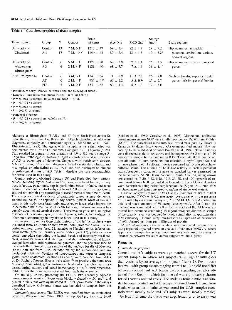
Materials and Methods Subjects. Postmortem-derived tissue samples from 59 patients, including 30 from the University of CincinnaIi (UC), 12 from the UnivwrsiQ of
6214 Scott et al. * NGF and Brain Cholinergic Innervation in AD
Table 1. Case demographics of tissue samples
Brain Storage Tissue source Group N Gender wt km) Age W PMD (hry (mo)6 Brain regions
University of Control 13 I M, 6 F 1211 k 47 68 -c 3.4 12 -c 1.7 28 5 7.2 Hippocampus, amygdala, Cincinnati AD 17 7M, IOF 1149 * 43 82 i 2.4’ 12 L 1.8 10 2 3.2d putamen, cerebellum, various
cortical regions
University of Control 6 5 M, 1 F 1328 ? 20 69 5 3.9 7 2 1.1 25 -t 3.3 Hippocampus, superior temporal Alabama at AD 6 2 M, 4 F 1128 -+ 40’ 68 -t 3.7 7 5 1.4 36 -c I.]/ gyms Birmingham
Rush Presbyterian Control 6 3 M, 3 F 1243 +- 64 71 F 2.9 11 2 2.3 26 2 7.8 Nucleus basalis, superior frontal AD 6 2 M, 4 F 983 t 57” 69 2 2.2 5 5 0.8’ 25 -c 2.1 gyrus, inferior parietal lobule PDs 5 3 M, 2 F 1331 2 58 69 2 1.4 6 2 1.3 17 k 5.6
d Postmortem delay (interval between death and freezing of tissue).
6 Length of time tissue was stored frozen (-80°C or below). ‘ P = 0.0018 vs control; all values are mean + SEM.
d P = 0.0172 vs control.
c P = 0.0012 “S control.
‘P = 0.0133 YS control.
ii Parkinson’s disease.
" P = 0.0122 vs control and 0.0022 vs. PD.
'P = 0.0394 vs control.
Alabama at Birmingham (UAB), and 17 from Rush-Presbyterian-St. Luke (Rush), were used in this study. Subjects classified as AD were diagnosed clinically and neuropathologically (McKhann et al., 1984; Khachaturian, 1985). The age ai which-symptoms were first noted was documented for 11 of 17 UC oatients. averaging 73 ? 3.4 vears (SEM). This resulted in a mean illne’ss duration of-6.q ? 0.9 years (range 31 13 years). Pathologic evaluation of aged controls revealed no evidence of AD or other type of dementia. Subjects with Parkinson’s disease, obtained through Rush, were diagnosed based on standard clinical and histological criteria (Mirra et al., 1993) and also displayed no clinical or pathological signs of AD. Table I displays the case demographics for tissue used in this study.
Control subjects obtained through UC and Rush died from various causes including myocardial infarction, congestive heart failure, urinary tract infection, pneumonia, sepsis, peritonitis, bowel infarcts, and renal failure. In contrast, control subjects from UAB all died from accidents, and did not exhibit any neurologic disease process at the time of death. There was no clinical evidence of metastatic tumor, seizure, dementia, alcoholism, AIDS, or hepatitis in any control patient. Most of the AD cases in this study were brain-only autopsies, so it was often impossible to determine the direct cause of death (although pneumonia was a fre- quently documented terminal event). There was no gross or histological evidence of neoplasia, spongy state, hypoxia, infarct, hemorrhage, or other such abnormality in any tissue block used in this study.
Bruin areas. Samples from eight regions of the left hemisphere were evaluated from UC brain donors: frontal pole (Brodmann area IO), su- perior temporal gyrus (area 22, anterior to Heschl’s gyri), inferior pa- rietal lobule (area 39), primary visual cortex (area l7), posterior baso- lateral amygdala (including the lateral, basal, and accessory basal nu- clei), Ammon’s horn and dentate gyrus of the mid-rostrocaudal hippo- campal formation, mid-rostrocaudal putamen, and the posterior lobe of the cerebellum. Snap-frozen samples of the nucleus basalis of Meynert (nbM), obtained from Rush, included mainly the anteromedial and an- terolateral subfields. Sections of hippocampus and superior temporal gyrus (same anatomical locations as above) were provided from UAB by Dr. Richard Powers. Blocks were taken from precisely the same area of every brain using gross anatomical landmarks. Samples were har- vested during autopsy and stored immediately at - 80°C until processed. Table 1 lists the brain areas obtained from each tissue source.
On the day or two preceding the ELISA, two coronally adjacent tissue samples were cut from each block, weighed (-100 mg), and placed in vials that were again stored at -80°C prior to use in the assays described below. Only gray matter was included in samples from the neocortex.
Immunologicnl assay. The ELISA was modified from an established protocol (Weskamp and Otten, 1987) as described previously in detail
(Saffran et al., 1989; Crutcher et al., 1993). Monoclonal antibodies raised against mouse NGF were kindly provided by Dr. William Mobley (UCSF). The polyclonal antiserum was raised in a goat by Hazelton Research Products, Inc. (Denver, PA) using purified mouse NGF ac- cording to an established protocol (Saffran et al., 1989). Tissue samples were dissected as described above and homogenized for 3 min at a 1: 10 dilution in sample buffer containing 0.1% Tween 20, 0.5% bovine se- rum albumin, 0.1 mM benzethonium chloride, 1 mg/ml aprotinin, and 0.1 mM phenylmethyl sulfonyl fluoride prepared in IO mM phosphate- buffered saline. The amount of NGF-like activity in each supernatant was subsequently calculated relative to standard curves generated on the same plates (NUNC, Irvine Scientific, Santa Ana, CA) using known concentrations (1.56, 3.12, 6.25, 12.5, 25, 50, and 100 pg/well) of re- combinant human NGF (provided by Genentech, Inc.). Optical densities were determined using orthophenylenediamine (Sigma, St. Louis MO) as chromagen and then converted to nglgm of tissue wet weight.
Choline ucetyltrunsferuse (ChAT) ussuy. Samples of brain extract were reacted (j7”C) with 0.2 mM acetyl Loenzyme A in the presence of 0.1 mu ohvsostigmine salicvlate. 2.0 mM EDTA. 8 mM choline io- dide, and t;ace amounts of 14C-acetyl coenzyme A. After 6 min the reaction was terminated with 2.0 M formic acid and the ACh was ex- tracted into octanone containing 15 mg/ml tetraphenylboron. A sample of the organic layer was counted by liquid scintillation at approximately 80% efficiency. Choline acetyltransferase was expressed as nanomoles of ACh formed per hour per milligram of protein.
Statistical analyses. Groups of data were compared parametrically using unpaired or paired t tests, or analysis of variance (ANOVA) where appropriate. Simple linear regression analyses were used to assess re- lationships between variables across subjects within a group.
Results Group demographics Control and AD subjects were age-matched except for the UC patient sample, in which AD subjects were significantly older than controls by an average of 14 years (Table 1). Postmortem interval, with group means ranging from 5 to 12 hr, did not differ between control and AD brains except regarding samples ob- tained from Rush, in which the interval was significantly shorter for AD versus control cases. The male-to-female ratio was sim- ilar between control and AD groups obtained from UC and from Rush, whereas an imbalance was noted for UAB samples (con- trols were mostly male and AD subjects were mostly female). The length of time the tissue was kept frozen prior to assay was
The Journal of Neuroscience, September 1995, 15(9) 6215
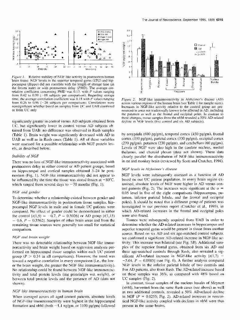
Figure I. Relative stability of NGF-like activity in postmortem human brain tissue. NGF levels in the superior temporal gyrus (STG) and hip- pocampus (Hippo) did not correlate with the length of storage time (in the frozen state) or with postmortem delay (PM@. The average cor- relation coefficient concerning PMD was 0.13, with P values ranging from 0.42 to 0.99 (-I8 subjects per comparison). Regarding storage time, the average correlation coefficient was 0.18 with P values ranging from 0.26 to 0.96 (-20 subjects per comparison). Correlations were nonsignificant whether based on samples from UC and UAB combined or from UC only.
significantly greater in control versus AD subjects obtained from UC, but significantly lower in control versus AD subjects ob- tained from UAB; no difference was observed in Rush samples (Table 1). Brain weight was significantly decreased with AD in UAB as well as in Rush cases (Table 1). All of these variables were assessed for a possible relationship with NGF protein lev- els, as described below.
Stability of NGF
There was no loss of NGF-like immunoreactivity associated with postmortem delay in either control or AD patient groups, based on hippocampal and cortical samples obtained 3-24 hr post- mortem (Fig. 1). NGF-like immunoreactivity did not appear to be influenced by the time the tissue was stored frozen at -80°C which ranged from several days to -70 months (Fig. 1).
NGF and gender
To determine whether a relationship existed between gender and NGF-like immunoreactivity in postmortem tissue samples, hip- pocampal NGF levels in male and in female UC patients were compared. No effect of gender could be demonstrated in either the control [t( 1,9) = -0.7, P = 0.50261 or AD group [t( 1,15) = 0.6, P = 0.5562]. Samples of other brain areas and from the remaining tissue sources were generally too small for statistical comparison.
NGF and brain weight
There was no detectable relationship between NGF-like immu- noreactivity and brain weight based on regression analyses per- formed on hippocampal tissue samples within each UC patient group (P > 0.34 in all comparisons). However, the trend was toward a negative correlation in every comparison (i.e., the low- er the brain weight, the greater the NGF-like immunoreactivity). No relationship could be found between NGF-like immunoreac- tivity and total protein levels (mg protein/gm wet weight), or between total protein levels and the presence of AD (data not shown).
NGF-like immunoreactivity in human brain
When averaged across all aged control patients, absolute levels of NGF-like immunoreactivity were highest in the hippocampal formation and nbM (both -1 .I ng/gm, or 1100 pg/gm) followed
Figure 2. NGF-like immunoreactivity in Alzheimer’s disease (AD) across various regions of the human brain (see Table 1 for sample sizes). Increases in NGF-like activity relative to the control group are pro- nounced in areas not traditionally known to be affected in AD, including the putamen as well as the frontal and occipital poles. In contrast to these changes, tissue samples from the nbM revealed a 50% AD-related decline in NGF levels (five control and six AD subjects).
by amygdala (690 pg/gm), temporal cortex (450 pg/gm), frontal cortex (330 pg/gm), parietal cortex (330 pg/gm), occipital cortex (270 pg/gm), putamen (230 pg/gm), and cerebellum (60 pg/gm). Levels of NGF were also high in the caudate nucleus, medial thalamus, and choroid plexus (data not shown). These data closely parallel the distribution of NGF-like immunoreactivity in rat and monkey brain (reviewed by Scott and Crutcher, 1994).
NGF levels in Alzheimer’s disease
NGF levels were subsequently assessed as a function of AD based on our UC patient population. In every brain region ex- amined, absolute levels of NGF were higher in AD versus con- trol patients (Fig. 2). The increases were significant at the (Y = 0.05 level in five of the eight comparisons (hippocampus, pu- tamen, inferior parietal lobule, and the frontal and occipital poles). It should be noted that a different group of patients was investigated in our previous report (Crutcher et al., 1993) in which AD-related increases in the frontal and occipital poles were also found.
Tissues were subsequently acquired from UAB in order to determine whether the AD-related increases in hippocampus and superior temporal gyrus would be present in tissue from another source. Based on six AD and six age-matched control subjects, we confirmed a significant AD-related increase in NGF-like ac- tivity. This increase was bilateral (see Fig. 5B). Additional sam- ples of the superior frontal gyrus, obtained from six AD and three age-matched controls through Rush, also revealed a sig- nificant AD-related increase in NGF-like activity [t( 1,7) = -5.04, P = O.OOlS] (see Fig. 6). A further analysis compared NGF levels in the inferior parietal lobule of two controls and five AD patients, also from Rush. The AD-related increase based on these samples was 36%, as compared with 48% based on UC samples (Fig. 2).
In contrast, tissue samples of the nucleus basalis of Meynert (nbM), harvested from the same Rush cases (see above) as well as two additional controls, revealed a 50% AD-related decline in NGF (P = 0.0255; Fig. 2). AD-related increases in neocor- tical NGF-like activity coupled with declines in nbM were thus present in the same brains.
6216 Scott et al. * NGF and Brain Cholinergic Innervation in AD
-o- Control -o- AD
LH LH LH LH LH LH LH LH
Neocortex
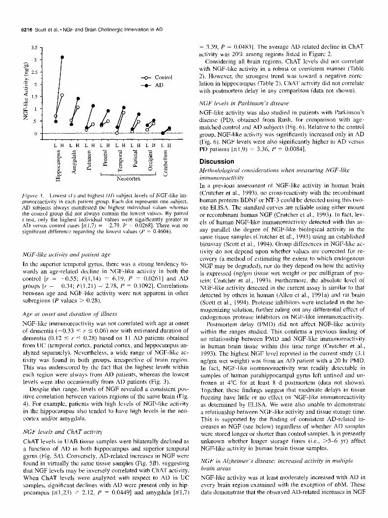
E&we 3. Lowest (L) and highest (H) subject levels of NGF-like im- munoreactivity in each patient group. Each dot represents one subject. AD subjects always manifested the highest individual values whereas the control group did not always contain the lowest values. By paired t test, only the highest individual values were significantly greater in AD versus control cases [t(l,7) = -2.79, P = 0.0268]. There was no significant difference regarding the lowest values (P = 0.4604).
NGF-like activity and patient age
In the superior temporal gyrus, there was a strong tendency to- wards an age-related decline in NGF-like activity in both the control [Y = -0.55; F(l,l4) = 6.19, P = 0.0261] and AD groups [v = -0.34; F(1,21) = 2.78, P = 0.10921. Correlations between age and NGF-like activity were not apparent in other subregions (P values > 0.28).
Age at onset and duration qf illness
NGF-like immunoreactivity was not correlated with age at onset of dementia (-0.33 5 r 5 0.06) nor with estimated duration of dementia (0.12 5 r 5 0.28) based on 1 1 AD patients obtained from UC (temporal cortex, parietal cortex, and hippocampus an- alyzed separately). Nevertheless, a wide range of NGF-like ac- tivity was found in both groups, irrespective of brain region. This was underscored by the fact that the highest levels within each region were always from AD patients, whereas the lowest levels were also occasionally from AD patients (Fig. 3).
Despite this range, levels of NGF revealed a consistent pos- itive correlation between various regions of the same brain (Fig. 4). For example, patients with high levels of NGF-like activity in the hippocampus also tended to have high levels in the neo- cortex and/or amygdala.
NGF levels and ChAT activity
ChAT levels in UAB tissue samples were bilaterally declined as a function of AD in both hippocampus and superior temporal gyrus (Fig. 5A). Conversely, AD-related increases in NGF were found in virtually the same tissue samples (Fig. SB), suggesting that NGF levels may be inversely correlated with ChAT activity. When ChAT levels were analyzed with respect to AD in UC samples, significant declines with AD were present only in hip- pocampus [t(l,23) = 2.12, P = 0.04491 and amygdala [t( 1,7)
= 3.39, P = 0.04831. The average AD-related decline in ChAT activity was 20% among regions listed in Figure 2.
Considering all brain regions, ChAT levels did not correlate with NGF-like activity in a robust or consistent manner (Table 2). However, the strongest trend was toward a negative corre- lation in hippocampus (Table 2). ChAT activity did not correlate with postmortem delay in any comparison (data not shown).
NGF levels in Parkinson’s disease
NGF-like activity was also studied in patients with Parkinson’s disease (PD), obtained from Rush, for comparison with age- matched control and AD subjects (Fig. 6). Relative to the control group, NGF-like activity was significantly increased only in AD (Fig. 6). NGF levels were also significantly higher in AD versus PD patients [t( 1,9) = 3.36, P = 0.0084].
Discussion Methodological considerations when measuring NGF-like immunoreactivity In a previous assessment of NGF-like activity in human brain (Crutcher et al., 1993) no cross-reactivity with the recombinant human proteins BDNF or NT-3 could be detected using this two- site ELISA. The standard curves are reliable using either mouse or recombinant human NGF (Crutcher et al., 1993). In fact, lev- els of human NGF-like immunoreactivity detected with this as- say parallel the degree of NGF-like biological activity in the same tissue samples (Crutcher et al., 1993) using an established bioassay (Scott et al., 1994). Group differences in NGF-like ac- tivity do not depend upon whether values are corrected for re- covery (a method of estimating the extent to which endogenous NGF may be degraded), nor do they depend on how the activity is expressed (ng/gm tissue wet weight or per milligram of pro- tein; Crutcher et al., 1993). Furthermore, the absolute level of NGF-like activity detected in the current assay is similar to that detected by others in human (Allen et al., 1991a) and rat brain (Scott et al., 1994). Protease inhibitors were included in the ho- mogenizing solution, further ruling out any differential effect of endogenous protease inhibitors on NGF-like immunoreactivity.
Postmortem delay (PMD) did not affect NGF-like activity within the ranges studied. This confirms a previous finding of no relationship between PMD and NGF-like immunoreactivity in human brain tissue within this time range (Crutcher et al., 1993). The highest NGF level reported in the current study (3.1 ng/gm wet weight) was from an AD patient with a 20 hr PMD. In fact, NGF-like immunoreactivity was readily detectable in samples of human parahippocampal gyrus left unfixed and un- frozen at 4°C for at least 8 d postmortem (data not shown). Together, these findings suggest that moderate delays in tissue freezing have little or no effect on NGF-like immunoreactivity as determined by ELISA. We were also unable to demonstrate a relationship between NGF-like activity and tissue storage time. This is supported by the finding of consistent AD-related in- creases in NGF (see below) regardless of whether AD samples were stored longer or shorter than control samples. It is presently unknown whether longer storage times (i.e., >5-6 yr) affect NGF-like activity in human brain tissue samples.
NGF in Alzheimer’s disease: increased uctivity in multiple brain areus
NGF-like activity was at least moderately increased with AD in every brain region examined with the exception of nbM. These data demonstrate that the observed AD-related increases in NGF

The Journal of Neuroscience, September 1995, 15(9) 6217
0 &i$q
0 Control
0 0 k!L
.4 .6 .8
Temporal Cortex 1 1.2
l-
@ .8-
3 .6- 3 r, .4-
g G .2- 0
0 ”
-. 2 1 I I I I I I I
0 .5 1 1.5 2 2.5 3 3.5 Hippocampus
1.6 -
1.4 - 0
0
cd 1.2-
2 2
l-
E .8- 4
.6 -
.4 - e
.2 I I I I I I I I I .6 .8 1 1.2 1.4 1.6 1.8 2 2.2
Hippocampus
0 0 0 I I I I I I I I
0 .5 1 1.5 2 2.5 3 3.5 Hippocampus
Figure 4. Scatterplots showing the relationship between NGF levels (ng/gm) within different regions of the same brain (each dot represents one brain). The average correlation coefficient for control groups was 0.41 compared with 0.62 for AD subjects (three of the four AD correlations were significant at the o( = 0.05 level). These data suggest that AD-related increases in NGF represent a widespread response to a primary underlying disease process.
are not unique to a particular tissue source, nor to the frontal and occipital poles of the neocortex. The AD-related increases in NGF were not confounded by group differences in subject age, as indicated previously by covariance analyses (Crutcher et al., 1993) in which a correlation between patient age and NGF is assumed. AD-related increases in NGF were also observed when age-matched control subjects were utilized. In addition, levels of NGF in the frontal cortex of AD patients were high when compared with age-matched PD cases, suggesting that not all age-related neurodegenerative conditions are characterized by increased NGE Assessment of additional subjects, brain areas, and neurological disorders will be necessary before drawing firm conclusions regarding the latter possibility.
The current findings along with a previous report (Crutcher et al., 1993) are in general agreement with Allen et al., (1991) whose data suggested moderate, though statistically insignifi- cant, AD-related increases in NGF-like immunoreactivity in sev- eral cortical regions. Murase et al. (1993) described no effect of AD on NGF-like immunoreactivity in hippocampus and parietal cortex although the AD means were higher in both comparisons (see Scott and Crutcher, 1994). In contrast, a recent abstract by Hamill et al., (1993a) described a significant AD-related reduc- tion in NGF-like immunoreactivity within the superior temporal gyrus. Several methodological factors (e.g., differences in ELI- SA protocol or tissue storage time) could account for this dis- crepancy. The fact that all AD patients in that study were in a
“vegetative state” prior to death (MD Lindner, personal com- munication) may also be relevant. In order to consider possible differences in patient populations as a function of tissue source, we analyzed samples obtained from three different brain banks. The fact that AD-related increases in NGF were observed re- gardless of the source suggests that increased cortical NGF-like activity is a general characteristic of AD.
Increased NGF in response to denervation
Destruction of the septohippocampal pathway in adult rodents leads to a significant increase in hippocampal NGF-like activity (Collins and Crutcher, 1985; Gasser et al., 1986; Korsching et al., 1986; Weskamp et al., 1986a,b; Larkfors et al., 1987; Collins and Crutcher, 1989; Lindefors et al., 1992; Scott et al., 1994). However, hippocampal NGF mRNA levels show no change fol- lowing septal denervation in adult animals (Goedert et al., 1986; Korsching et al., 1986; Whittemore et al., 1986) and cortical levels are stable following nbM lesioning (Goedert et al., 1986). These data provide a model in which NGF protein levels pas- sively accumulate within the innervation territories of damaged cholinergic basal forebrain neurons. Considering the loss of NGF-consumer basal forebrain neurons in AD (Hefti and Mash, 1989; Kordower et al., 1989b; Mufson et al., 1989a; Loy et al., 1990; Strada et al., 1992; Mufson et al., 1995) we hypothesized that NGF levels would be increased within the target tissues of these neurons.

6218 Scott et al. * NGF and Brain Cholinergic Innervation in AD
A. Table 2. Correlation between NGF levels and CbAT activity
.16 -
s .14 - s
g.12 - M
5 .l -
E 08 - 9. _ ,x 12 06 -
2 .04 -
2 8 .02 -
B.
Left Right Hippocampus
Left Superior Temporal Gyrus
Left Right Left Right
* P < 0.05
Hippocampus Superior Temporal Gyrus
Figure 5. ChAT activity and NGF-like immunoreactivity (mean + SEM) measured in the same tissue samples from UAB patients with and without AD. AD was accompanied by a bilateral decline in ChAT activity (A) with the greatest reduction occurring in the superior tem- poral gyrus (P < 0.001). Conversely, NGF-like activity was signifi- cantly increased [B; F(l,lO) = 9.27, P = 0.0124]. NGF levels were significantly higher in hippocampus versus superior temporal gyrus [F(l,l 1) = 79.21, P = O.OOOl]. In addition, there was a nonsignificant tendency for greater NGF levels to occur in the left hemisphere [F( 1,l 1) = 3.08, P = 0.1070].
However, NGF levels were also elevated in the putamen, a region that does not receive nbM input. The frontal and occipital cortical poles receive only modest innervation from the basal forebrain (Mesulam et al., 1983) but show significant increases in NGF-like activity. Combined with the tendency for multiple regions to display increased NGF in a given AD patient (Fig. 4), these data indicate a widespread increase in NGF irrespective of nbM input.
It has been known for some time that NGF binds to a low affinity receptor (~75 NGFR; Bothwell, 199 1). While ~75 NGFR likely plays a role in the activity of several neurotrophins, signal
Brain region Grouo Y Sample (n) P value
Hippocampus (UAB) C -0.85 6 0.034* AD -0.74 6 0.094
Hippocampus C -0.48 10 0.132 AD +0.09 14 0.765
Amygdala C -0.77 4 0.228 AD -0.12 5 0.849
Frontal pole C 1-0.57 6 0.238 AD +0.56 6 0.243
Sup temp. gyrus (UAB) C -0.57 6 0.240 AD +0.19 6 0.725
Sup. temp. gyrus C +0.47 IO 0.167 AD +0.31 13 0.297
Inf. parietal lobule C +0.35 10 0.322 AD +0.06 13 0.839
Primary visual cortex C +0.13 6 0.805 AD -0.38 6 0.464
Data show relationship between NGF-like activity and ChAT levels in left hemispheric tissue samples obtained either from UC or UAB (UC if not spec- ified). Samples from both sources suggested an inverse relationship between NGF and ChAT activity in hippocampus. Samples from the right hemisphere of UAB patients also manifested the same trend (r = 0.46 and 0.5 1 in control and AD groups, respectively; data not shown). C, Control subjects; AD, Alz- heimer subjects; *, P < 0.05.
transduction for NGF specifically requires interaction with a high affinity tyrosine kinase receptor, referred to as trkA (Both- well, 1991). Recent immunohistochemical (Steininger et al., 1993; Sobreviela et al., 1994) and in situ hybridization experi- ments (Gibbs and Pfaff, 1994; Holtzmann et al., 1995) reveal a widespread distribution of trkA-containing neurons throughout the brain including, for example, the cholinergic perikarya of the putamen. Abnormalities in the utilization, internalization, or transport of NGF by this region as well as others could thus play
.2
n Control Alzheimer Parkinsonian
Subjects Subjects Subjects
* p = 0.0015 vs. control group and 0.0084 vs. Parkinson group
Figure 6. NGF-like activity in the superior frontal gyrus (mean -C SEM). Increased levels were observed in superior frontal gyrus from Alzheimer’s but not Parkinson’s patients when compared with age- matched control tissue. All samples were obtained from Rush Presby- terian (see text).
The Journal of Neuroscience, September 1995, 15(9) 6219
a role in the widespread increase in NGF-like activity in patients with AD.
Loss qf’ NGF in basal @rebrain
If AD-related increases in cortical NGF result from impaired retrograde transport to cholinergic basal forebrain neurons (Muf- son et al., 1994; Scott and Crutcher, 1994; Sobreviela et al., 1994), one would predict a substantial loss of NGF in nbM. In the present study, the AD-related decline in NGF within nbM was calculated at 50%. However, this could reflect either loss of NGF-responsive nbM neurons, impairment in the retrograde transport of NGE or both. Recent data (Mufson et al., 1995) show that in normal aged individuals, virtually all p75 NGFR- containing nbM neurons are also immunoreactive for NGE In contrast, in AD there is a marked dissociation, with many ~75 NGFR-immunoreactive neurons containing little or no NGF-like immunoreactivity. This finding provides indirect support for im- paired retrograde transport which may, in part, explain the over- all reduction of NGF in this brain structure in AD.
NGF and ChAT activity
It was further postulated that ChAT levels would correlate in- versely with NGF-like activity. The rationale behind this pre- diction was that AD-related loss of cholinergic fibers should result in reduced ChAT activity, along with increased levels of NGF (since the NGF cannot be retrogradely transported). There- fore, the greater the loss in ChAT activity, the greater the in- crease in NGE The current results generally failed to support this hypothesis (Table 2). In a previous study of NGF levels in young and old rats (Hellweg et al., 1990) NGF and ChAT also did not correlate in any brain region when performed across rats in each age group. However, the absence of a linear correlation does not rule out the possibility of a more complex relationship. A negative linear correlation would suggest that NGF levels con- tinue to accumulate as AD-related cholinergic denervation of the cortex proceeds. It is possible that the NGF protein is also being removed or broken down during this hypothetical process. Al- ternatively, levels of cortical NGF may simply be unrelated to the degree of nbM cholinergic input. The fact that relatively small sample sizes were used (Table 2) should also be taken into account.
Considering AD patients from UC, most of whom were late- onset cases, ChAT activity was significantly declined only in hippocampus and amygdala. In contrast, early-onset AD patients (from UAB) manifested dramatic losses in ChAT activity in the superior temporal gyrus. These observations agree with a pre- vious study (Bird et al., 1983) in which early-onset AD patients showed widespread declines in ChAT activity, whereas declines in late-onset patients were present only in hippocampus. Nev- ertheless, both groups in the present study (AD patients from UC as well as from UAB) manifested increased levels of hip- pocampal NGF-like activity relative to controls.
Alternative mechanisms ,for increased NGF-like activity in AD
It is possible that NGF may be actively produced as a conse- quence of AD. Hippocampal NGF protein and/or mRNA levels in rodents can be upregulated by a variety of means, including deafferentation, electrical stimulation, and treatment with neu- rotoxins (Scott and Crutcher, 1994). Although most available evidence indicates that brain NGF is synthesized by neurons, abundant evidence exists for NGF synthesis and secretion by glial cells in culture. The substantial gliosis observed in AD is
consistent with the possibility that glial synthesis contributes to increased NGF-like activity.
Many lines of evidence suggest that the immune system may play a role in NGF production, including in AD (Scott and Crutcher, 1994). Several other immune disorders in humans are characterized by increased levels of NGF (Dicou et al., 1993). These data collectively suggest that supranormal NGF levels may constitute a symptom in a host of diseases involving altered immune responses.
The rationale for treating AD with NGF
Much experimental evidence has recently been generated in fa- vor of treating AD with exogenous NGE This evidence shows that NGF stimulates central cholinergic neurons under a variety of circumstances (Hefti et al., 1989; Scott and Crutcher, 1994), which takes on clinical significance under the premise that de- generation of cholinergic basal forebrain neurons lies at the heart of AD symptomatology. Most of these neurons express ~75 NGFR (Hefti et al., 1986; Kordower et al., 1988; Batchelor et al., 1989; Hefti and Mash, 1989; Kordower et al., 1989b; Muf- son et al., 1989b; Allen et al., 1990). The specific retrograde transport of NGF by these neurons is well established (Scott and Crutcher, 1994). These findings are consistent with the sugges- tion that NGF be used to treat AD and, in fact, at least one AD patient has already received intracerebroventricular NGF infu- sion, with mixed results (Olson et al., 1992).
However, no published study has shown that the AD brain contains less NGF than normal, either at the protein (Allen et al., 1991a; Crutcher et al., 1993; Murase et al., 1993; the current study) or mRNA level (Goedert et al., 1986; Ernfors et al., 1990; Phillips et al., 1991; Jette et al., 1994). ChAT and ~75 NGFR neuronal staining remain colocalized in basal forebrain neurons in AD (Kordower et al., 1989b) and ~75 NGFR binding char- acteristics of these perikarya appear unchanged (Allen et al., 1991b; Strada et al., 1992). It is even unclear whether nbM levels of ~75 NGFR protein or its mRNA are altered overall in AD (Goedert et al., 1989; Higgins and Mufson, 1989) given the upregulation that seems to occur at the cellular level (Kordower et al., 1989; Ernfors et al., 1990; Mufson and Kordower, 1992). Combined with evidence for stimulation of ~75 NGFR synthesis by NGF (Gage et al., 1989; Higgins et al., 1989; Miller et al., 1994; Gibbs and Pfaff, 1994), these data strongly suggest that NGF protein is abundantly available in AD brain. The rationale for providing additional NGF to AD patients must also consider the possible adverse consequences of NGF delivery within the CNS (Butcher and Woolf, 1989; Crutcher et al., 1993; Scott and Crutcher, 1994).
The finding of an AD-related increase in NGF does not nec- essarily argue against using exogenous NGF to treat AD, since the observed increases are relatively small and may occur sec- ondary to another pathological process. Substantially higher dos- es of exogenous NGF, similar to those employed in animal mod- els, might still be expected to benefit a neuronal system that is both NGF-sensitive and selectively vulnerable in AD. Either way, the present findings support a model of impaired retrograde transport of target-derived NGF to basal forebrain cholinergic neurons in AD (Mufson et al., 1994, 1995; Scott and Crutcher, 1994; Sobreviela et al., 1994). This hypothesis should be taken into consideration in future developmental strategies aimed at treating the disorder.
6220 Scott et al. - NGF and Brain Cholinergic Innervation in AD
References
Allen SJ, Dawbarn D, MacGowan SH, Wilcock GK, Treanor JJS, Moss TH (1990) A auantitative moruhometric analvsis of basal forebrain . neurons express&ing P-NGF receptors in normal and Alzheimer’s dis- ease brains. Dementia I :125-l 37.
Allen SJ, MacGowan SH, Treanor JJS, Feeney R, Wilcock GK, Daw- barn D (199 1 a) Normal P-NGF content in Alzheimer’s disease ce- rebral cortex and hippocampus. Neurosci Lett I3 I : 135-139.
Allen SJ, Treanor JJS, MacGowan SH, Wilcock GK, Dawbarn D (199 1 b) Distribution and characterization of p-nerve growth factor receptors in Alzheimer’s disease. In: Growth factors and Alzheimer’s disease (Hefti E Brachet P, Will B, Christen Y, eds), pp 81-97. Berlin: Springer.
Appel SH (1981) A unifying hypothesis for the cause of amyotrophic lateral sclerosis, Parkinsonism, and Alzheimer disease. Ann Neurol 10:499%505.
Batchelor PE, Armstrong DM, Blaker SN, Gage FH (1989) Nerve growth factor receptor and choline acetyltransferase colocalisation in neurons within the rat forebrain: response to fimbria-fornix transec- tion. J Comp Neurol 284: 187-204.
Bird TD, Stranahan S, Sumi SM, Raskind M (1983) Alzheimer’s dis- ease: choline acetyltransferase activity in brain tissue from clinical and pathological subgroups. Ann Neurol 14:284-293.
Bothwell M (1991) Keeping track of neurotrophin receptors. Cell 65: 915-918.
Butcher LL, Woolf NJ (1989) Neurotrophic agents may exacerbate the pathologic cascade of Alzheimer’s disease. Neurobiol Aging 10:555- 570.
Collins F, Crutcher KA (1985) Neurotrophic activity in the adult rat hippocampal formation: regional distribution and increase after septal lesion. J Neurosci 5:2809-28 14.
Collins F, Crutcher KA (1989) Sustained elevation in hippocampal NGF-like biological activitv followine medial seotal lesions in the rat. Brain Res ~0:355~360~
Crutcher KA, Weingartner J (1991) Hippocampal NGF levels are not reduced in the aged Fischer 344 rat. Neurobiol Aging 12:449454.
Crutcher KA, Scott SA, Liang S, Everson WV, Weingartner J (1993) Detection of NGF-like activity in human brain tissue: increased levels in Alzheimer’s disease. J Neurosci 13:2540-2550.
Dicou E, Hurez D, Nerribe V (1993) Natural autoantibodies against the nerve growth factor in autoimmune diseases. J Neuroimmunol 47:159-168.
Ernfors P, Lindefors N, Chan-Palay V, Persson H (1990) Cholinergic neurons of the nucleus basalis express elevated levels of nerve growth factor receptor mRNA in senile dementia of the Alzheimer type. De- mentia 28: 138-145.
Gage FH, Batchelor PE, Chen KS, Chin D, Higgins GA, Koh S, Deputy S, Rosenberg MB, Fischer W, Bjiirklund A (1989) NGF receptor re- expression and NGF mediated cholinergic neuronal hypertrophy in the damaged adult neostriatum. Neuron 2:1177-l 185.
Gasser UE, Weskamp G, Otten U, Dravid AR (I 986) Time course of the elevation of nerve growth factor (NGF) content in the hippocam- pus and septum following lesions of the septohippocampal pathway in rats. Brain Res 376:351-356.
Gibbs RB, Pfaff DW (1994) In situ hybridization detection of trkA mRNA in brain: distribution, colocalization with ~75 NGFR and up- regulation by nerve growth factor. J Comp Neurol 341:324-339.
Goedert M, Fine A, Hunt Se Ullrich A (1986) Nerve growth factor mRNA in peripheral and central rat tissues and in the human central nervous system: lesion effects in the rat brain and levels in Alzhei- mer’s disease. Mol Brain Res 1:85-92.
Goedert M, Fine A, Dawbarn D, Wilcock GK, Chao MV (1989) Nerve growth factor receptor mRNA distribution in human brain: normal levels in basal forebrain in Alzheimer’s disease. Mol Brain Res 5:1-7.
Hamill RW, Lindner MD, Loy R (1993a) Decline in levels of NGF protein and ~75 receptor in Alzheimer’s diseased cortex. Sot Neu- rosci Abstr 19:191.
Hefti F (1983) Is Alzheimer’s disease caused by lack of nerve growth factor? Ann Neurol 13: 109-I 10.
Hefti F, Mash DC (1989) Localization of nerve growth factor receptors in the normal human brain and in Alzheimer’s disease. Neurobiol Aging lO:75-87.
Hefti F, Schneider LS (1989) Rationale for the planned clinical trials with nerve growth factor in Alzheimer’s disease. Psychiatr Dev 41297-315.
Hefti F, Weiner WJ (1986) Nerve growth factor and Alzheimer’s dis- ease. Ann Neurol 20:275-28 I.
Hefti F, Hartikka J, Salvatierra A, Weiner WJ, Mash DC (1986) Lo- calization of nerve growth factor receptors in cholinergic neurons of the human basal forebrain. Neurosci Lett 69:37-41.
Hefti E Hartikka J, Knusel B (1989) Function of neurotrophic factors in the adult and aging brain and their possible use in the treatment of neurodegenerative diseases. Neurobiol Aging lO:515-533.
Hellweg R, Fischer W, Hock C, Gage FH, BjGrklund A, Thoenen H (1990) Nerve growth factor levels and choline acetyltransferase ac- tivity in the brain of aged rats with spatial memory impairments. Brain Res 537:123-130.
Higgins GA, Mufson EJ (1989) NGF receptor gene expression is de- creased in the nucleus basalis in Alzheimer’s disease. Exp Neurbl 106:222-236.
Higgins GA, Koh S, Chen KS, Gage FH (1989) NGF induction of NGF receptor gene expression and cholinergic neuronal hypertrophy within the basal forebrain of the adult rat. Neuron 3:247-256.
Holtzman DM, Kilbridge J, Li Y, Cunningham ET Jr, Lenn NJ, Clary DO, Reichardt LF, Mobley WC (1995) TrkA expression in the CNS: evidence for the existence of several novel NGF-responsive CNS neurons. J Neurosci 15:1567-1576.
Jette N, Cole MS, Fahnestock M (1994) NGF mRNA is not decreased in frontal cortex from Alzheimer’s disease patients. Mol Brain Res 251242-250.
Khachaturian ZS (1985) Diagnosis of Alzheimer’s disease. Arch Neu- rol 42:1097-l 105.
Kordower JH, Bartus RT, Bothwell M, Schatteman G, Gash DM (1988) Nerve growth factor receptor immunoreactivity in the nonhuman pri- mate (cebus appela): distribution, morphology, and colocalization with cholinergic enzymes. J Comp Neurol 277:465-486.
Kordower JH, Gash DM, Bothwell M, Hersh L, Mufson EJ (1989) Nerve growth factor receptor and choline acetyltransferase remain colocalized in the nucleus basalis (CH4) of Alzheimer’s patients. Neurobiol Aging 10:67-74.
Korsching S, Heumann R, Thoenen H, Hefti F (1986) Cholinergic denervation of the rat hippocampus by fimbrial transection leads to a transient accumulation of nerve growth factor (NGF) without a change in mRNA NGF content. Neurosci Lett 66: 175-l 80.
Larkfors L, Stromberg I, Ebendal T, Olson L (1987) Nerve growth factor protein level increases in the adult rat hippocampus after a specific cholinergic lesion. J Neurosci Res 18:525-531.
Larkfors L, Ebendal T, Whittemore SR, Persson H, Hoffer B, Olson L (1988) Developmental appearance of nerve growth factor in the rat brain: significant deficits in the aged forebrain. Prog Brain Res 78: 27-3 1.
Lindefors N, Ernfors P, Falkenberg T, Persson H (I 992) Septal cholin- ergic afferents regulate expression of brain-derived neurotrophic fac- tor and p-nerve growth factor mRNA in rat hippocampus. Exp Brain Res 88:78-90.
Loy R, Heyer D, Clagett-Dame M, DiStefano PS (1990) Localization of NGF receptors in normal and Alzheimer’s basal forebrain with monoclonal antibodies against the truncated form of the receptor. J Neurosci Res 27165 l-664.
McKhann G, Drachman DA, Folstein M, Katzman R, Price D, Stadlan EM (1984) Clinical diagnosis of Alzheimer’s disease: report of the NINCDS-ADRDA Work Group under the auspices of Department of Health and Human Services Task Force on Alzheimer’s Disease. Neu- rology 34:939-944.
Mesulam M, Mufson EJ, Levey Al, Wainer B (1983) Cholinergic in- nervation of cortex by the basal forebrain: cytochemistry and cortical connections of the septal area, diagonal band nuclei, nucleus basalis (substantia innominata), and hypothalamus in the rhesus monkey. J Comp Neurol 214:170-197.
Miller FD, Speelman A, Mathew TC, Fabian J, Chang E, Pozniak C, Toma JG (I 994) Nerve growth factor derived from terminals selec- tively increases the ratio of ~75 to trkA NGF receptors on mature sympathetic neurons. Dev Biol 161:206-217.
Mirra SS, Hart MN, Terry RD (1993) Making the diagnosis of Al- zheimer’s disease. Arch Pathol 117: 132-144.
Mufson EJ, Kordower JH (1992) Cortical neurons express nerve growth factor receptors in advanced age and Alzheimer disease. Proc Nat1 Acad Sci USA 89:569-573.
Mufson EJ, Bothwell M, Kordower JH (1989a) Loss of nerve growth factor receptor-containing neurons in Alzheimer’s disease: a quanti-
The Journal of Neuroscience, September 1995, 15(9) 6221
tative analysis across subregions of basal forebrain. Exp Neural 105: 221-232.
Mufson EJ, Bothwell MA, Hersh LB, Kordower JH (1989b) Nerve growth factor receptor immunoreactive profiles in the normal aged human basal forebrain: colocalization with cholinergic neurons. J Comp Neural 285: 196-217.
Mufson EJ, Conner JM, Varon S, Kordower JH (1994) Nerve growth factor-like immunoreactive profiles in the primate basal forebrain and hippocampal formation. J domp Neural j41:507-5 18.
Mufson EJ, Connor JM, Kordower JH (1995) Nerve growth factor in Alzheimer’s disease: defective retrograde transport to nucleus basalis. Neuroreport 6: in press.
Murase K, Nabeshima T, Robitaille Y, Quirion R, Ogawa M, Hayashi K (1993) NGF level is not decreased in the serum, brain-spinal fluid, hippocampus, or parietal cortex of individuals with Alzheimer’s dis- ease. Biochem Bioohvs Res Commun 193: 198-203.
Olson L, Nordberg A: \ion Holst H, BBckman L, Ebendal T, Alafuzoff I, Amber-la K, Hartvig P, Herlitz A, Lilja A, Lundqvist H, L$gstrGm B, Meyerson B, Persson A, Viitanen M, Winblad B, Seiger A (1992) Nerve growth factor affects “C-nicotine binding, blood flow, EEG, and verbal episodic memory in an Alzheimer patient (case report). J Neural Trans Park Disease Dementia Section 4:79-95.
Phelps CH, Gage FH, Growdon JH, Hefti F, Harbaugh R, Johnston MV, Khachaturian ZS, Mobley WC, Price DL, Raskind M, Simpkins J, Thal LJ, Woodcock J (1989) Potential use of nerve growth factor in Alzheimer’s disease. Neurobiol Aging 10:205-207. -
Phillios HS. Hains JM. Armanani M. Laramee GR. Johnson SA. Win- &
slow JW (1991) BDNF mRNA is decreased in the hippocampus in individuals with Alzheimer’s disease. Neuron 7:695-702.
Saffran BN (19921 Should intracerebroventricular nerve growth factor be used to‘treat Alzheimer’s disease. Perspect Biol Med-35:471-486.
Saffran BN. Woo JE. Moblev WC. Crutcher KA (1989) Intraventricular NGF infhsion in ‘the maiure rat brain enhances simpathetic inner- vation of cerebrovascular targets but fails to elicit sympathetic in- growth. Brain Res 492:245-254.
Scott SA, Crutcher KA (1994) Nerve growth factor and Alzheimer’s disease. Rev Neurosci 5: 179-2 I 1.
Scott SA, Weingartner JA, Liang S, Crutcher KA (1994) Increased NGF-like activity in young but not aged rat hippocampus after septal lesions. Neurobiol Aging 15:337-346.
Sobreviela T, Clary DO, Reichardt LF, Brandabur MM, Kordower JH, Mufson EJ (1994) TrkA-immunoreactive profiles in the central ner- vous system: colocalization with neurons containing ~75 nerve growth factor receptor, choline acetyltransferase, and serotonin. J Comp Neural 350:587-611.
Steininger TL, Wainer BH, Klein R, Barbacid M, Palfrey HC (1993) High affinity nerve growth factor receptor (trk) immunoreactivity is localized in cholinergic neruons of the basal forebrain and striatum of the rat. Brain Res 612:330-335.
Strada 0, Hirsch EC, Javoy-Agid E LehCricy S, Ruberg M, Hauw J-J, Agid Y (1992) Does loss of nerve growth factor receptors precede loss of cholinergic neurons in Alzheimer’s disease? An autoradio- graphic study in the human striatum and basal forebrain. J Neurosci 1214766-4774.
Weskamp G, Otten U (1987) Enzyme-linked immunoassay for nerve growth factor (NGF): a tool for studying regulatory mechanisms in- volved in NGF production in brain and in peripheral tissues. J Neu- rochem 48: 1779-l 786.
Weskamp G, Gasser UE, Dravid AR, Otten U (1986a) Fimbria-fornix lesion increases nerve growth factor content in adult rat septum and hippocampus. Neurosci Lett 70: 12 1-126.
Weskamp G, Lorez HP, Keller HH, Otten U (1986b) Cholinergic but not monaminergic denervation increases nerve growth factor content in the adult rat hippocampus and cerebral cortex. Arch Pharmacol 334:346-35 1.
Whittemore SR, Ebendal T, Larkfors L, Olson L, Seiger A, Stromberg I, Persson H (1986) Developmental and regional expression of p-nerve growth factor messenger RNA and protein in the rat central nervous system. Proc Nat] Acad Sci USA 83:s 17-821.