neotropical bats that co-habit with humans function as … · neotropical bats that co-habit with...
TRANSCRIPT

RESEARCH ARTICLE
Neotropical bats that co-habit with humans
function as dead-end hosts for dengue virus
Amanda Vicente-Santos1,2, Andres Moreira-Soto1,3, Claudio Soto-Garita1, Luis
Guillermo Chaverri4, Andrea Chaves2, Jan Felix Drexler3,5, Juan Alberto Morales6,
Alejandro Alfaro-Alarcon6, Bernal Rodrıguez-Herrera2, Eugenia Corrales-Aguilar1*
1 Virology-CIET (Research Center for Tropical Diseases), Microbiology, University of Costa Rica, San Jose,
Costa Rica, 2 Biology, University of Costa Rica, San Jose, Costa Rica, 3 Institute of Virology, University of
Bonn Medical Centre, Bonn, Germany, 4 Exact and Natural Sciences School, National Distance Education
University, San Jose, Costa Rica, 5 German Centre for Infection Research, Bonn-Cologne, Germany,
6 Department of Pathology, School of Veterinary Medicine, National University, Heredia, Costa Rica
Abstract
Several studies have shown Dengue Virus (DENV) nucleic acids and/or antibodies present
in Neotropical wildlife including bats, suggesting that some bat species may be susceptible
to DENV infection. Here we aim to elucidate the role of house-roosting bats in the DENV
transmission cycle. Bats were sampled in households located in high and low dengue inci-
dence regions during rainy and dry seasons in Costa Rica. We captured 318 bats from 12
different species in 29 households. Necropsies were performed in 205 bats to analyze virus
presence in heart, lung, spleen, liver, intestine, kidney, and brain tissue. Histopathology
studies from all organs showed no significant findings of disease or infection. Sera were
analyzed by PRNT90 for a seroprevalence of 21.2% (51/241), and by PCR for 8.8% (28/318)
positive bats for DENV RNA. From these 28 bats, 11 intestine samples were analyzed by
RT-PCR. Two intestines were DENV RNA positive for the same dengue serotype detected
in blood. Viral isolation from all positive organs or blood was unsuccessful. Additionally, viral
load analyses in positive blood samples by qRT-PCR showed virus concentrations under
the minimal dose required for mosquito infection. Simultaneously, 651 mosquitoes were col-
lected using EVS-CO2 traps and analyzed for DENV and feeding preferences (bat cyto-
chrome b). Only three mosquitoes were found DENV positive and none was positive for bat
cytochrome b. Our results suggest an accidental presence of DENV in bats probably caused
from oral ingestion of infected mosquitoes. Phylogenetic analyses suggest also a spillover
event from humans to bats. Therefore, we conclude that bats in these urban environments
do not sustain DENV amplification, they do not have a role as reservoirs, but function as epi-
demiological dead end hosts for this virus.
Author summary
Dengue is the most important human vector-borne disease. Several studies have shown
DENV presence in mammalian wildlife such as bats, thus considering them putative
PLOS Neglected Tropical Diseases | https://doi.org/10.1371/journal.pntd.0005537 May 18, 2017 1 / 18
a1111111111
a1111111111
a1111111111
a1111111111
a1111111111
OPENACCESS
Citation: Vicente-Santos A, Moreira-Soto A, Soto-
Garita C, Chaverri LG, Chaves A, Drexler JF, et al.
(2017) Neotropical bats that co-habit with humans
function as dead-end hosts for dengue virus. PLoS
Negl Trop Dis 11(5): e0005537. https://doi.org/
10.1371/journal.pntd.0005537
Editor: Robert C Reiner, University of Washington,
UNITED STATES
Received: September 13, 2016
Accepted: March 29, 2017
Published: May 18, 2017
Copyright: © 2017 Vicente-Santos et al. This is an
open access article distributed under the terms of
the Creative Commons Attribution License, which
permits unrestricted use, distribution, and
reproduction in any medium, provided the original
author and source are credited.
Data Availability Statement: All Supporting
Information files are available from the GenBank
database (accession numbers KY461756-
KY461776 and KY474382).
Funding: This work was supported by FEES-
CONARE VI-803-B4-656, the Graduate Studies
System Fund of the University of Costa Rica (SEP-
FR-082), and the Incentive Fund from the
Postgraduate Studies Program of the National
Council for Scientific and Technological Research
(CONICIT). This work was also supported by the

reservoirs or hosts. We aimed to elucidate if bats that cohabit in houses in close proximity
with humans may be involved in a dengue transmission cycle. We sampled bats in low
and high dengue incidence areas during the dry (low mosquito abundance) and wet (high
mosquito abundance) seasons. We analyzed blood and several organs. As previously
reported, we found DENV nucleic acid and neutralizing antibodies in a small percentage
of blood samples, but virus detection in all organs was negative. We were able to show
that dengue found in all positive samples was in low concentration and thus virus isolation
was unsuccessful. We found positive intestine samples which may suggest infection
through DENV-positive mosquito ingestion. Furthermore, mosquitoes sampled in close
vicinity of bats’ roosting place were not feeding on these mammals. Virus sequence analy-
sis from bats and humans show a spillover effect from humans to bats. Taken together,
our results indicate that bats do not sustain sufficient virus amplification in order to func-
tion as reservoirs and exclude them as players in the dengue virus transmission cycle.
Introduction
Dengue is the most important arthropod-borne viral infection of humans, it is established in
the tropics worldwide, and its geographical expansion is expected to increase due to factors
such as modern dynamics of climate change, globalization, travel, trade, poverty, unplanned
urbanization, and viral evolution [1]. The World Health Organization (WHO) estimates that
2.5 billion people are at risk of infection, with 50–100 million infections per year [2].
Currently, two distinct and independent DENV transmission cycles occur: (i) Endemic
DENV circulates among humans functioning as reservoirs and amplification hosts, transmit-
ted mainly by peridomestic Ae. aegypti and Ae. albopictusmosquitoes; and (ii) Sylvatic DENV
that circulates among non-human primate reservoir hosts transmitted by several different
Aedes mosquitoes found in forested habitats of West Africa and Southeast Asia [3]. In the New
World, dengue infection is thought to be absent in wildlife, as the endemic transmission cycle
was introduced by its vectors Ae. aegypti and Ae. albopictus to the Neotropics [3]. However,
several studies have documented the presence of DENV RNA and/or antibodies against
DENV in mammalian wildlife in the Neotropics [4–10]. However which mosquito species
might function as transmission vectors in the forest/jungle are yet unknown, since Ae. aegyptiis predominantly associated with urban sites [8]. Antibodies against DENV were found in bat
sera by plaque reduction neutralization test (PRNT) in a study conducted in Costa Rica and
Ecuador [4]. They observed that Ae. aegypti fed on bats in controlled laboratory conditions,
suggesting that these mammals could play a role in the virus cycle as possible reservoirs. In
French Guiana, another study determined the presence of neutralizing antibodies against flavi-
virus in wild mammals (armadillos, porcupines, opossums, agoutis, and wild goats) of pristine
forests [6]. The authors proposed that wild animals can be exposed to DENV thus functioning
as temporary reservoirs. A study conducted in high dengue incidence areas from the Mexican
Pacific coast found four bats species positive for DENV-2 using different techniques [7], sug-
gesting their role in maintaining DENV in nature. Another study in French Guiana detected
all four serotypes in liver samples and/or serum of 92 wild mammals (bats, rodents and marsu-
pials) [5,8]. By short sequence analysis of the C/prM region they determined that DENV-1,
DENV-3, and DENV-4 were different from those DENV co-circulating in humans in the same
geographical area. Furthermore, the analysis of DENV-2 short sequences found in mammals
showed that some wild DENV strains seemed to diverge from concurrent human strains,
though others were identical. They indicated that neotropical mammals living in peri-urban
Neotropical bats in households are not dengue seservoirs
PLOS Neglected Tropical Diseases | https://doi.org/10.1371/journal.pntd.0005537 May 18, 2017 2 / 18
European Union’s Horizon 2020 research and
innovation programme (ZIKAlliance grant
agreement no. 734548). The funders had no role in
study design, data collection and analysis, decision
to publish, or preparation of the manuscript.
Competing interests: The authors have declared
that no competing interests exist.

areas may encounter DENV strains circulating in humans, and under pressure from a strong
epidemic event, urban strains could be introduced into the forest and infect wildlife fauna
[5,8]. The authors emphasize that it is important to consider wildlife not only as potential res-
ervoirs for DENV, but also as potential hosts sensitive to infections by human pathogens [5].
They suggested that these species can be an epidemic dead end or play a role in maintaining
the virus during epidemic inter-periods. Another serological survey conducted in Mexico
found flavivirus-specific antibodies in 19% of bats tested [10]. Here the PRNT titers against
DENV were higher than for other flaviviruses, however since all neutralization titers were
considered low this prompted them to the conclusion that bats may have been infected with
another flavivirus not included in the analysis [10]. Additionally, DENV-2 was found in
spleens of 6 bats captured from anthropogenically changed and unaltered landscapes in south-
ern Mexico [9]. They do not report any effect of anthropogenic disturbance on the occurrence
of DENV.
Two recent studies have tested the susceptibility of bats for DENV infection in laboratory
controlled conditions. Perea-Martınez et al. [11] showed that after intraperitoneally inocula-
tion, DENV-2 replicates poorly on Artibeus intermedius bats suggesting that they are not suit-
able hosts for this virus. In addition, another study inoculated Artibeus jamaicensis bats with
DENV-1 and DENV-4 using different routes: subcutaneously, intraperitoneally, and bitten by
infected Ae. aegypti. They detected DENV RNA (6/22) in spleen and NS1 (8/22) in serum in
some cases, though in low concentrations and in a non-reproducible manner [12]. The authors
concluded that bats are thus incapable of sustaining DENV replication and are unlikely to act
as reservoirs for this virus.
More than 200 viruses from 27 different families, including Flaviviridae, have been isolated
or detected in bats [13]. However, whether bats are simply incidental virus hosts or serve as
competent reservoirs able to transmit these viruses to other vertebrates are open questions that
must be carefully addressed [13–20]. Bats are extremely important components of biodiversity:
their role in forest regeneration and insect pest control is well known [21,22]. The privileged
geographical location of Costa Rica favors a rich bat biodiversity, representing this land’s larg-
est order of mammals [23]. In Costa Rica, 10% of houses (mainly ceilings and rooftops) are
colonized by bats, and community reports of bats colonization in neighboring buildings such
as schools and churches are common [24]. This indicates a close proximity of humans with
these wild animals in this country.
The present study aims to elucidate a putative cycle of dengue viral transmission involving
humans, mosquitoes and bats in places where they could interact: household environments.
Different angles of this possible role were comprehensively achieved by molecular and serolog-
ical diagnosis, histopathological analyses, phylogenetic relationship of the viral agent, and inte-
gration with anthropogenic and ecological factors.
Methods
Ethics statement
All bat specimens were collected following the recommendations of the Institutional Commit-
tee of Care and Use of Animals of the University of Costa Rica (IACUC) (CICUA-36-13)
according to national guidelines for animal caring described in the Costa Rica National Law
for Animal Welfare 7451. After signing an informed consent approved by the University of
Costa Rica’s Scientific Ethics Committee (CEC) according to the principles expressed in the
Declaration of Helsinki (CEC VI-3970-2013), a blood sample from humans was taken for IgG
ELISA analysis.
Neotropical bats in households are not dengue seservoirs
PLOS Neglected Tropical Diseases | https://doi.org/10.1371/journal.pntd.0005537 May 18, 2017 3 / 18

Bats, mosquitoes, and humans sampling
Sampling was performed during the rainy (more than 250 mm rainfall/month) and dry (less
than 100 mm rainfall/month) [25] seasons of 2013 and 2014 in three different locations with
dengue low and high incidence according to the reported dengue cases by the National Minis-
try of Health [26] (S1 Fig). The first site, La Virgen from Sarapiquı (10˚24’20”N, 84˚8’3”W), is
a rural area surrounded by rainforest and agricultural fields with high incidence of dengue.
The second site, Nicoya (10˚9’42”N, 85˚26’48”W) is a peri-urban area located between pas-
tures and dry forest with dengue high incidence. The third site was the Central Valley (9˚
55’42”N, 84˚8’35”W), where the Great Metropolitan Urban area is found and has low inci-
dence of dengue. At each site, at least 5 houses where humans and bats cohabit were located
using a snowball sampling strategy and sampled during both seasons. Bats were captured with
mist nets positioned at their root’s exit or directly by hand from the ceiling. Captured animals
were taxonomically identified [27]. Age, sex, and reproductive status were also determined.
Five bats per household were euthanized by intra-muscular anesthesia overdose (ketamine 10
mg/kg + xylazine 1 mg/kg). Complete gross necropsies were performed from aseptically col-
lected heart, lung, liver, spleen, kidney, intestine, and brain. A segment of each organ was pre-
served at -80˚C with 200 μl of RNAlater Stabilization Solution (Life Technologies, Thermo
Fisher Scientific Inc.). Another segment was preserved in 10% neutral buffered formalin for
histopathology analyses. Remaining bats captured from each household were released after
blood sampling by puncture of a branchial vein. Coagula and plasma were stored at -80˚C for
later analysis.
In parallel, four EVS-CO2 Traps (BioQuip Products, CA, USA) were placed inside and out-
side of each sampled household during 16–20 hours. Collected mosquitoes were frozen in dry
ice, identified to species or genus [28], and stored at -80˚C for later PCR analysis. Mosquito
breeding sites near or inside the households were also located for larvae collection.
A survey was conducted in each household to determine previously diagnosed or suspected
human dengue infections, social and economic aspects, and their interaction with bats.
Molecular methods, histopathology, viral isolation and
microneutralization assays of collected bat samples
Viral RNA was extracted from blood and from a pool of collected organs using TRIzol Reagent
(Invitrogen, Carlsbad CA, USA) according to the manufacturer’s instructions. cDNA was syn-
thesized using RevertAid H Minus Kit (Fermentas, ThermoFisher Scientific, USA) with ran-
dom hexamers or the D1 forward primer [29], according to the manufacturer’s instructions.
All primers used and their references are found in the S1 Table. A seminested-PCR was per-
formed following the protocol previously described [29]. Briefly, PCR was performed using
cDNA and the D1 and D2 primers, amplifying a fragment of 511 bp from the capsid and pre-
membrane (C/prM) genes. The second amplification was performed using a dilution of the
first PCR product, and the primers D1 and TS1-TS2-TS3-TS4, generating PCR products of dif-
ferent sizes for each serotype (482bp for DENV-1, 119bp for DENV-2, 290bp for DENV-3,
and 392bp for DENV-4). Positive controls (DENV-1 Angola (D1/AO/XX/1988), DENV-2
Jamaica (D2/JM/1409/1983), DENV-3 Nicaragua (D3/NI/30-94/1994), DENV-4 Dominica
(D4/DM/ 814669/1981)) and negative control (water) were present in each run for serotype
confirmation and to rule out cross-contamination. If whole blood was found positive for
DENV RNA, single organ PCR back analyses were then performed in heart, lung, liver, spleen,
kidney, brain, and intestine (when collected) separately.
Positive blood samples for DENV RNA were also analyzed by a quantitative Real Time
PCR (qRT-PCR) assay as described elsewhere [30]. A StepOne RT-PCR System (Applied
Neotropical bats in households are not dengue seservoirs
PLOS Neglected Tropical Diseases | https://doi.org/10.1371/journal.pntd.0005537 May 18, 2017 4 / 18
Biosystems, ThermoFisher Scientific) with the SuperScript III OneStep RT-PCR System, with
Platinum Taq kit (Invitrogen, ThermoFisher Scientific), and the primers and probe (DEN
IVT) designed by Drosten et al. [30] were used. Quantification was performed using a standard
curve generated with log10 probe dilutions.
Tissue samples fixed in 10% neutral buffered formalin were embedded in paraffin, sec-
tioned at 3 μm, and stained following standard procedures [31]. Complete histopathological
examination of tissues was done.
Viral isolation was attempted from DENV PCR positive blood samples. 48-well flat-bot-
tomed cell culture plates were seeded with 2.5 x 10E5 C6/36 (Aedes albopictus cell line ATCC
Number: CRL-1660) in RPMI 1640 medium with GlutaMAX-I (Gibco, BRL) supplemented
with 2% fetal bovine serum (Gibco, BRL), penicillin (100 units/ml) and streptomycin (100 μg/
ml) (Sigma-Aldrich, USA). Coagula with plasma rest were washed with 50 μl sterile PBS, cen-
trifuged and 20 μl of the supernatant was inoculated in each duplicate well. Cells were incu-
bated at 28˚C in a 5% CO2 atmosphere during 24 hours for virus adsorption, then medium
was changed and further incubated during 15 days. Afterwards, cells were passaged into 25cm2
cell culture flasks, and incubated for 30 more days. Cells were observed daily for appearance of
viral cytopathic effect (CPE), and analyzed periodically for DENV RNA by PCR.
Bat sera were analyzed in microneutralization assays performed in 96-well, flat-bottomed
tissue culture plates with Vero cells (ATCC Number: CCL-81). Serum samples were heat-inac-
tivated at 57˚C for 30 min, and diluted 1:10 in MEM 2% FCS. Only one dilution was tested
due to limited sera amount from some bat species. 30 μl of virus inoculum with different PFU
amounts (DENV-1 Angola (D1/AO/XX/1988), 100 PFU; DENV-2 Jamaica (D2/JM/1409/
1983), 150 PFU; DENV-3 Nicaragua (D3/NI/30-94/1994), 150 PFU; DENV-4 Dominica (D4/
DM/ 814669/1981), 200 PFU) were mixed with an equal volume of serum dilution and incu-
bated 1 h at 37˚C. Then, 50 μl of the serum-virus mixture was placed into Vero cells and incu-
bated 90 min at 37˚C. After adsorption, the serum-virus inoculum was removed and 100 μl of
1.5% carboxymethylcellulose (CMC) (Sigma-Aldrich, USA) overlay medium were added.
Plates were incubated at 37˚C in an atmosphere of 5% CO2 for 72 hours for DENV-1, DENV-
2, and DENV-3, and for 48 hours for DENV-4. As assay controls, a positive human serum
pool (sera previously determined to possess high ELISA titers against all four DENV sero-
types), negative human serum, and mock cell controls were included. After the incubation
period, the CMC overlay medium was removed, cells were fixed with methanol at -20˚C and
stained with the monoclonal Dengue virus 1, 2, 3 & 4 antibody [D1-11(3)] (GeneTex, CA,
USA) for DENV-1, DEN-2 and DENV-3 and with the 4G2 antibody (Hennessey Research,
Inc.) for DENV-4. Foci were counted visually. A 90% reduction of foci number at the 1:20
serum dilution was considered positive.
DENV RNA presence and blood meal preferences from collected
mosquitoes
Mosquitoes were sorted by date, house, trap and species [28]. Female mosquitoes were dis-
sected in heads and abdomens. Forceps and surgical blades used were sterilized in ethanol,
flamed and immersed in DNA Away (Molecular Bioproducts Inc., CA, USA) between dissec-
tions to avoid cross-contamination. Pools of 25 or fewer individuals were macerated and
homogenized in 200 μl of RNAlater Stabilization Solution (Life Technologies, Thermo Fisher
Scientific Inc.). Viral RNA extraction, cDNA retro-transcription and DENV PCR were per-
formed as described above.
For blood meal preference analysis of mosquitoes, DNA was extracted from gut pools using
NucleoSpin Tissue (Macherey-Nagel, Germany) according to the manufacturer’s instructions.
Neotropical bats in households are not dengue seservoirs
PLOS Neglected Tropical Diseases | https://doi.org/10.1371/journal.pntd.0005537 May 18, 2017 5 / 18

Gene segments were amplified with two sets of primers that amplify overlapping regions of
mitochondrial cytochrome oxidase subunit I (COI), COI_short and COI_long, and one
primer set for cytochrome b (Cyt b) as described in Townzen et al. [32]. PCR products were
purified using Exonuclease I and Thermo Scientific FastAP Thermosensitive Alkaline Phos-
phatase (Thermo Fisher Scientific Inc.) following the manufacturer’s protocol. Both strands
of the amplicons were sequenced by Macrogen Inc. (Seoul, South Korea). Each obtained
sequence was compared with entries in GenBank using the nucleotide basic local alignment
search tool (BLASTn) (http://www.ncbi.nlm.nih.gov/).
DENV IgG detection in human serum samples
DENV-specific IgG titers in human serum samples were determined by a sandwich-like
enzyme-linked immunosorbent assay (ELISA) system (AccuDiag Dengue IgG ELISA kit;
Diagnostic Automation, Inc., Calabasas, CA). Analysis of the samples and the interpretation of
positive or negative ELISA reactions were made according to the manufacturer’s instructions.
Phylogenetic analysis of obtained DENV sequences
After dengue serotype identification of positive samples, cDNA segments were amplified by
two different methodologies for phylogenetic analysis. First, cDNA segments between 2,474
and 2,577 nucleotides encompassing the prM and E genes of DENV were amplified by PCR
using the consensus primer D1 and serotype-specific reverse primers as in Dıaz et al. [33] with
modifications for DENV-3, since the annealing temperatures were changed to 51˚C and 60˚C.
Additionally, cDNA segments between 424 bp and 461 bp including the C/prM region or
DENV were amplified using the first PCR products from the [29] seminested-PCR for another
seminested-PCR amplification using D1 and one of serotype-specific reverse primers as
described elsewhere [5]. PCR products were purified using Exonuclease I and Thermo Scien-
tific FastAP Thermosensitive Alkaline Phosphatase (Thermo Fisher Scientific Inc.) following
the manufacturer’s protocol. Both strands of the amplicons were sequenced in Macrogen Inc.
(Seoul, South Korea), using either the primers from Dıaz et al. [33] or the amplification prim-
ers from de Thoisy et al. [5], respectively. Obtained sequences from bats, mosquitoes, and
from already published dengue isolates in Costa Rica [34] were aligned with previously pub-
lished sequences of dengue virus in GenBank databases (S4 and S5 Tables) using MEGA v6.0
software. Alignments were checked manually. The identification of the best nucleotide substi-
tution model and the construction of phylogenetic trees using the maximum likelihood statis-
tical method were performed using MEGA v6.0 (www.megasoftware.net). The robustness of
the resulting tree was established by bootstrap analysis with 1,000 replications.
Statistical analysis
For the DENV RNA results, factors such as gender, sampling site (Sarapiquı, Nicoya or Central
Valley), and season (dry or rainy) were subjected to the chi-squared test (χ2). While for the
seropositivity, factors such as gender, age (juvenile or adult), reproductive status (inactive,
pregnancy, lactation), and sampling site were tested. A GLMM Binomial was performed in
order to test correlation between human IgG anti-DENV presence and bat DENV RNA pres-
ence. Mosquito abundance was tested by sampling site and season with Kruskal-Wallis. Analy-
ses of the data were done using R v3.2.1 software [35].
Neotropical bats in households are not dengue seservoirs
PLOS Neglected Tropical Diseases | https://doi.org/10.1371/journal.pntd.0005537 May 18, 2017 6 / 18

Results
A limited number of bats roosting in households have DENV RNA or
antibodies against DENV in blood
A total of 318 bats from twelve different species were captured: Balantiopteryx plicata (5), Epte-sicus fuscus (3), Eumops glaucinus (3), Glossophaga soricina (10),Molossus pretiosus (10),
Molossus rufus (54),Molossus sinaloae (207),Myotis elegans (1),Myotis nigricans (3), Rhogeessaio (1), Rhogeessa bickhami (20), and Uroderma convexum (1). Part (n = 205) was collected and
euthanized, and blood samples was taken from the rest. The blood positive samples (8.8%, 28/
318; Fig 1A, S2 Table) were confirmed by sequencing. We found DENV RNA in the species E.
glaucinus (1/3), G. soricina (1/10),M. pretiosus (1/10),M. rufus (5/54),M. sinaloae (17/207),
and R. bickhami (6/20). RNA of DENV-1 was present in 17.8% (5/28), DENV-2 in 50% (14/
28), DENV-3 in 7% (2/28), and DENV-4 in 35.7% (10/28) of the positive blood samples. Inter-
estingly, we found two individuals exposing double RNA presence with DENV-2 and DENV-
4 (M. sinaloae), and one with DENV-2 and DENV-3 (M. rufus).Considering geographic samples, at Sarapiquı 7.8% of the bats sampled (8/102) were
DENV-2 (3/8), DENV-3 (2/8), and DEN-4 (3/8) positive. At Nicoya, 6.1% of the bats sampled
(6/98) were DENV-2 (3/6) and DENV-4 (3/6) positive. Finally, at the Central Valley, 14.4% of
the bats sampled (17/118) had DENV-1 (5/17), DENV-2 (8/17) and DENV-4 (4/17) RNA in
blood samples. Surprisingly, the Central Valley was the only site where DENV RNA was
detected during the dry season (7 positive bats) even though it is considered to be a low dengue
incidence region. We found no differences of DENV positivity between gender (χ2 = 0.09,
df = 1, p = 0.76), sampling site (χ2 = 3.40, df = 2, p = 0.18) or season (χ2 = 1.87, df = 1,
p = 0.17).
To detect putative viral replication sites, bat organ pools were analyzed for DENV RNA
presence. We did not detect any DENV RNA in any organ pool. To exclude possible dilution
of the DENV RNA in the pool hence the negative results, we analyzed individually then each
organ from the 28 DENV RNA in blood positive bats. Interestingly, nor heart, lung, liver,
spleen, kidney, or brain were found positive for viral RNA. No pathological lesions in the tissue
organs sampled were observed. Additionally, we had collected some intestines (11) from the
28 DENV RNA in blood positive bats. Surprisingly, after DENV RNA analysis two individuals
had positive DENV RNA PCR results, coinciding with the previously detected serotype in
their respective blood. Nevertheless, we are not able to exclude false positives from intestinal
content rests which may include DENV-positive mosquitoes, since both were insectivorous
bats (M. sinaloae).No dengue virus was successfully isolated in C6/36 cells from DENV RNA positive blood
samples. RT-PCR analysis of all supernatants and cells resulted negative even at 45 days after
sample inoculation. To assess the possibility that virus present in the blood was in low quantity
and therefore successful isolation was precluded, a quantitative RT-PCR was done. Even
though the qRT-PCR was able to detect highly diluted DENV-1 through DENV-4 virus con-
trols, the qRT-PCR of all positive samples showed to be under the detection limit (16.4 RNA
copies per reaction) [30], which indicates therefore a probably low viremia in the sampled
bats.
For detecting the presence of anti-dengue antibodies, a microneutralization test was per-
formed with 241 bat sera samples with a final dilution of 1:20. We found an all-around DENV
seroprevalence of 21.2% (51/241; Fig 1B, S3 Table). There were no differences in the seropreva-
lence found between males and females (χ2 = 0.35, df = 1, p = 0.55) nor juveniles and adults
(χ2 = 0.03, df = 1, p = 0.86). We found antibodies against DENV in the species B. plicata (1/4),
G. soricina (1/5),M. pretiosus (1/8),M. rufus (5/35),M. sinaloae (50/160), R. io (1/1), and R.
Neotropical bats in households are not dengue seservoirs
PLOS Neglected Tropical Diseases | https://doi.org/10.1371/journal.pntd.0005537 May 18, 2017 7 / 18

Fig 1. Detection of DENV RNA and antibodies against DENV in the captured bats in the 3 sites of study in Costa Rica (Nicoya,
Sarapiquı and Central Valley, S1 Fig) during the dry and rainy season, 2013–2014. (A) Prevalence of DENV RNA obtained by
PCR from each serotype of DENV in positive bat species. *Two individuals with joint detection of DENV-2 and DENV-4. **One
individual with joint detection of DENV-2 and DENV-3. (B) Seroprevalence against each dengue serotype obtained from serum diluted
1:20 by PRNT90 from positive bat species. Twelve individuals presenting antibodies against more than one serotype: † DENV-1 &
DENV-3 (3) and DENV-2 & DENV-3 (4); † † DENV-1 & DENV-3 (2) and DENV-1 & DENV-4 (1); † † † DENV-1 & DENV-3 (2).
https://doi.org/10.1371/journal.pntd.0005537.g001
Neotropical bats in households are not dengue seservoirs
PLOS Neglected Tropical Diseases | https://doi.org/10.1371/journal.pntd.0005537 May 18, 2017 8 / 18

bickhami (4/19). These bats belong to 4 different bat families which are strictly insectivorous,
with the exception of Glossophaga, which is nectarivorous but also consumes fruit and insects
[23]. Antibodies were present against DENV-1 in 25.5% (13/51), against DENV-2 in 23.5%
(12/51), against DENV-3 in 70.6% (36/51), and against DENV-4 in 3.9% (2/51) of positive
bats, with twelve individuals presenting antibodies against more than one serotype. Sarapiquı
bats presented higher seroprevalence (χ2 = 7.31, df = 2, p = 0.03) with 30.67% (23/75), against
DENV-1 (5/23), DENV-2 (7/23), and DENV-3 (18/23). Seroprevalence of Nicoya bats was
lower at 17.1% (14/82), with the presence of antibodies against all four serotypes, DENV-1 (5/
14), DENV-2 (1/14), DENV-3 (9/14), and DENV-4 (2/14). DENV seroprevalence of Central
Valley bats was slightly lower at 16.67% (14/84), presenting antibodies against DENV-1 (3/14),
DENV-2 (4/14), and DENV-3 (9/14). Interestingly, six individuals presented antibodies and
DENV RNA in blood simultaneously, with 3 bats presenting antibodies against the same sero-
type detected in their blood that may suggest a putative incipient infection, and 3 bats with
antibodies against a different serotype showing probably a potential secondary infection. Tak-
ing together, different rates of DENV seroprevalence in bats are observed in the three distinct
locations sampled in Costa Rica.
Captured mosquitoes PCR analyses show low dengue positivity and
absence of bat blood feeding
To collect mosquitoes for dengue analyses and to determine if they were able to feed on bats,
we set EVS CO2 traps in each sampled for bats household. We captured 651 mosquitoes, 121
males and 531 females from the following species: Culex quinquefasciatus (202), C. nigripalpus(315), C.mollis (1), C. lactator (16), Culex sp. (29), Aedes aegypti (54), Ae. albopictus (4), Aedessp. (21), Trichoprosopon digitatum (2), Limatus durhamii (1), Anopheles apicimacula (2), An.
neivai (1), and Anopheles sp. (3). Aedes mosquitoes were present in all sampling sites, though
Ae. albopictus was only found in Sarapiquı, confirming its already reported presence in that
particular area [36,37]. Most Ae. aegyptiwere collected in Nicoya during the rainy season, with
just two exemplars captured in the Central Valley. Though measuring mosquito abundance
was not our goal, no difference in abundance between sites was observed (Kruskal-Wallis,
χ2 = 3.99, df = 2, p = 0.14). As expected but not statistically significant (Kruskal-Wallis,
χ2 = 3.99, df = 2, p = 0.14), more mosquitoes were collected during rainy season, with the
exception of the Central Valley where more mosquitoes were collected during the dry season.
We pooled males (39 pools) and females (108 pools) corresponding to household and species
sampled, hence number of mosquitoes in each pool differed. All male pools were negative for
DENV RNA. Female pools were subdivided into heads and bodies for PCR analyses. DENV
RNA presence in female mosquitoes was scarce. DENV-1 was detected only in one Ae. aegypti(1 head) and DENV-2 in a Culex sp. (1 body), both collected from the same household located
in the Central Valley (low dengue incidence in humans). Also, DENV-3 was detected in a head
pool of Culex sp. from Sarapiquı. All female body pools were tested for mosquito blood meal
preference by detection of COI and Cyt b. After sequencing and blasting analyses, we detected
human, dog, cat, rooster, horse, cattle, and rat blood, but no bat blood was found, suggesting
that at least for the collected mosquitoes, bats that roost in houses with a given human proxim-
ity are not the main or even a source of feeding compared to other taxa.
DENV seroprevalence of humans cohabiting with bats mirrors officially
reported incidence in each area
No person surveyed presented any dengue symptomatic infection at sampling time. As
expected, Nicoya presented higher seroprevalence in humans with a positivity of 82.6% (19/23).
Neotropical bats in households are not dengue seservoirs
PLOS Neglected Tropical Diseases | https://doi.org/10.1371/journal.pntd.0005537 May 18, 2017 9 / 18

In Sarapiquı the seroprevalence was 16.7% (4/24) and in the Central Valley was 8.3% (1/12).
Our results go in agreement with the epidemiological data published by the Ministry of Health,
where in 2013 the incidence rate (number of new cases per 1000 persons at risk in a year) was
6592.1 in Nicoya, 2236.6 in Sarapiquı, and 2023.6 in the Central Valley (in the districts where
we sampled). Whereas in 2014, the incidence was 677.4 in Nicoya, 773.8 in Sarapiquı, and 134
in the Central Valley [26] (S1 Fig). Therefore, high and low dengue incidence sampling site clas-
sification was performed appropriately. However, we found no correlation between human
anti-dengue seroprevalence and bat dengue positivity among the sampled houses (GLMM
Binomial, Z = -0.964, P>0.05, N = 235).
Phylogenetic analyses suggest a spillover event from humans to bats
In order to compare if viruses detected in bats, mosquitoes, and humans were similar, a phylo-
genetic analysis was performed. The short fragment obtained from the Dıaz et al. screening
PCR [33] is not suitable for genotyping, therefore we used a genotyping method described by
de Thoisy et al. [5] and were able to obtain only C/prM sequences from DENV-2 (10 from bats
and one from mosquito) and from DENV-4 (8 from bats). Phylogenetic trees were assembled
using the maximum likelihood (ML) statistical method based on K2 + I for DENV-2 and K2 for
DENV-4. A total of 38 DENV-2 sequences were used for this analysis: 24 from GenBank, 3
from a past outbreak [34] and 11 new sequences from this study (Fig 2; S4 Table). The retrieved
sequences cluster together in the Asian/American genotype, the same reported genotype of
strains from Nicaragua and Costa Rica. A total of 24 C/prM sequences of DENV-4 were
included in the phylogenetic analysis: 16 from GenBank and 8 from bats (Fig 3; S5 Table). The
Fig 2. Maximum likelihood tree of 38 DENV-2 C/prM gene sequences (390 bp) including 10 from bats, 1 from a mosquito, and 3 from humans
from former outbreaks in Costa Rica. Black rectangles (■) indicate the strains from Costa Rica. Bootstrap values are indicated at the respecting nodes.
The sequences were named according to reference number/country/year of collection or detection.
https://doi.org/10.1371/journal.pntd.0005537.g002
Neotropical bats in households are not dengue seservoirs
PLOS Neglected Tropical Diseases | https://doi.org/10.1371/journal.pntd.0005537 May 18, 2017 10 / 18
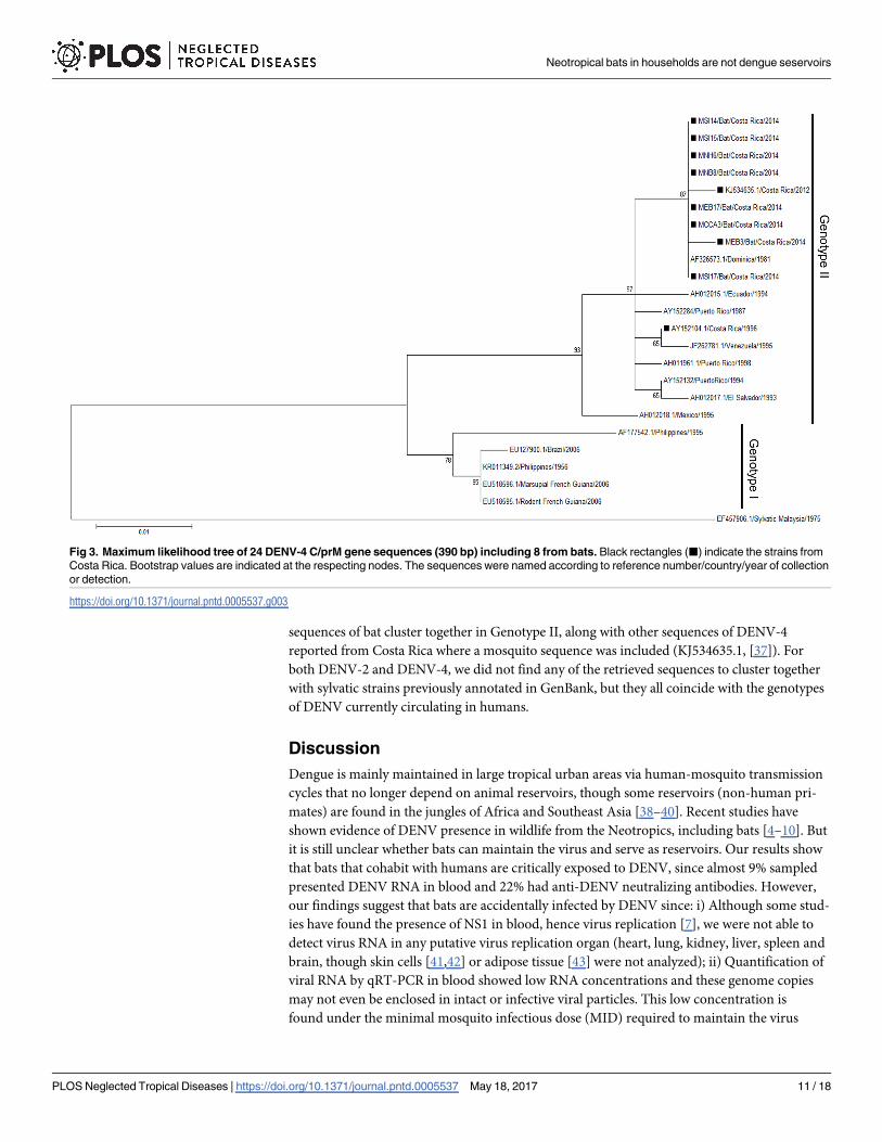
sequences of bat cluster together in Genotype II, along with other sequences of DENV-4
reported from Costa Rica where a mosquito sequence was included (KJ534635.1, [37]). For
both DENV-2 and DENV-4, we did not find any of the retrieved sequences to cluster together
with sylvatic strains previously annotated in GenBank, but they all coincide with the genotypes
of DENV currently circulating in humans.
Discussion
Dengue is mainly maintained in large tropical urban areas via human-mosquito transmission
cycles that no longer depend on animal reservoirs, though some reservoirs (non-human pri-
mates) are found in the jungles of Africa and Southeast Asia [38–40]. Recent studies have
shown evidence of DENV presence in wildlife from the Neotropics, including bats [4–10]. But
it is still unclear whether bats can maintain the virus and serve as reservoirs. Our results show
that bats that cohabit with humans are critically exposed to DENV, since almost 9% sampled
presented DENV RNA in blood and 22% had anti-DENV neutralizing antibodies. However,
our findings suggest that bats are accidentally infected by DENV since: i) Although some stud-
ies have found the presence of NS1 in blood, hence virus replication [7], we were not able to
detect virus RNA in any putative virus replication organ (heart, lung, kidney, liver, spleen and
brain, though skin cells [41,42] or adipose tissue [43] were not analyzed); ii) Quantification of
viral RNA by qRT-PCR in blood showed low RNA concentrations and these genome copies
may not even be enclosed in intact or infective viral particles. This low concentration is
found under the minimal mosquito infectious dose (MID) required to maintain the virus
Fig 3. Maximum likelihood tree of 24 DENV-4 C/prM gene sequences (390 bp) including 8 from bats. Black rectangles (■) indicate the strains from
Costa Rica. Bootstrap values are indicated at the respecting nodes. The sequences were named according to reference number/country/year of collection
or detection.
https://doi.org/10.1371/journal.pntd.0005537.g003
Neotropical bats in households are not dengue seservoirs
PLOS Neglected Tropical Diseases | https://doi.org/10.1371/journal.pntd.0005537 May 18, 2017 11 / 18

transmission cycle [44]. Moreover, the low concentration of virus RNA in blood precluded
amplification of the 2.5 kbp region encompassing the prM/E genes [33]. Thus we were able
only to obtain short length sequences (424–461 bp) from the C/prM region with a seminested
PCR for sequencing and phylogenetic analyses [5]. Finally, iii) even if virus isolation is not
always successful, we failed in attempts to isolate the virus from positive blood samples, sug-
gesting no intact or infectious virus present. Taken together, under the current conditions of
this study our results suggest that bats in these environments (sampled households) do not
show sufficient virus replication, excluding them as potential hosts or reservoirs with no role
in the transmission cycle, and making them feasible dead-end hosts for the virus. This con-
firms results obtained from previous independent studies where bats infected with DENV in
controlled laboratory conditions failed to show viral amplification [11,12].
DENV RNA detected from the positive individuals corresponded to the DENV four sero-
types. While DENV-1, -2 and -3 were causing dengue outbreaks in Costa Rica during 2013–
2015, DENV-4 has not been reported in human samples from Costa Rica since 2002 [26]. Fur-
thermore, in the human sera tested for this study and other serological studies performed in
our lab, no human sera has shown monotypic anti-DENV-4 neutralizing response. However,
DENV-4 was detected in an Ae. albopictus collected during 2015 from a pineapple field in Sar-
apiquı [37]. Besides competing with the other three serotypes, DENV-4 is the least frequently
isolated serotype, it has not been associated with severe dengue outbreaks, and causes most of
the clinically mild cases after dengue infection in humans [45,46]. Thus, DENV-4 may circu-
late unnoticed without its detection in health care facilities, and bats may function as sentinels
showing exposure to this serotype.
We predicted to find more DENV positive bats in high human dengue incidence locations,
yet we found positive bats in all sites sampled [26]. Also, we did not find any correlation
between the DENV seroprevalence in humans sampled in the timespan this research was done
and the presence of DENV positivity in bats. We also expected to find more positive individu-
als during the rainy season since the mosquito population augments and human cases increase
considerably [26,47], but surprisingly in the Central Valley (low human incidence location) we
found positive bats during both seasons. Furthermore, despite that we did not find a significant
difference in the quantity of mosquitoes between seasons; as expected we collected more mos-
quitoes overall during the rainy season. Though, interestingly in the Central Valley, more mos-
quitoes were collected during the dry season. The presence of vector mosquitoes sustaining the
viral transmission during the dry season has been associated with human habits such as saving
water for the drought in artificial containers without proper management, presence of other
type of containers such as flower vases, used tires, garbage and rubbish, therefore becoming all
potential breeding sites [48]. The hyper endemic circulation of dengue and the presence of the
mosquito vector in sampled sites may indicate enough potential sources for bats dengue expo-
sure. Furthermore, the high seroprevalence observed in bats suggests a high exposure and rate
of contact between bats and DENV. We detected more antibodies against DENV-3 (67.9%)
and most of the bat sera studied showed a relative maturation in their neutralization response.
Differences between age and gender have been reported in the immunological response
against other viruses [49]. We found no significant difference, suggesting that males and
females, adults and juveniles, are equally exposed to DENV. We did find higher bat seropreva-
lence in Sarapiquı compared to the other two sites. Sarapiquı has a greater rainfall amount
yearly with a less drastic dry season, in comparison to Nicoya where human incidence is
higher. This amount of rainfall nourishes populations of mosquitoes which may sustain viral
transmission throughout the entire year. In six individuals we found concomitantly DENV
RNA in blood and antibodies against DENV. Half of the individuals presented antibodies
against the same serotype detected in blood, suggesting a previous or parallel immunological
Neotropical bats in households are not dengue seservoirs
PLOS Neglected Tropical Diseases | https://doi.org/10.1371/journal.pntd.0005537 May 18, 2017 12 / 18

response. The other three individuals presented antibodies against a distinct serotype suggest-
ing a potential secondary infection. Studies indicate that even after a controlled infection with
a virus, bats do not always produce antibodies [50,51]. This observation displays how complex
the humoral immunological response is in bats, thus making serological results difficult to
interpret. Also, we have to take into account that so far, at least 19 different flaviviruses have
been associated with bats [10,13,49–61]. Therefore, although PRNT is the gold standard for
the serological diagnosis of flavivirus infections, results interpretation must be made with cau-
tion, and simultaneous assessment against all endemic flaviviruses must be performed for
comparison of end-point titers to assure specific anti-DENV neutralizing antibodies [62].
Recently, Cabrera-Romo et al. [63] explored the role of bats as part of putative DENV sylvatic
cycles in Mexico. They collected more than 200 bats of 18 different species from contrasting
ecological settings with concurrent human DENV activity. RNA extracted from liver or spleen
failed to show evidence for the presence of DENV nucleic acids, in agreement with our results.
Nevertheless, their PRNT analyses showed no evidence of neutralizing anti-DENV antibodies.
These contrasting results may be due to i) presence of anti-DENV neutralizing antibodies as a
result of cross-reaction against other bat specific or human flaviviruses [62] as explained
above; or ii) disparities in the bats species collected. The most frequent bat species collected by
us belongs to the molossids, bat species absent from the Mexican study. In another of our
recent publications [64], after primary embryonic cell culture of three different bat species, we
observed a limited serotype-, organ-, and bat species- specific dengue susceptibility. Only
someMolossus- but not Artibeus or Desmodus-derived primary cells sustained solely and
poorly initial DENV-1 replication, though it was latter absent. These results confirm our cur-
rent observations in molossids and reinforce the Cabrera-Romo et al. [63] findings; but note-
worthy denote the importance on which bat species and which dengue serotype should be
taken in account for further (if any) studies.
We hypothesized that bat infection will occur through an infected mosquito bite, but no
mosquito was found positive for bat cytochrome b. However, our findings surmise that infec-
tion may occur through the oral ingestion of an infected mosquito. Studies have shown that
bat infection is plausible after ingestion of mosquitoes infected with flaviviruses such as Yellow
Fever Virus (YFV) and WNV [20,50,65]. Additionally, albeit we could not detect the virus in
any putative replication organ, we detected two intestines fromM. sinaloae positive for the
same DENV serotype as found in their respective blood. Although this result may be caused by
the presence of a positive dengue mosquito in the intestine lumen, it is tempting to speculate
that some limited local viral replication in the bat intestine endothelial cells may be occurring.
Supporting this conjecture, failure in recent studies attempting DENV infection of bats
through a mosquito bite or virus inoculation may support an oral infection route [11,12].
Accordingly, the collected mosquitoes feeding preference did not indicate presence of bat
blood, even though the EVC-CO2 traps were placed in close proximity to the bats roosting
area. Moreover, the majority of the bats sampled by us are insectivorous, and even though the
preferential food source for the molossids and other bigger bats may not be mosquitoes, it is
possible that they will feed on them due to increased abundance [22,66]. Likewise, a nectari-
vorous bat as G. soricina could accidentally feed on dengue positive male mosquitoes while
functioning as pollinators from flowers. Although the Aedes mosquitoes are diurnal, other
nocturnal mosquitoes as Culex. which show limited virus replication in the gut [67] may be
also a feeding and infection source. As well, bats could have been exposed to the virus in a dif-
ferent environment far from the roosting household.
Nevertheless, even though bats seem to get infected with DENV, they do not amplify the
virus to a considerable extent to be able to transmit it to a mosquito. It seems that the expo-
sure of bats to DENV is accidental, becoming an example of spill over from humans to bats
Neotropical bats in households are not dengue seservoirs
PLOS Neglected Tropical Diseases | https://doi.org/10.1371/journal.pntd.0005537 May 18, 2017 13 / 18

as reported by de Thoisy et al. [5] with samples of wildlife taken in close proximity to human
settlements where dengue outbreaks ensue. This is supported not only by our results in the
phylogenetic analysis, where the dengue strains sequenced from bats and mosquitoes cluster
together in close relation with the reported strains of dengue in this and neighboring coun-
tries (Figs 2 and 3), but also by not showing any histopathological findings suggestive of
infection in all analyzed tissues. Even if we cannot determine the route of contact occurring
between the virus and the bat, our results suggest that bats are an epidemic dead end for this
virus
Several viruses have been detected in bat tissues or excreta; however, this does not prove
causation of disease [16]. Some of these viruses or viral sequences might have been acquired
from food eaten by bats and could be irrelevant with respect to viral disease epidemiology.
Many knowledge gaps connecting bats and zoonotic viruses exist, thus linking bats with
these events without strong evidence is a disservice with negative consequences [13]. For
example, investing efforts in controlling the wrong reservoir can postpone suitable mitiga-
tion actions that could prevent deaths or interrupt disease spread; and a potential ‘pest con-
trol’ of bat populations may deny us their important ecosystemic services [13]. Therefore as
stated by Moratelli and Calisher, after understanding the role of bats (or wildlife) in the
maintenance and circulation of pathogens and the mechanisms underlying the emergence
of zoonotic diseases, wildlife biologists and epidemiologists should work together develop-
ing appropriate management plans to control virus circulation [13]. Just then, risks of
human infection without causing significant biases against specific animal populations will
be minimized.
Supporting information
S1 Fig. Sampling sites in Costa Rica and their dengue incidence during study years, 2013
and 2014. The black squares, diamonds and triangles (■,♦,▲) represent a sampled house-
hold. The incidence values were retrieved from epidemiological surveillance done by the Min-
istry of Health [26]. Map was created using QGIS 2.14.3 (http://www.qgis.org/en/site/) and
DIVA GIS maps (http://www.diva-gis.org). Baselayer data was obtained from http://www.
diva-gis.org/gdata.
(TIF)
S1 Table. Primers used in the study.
(DOCX)
S2 Table. Prevalence of DENV RNA obtained by PCR from each serotype (D1-4) in all cap-
tured bat species in the 3 sites of study in Costa Rica (Nicoya, Sarapiquı and Central Val-
ley) during the dry and rainy season, 2013–2014.
(DOCX)
S3 Table. Seroprevalence against each dengue serotype obtained from serum diluted 1:20
by PRNT90 from bats captured in the 3 sites of study in Costa Rica (Nicoya, Sarapiquı and
Central Valley) during 2013–2014.
(DOCX)
S4 Table. Information of DENV-2 sequences used in phylogenetic analysis.
(DOCX)
S5 Table. Information of DENV-4 sequences used in phylogenetic analysis.
(DOCX)
Neotropical bats in households are not dengue seservoirs
PLOS Neglected Tropical Diseases | https://doi.org/10.1371/journal.pntd.0005537 May 18, 2017 14 / 18

Acknowledgments
We are grateful to E. Rojas, T. Boring, G. Nuñez, A. Gamboa, C. Lavin, L. Lopez, A. Montero,
and D. Villalobos for their appreciated assistance in the field and in the lab. We also thank
Francisco Vega, Geovanni Vargas and Carlos Vargas for valuable technical assistance, and
Alexandra Rucavado (Instituto Clodomiro Picado, Facultad de Microbiologıa, Universidad de
Costa Rica) for providing help regarding some methodologies. We acknowledge B. Willink
and D. Lorıa for discussing ideas and providing help in the analysis. Most importantly, we will
like to thank all the people who allowed us to sample their homes in Sarapiquı, Nicoya, and the
Central Valley.
Author Contributions
Conceptualization: AVS AMS AC BRH ECA.
Data curation: AVS.
Formal analysis: AVS AMS CSG.
Funding acquisition: AVS AMS AC ECA.
Investigation: AVS AMS CSG AC LGC JAM AAA ECA.
Methodology: AVS AMS CSG LGC AC JFD JAM AAA BRH ECA.
Project administration: AVS AC ECA.
Resources: AVS LGC JFD ECA.
Supervision: JFD AC BRH ECA.
Validation: AVS AMS CSG JFD ECA.
Visualization: AVS AMS CSG.
Writing – original draft: AVS AMS CSG AC JFD ECA.
Writing – review & editing: AVS ECA.
References1. Murray NEA, Quam MB, Wilder-Smith A. Epidemiology of dengue: past, present and future prospects.
Clin Epidemiol. 2013; 5: 299–309. https://doi.org/10.2147/CLEP.S34440 PMID: 23990732
2. OMS, TDR. Dengue: guias para el diagnostico, tratamiento, prevencion y control. 2009. 1. OPS, OMS,
editors. Bolivia: OMS; 2009.
3. Weaver SC, Vasilakis N. Molecular evolution of dengue viruses: contributions of phylogenetics to
understanding the history and epidemiology of the preeminent arboviral disease. Infect Genet Evol.
2009; 9: 523–40. https://doi.org/10.1016/j.meegid.2009.02.003 PMID: 19460319
4. Platt KB, Mangiafico JA, Rocha OJ, Zaldivar ME, Mora J, Trueba G, et al. Detection of dengue virus
neutralizing antibodies in bats from Costa Rica and Ecuador. J Med Entomol. 2000; 37: 965–7. Avail-
able: http://www.ncbi.nlm.nih.gov/pubmed/11126559 PMID: 11126559
5. de Thoisy B, Lacoste V, Germain A, Muñoz-Jordan J, Colon C, Mauffrey J-F, et al. Dengue infection in
neotropical forest mammals. Vector Borne Zoonotic Dis. 2009; 9: 157–70. https://doi.org/10.1089/vbz.
2007.0280 PMID: 18945183
6. de Thoisy B, Dussart P, Kazanji M. Wild terrestrial rainforest mammals as potential reservoirs for flavivi-
ruses (yellow fever, dengue 2 and St Louis encephalitis viruses) in French Guiana. Trans R Soc Trop
Med Hyg. 2004; 98: 409–12. https://doi.org/10.1016/j.trstmh.2003.12.003 PMID: 15138077
7. Aguilar-Setien A, Romero-Almaraz ML, Sanchez-Hernandez C, Figueroa R, Juarez-Palma LP, Garcıa-
Flores MM, et al. Dengue virus in Mexican bats. Epidemiol Infect. 2008; 136: 1678–83. https://doi.org/
10.1017/S0950268808000460 PMID: 18325131
Neotropical bats in households are not dengue seservoirs
PLOS Neglected Tropical Diseases | https://doi.org/10.1371/journal.pntd.0005537 May 18, 2017 15 / 18

8. Lavergne A, Lacoste V, Germain A, Matheus S, Dussart P, Deparis X, et al. Infection par le virus de la
dengue de mammifères sauvages en region neotropicale: hotes accidentels ou reservoirs potentiels?
Medecine Trop. 2009; 69: 345–350.
9. Sotomayor-Bonilla J, Chaves A, Rico-Chavez O, Rostal MK, Ojeda-Flores R, Salas-Rojas M, et al. Den-
gue virus in bats from southeastern Mexico. Am J Trop Med Hyg. 2014; 91: 129–31. https://doi.org/10.
4269/ajtmh.13-0524 PMID: 24752688
10. Machain-Williams C, Lopez-Uribe M, Talavera-Aguilar L, Carrillo-Navarrete J, Vera-Escalante L,
Puerto-Manzano F, et al. Serologic evidence of flavivirus infection in bats in the Yucatan Peninsula of
Mexico. J Wildl Dis. 2013; 49: 684–9. https://doi.org/10.7589/2012-12-318 PMID: 23778622
11. Perea-Martınez L, Moreno-Sandoval HN, Moreno-Altamirano MM, Salas-Rojas M, Garcıa-Flores MM,
Arechiga-Ceballos N, et al. Experimental infection of Artibeus intermedius bats with serotype-2 dengue
virus. Comp Immunol Microbiol Infect Dis. Elsevier Ltd; 2013; 36: 193–8. https://doi.org/10.1016/j.cimid.
2012.12.002 PMID: 23312108
12. Cabrera-Romo S, Recio-Totoro B, Alcala AC, Lanz H, Del Angel RM, Sanchez-Cordero V, et al. Experi-
mental inoculation of Artibeus jamaicensis bats with dengue virus serotypes 1 or 4 showed no evidence
of sustained replication. Am J Trop Med Hyg. 2014; 91: 1227–34. https://doi.org/10.4269/ajtmh.14-
0361 PMID: 25311698
13. Moratelli R, Calisher CH. Bats and zoonotic viruses: Can we confidently link bats with emerging deadly
viruses? Mem Inst Oswaldo Cruz. 2015; 110: 1–22. https://doi.org/10.1590/0074-02760150048 PMID:
25742261
14. Dobson AP. Virology. What links bats to emerging infectious diseases? Science. 2005; 310: 628–9.
https://doi.org/10.1126/science.1120872 PMID: 16254175
15. Calisher CH, Childs JE, Field HE, Holmes K V, Schountz T. Bats: important reservoir hosts of emerging
viruses. Clin Microbiol Rev. 2006; 19: 531–45. https://doi.org/10.1128/CMR.00017-06 PMID: 16847084
16. Calisher CH, Holmes K V, Dominguez SR, Schountz T, Cryan P. Bats Prove To Be Rich Reservoirs.
Microbe. 2008; 3: 521–528.
17. Bennett M. Bats and human emerging diseases. Epidemiol Infect. 2006; 134: 905–7. https://doi.org/10.
1017/S0950268806006674 PMID: 16740197
18. Wong S, Lau S, Woo P, Yuen K-Y. Bats as a continuing source of emerging infections in humans. Rev
Med Virol. 2007; 17: 67–91. https://doi.org/10.1002/rmv.520 PMID: 17042030
19. Wibbelt G, Moore MS, Schountz T, Voigt CC. Emerging diseases in Chiroptera: why bats? Biol Lett.
2010; 6: 438–40. https://doi.org/10.1098/rsbl.2010.0267 PMID: 20427329
20. Melaun C, Werblow A, Busch MW, Liston A, Klimpel S. Bats (Chiroptera) as Vectors of Diseases and
Parasites [Internet]. Klimpel S, Mehlhorn H, editors. Berlin, Heidelberg: Springer Berlin Heidelberg;
2014. https://doi.org/10.1007/978-3-642-39333-4
21. Melo FPL, Rodrıguez-Herrera B, Chazdon RL, Medellın RA, Ceballos GG. Small tent-roosting bats pro-
mote dispersal of large-seeded plants in a neotropical forest. Biotropica. 2009; 41: 737–743.
22. Boyles JG, Cryan PM, McCracken GF, Kunz TH. Conservation: economic importance of bats in agricul-
ture. Science (80-). 2011; 332: 41–42. https://doi.org/10.1126/science.1201366 PMID: 21454775
23. LaVal R, Rodrıguez-Herrera B. Mucielagos de Costa Rica. 1st ed. Heredia, Costa Rica: Instituto
Nacional de Biodiversidad (InBio); 2002.
24. Drews C. Convivencia con murcielagos de Costa Rica. Ambientico. 2002; 12–13.
25. Borchert R, Meyer SA, Felger RS, Porter-Bolland L. Environmental control of flowering periodicity in
Costa Rican and Mexican tropical dry forests. Glob Ecol Biogeogr. 2004; 13: 409–425.
26. Ministerio de Salud de Costa Rica. Analisis de la situacion de salud: Dengue 2015 [Internet]. 2015
[cited 1 Jan 2016]. http://www.ministeriodesalud.go.cr/index.php/vigilancia-de-la-salud/analisis-de-
situacion-de-salud.
27. Timm RM, LaVal RK, Rodrıguez-Herrera B. Clave de Murcielagos de Costa Rica. Brenesia. 2000; 1–
32.
28. Chaverri LG. Culicidae (Mosquitos, Zancudos). In: Brown BV, Borkent A, Cumming JM, Wood DM,
Woodley NE, Zumbado MA, editors. Manual of Central American Diptera, Vol 1. Ontario, Canada:
NCR Research Press; 2009. pp. 369–388.
29. Lanciotti R, Calisher C, Gubler D, Chang G, Vorndam A. Rapid detection and typing of dengue viruses
from clinical samples by using reverse transcriptase-polymerase chain reaction. J Clin Microbiol. 1992;
30: 545–51. Available: http://www.pubmedcentral.nih.gov/articlerender.fcgi?artid=265106&tool=
pmcentrez&rendertype=abstract PMID: 1372617
30. Drosten C, Gottig S, Schilling S, Panning M, Schmitz H, Asper M, et al. Rapid Detection and Quantifica-
tion of RNA of Ebola and Marburg Viruses, Lassa Virus, Crimean-Congo Hemorrhagic Fever Virus, Rift
Neotropical bats in households are not dengue seservoirs
PLOS Neglected Tropical Diseases | https://doi.org/10.1371/journal.pntd.0005537 May 18, 2017 16 / 18

Valley Fever Virus, Dengue Virus, and Yellow Fever Virus by Real-Time Reverse Rapid Detection and
Quantification of RNA. J Clin Microbiol. 2002; 40: 2323–2330. https://doi.org/10.1128/JCM.40.7.2323-
2330.2002 PMID: 12089242
31. Aughey E, Frye F. Comparative veterinary histology with clinical correlates. Illustrate. CRC Press, edi-
tor. FL, USA; 2001.
32. Townzen JS, Brower AV, Judd DD. Identification of mosquito bloodmeals using mitochondrial cyto-
chrome oxidase subunit I and cytochrome b gene sequences. Med Vet Entomol. 2008; 22: 386–393.
https://doi.org/10.1111/j.1365-2915.2008.00760.x PMID: 19120966
33. Dıaz FJ, Black WC, Farfan-Ale JA, Loroño-Pino MA, Olson KE, Beaty BJ. Dengue virus circulation and
evolution in Mexico: a phylogenetic perspective. Arch Med Res. 2006; 37: 760–73. https://doi.org/10.
1016/j.arcmed.2006.02.004 PMID: 16824937
34. Soto-Garita C, Tsomogyi T, Vicente-Santos A, Corrales-Aguilar E. Molecular Characterization of Two
Major Dengue Outbreaks in Costa Rica. Am J Trop Med Hyg. 2016; 1: 201–205. https://doi.org/10.
4269/ajtmh.15-0835 PMID: 27139442
35. R Core Team. R: A language and environment for statistical computing [Internet]. Vienna, Austria: R
Foundation for Statistical Computing; 2013. http://www.r-project.org/
36. Calderon-Arguedas O, Troyo A, Avendaño A, Gutierrez M. Aedes albopictus (Skuse) en la Region Hue-
tar Atlantica de Costa Rica. Rev Costarric Salud Publica. 2012; 21: 76–80.
37. Calderon-Arguedas O, Troyo A, Moreira-Soto RD, Marın R, Taylor L. Dengue viruses in Aedes albopic-
tus Skuse from a pineapple plantation in Costa Rica. J Vector Ecol. 2015; 40: 184–186. https://doi.org/
10.1111/jvec.12149 PMID: 26047200
38. Gubler DJ. Human arbovirus infections worldwide. Ann N Y Acad Sci. 2001; 951: 13–24. Available:
http://www.ncbi.nlm.nih.gov/pubmed/11797771 PMID: 11797771
39. Gubler DJ. The global emergence/resurgence of arboviral diseases as public health problems. Arch
Med Res. 2002; 33: 330–42. Available: http://www.ncbi.nlm.nih.gov/pubmed/12234522 PMID:
12234522
40. Mackenzie JS, Gubler DJ, Petersen LR. Emerging flaviviruses: the spread and resurgence of Japanese
encephalitis, West Nile and dengue viruses. Nat Med. 2004; 10: S98–109. https://doi.org/10.1038/
nm1144 PMID: 15577938
41. Wu S-JL, Grouard-Vogel G, Sun W, Mascola JR, Brachtel E, Putvatana R, et al. Human skin Langer-
hans cells are targets of dengue virus infection. Nat Med. 2000; 6: 816–820. https://doi.org/10.1038/
77553 PMID: 10888933
42. Schmid MA, Harris E. Monocyte Recruitment to the Dermis and Differentiation to Dendritic Cells
Increases the Targets for Dengue Virus Replication. PLoS Pathog. 2014; 10: e1004541. https://doi.org/
10.1371/journal.ppat.1004541 PMID: 25474197
43. Davis AD, Morgan SMD, Dupuis M, Poulliott CE, Jarvis JA, Franchini R, et al. Overwintering of Rabies
Virus in Silver Haired Bats (Lasionycteris noctivagans). PLoS One. 2016; 11: e0155542. https://doi.org/
10.1371/journal.pone.0155542 PMID: 27195489
44. Pongsiri A, Ponlawat A, Thaisomboonsuk B, Jarman RG, Scott TW, Lambrechts L. Differential suscepti-
bility of two field Aedes aegypti populations to a low infectious dose of dengue virus. PLoS One. 2014;
9: 3–8. https://doi.org/10.1371/journal.pone.0092971 PMID: 24664142
45. Klungthong C, Putnak R, Mammen MP, Li T, Zhang C. Molecular genotyping of dengue viruses by phy-
logenetic analysis of the sequences of individual genes. J Virol Methods. 2008; 154: 175–181. https://
doi.org/10.1016/j.jviromet.2008.07.021 PMID: 18778736
46. Kyle JL, Harris E. Global spread and persistence of dengue. Annu Rev Microbiol. 2008; 62: 71–92.
https://doi.org/10.1146/annurev.micro.62.081307.163005 PMID: 18429680
47. Feng Z, Velasco-Hernandez JX. Competitive exclusion in a vector-host model for the dengue fever. J
Math Biol. 1997; 35: 523–544. PMID: 9145954
48. Trewin BJ, Kay BH, Darbro JM, Hurst TP. Increased container-breeding mosquito risk owing to
drought-induced changes in water harvesting and storage in Brisbane, Australia. Int Health. 2013; 5:
251–258. https://doi.org/10.1093/inthealth/iht023 PMID: 24225151
49. Thompson NN, Auguste AJ, Travassos da Rosa APA, Carrington CVF, Blitvich BJ, Chadee DD, et al.
Seroepidemiology of selected alphaviruses and flaviviruses in bats in Trinidad. Zoonoses Public Health.
2015; 62: 53–60. https://doi.org/10.1111/zph.12118 PMID: 24751420
50. Davis A, Bunning M, Gordy P, Panella N, Blitvich B, Bowen R. Experimental and natural infection of
north american bats with West Nile Virus. Am J Trop Med Hyg. 2005; 73: 467–469. PMID: 16103624
51. Van Den Hurk AF, Smith CS, Field HE, Smith IL, Northill JA, Taylor CT, et al. Transmission of Japanese
encephalitis virus from the black flying fox, Pteropus alecto, to Culex annulirostris mosquitoes, despite
Neotropical bats in households are not dengue seservoirs
PLOS Neglected Tropical Diseases | https://doi.org/10.1371/journal.pntd.0005537 May 18, 2017 17 / 18

the absence of detectable viremia. Am J Trop Med Hyg. 2009; 81: 457–462. 81/3/457 [pii] PMID:
19706915
52. Charlier N, Leyssen P, Pleij CWA, Lemey P, Billoir F, Van Laethem K, et al. Complete genome
sequence of Montana Myotis leukoencephalitis virus, phylogenetic analysis and comparative study of
the 3 h untranslated region of flaviviruses with no known vector. J Gen Virol. 2002; 1875–1885. https://
doi.org/10.1099/0022-1317-83-8-1875 PMID: 12124451
53. de Lamballerie X, Crochu S, Billoir F, Neyts J, de Micco P, Holmes EC, et al. Genome sequence analy-
sis of Tamana bat virus and its relationship with the genus Flavivirus. J Gen Virol. 2002; 83: 2443–2454.
https://doi.org/10.1099/0022-1317-83-10-2443 PMID: 12237426
54. Tajima S, Takasaki T, Matsuno S, Nakayama M, Kurane I. Genetic characterization of Yokose virus, a
flavivirus isolated from the bat in Japan. Virology. 2005; 332: 38–44. https://doi.org/10.1016/j.virol.2004.
06.052 PMID: 15661139
55. Kuno G, Chang GJ. Characterization of Sepik and Entebbe Bat viruses closely related to Yellow Fever
Virus. Am J Trop Med Hyg. 2006; 75: 1165–1170. PMID: 17172387
56. Cui J, Counor D, Shen D, Sun G, He H, Deubel V, et al. Detection of Japanese Encephalitis Virus anti-
bodies in bats in Southern China. Am J Trop Med Hyg. 2008; 78: 1007–1011. PMID: 18541785
57. Wang J-L, Pan X-L, Zhang H-L, Fu S-H, Wang H-Y, Tang Q, et al. Japanese encephalitis viruses from
bats in Yunnan, China. Emerg Infect Dis. 2009; 15: 939–42. https://doi.org/10.3201/eid1506.081525
PMID: 19523297
58. Epstein JH, Quan P-L, Briese T, Street C, Jabado O, Conlan S, et al. Identification of GBV-D, a novel
GB-like flavivirus from Old World frugivorous bats (Pteropus giganteus) in Bangladesh. Parrish C, edi-
tor. PLoS Pathog. 2010; 6: e1000972. https://doi.org/10.1371/journal.ppat.1000972 PMID: 20617167
59. Volkova E, Tesh RB, Monath TP, Vasilakis N. Full genomic sequence of the prototype strain (M64) of
Rio Bravo virus. J Virol. 2012; 86: 4715. https://doi.org/10.1128/JVI.00331-12 PMID: 22457530
60. Quan P, Firth C, Conte JM, Williams SH, Zambrana-Torrelio CM, Anthony SJ, et al. Bats are a major
natural reservoir for hepaciviruses and pegiviruses. Proc Natl Acad Sci J. 2013; 10: 3–8199. doi: 10.
1073/pnas.1303037110/-/DCSupplemental.www.pnas.org/cgi/doi/10.1073/pnas.1303037110
61. Cadar D, Becker N, de Mendonca Campos R, Borstler J, Jost H, Schmidt-Chanasit J. Usutu Virus in
Bats, Germany, 2013. Emerg Infect Dis. 2014; 20: 1771–1773. https://doi.org/10.3201/eid2010.140909
PMID: 25271769
62. Hobson-Peters J. Approaches for the development of rapid serological assays for surveillance and
diagnosis of infections caused by zoonotic flaviviruses of the japanese encephalitis virus serocomplex.
J Biomed Biotechnol. 2012; 2012. https://doi.org/10.1155/2012/379738 PMID: 22570528
63. Cabrera-Romo S, Max Ramirez C, Recio-Totoro B, Tolentino-Chi J, Lanz H, del Angel RM, et al. No Evi-
dence of Dengue Virus Infections in Several Species of Bats Captured in Central and Southern Mexico.
Zoonoses Public Health. 2016; 63: 579–583. https://doi.org/10.1111/zph.12276 PMID: 27357156
64. Moreira-Soto A, Soto-Garita C, Corrales-Aguilar E. Neotropical primary bat cell lines show restricted
dengue virus replication. Comp Immunol Microbiol Infect Dis. Elsevier Ltd; 2017; 50: 101–105. https://
doi.org/10.1016/j.cimid.2016.12.004 PMID: 28131369
65. Scott TW. Are Bats really involved in Dengue virus transmission? / Bats might be involved in the natural
history of Dengue virus. J Med Entomol. 2001; 38: 771–773. PMID: 11761372
66. Gonsalves L, Bicknell B, Law B, Webb C, Monamy V. Mosquito Consumption by Insectivorous Bats:
Does Size Matter? PLoS One. 2013; 8: 1–11. https://doi.org/10.1371/journal.pone.0077183 PMID:
24130851
67. Vazeille-Falcoz M, Rosen L, Mousson L, Rodhain F. Replication of dengue type 2 virus in Culex quin-
quefasciatus (Diptera: Culicidae). Am J Trop Med Hyg. 1999; 60: 319–21. Available: http://www.ncbi.
nlm.nih.gov/pubmed/10072159 PMID: 10072159
Neotropical bats in households are not dengue seservoirs
PLOS Neglected Tropical Diseases | https://doi.org/10.1371/journal.pntd.0005537 May 18, 2017 18 / 18