neomycin-induced hair cell death and rapid regeneration in the lateral line of zebrafish
TRANSCRIPT

Neomycin-Induced Hair Cell Death and RapidRegeneration in the Lateral Line of Zebrafish(Danio rerio)
JULIE A. HARRIS,1,2 ALAN G. CHENG,1 LISA L. CUNNINGHAM,1 GLEN MACDONALD,1,4
DAVID W. RAIBLE,2,3AND EDWIN W RUBEL
1,2
1Virginia Merrill Bloedel Hearing Research Center and Department of Otolaryngology–Head and Neck Surgery,University of Washington, Seattle, WA 98195, USA2Graduate Program in Neurobiology and Behavior, University of Washington, Seattle, WA 98195, USA3Department of Biological Structure, University of Washington, Seattle, WA 98195, USA4Core for Communication Research, University of Washington, Box 357923, Seattle, WA 98195, USA
Received: 16 May 2002; Accepted: 5 October 2002; Online publication: 6 May 2003
ABSTRACT
Mechanoreceptive hair cells are extremely sensitive toaminoglycoside antibiotics, including neomycin. Haircell survival was assessed in larval wild-type zebrafishlateral line neuromasts 4 h after initial exposure to arange of neomycin concentrations for 1 h. Each ofthe lateral line neuromasts was scored in live fish forthe presence or absence of hair cells using the fluo-rescent vital dye DASPEI to selectively label hair cells.All neuromasts were devoid of DASPEI-labeled haircells 4 h after 500 lM neomycin exposure. VitalDASPEI staining was proportional to the number ofhair cells per neuromast identified in fixed larvaeusing immunocytochemistry for acetylated tubulinand phalloidin labeling. The time course of hair cellregeneration in the lateral line neuromasts was alsoanalyzed following neomycin-induced damage. Re-generated hair cells were first observed using liveDASPEI staining 12 and 24 h following neomycintreatment. The potential role of proliferation in re-generating hair cells was analyzed. A 1 h pulse-fixprotocol using bromodeoxyuridine (BrdU) incorpo-ration was used to identify S-phase cells in neuro-masts. BrdU incorporation in neomycin-damaged
neuromasts did not differ from control neuromasts 4h after drug exposure but was dramatically upregu-lated after 12 h. The proliferative cells identifiedduring a 1 h period at 12 h after neomycin treatmentwere able to give rise to new hair cells by 24–48 h afterdrug treatment. The results presented here provide astandardized preparation for studying and identifyinggenes that influence vertebrate hair cell death, sur-vival, and regeneration following ototoxic insults.
Keywords: hair cell death, lateral line, aminogly-cosides, hair cell regeneration, zebrafish, geneticscreening
INTRODUCTION
The degeneration of mechanosensory hair cells ofthe inner ear sensory epithelia is the primary cause ofboth deafness and balance disorders. Hair cells arecommonly lost through aging, as well as noise- anddrug-induced trauma. In particular, hair cells are verysusceptible to aminoglycosides. While the exactmechanisms of aminoglycoside ototoxicity remainunknown, it appears to involve both known apoptoticcell death pathways and the formation of free radicals(for review see Forge and Schacht 2000). For exam-ple, the apoptotic cell death proteases, caspases, areactivated in hair cells following aminoglycoside
Correspondence to: Edwin W Rubel Æ University of Washington Æ VMBHearing Research Center Æ Box 357923 Æ Seattle, WA 98195-7293.Telephone: (206) 543-8360; fax: (206) 221-5685; email: [email protected]
219
JARO 04: 219–234 (2003)DOI: 10.1007/s10162-002-3022-x JARO
Journal of the Association for Research in Otolaryngology

exposure and inhibiting caspase activation reduceshair cell death (Li et al. 1995; Forge and Li2000; Williams and Holder 2000; Cheng et al.2001; Cunningham et al. 2002; Matsui et al. 2002).In addition, the c-Jun N-terminal kinase (JNK) sign-aling cascade, implicated in apoptotic death follow-ing a variety of insults including oxidative stress, isactivated in auditory hair cells following aminoglyco-side treatment (Pirvola et al. 2000; Ylikoski et al.2002).
One approach to conserve sensory function fol-lowing ototoxicity is to identify molecules necessaryfor inducing hair cell regeneration. Hair cells are notreplaced in mammalian auditory organs followingototoxic insults (Schuknecht 1974; Roberson andRubel 1994; Chardin and Romand 1995). Nonmam-malian vertebrate hair cells, however, can regeneratefollowing noise- or drug-induced hair cell death, andregeneration restores sensory function (Cotanche1987; Cruz et al. 1987; Weisleder and Rubel 1993;Cotanche et al. 1994; Carey et al. 1996; Smolders1999; Stone and Rubel 2000a). A second approachtoward reducing hearing and balance disorders is toidentify molecules that can protect, or prevent, haircells from undergoing death following ototoxicstimuli. In vitro preparations are useful for applyingcandidate protective molecules, such as caspase in-hibitors, directly to vertebrate hair cells, but for themost part they lack the capacity to identify novel en-dogenous genes involved in cell death and survival. Itis well known that there is a genetic contribution toaminoglycoside-induced deafness, and, therefore, it isof importance to identify the candidate genes thatincrease (or decrease) susceptibility to ototoxicity(Fischel–Ghodsian 1999).
The zebrafish (Danio rerio) is emerging as a pow-erful preparation for identifying essential vertebrategenes and their functions. Following random muta-genesis, phenotypes can be rapidly analyzed andmutations assigned to specific chromosomal locationsusing molecular mapping techniques. In addition tothe inner ear, fish have a second mechanosensoryorgan, the lateral line. The lateral line consists ofneuromasts that reside along the head and body.Each neuromast contains a group of hair cells thatfunction to detect water current relative to the ani-mal’s body via movement of their stereocilia (Kalmijn1989; Montgomery et al. 2000). Lateral line hair cellsof zebrafish larvae are easily accessed making themideal for experimental manipulation.
The present study was designed to provide a stand-ardized model for studying lateral line hair cell deathand regeneration. First, a dose–response relationshipbetween lateral line hair cell survival and neomycinconcentration was established in zebrafish larvae.Second, the time frame of lateral line hair cell re-
generation in larval zebrafish following aminoglyco-side-induced damage was assessed. The long-termgoal of this research is to understand the interactionof genetic factors and environmental insultssuch as ami-noglycoside antibiotics on hair cell lossand renewal.
MATERIALS AND METHODS
Animals
Zebrafish embryos (Danio rerio) were produced bypaired matings of fish raised at 28.5�C (Westerfield2000). Animals were maintained at 28.5�C until fouror five days postfertilization (p4 and p5) when theexperiments were conducted. All animal procedureswere approved by the University of Washington In-stitutional Animal Care Committee.
Dose–response relationship
Aminoglycoside treatment. Figure 1A shows the experi-mental paradigm for this study. Neomycin sulfate(Pharma-Tek, Huntington, NY) was added to 6 ml ofembryo medium [1 mM MgSO4, 120 lM KH2PO4, 74lM Na2HPO4 1 mM CaCl2, 500 lM KCl, 15 mM NaCl,and 500 lM NaHCO3 in dH2O (Westerfield 2000)] ineach well of a six-well tissue culture plate to make finalconcentrations of 0, 10, 50, 100, 125, 250, 300, or 500lM. Zebrafish larvae (n ‡ 6/well/trial) were placedinto a 50 ml tube which had one end cut off and amesh cover at the bottom to be used as a transferdevice. Larvae were moved from fresh medium, im-mersed in neomycin-containing medium, and incu-bated (28.5�C) for 1 h. Larvae of different ages (p4 orp5) were treated separately. After 1 h, animals wererinsed three times quickly in fresh medium and re-turned to the incubator in normal embryo mediumfor three additional hours at 28.5�C. Each treatmentgroup was replicated 2–5 times (n = 18–45 fish/treatment group, n = 232 zebrafish total).
Neuromast staining. The fluorescent dye 2-[4-(di-methylamino)styryl]-N-ethylpyridinium iodide (DAS-PEI; Molecular Probes, Eugene, OR) was used as avital dye to stain hair cells within neuromasts. Larvaewere incubated in embryo medium containing0.005% DASPEI for 15 min. The larvae were anes-thetized in MS222 (10 lg/ml, 3-aminobenzoic acidethyl ester, methanesulfonate salt; Sigma, St. Louis,MO) for 5 min. The zebrafish were then rinsed oncein fresh embryo medium and analyzed under anepifluorescence dissecting microscope (Leica MZ-12FL111) equipped with a DASPEI filter set (excitation450–490 nM and barrier 515 nM; Chroma Technol-ogies, Brattleboro, VT).
220 HARRIS ET AL.: Lateral Line Hair Cell Death

Neuromast scoring and data analysis. At four and fivedays postfertilization, larvae are free swimming andhave 18 head and 9 trunk neuromasts on each side ofthe body in the several lines that make up the lateralline system. The location of each of these 27 neuro-masts is shown in Figure 1B (Metcalfe et al. 1985; Ra-ible and Kruse 2000). This schematic was used as thedata sheet for scoring neuromast hair cell survival.Individual neuromasts were scored as having normalstaining indicating hair cells are present (DASPEIscore = 2), reduced staining (DASPEI score = 1), orabsent staining (DASPEI score = 0). Total neuromastscores were tabulated for one side of each fish. Themaximum score possible was 54, or 27 neuromastsmultiplied by the high score of 2 which indicatesnormal staining of each neuromast. The average scorefrom 37 untreated animals was 52.3. The scores fromthe treated groups of fish were expressed as a pro-portion of this average neuromast score from un-treated fish. Data were subjected to a one-way or two-way analysis of variance (Statview 5.0 for Macintosh,Abacus Concepts, Berkeley, CA) to assess the effects ofneomycin concentration, age, or both on neuromastsurvival. The scores were also analyzed separately foreach neuromast to determine if individual neuromastsshowed differential sensitivities to neomycin.
Hair cell regeneration
Neuromast scoring and data analysis. The time frame ofhair cell regeneration in p4 and p5 zebrafish was in-vestigated using the aminoglycoside treatment
schedule described above (Fig. 1A) but with addi-tional time points of assessment at 12 and 24 h afterinitial neomycin exposure. These experiments in-cluded groups treated with 0, 125, 250, and 500 lMneomycin. At 4 h after initial drug treatment, 2–3 fishper group were scored using DASPEI as a positivecontrol for the damage protocol. Remaining fish werescored at either 12 or 24 h. These groups were rep-licated 2 times (12 h; n = 15–24 fish/treatmentgroup) or 3 times (24 h; n = 11–29 fish/treatmentgroup). Total neuromast scores based on DASPEIstaining were subjected to analysis of variance foroverall and interaction effects of time and neomycinconcentration on hair cell regeneration.
Cell proliferation and analysis. A pulse-fix protocolusing 5-bromo-2-deoxyuridine (BrdU; Sigma) wasused to identify S-phase cells in lateral line neuro-masts. Larvae were either untreated (0 lM neomycin,n = 19) or treated (500 lM neomycin n = 17) for 1 hand then allowed to recover for 4, 12, or 24 h afterinitial exposure as described above. At these times,larvae were placed into 10 mM BrdU with 1% di-methylsulfoxide (DMSO) in embryo medium for 1 hat 28.5�C. They were then anesthetized in MS222 andfixed with 4% paraformaldehye (PFA) in phosphate-buffered saline (PBS) for 2 h at room temperature(RT).
BrdU incorporation was detected by immunocyto-chemistry. The fixed larvae were washed in PBS with1% DMSO and 0.1% Tween-20 (PBDT) and placed inmethanol for 1 h at )20�C. Larvae were then rehy-drated in a graded methanol series and washed inPBDT for 20 min. Following rehydration, all larvaewere digested with Proteinase K (10 lg/ml; GIBCO,Carlsbad, CA) for 20 min, washed in PBDT, and re-fixed in 4% PFA for 20 min. After another washing inPBDT, the larvae were placed into 2N HCl for 1 h atRT. Larvae were washed again in PBDT and nonspe-cific binding was blocked in 10% normal goat serumin PBDT for 1 h at RT. The larvae were incubated inmouse anti-BrdU IgG in blocking serum (1:100;Becton Dickinson, San Jose, CA) overnight at 4�C.Larvae were washed for 1 h in PBDT and incubated inAlexa 594-conjugated goat antimouse IgG for 5 h atRT (1:200; Molecular Probes). The larvae were wholemounted and coverslipped with Vectashield (Vector,Burlingame, CA). BrdU-labeled cells in neuromastswere counted on a fluorescence light microscopeusing a 40X objective and Nomarski optics (LeitzAristoplan). These counts were subjected to a two-wayanalysis of variance for effects of recovery time andneomycin treatment on the number of proliferativecells.
To determine if cells labeled with BrdU at 12 hafter neomycin treatment could develop into maturehair cells, larvae were treated as above but then sur-
FIG. 1. Experimental protocol. A. Larvae were exposed to neo-mycin for 1 h. Neuromast hair cell survival was analyzed 4, 12, and24 h following initial neomycin exposure. B. The stereotyped posi-tions of lateral line neuromasts in p4 and p5 zebrafish larvae(adapted from Metcalfe et al. 1985; Raible and Kruse 2000). Theposition names are based on the head and trunk lateral lines. Ab-breviations: D = dorsal trunk, IO = infraorbital, M = mandibular,MI = middle, O = otic, OC = occipital, OP = opercular, P = poste-rior, PO = preoptic, SO =supraorbital.
HARRIS ET AL.: Lateral Line Hair Cell Death 221
vived for 12 or 36 h after the 1 h BrdU exposure. Inthis protocol, if cells that are in S-phase during theBrdU exposure (at 12 h after neomycin) have differ-entiated into hair cells, they will be double-labeled forBrdU and a hair cell-specific protein. It is importantto recognize that only a portion of the new hair cellsare expected to be double-labeled for BrdU becausethe BrdU is available for only a short time (1 h), andwe do not expect that the mitotic divisions of theprecursor population will be precisely synchronized.Whole mounts were double-immunolabeled for BrdUand acetylated tubulin. The fixed larvae were rinsedin PBS and incubated in acetone for 7 min at )20�C.They were then rehydrated in distilled H2O andPBS followed by 3 rinses in 1% bovine serum albu-min, 1% DMSO, and 0.1% TritonX-100 in PBS(PBT). Nonspecific binding was blocked by incubat-ing larvae in 10% normal goat serum in BSA/DMSO/PBT for 1 h at RT. Larvae were incubated in mouseantiacetylated tubulin IgG (1:1000, Sigma) in block-ing solution overnight at 4�C. They were rinsed againin BSA/DMSO/PBT for 2 h and incubated in Alexa594 conjugated goat antimouse IgG for 5 h at RT(1:300, Molecular Probes). The same procedure
described above for BrdU labeling was then fol-lowed except that rat-anti-BrdU (1:100, AccurateChemical & Scientific Corp. Westbury, NY) wasused in the double-labeling experiment followed bythe secondary antibody Alexa 488 conjugated goatantirat IgG (Molecular Probes). Double labeling wasassessed on a confocal microscope (Bio-Rad MRC-1024UV).
Hair cell counts
Number of hair cells within normal neuromasts of p4 and
p5 zebrafish larvae. To determine the average numberof hair cells located within individual neuromasts ofthe larval lateral line at p4 and p5, hair cell stereociliawere stained for F-actin with fluorescently conjugatedphalloidin (Whitfield et al. 1996; Haddon et al. 1999;Ernest et al. 2000; Williams and Holder 2000; Banget al. 2001). Larvae were anesthetized with MS222and fixed in 4% PFA for 2 h at RT. The fixed larvaewere rinsed 3 times in PBS with 0.5% TritonX-100.Whole zebrafish larvae were then incubated inrhodamine-conjugated phalloidin (1:100, MolecularProbes) for 2 h at RT, followed by 3 rinses in PBS.
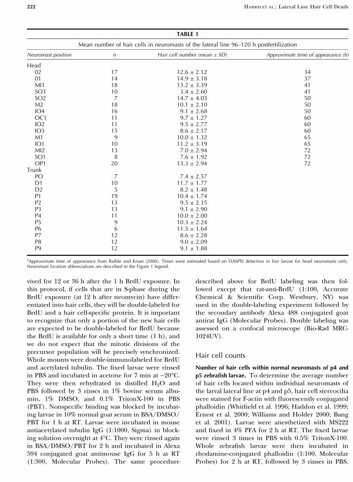
TABLE 1
Mean number of hair cells in neuromasts of the lateral line 96–120 h postfertilization
Neuromast position n Hair cell number (mean ± SD) Approximate time of appearance (h)
Head02 17 12.6 ± 2.12 3401 14 14.9 ± 3.18 37MI1 18 13.2 ± 3.39 41SO3 10 3.4 ± 2.60 41SO2 7 14.7 ± 4.03 50M2 18 10.1 ± 2.10 50IO4 16 9.1 ± 2.68 50OC1 11 9.7 ± 1.27 60IO2 11 9.5 ± 2.77 60IO3 15 8.6 ± 2.17 60M1 9 10.0 ± 1.32 65IO1 10 11.2 ± 3.19 65MI2 13 7.0 ± 2.94 72SO1 8 7.6 ± 1.92 72OP1 20 13.3 ± 2.94 72
TrunkPO 7 7.4 ± 2.57D1 10 11.7 ± 1.77D2 5 8.2 ± 1.48P1 19 10.4 ± 1.74P2 13 9.5 ± 2.15P3 13 9.1 ± 2.90P4 11 10.0 ± 2.00P5 9 10.3 ± 2.24P6 6 11.5 ± 1.64P7 12 8.6 ± 2.28P8 12 9.0 ± 2.09P9 12 9.1 ± 1.88
aApproximate time of appearance from Raible and Kruse (2000). Times were estimated based on DASPEI detection in live larvae for head neuromasts only.Neuromast location abbreviations are described in the Figure 1 legend.
222 HARRIS ET AL.: Lateral Line Hair Cell Death

Larvae were mounted onto glass slides using Vecta-shield (Vector) and coverslipped. Hair cell countswere made in a total of 326 neuromasts. Each neu-romast position was analyzed in 5 to 20 fish (see Table1) under epifluorescent illumination using a 63X oilimmersion objective (Leitz Aristoplan).
Correspondence of DASPEI, actin, andacetylated tubulin staining
To verify that the DASPEI score that was assigned tospecific neuromasts in the neomycin toxicity experi-ments was proportional to the number of hair cellspresent after aminoglycoside treatment, hair cell cil-iary bundles were stained for F-actin with phalloidinin the same animals that were scored by DASPEI.Animals from all treatment groups at 4 h after neo-mycin exposure were included for analysis. Thenumber of phalloidin-stained hair cell bundles inindividual neuromasts (n = 388) on each of 31 ze-brafish were counted. The analysis was blinded. Thecounts were subjected to analysis of variance as afunction of DASPEI scoring group (0, 1, or 2).
Hair cell somata were immunolabeled with anti-acetylated tubulin to compare cell survival withDASPEI staining after aminoglycoside treatment. Forthis analysis, animals from control and 500 lMtreatment groups (n = 5/treatment group) werescored using DASPEI under an epifluorescence dis-secting microscope. The fish were then immediatelyanesthetized and fixed. Hair cells were labeled withmouse antiacetylated tubulin as described above.Immunoreactive cells were assessed using a Bio-RadMRC-1024UV confocal microscope.
In an additional experiment to quantitativelycompare DASPEI scores with the number of hair cellspresent in a neuromast, larvae (n = 9–20 per group)were exposed to a range of neomycin doses (0, 125,250, and 500 lM) for 1 h. Four hours after the ini-tial drug exposure, head neuromasts were scoredby labeling with DASPEI. The larvae were thenanesthetized and fixed and hair cells were double-labeled first for acetylated tubulin and second withphalloidin, similar to methods described above ex-cept acetone was omitted from the procedure andAlexa 488 conjugated phalloidin was used (MolecularProbes). Hair cell counts were made independ-ently using tubulin and phalloidin labeling in thesame head neuromasts that were previously scoredlive using DASPEI. All hair cell counts were con-ducted with the experimenter ‘‘blinded’’ with respectto treatment group and to scores using the other twomethods. Each neuromast was analyzed under epi-fluorescent illumination using a 60X oil immersionobjective.
The hair cell counts and DASPEI scores were thensubjected to a two-way ANOVA for analysis type andneomycin concentration.
Scanning electron microscopy
Anesthetized larvae were immersion fixed in 4%glutaraldehyde in 0.1 M sodium cacodylate (pH 7.4)and 8 mM CaCl2 on a shaker table for 1 h at 25�Cand then overnight at 4�C. Following three 10-minwashes in 0.1 M cacodylate buffer, larvae were post-fixed in 1% OsO4 in 0.1 M cacodylate and 8 mMCaCl2 on ice for 1 h on the shaker table. Larvae werewashed again three times at 10 min each and dehy-drated through a graded ethanol series: 35%, 70%,95%, 100%, and 100% for 10 min each, then criticallypoint dried using CO2. Zebrafish were then mountedonto SEM specimen mounts using double-sided car-bon adhesive tape and sputter-coated with Au/Pd.Specimens were examined and photographed with a
FIG. 2. Lateral line neuromasts stained with DASPEI. A, C. Liveuntreated p5 zebrafish larvae. B, D. Representative live p5 larvae 4 hafter initial treatment with 500 lM neomycin for 1 h. A–D are lateralviews with anterior to the left and dorsal up. The nasal epithelium isalso stained with DASPEI in both groups (arrows). E. A higher-mag-nification view of a DASPEI-stained neuromast from a live controlanimal shows the rosette pattern of hair cells. F. A neuromast from afixed larva containing hair cells labeled for f-actin with phalloidin.The neuromasts shown in E and F are not identical neuromasts andthus may have a different average number of hair cells (see Table 1).Scale bars = 0.5 mm (A and B in B; C and D in C) and 25 lm(E and F).
HARRIS ET AL.: Lateral Line Hair Cell Death 223
JEOL JSM 6300F field emission scanning electronmicroscope.
RESULTS
Lateral line hair cell labeling in larval zebrafish
The voltage-sensitive dye DASPEI specifically labelssensory hair cells of the lateral line following 15 minof incubation. DASPEI has previously been shown tolabel nerve fibers and neurons which contact the haircells with longer incubation times and at higherconcentrations than used here (Alexandre and Ghy-sen 1999). In addition to labeling hair cells, DASPEIis also selectively taken up by the nasal epithelium(Alexandra and Ghysen 1999). Figures 2A and C showthe stereotypical distribution of neuromasts (sche-matized in Fig. 1B) in representative live untreatedzebrafish larvae incubated in DASPEI. At the level ofmagnification shown in Figures 2A–D, individual haircells within a given neuromast cannot be visualized.At higher magnification, individual DASPEI-stainedhair cells are centrally located in the neuromaststructures and easily observable, as illustrated in Fig-ure 2E.
To quantify the number of hair cells present inlateral line neuromasts at these ages, hair cell ciliarybundles were labeled with phalloidin, as shown inFigure 2F. On average, there are 10 hair cells perneuromast in the lateral line of untreated p4 and p5larvae. However, the mean numbers (± SD) of haircells contained in each neuromast range from3.4 ± 2.60 to 14.7 ± 4.03 and depend on the neuro-mast location in the lateral line system (Table 1).Neuromasts that appear earlier in development, suchas O1, O2, and MI1 (see Fig. 1B), generally containedhigher numbers of hair cells per neuromast at thisage than neuromasts that develop later, although thisrelationship was not true for all neuromasts (Raibleand Kruse 2000).
Neomycin toxicity of lateral line hair cells
Neomycin exposure induced lateral line hair celldeath in p4 and p5 zebrafish larvae in a dose-de-pendent manner. Figures 2B and D illustrate that ahigh dose of 500 lM neomycin for 1 h effectivelyeliminated DASPEI-stained lateral line hair cells 4 hlater in all neuromasts, regardless of their position.Staining of the sensory nasal epithelium was unaf-fected by exposure to this concentration of neomy-cin; this served as an internal control for thereliability of DASPEI labeling.
Having established a dose sufficient to eliminatelateral line hair cells stained with DASPEI at 4 h after
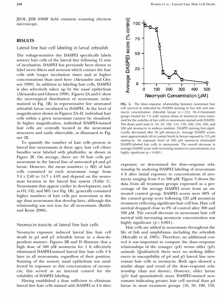
exposure, we determined the dose–response rela-tionship by analyzing DASPEI labeling of neuromasts4 h after initial exposure to concentrations of neo-mycin ranging from 0 to 500 lM. Figure 3 shows thedata from all treatment groups expressed as a per-centage of the average DASPEI score from an un-treated control group. DASPEI scores were 60% ofthe control group score following 125 lM neomycintreatment reflecting significant hair cell loss. Hair cellsurvival dropped close to 0% of control after 300 and500 lM. The overall decrease in neuromast hair cellsurvival with increasing neomycin concentration washighly significant (p < 0.001).
Hair cells are added in neuromasts throughout thelife of fish and amphibians, including the zebrafish(Metcalfe et al. 1985). Therefore, an additional con-trol it was important to compare the dose–responserelationships of the younger (p4) versus older (p5)animals. Qualitatively there were no striking differ-ences in susceptibility of p4 and p5 lateral line neu-romast hair cells to neomycin. Both ages showed asimilar shape and trend of the dose–response rela-tionship (data not shown). However, older larvae(p5) had quantitatively more DASPEI–stained neu-romasts indicating greater hair cell survival than p4larvae in most treatment groups (10, 50, 100, 150,
FIG. 3. The dose–response relationship between neuromast haircell survival as indicated by DASPEI staining in live fish and neo-mycin concentration. Zebrafish larvae (n = 232; 18–41/treatmentgroup) treated for 1 h with various doses of neomycin were exam-ined for the viability of hair cells in neuromasts stained with DASPEI.The doses used were 0, 10, 50, 100, 125, 150, 200, 250, 300, and500 lM neomycin in embryo medium. DASPEI staining first signif-icantly decreased after 50 lM neomycin. Average DASPEI scoreswere approximately 60 of control levels in larvae exposed to 125 lMneomycin. An exposure level of 500 lM neomycin eliminatedDASPEI-labeled hair cells in neuromasts. The overall decrease inaverage DASPEI score with increasing neomycin concentrations washighly significant (p < 0.001).
224 HARRIS ET AL.: Lateral Line Hair Cell Death
200, and 250 lM neomycin). This difference wassmall, usually 3–4 neuromasts, but statistically signif-icant (data not shown). There were no reliable dif-ferences in neuromast hair cell survival as a functionof age following the highest neomycin doses (300 and500 lM). It is possible that p5 neuromasts retainmore DASPEI-labeled hair cells at intermediate con-centrations because they have a greater number ofmature hair cells to begin with. We checked this bycomparing phalloidin-labeled hair cell counts foridentical neuromasts at p4 and p5 in untreated lar-vae. The average number of hair cells containedwithin a specific neuromast of the lateral line at p5was slightly greater than at p4 in almost all neuro-masts. The mean difference between ages was 0.71
phalloidin-labeled hair cells per neuromast. However,this difference was not statistically significant(p = 0.06).
We also assessed the possibility that individualneuromasts of the head and trunk lateral lines aredifferentially sensitive to neomycin treatment. Dose–response curves were constructed based on DASPEIstaining of neuromasts of the head or trunk sepa-rately and for each of the 27 neuromasts. Neuromasts
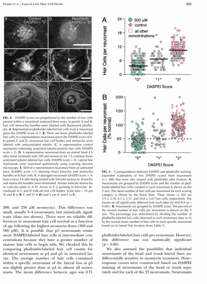
FIG. 4. DASPEI scores are proportional to the number of hair cellspresent within a neuromast analyzed three ways. In panels A and B,hair cell stereocilia bundles were labeled with fluorescent phalloi-din. A. Representative phalloidin-labeled hair cells from a neuromastgiven the DASPEI score of 2. B. There are fewer phalloidin-labeledhair cells in a representative neuromast given the DASPEI score of 0.In panels C and D, neuromast hair cell bodies and stereocilia werelabeled with antiacetylated tubulin. C. A representative controlneuromast containing acetylated tubulin-positive hair cells (DASPEIscore = 2). D. A representative neuromast from an animal fixed 4 hafter initial treatment with 500 lM neomycin for 1 h contains feweracetylated tubulin-labeled hair cells (DASPEI score = 0). Lateral lineneuromasts were examined qualitatively using scanning electronmicroscopy. E. SEM of a representative neuromast from an untreatedlarva (DASPEI score = 2) showing intact kinocilia and stereociliabundles of its hair cells. F. A damaged neuromast (DASPEI score = 0)from a larva 4 h after being treated with 500 lM neomycin. Kinociliaand stereocilia bundles were eliminated. Arrows indicate stereociliaor cuticular plate in A–D. Arrow in E is pointing to kinocilia. Ar-rowheads in C and D indicate hair cell bodies. Scale bars = 10 lm(A and B in B, C and D in D) and 5 lm (E and F in F).
FIG. 5. Correspondence between DASPEI and phalloidin staining.Expanded scatterplots of live DASPEI scores from neuromasts(n = 388) that were also stained with phalloidin after fixation. A.Neuromasts are grouped by DASPEI score and the number of phal-loidin-labeled hair cells counted in each neuromast is shown on theY axis. The mean number of hair cells per neuromast for each scoringcategory is shown by the black lines. These means (± SD) are3.9 ± 2.70, 6.5 ± 3.37, and 10.8 ± 3.07 hair cells, respectively. Themeans are all significantly different from each other by ANOVA (p <0.001). B. Neuromasts are grouped by DASPEI score. The percent ofthe normal number of hair cells per neuromast is shown on the Yaxis. This percentage was determined by dividing the number ofphalloidin-labeled hair cells observed in each neuromast (data in A)by the normal mean number of hair cells in each specific neuromastbased on its lateral line location (from Table 1).
HARRIS ET AL.: Lateral Line Hair Cell Death 225

of the head and trunk did not differ in their suscep-tibility to neomycin-induced damage, nor did anysingle neuromast show reliably greater sensitivity thanany other neuromast (data not shown).
Correspondence between DASPEI, actin, andacetylated tubulin staining
We assessed the validity of DASPEI labeling as an in-dicator of the number of hair cells present in threeways: (1) f-actin staining of stereocilia with phalloidin,(2) immunocytochemistry with a hair cell selectiveantibody, acetylated tubulin (Raible and Kruse 2000),and (3) scanning electron microscopy (SEM) obser-vations. Representative examples of normal neuro-masts and neuromasts treated with 500 lM neomycinare shown in Figure 4. Figures 4A and B show a rep-resentative neuromast from an untreated (control)larva that had received a live DASPEI score of 2 and arepresentative neuromast that had received a DASPEIscore of 0 before being fixed and labeled with phal-loidin. Many more phalloidin-labeled hair cells areobserved in neuromasts scored as 2 than 0 usingDASPEI. Figures 4C and D show representative neu-romasts from a control animal and an animal 4 hafter exposure to 500 lM neomycin (DASPEIscore = 0) labeled with acetylated tubulin. Hair cellbodies as well as stereocilia bundles are lost in pro-portion to the loss of DASPEI labeling.
As a third method of DASPEI validation, untreatedand neomycin-treated neuromasts were examinedusing scanning electron microscopy. A normal neu-romast that had received a DASPEI score of 2 isshown in Figure 4E. The neuromast has a character-istic dome- or volcano-shaped structure protrudingfrom the body surface. Extending from the ‘‘crater’’
are the long kinocilia from the resident hair cells.Less apparent at this level of scanning electronmicroscopy examination are the short stereociliabundles of the hair cells. All of the normal, untreatedneuromasts observed had this same morphology re-gardless of position in the lateral line system atthis age. Figure 4F shows a representative neuromastthat was fixed 4 h following the initiation of a 1 h500 lM neomycin treatment and had received aDASPEI score of 0. The long kinocilia have beeneliminated. Additional studies by transmission elec-tron microscopy confirm the loss of kinocilia andhair cells following 500 lM neomycin treatment(Pujol et al. 2002). These experiments support thevalidity of using DASPEI in a screening protocol toquickly assess lateral line hair cell integrity in livinganimals.
Using each method, more hair cells were presentin neuromasts that had previously received a DASPEIscore of 2 rather than 1 or 0. On the other hand, it isimportant to note that screening for hair cells usingDASPEI is not perfect; some neuromast hair cellswere present in the majority of neuromasts scored as0. To quantify these relationships, DASPEI stainingwas scored in live animals’ neuromasts at relativelylow magnification under a fluorescence dissectionmicroscope and then the number of phalloidin-stained hair cells were counted in the same neuro-masts after the fish were fixed. Fish from all neomycindosage groups were included in this analysis. Phal-loidin cell counts were grouped according to theirprevious DASPEI scores, as shown in Figure 5A. Themean number (± SD) of phalloidin-labeled hair cellsper neuromast for a DASPEI score of 0, 1, or 2 was3.9 ± 2.70, 6.5 ± 3.37, and 10.8 ± 3.07 hair cells, re-spectively. While there is some overlap in the distri-
FIG. 6. A representative neuromast from a control zebrafishdouble-labeled with antiacetylated tubulin and phalloidin. A.Acetylated tubulin labels the kinocilia (indicated with an asterisk)and extends toward the nuclei of the cells. Two labeled hair cells areindicated with arrows. B. Phalloidin labels the stereocilia bundles.
The same two hair cells indicated in A are shown here labeled withphalloidin indicated by the arrows. C. A composite image ofacetylated tubulin label (red) and phalloidin (green). Scale bar = 10lM.
226 HARRIS ET AL.: Lateral Line Hair Cell Death
butions of these scoring categories, the means are allsignificantly different from each other (p < 0.001).The overlap among categories is considerably re-
duced when data points from control and 500 lM-treated neuromasts are the only groups included foranalysis as shown in Figure 5A represented by thegreen- and red-filled symbols.
The number of hair cells per neuromast in un-treated zebrafish at p4 and p5 varies according toneuromast position in the lateral line system (shownin Table 1). To more accurately represent changes inhair cell number, we used the mean number of haircells for each neuromast position (Table 1) to recal-culate the data in Figure 5A. An expanded scatterplotof DASPEI scoring category against the percentage ofnormal hair cells per neuromast is shown in Figure5B. Importantly, there were only 2 out of 167 neu-romasts scored as 2 that contained less than 50% ofthe normal mean hair cell number. The majority ofneuromasts (128 out of 175; 73%) scored as 0 con-tained less than 50% of normal hair cell numbers forthat neuromast.
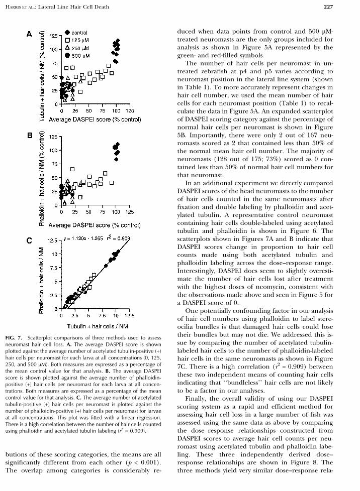
In an additional experiment we directly comparedDASPEI scores of the head neuromasts to the numberof hair cells counted in the same neuromasts afterfixation and double labeling by phalloidin and acet-ylated tubulin. A representative control neuromastcontaining hair cells double-labeled using acetylatedtubulin and phalloidin is shown in Figure 6. Thescatterplots shown in Figures 7A and B indicate thatDASPEI scores change in proportion to hair cellcounts made using both acetylated tubulin andphalloidin labeling across the dose–response range.Interestingly, DASPEI does seem to slightly overesti-mate the number of hair cells lost after treatmentwith the highest doses of neomycin, consistent withthe observations made above and seen in Figure 5 fora DASPEI score of 0.
One potentially confounding factor in our analysisof hair cell numbers using phalloidin to label stere-ocilia bundles is that damaged hair cells could losetheir bundles but may not die. We addressed this is-sue by comparing the number of acetylated tubulin-labeled hair cells to the number of phalloidin-labeledhair cells in the same neuromasts as shown in Figure7C. There is a high correlation (r2 = 0.909) betweenthese two independent means of counting hair cellsindicating that ‘‘bundleless’’ hair cells are not likelyto be a factor in our analyses.
Finally, the overall validity of using our DASPEIscoring system as a rapid and efficient method forassessing hair cell loss in a large number of fish wasassessed using the same data as above by comparingthe dose–response relationships constructed fromDASPEI scores to average hair cell counts per neu-romast using acetylated tubulin and phalloidin labe-ling. These three independently derived dose–response relationships are shown in Figure 8. Thethree methods yield very similar dose–response rela-
FIG. 7. Scatterplot comparisons of three methods used to assessneuromast hair cell loss. A. The average DASPEI score is shownplotted against the average number of acetylated tubulin-positive (+)hair cells per neuromast for each larva at all concentrations (0, 125,250, and 500 lM). Both measures are expressed as a percentage ofthe mean control value for that analysis. B. The average DASPEIscore is shown plotted against the average number of phalloidin-positive (+) hair cells per neuromast for each larva at all concen-trations. Both measures are expressed as a percentage of the meancontrol value for that analysis. C. The average number of acetylatedtubulin-positive (+) hair cells per neuromast is plotted against thenumber of phalloidin-positive (+) hair cells per neuromast for larvaeat all concentrations. This plot was fitted with a linear regression.There is a high correlation between the number of hair cells countedusing phalloidin and acetylated tubulin labeling (r2 = 0.909).
HARRIS ET AL.: Lateral Line Hair Cell Death 227

tionships at low to midrange concentrations. Howev-er, at the highest concentrations some cells appear tobe resistant to neomycin-induced death when studiedby phalloidin or acetylated tubulin labeling. Theseconclusions were supported by statistical analysis us-ing two-way ANOVA (dosage by analysis method)followed by appropriate individual comparisons be-tween groups. The overall analysis showed significanteffects of dosage (p < 0.001), analysis method (p <0.001), and the interaction term (p < 0.01). Therewere no significant differences between DASPEI scoreand hair cell counts by either method after 0 and 125lM neomycin exposure. Individual comparisons re-vealed that the methods do yield significant dif-ferences at the highest doses, 250 and 500 lM, witha mean of approximately 2.5 hair cells per neuro-mast remaining by both acetylated tubulin andphalloidin labeling, while average DASPEI scores are13% and 4.6%, respectively, of the average DASPEIcontrol score. Scores as a function of neomycin con-centration yielded significant differences at eachsuccessive dosage (p < 0.001) except for the com-parisons between 250 and 500 lM using phalloidin oracetylated tubulin labeling, which revealed equivalenteffects.
Lateral line hair cells at 12 and 24 hafter neomycin treatment
Time course of DASPEI- and phalloidin-stained lateral line
hair cell loss and recovery. It has been demonstratedpreviously that lateral line hair cells can regeneratefollowing damage (Jorgensen 1991; Song et al. 1995;Jones and Corwin 1996). We examined neuromasthair cells at 12 and 24 h after neomycin exposureusing DASPEI to further understand the changes inhair cell populations in p4 and p5 zebrafish larvaeafter aminoglycoside treatments. Figures 9A and Bshow DASPEI staining in representative larvae 4 hafter initial incubation in either control media (Fig.9A) or media containing 500 lM neomycin (Fig. 9B).The neomycin treatment eliminated all DASPEIstaining. When animals were allowed an additional 20h of recovery time and stained with DASPEI, thestaining pattern looked remarkably normal, as illus-trated in Figure 9C.
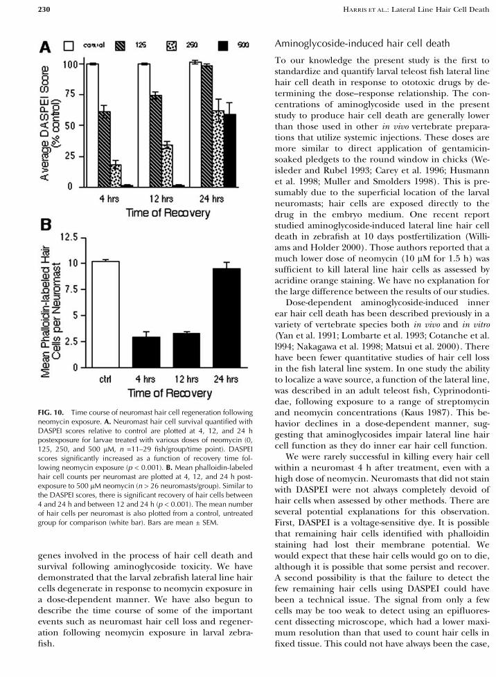
We quantified neuromast hair cell loss and recov-ery using the DASPEI scoring system at 12 and 24 hfor four different doses of neomycin (0, 125, 250, and500 lM). We also analyzed phalloidin labeling atthese times for fish treated with 500 lM neomycin.Figure 10A shows that DASPEI-stained lateral lineneuromasts increased in each treatment group as afunction of the period of recovery. At 12 h followingneomycin exposure, DASPEI scores were alreadygreater than those at 4 h for intermediate concen-trations. Animals that had received the highest neo-mycin dose (500 lM) did not show any recovery ofDASPEI staining when analyzed 12 h after the initialaminoglycoside exposure. Hair cell staining recov-ered to control levels by 24 h after 125 lM neomycintreatments. Allowing the zebrafish to recover for 24 hfollowing initial treatment with either 250 or 500 lMneomycin resulted in DASPEI labeling of approxi-mately 60% of control levels.
Figure 10B shows the mean number of phalloidin-labeled hair cells per neuromast from the 500 lM-treated groups at 4, 12, and 24 h after aminoglycosidetreatment. In the 500 lM-treated group, there was nofurther loss in the average number of hair cells perneuromast between 4 and 12 h. The number ofphalloidin-labeled hair cells returned to the averagecontrol level after 24 h of recovery time. There ap-pears to be more recovery of hair cell numbers whenassessed using phalloidin labeling compared withDASPEI staining at 24 h after 500 lM neomycin ex-posure.
BrdU incorporation in neuromast cells.. To furtherexplore the events during the 24 h following amino-glycoside toxicity, we assessed the relative amount ofcell proliferation occurring in neuromasts in controlanimals and at 4, 12, and 24 h following 500 lM
FIG. 8. Dose–response relationships of neuromast hair cell survivaland neomycin concentration determined by three different methods(from the same data as shown in Fig. 7). The average DASPEI scoreexpressed as a percentage of the control value is plotted on the left Yaxis. The average number of hair cells per neuromast counted usingphalloidin and acetylated tubulin labeling is plotted using the right Yaxis. The overall decrease in DASPEI scores and hair cell counts washighly significant (p < 0.001) and all doses were significantly dif-ferent from each other (p’s < 0.001), except scores for 250 vs. 500lM using phalloidin and acetylated tubulin staining. There was nosignificant difference between phalloidin and acetylated tubulin cellcounts at any concentration. DASPEI scores differed significantlyfrom both other methods at the highest doses, 250 and 500 lM (p’s <0.001), but they did not differ at the intermediate dose, 125 lM.Hence the change in DASPEI scores is proportional to hair cellcounts across the dose–response range but slightly overestimates haircell loss at the high doses.
228 HARRIS ET AL.: Lateral Line Hair Cell Death

neomycin treatment. This was accomplished by asingle exposure of larvae to BrdU in the embryomedium for 1 h at the time points of interest. The fishwere fixed immediately after the BrdU exposure(pulse-fix protocol). This BrdU-labeling protocol willassess only cells that are in S-phase during the 1 hexposure. The number of neuromast cells in S-phasewas counted. Figures 11A and B show representativeneuromasts from a control animal and an animal 12 hafter 500 lM neomycin treatment. Treated animalshad many more BrdU-labeled cells within individualneuromasts than untreated animals. The labeled cellsoccurred predominantly in the periphery of theneuromast structure, suggesting that they are likelymantle support cells because of their location in theneuromast as well as the large size of the nuclei incomparison to the hair cell nuclei. A minority of thelabeled cells in the periphery appear to be muchlarger than expected for BrdU-labeled nuclei. Thishas been observed previously (Williams and Holder2000) and interpreted as BrdU labeling in the cyto-plasm as well as the nuclei. As expected, there isongoing proliferation in neuromasts of controlanimals.
Figure 12 shows the average number of neuromastcells in S-phase from control larvae and at 4, 12, and24 h following 500 lM neomycin treatment. In con-trol animals the mean number of BrdU-labeled cellsafter 1 h of BrdU exposure was approximately 1 cellper neuromast. At 4 h after neomycin exposure, therewas a slight increase in the number of BrdU-labeledcells per neuromast relative to control levels, but thisdifference was not significant. However, proliferationwas dramatically upregulated at 12 h following neo-mycin exposure with an approximate four-fold in-
crease over control levels of neuromast cells in S-phase. By 24 h after aminoglycoside toxicity, thenumber of proliferative cells remained higher thancontrol levels but lower than the number of cells in-corporating BrdU at 12 h following 500 lM neomycinexposure. Both the neomycin treatment and the in-teraction effect of treatment and time of recovery onthe number of BrdU-labeled cells were highly signif-icant (p < 0.005). Differences between treatment andcontrols were significant at 12 and 24 h (p < 0.005),and treatment groups differed significantly at eachrecovery time (p < 0.001).
We also assessed whether the increase in neuromastcells in S-phase at 12 h after neomycin exposure re-sulted in the production of new hair cells. Larvae wereexposed to BrdU for 1 h 12 h following 500 lM neo-mycin treatment. They were then allowed to recoverfor an additional 0, 12, or 36 h. Hair cells were iden-tified using acetylated tubulin immunocytochemistry.Neuromast cells double-labeled for acetylated tubulinand BrdU represented hair cells assumed to be‘‘born’’ around 12 h after neomycin exposure. We didnot observe double-labeled cells in neuromasts fromeither control or neomycin-treated larvae in the 12 hpulse-fix experiment as exemplified in Figures 13 Aand B. As shown previously, BrdU incorporation at 12h after 500 lM neomycin exposure is significantlygreater than the amount of ongoing proliferationseen in a control neuromast (compare BrdU label in11B to 11A). A very low number of neuromasts con-taining cells double-labeled for acetylated tubulin andBrdU were observed in both control and neomycin-treated animals at 24 h after neomycin exposure orexperimental initiation (12 h after BrdU exposure),and there appeared to be more neuromasts contain-ing new hair cells in the drug-treated group. By 36 hafter BrdU exposure in untreated animals, approxi-mately one-third of all neuromasts contained 1–2double-labeled cells. In neomycin-treated animals thatrecovered for 36 h after BrdU exposure (48 h afterneomycin treatment), 1–4 double-labeled hair cellswere observed within most neuromasts. Figures 13Cand D show two examples from animals exposed toneomycin that were allowed to recover for 48 h (36 hafter BrdU). In summary, the upregulation of prolif-eration observed during this particular 1 h period, 12h after aminoglycoside exposure, results in the pro-duction of some new hair cells 12 h later. By 36 h afterS-phase, a large number of these cells are expressing ahair cell phenotype.
DISCUSSION
The goal of our experiments was to develop andcommunicate an efficient preparation for studying
FIG. 9. Hair cell regeneration in neomycin-treated neuromasts. A.An untreated larva showing typical DASPEI staining of neuromasts.B. A DASPEI-stained larva 4 h after exposure to 500 lM neomycin.DASPEI staining is eliminated in neuromasts. C. A DASPEI-stainedlarva 24 h following exposure to 500 lM neomycin. Note the returnof a normal DASPEI staining pattern. A, C insets: DASPEI-labeledcontrol (A) and regenerated (C) neuromasts appear similar at highermagnification. Scale bar = 0.5 mm for all panels excluding insets.
HARRIS ET AL.: Lateral Line Hair Cell Death 229
genes involved in the process of hair cell death andsurvival following aminoglycoside toxicity. We havedemonstrated that the larval zebrafish lateral line haircells degenerate in response to neomycin exposure ina dose-dependent manner. We have also begun todescribe the time course of some of the importantevents such as neuromast hair cell loss and regener-ation following neomycin exposure in larval zebra-fish.
Aminoglycoside-induced hair cell death
To our knowledge the present study is the first tostandardize and quantify larval teleost fish lateral linehair cell death in response to ototoxic drugs by de-termining the dose–response relationship. The con-centrations of aminoglycoside used in the presentstudy to produce hair cell death are generally lowerthan those used in other in vivo vertebrate prepara-tions that utilize systemic injections. These doses aremore similar to direct application of gentamicin-soaked pledgets to the round window in chicks (We-isleder and Rubel 1993; Carey et al. 1996; Husmannet al. 1998; Muller and Smolders 1998). This is pre-sumably due to the superficial location of the larvalneuromasts; hair cells are exposed directly to thedrug in the embryo medium. One recent reportstudied aminoglycoside-induced lateral line hair celldeath in zebrafish at 10 days postfertilization (Willi-ams and Holder 2000). Those authors reported that amuch lower dose of neomycin (10 lM for 1.5 h) wassufficient to kill lateral line hair cells as assessed byacridine orange staining. We have no explanation forthe large difference between the results of our studies.
Dose-dependent aminoglycoside-induced innerear hair cell death has been described previously in avariety of vertebrate species both in vivo and in vitro(Yan et al. 1991; Lombarte et al. 1993; Cotanche et al.l994; Nakagawa et al. 1998; Matsui et al. 2000). Therehave been fewer quantitative studies of hair cell lossin the fish lateral line system. In one study the abilityto localize a wave source, a function of the lateral line,was described in an adult teleost fish, Cyprinodonti-dae, following exposure to a range of streptomycinand neomycin concentrations (Kaus 1987). This be-havior declines in a dose-dependent manner, sug-gesting that aminoglycosides impair lateral line haircell function as they do inner ear hair cell function.
We were rarely successful in killing every hair cellwithin a neuromast 4 h after treatment, even with ahigh dose of neomycin. Neuromasts that did not stainwith DASPEI were not always completely devoid ofhair cells when assessed by other methods. There areseveral potential explanations for this observation.First, DASPEI is a voltage-sensitive dye. It is possiblethat remaining hair cells identified with phalloidinstaining had lost their membrane potential. Wewould expect that these hair cells would go on to die,although it is possible that some persist and recover.A second possibility is that the failure to detect thefew remaining hair cells using DASPEI could havebeen a technical issue. The signal from only a fewcells may be too weak to detect using an epifluores-cent dissecting microscope, which had a lower maxi-mum resolution than that used to count hair cells infixed tissue. This could not have always been the case,
FIG. 10. Time course of neuromast hair cell regeneration followingneomycin exposure. A. Neuromast hair cell survival quantified withDASPEI scores relative to control are plotted at 4, 12, and 24 hpostexposure for larvae treated with various doses of neomycin (0,125, 250, and 500 lM, n =11–29 fish/group/time point). DASPEIscores significantly increased as a function of recovery time fol-lowing neomycin exposure (p < 0.001). B. Mean phalloidin-labeledhair cell counts per neuromast are plotted at 4, 12, and 24 h post-exposure to 500 lM neomycin (n > 26 neuromasts/group). Similar tothe DASPEI scores, there is significant recovery of hair cells between4 and 24 h and between 12 and 24 h (p < 0.001). The mean numberof hair cells per neuromast is also plotted from a control, untreatedgroup for comparison (white bar). Bars are mean ± SEM.
230 HARRIS ET AL.: Lateral Line Hair Cell Death

however, because as few as 2 cells were able to give aDASPEI signal scored as 1 (see Fig. 5A).
A third possible explanation is that ongoing pro-liferation is occurring in neuromasts during our ex-perimental paradigm. As a result it could bepresumed that there are hair cells at several stages ofdevelopment within any given neuromast at the timethey are treated with neomycin. Aminoglycosides maynot be effective in inducing death in hair cells thatare immature (Rubel 1978; Duckert and Rubel 1990;
Hashino and Salvi 1996). Therefore, we might expectto see a low number of hair cells, possibly the‘‘youngest’’ within a given neuromast, that survivedrug treatment. A final possibility is that there may behair cell heterogeneity within a neuromast at thispoint in lateral line development. Based on differ-ential ototoxic sensitivity, type I and type II hair cellshave been described in the fish inner ear as well as incanal and superficial neuromasts, respectively, of theadult lateral line (Yan et al. 1991; Song et al. 1995).Differentiating among these possibilities will requirefurther research. In summary, DASPEI scores re-corded from low-power images through a dissectingmicroscope did not perfectly predict the number ofhair cells present within a neuromast as assessed bythese other methods. However, DASPEI scores areproportional to the number of hair cells present us-ing a variety of methods, including phalloidin andacetylated tubulin labeling. Of particular importance
FIG. 12. Time course of neuromast cells entering S-phase following500 lM neomycin exposure. Mean BrdU-positive cells per neuro-mast are plotted for neuromasts exposed to BrdU in the embryomedium for 1 h 4, 12, and 24 h after neomycin exposure (n = 173neuromasts, 19–41 neuromasts/group and 3–7 neuromasts/fish). Thenumber of S-phase cells in a neuromast is upregulated at 12 and 24 hafter neomycin exposure. The time of BrdU exposure and neomycintreatment had significant effects on the number of proliferating cells(two-way ANOVA, p < 0.005). Bars are mean ± SEM.
FIG. 13. New hair cells produced by neuromast cells in S-phase at12 h after 500 lM neomycin exposure. Hair cells were identifiedusing immunocytochemistry (ICC) for acetylated tubulin (shown inred). BrdU-positive cells were also detected with ICC (shown ingreen). A. A representative neuromast from a control animal pulsedwith BrdU for 1 h 12 h after the experiment was initiated and fixedimmediately. There are few S-phase cells within the neuromast. B. Arepresentative neuromast exposed to 500 lM neomycin, pulsed withBrdU 12 h later, and then immediately fixed. This neuromast con-tains many more S-phase cells but few hair cells. C, D. Two exam-ples of neuromasts treated with 500 lM neomycin, exposed to BrdUfor 1 h 12 h after neomycin exposure, and then fixed 36 h later (48 hafter neomycin treatment). These neuromasts contained new haircells double-labeled for acetylated tubulin and BrdU as well as haircells without BrdU labeling and BrdU-labeled cells without acetyl-ated tubulin labeling. Asterisks indicate hair cells, arrowheads in-dicate BrdU labeling, and the arrows indicate ‘‘new’’ hair cells.Scale bar = 10 lm.
FIG. 11. Neomycin-induced upregulation of proliferation in neu-romasts. A. A representative neuromast from an untreated controllarva that was exposed to BrdU 12 h after the initiation of the ex-periment contains few BrdU-labeled cells. B. A neuromast from ananimal treated with 500 lM neomycin contains many more S-phasecells than the control neuromast when exposed to BrdU for 1 h 12 hafter treatment. The BrdU nuclei (green) are overlaid onto a gray DICimage of the neuromast. Each fluorescent image is from a flattened z-series through the neuromast. Scale bar = 10 lm.
HARRIS ET AL.: Lateral Line Hair Cell Death 231

is the fact that none of the control fish were given aDASPEI score of 0. Therefore, DASPEI scoring pro-vides a rapid, reliable screening method for assess-ment of lateral line hair cell survival.
Lateral line hair cell regeneration
Lateral line hair cells of larval zebrafish regeneratefollowing aminoglycoside-induced degeneration.There are at least two possible mechanisms for re-placing hair cells in sensory epithelia. New hair cellscan be produced by proliferation of precursor cellsthat then differentiate into hair cells. Alternatively,postmitotic cells can be induced to differentiate intohair cells with no intervening mitosis in a processtermed direct transdifferentiation (Adler and Ra-phael 1996; Baird et al. 1996; Jones and Corwin 1996;Roberson et al. 1996; Steyger et al. 1997).
We investigated the potential role of proliferationin generating new hair cells following maximal haircell loss. Neuromast cell entry into S-phase is dra-matically upregulated 12 h after the onset of amino-glycoside toxicity. We observed that some cellslabeled with the S-phase marker BrdU 12 h afterneomycin exposure assumed a hair cell phenotypewithin the next 12–36 h. Therefore, it is likely thatdeath-induced upregulation of proliferative eventscontributes to at least some of the new hair cells seenat 24 h using DASPEI and phalloidin labeling. A moreintensive study involving BrdU exposure at times be-tween 4 and 12 h will be necessary to determine whatproportion of hair cell replacement observed by 24 his due to induced proliferation versus ongoing pro-liferation or induced transdifferentiation.
The observed time course of larval lateral line haircell regeneration is extraordinarily fast in comparisonto other systems. In the chick basilar papilla, new haircells are detected by 90–100 h after noise-induceddamage and several days following drug administra-tion (Girod et al. 1989; Duckert and Rubel 1990;Cotanche et al. 1994; Matsui et al. 2000; Stone andRubel 2000b). Jones and Corwin (1996) observedmature hair cells arising over one week in the axolotlafter laser-induced damage. However, in the adultoscar, a teleost fish, new hair cell stereocilia bundleslocated in canal neuromasts were observed as early astwo days following prolonged aminoglycoside expo-sure (Song et al. 1995). In addition, behavioral re-covery in adult Cyprinodontidae fish is reported tooccur one to three days after aminoglycoside treat-ment (Kaus 1987). Regeneration may occur morequickly in these systems as opposed to the chick bas-ilar papilla because cells are not mitotically quiescentunder normal developmental circumstances. In ad-dition, cell cycle length increases during zebrafishdevelopment, suggesting that proliferation and dif-
ferentiation could occur more quickly in the larvathan in adult systems (Kane and Kimmel 1993; Kim-mel et al. 1994).
Supporting cells appear to be the most likely can-didate for hair cell progenitors in the avian systemafter damaging stimuli (Corwin and Cotanche 1988;Girod et al. 1989; Weisleder and Rubel 1993; Stone etal. 1996; Warchol and Corwin 1996; Stone and Rubel2000b). In the axolotl lateral line, Jones and Corwin(1996) have provided direct evidence that new haircells arise from supporting cell divisions in neuro-masts after laser ablation of all preexisting hair cells.In the zebrafish lateral line, we observed that BrdU-labeled cells were generally located in the peripheryof the neuromast following aminoglycoside exposure,similar to the observations of Williams and Holder(2000). This location suggests that they may be sup-port cells, although other cell types are possible.Further investigation, including double-labelingtransmission electron microscopy studies, are inprogress to accurately and carefully describe hair cellregeneration and the progenitors in this model sys-tem (Pujol et al. 2002).
One application of this study is to begin a geneticscreen using randomly mutagenized zebrafish tosearch for genes that modulate hair cell susceptibilityto ototoxicity. Genetic screens in zebrafish have al-ready been successful in pulling out classes of genesthat are important for hair cell function and devel-opment, including myosin VIIA (Whitfield et al. 1996;Nicolson et al. 1998; Ernest et al. 2000). The finalmechanism of hair cell death following ototoxic in-sults appears to be similar in a variety of vertebratesincluding fish (Forge and Li 2000; Pirvola et al. 2000;Williams and Holder 2000). Therefore, we expect bothexogenous and endogenous molecules that influencelateral line hair cell death, survival, and regenerationin the zebrafish are likely to be effective in the innerear of other vertebrate species, including humans.
ACKNOWLEDGMENTS
The authors thank Dale Cunningham for his help withscanning electron microscopy, Laurie Johnson for manu-script preparation assistance, and Melinda Modrell andTiffany Kao for their excellent technical support. We alsothank Dr. Eric Bauer and Setsuko Murakami for additionalhistology and valuable help. This work was supported byNIDCD grants DC04661, DC00018, and DC02854, and theUniversity of Washington RRF.
REFERENCES
ADLER HJ, RAPHAEL Y. New hair cells arise from supporting cellconversion in the acoustically damaged chick inner ear. Neu-rosci. Lett. 205:17–20, 1996.
232 HARRIS ET AL.: Lateral Line Hair Cell Death

ALEXANDRE D, GHYSEN A. Somatotopy of the lateral line projectionin larval zebrafish. Proc. Natl. Acad. Sci. U.S.A. 96:7558–7562,1999.
BAIRD RA, STEYGER PS, SCHUFF NR. Mitotic and nonmitotic hair cellregeneration in the bullfrog vestibular otolith organs. Ann. N.Y.Acad. Sci. 781:59–70, 1996.
BANG PI, SEWELL WF, MALICKI JJ. Morphology and cell type hetero-geneities of the inner ear epithelia in adult and juvenile ze-brafish (Danio rerio). J. Comp. Neurol. 438:173–190, 2001.
CAREY JP, FUCHS AF, RUBEL EW. Hair cell regeneration and recoveryof the vestibuloocular reflex in the avian vestibular system.J. Neurophysiol. 76:3301–3312, 1996.
CHARDIN S, ROMAND R. Regeneration and mammalian auditory haircells. Science 267:707–711, 1995.
CHENG AG-L, SHANG J, RUBEL EW. Hair cell death in vitro in theavian BP: characterization of the model and caspase inhibition.Assoc. Res. Otolaryngol. 24:Abstr. 466, 2001.
CORWIN JT, COTANCHE DA. Regeneration of sensory hair cells afteracoustic trauma. Science 240:1772–1774, 1988.
COTANCHE DA. Regeneration of hair cell stereociliary bundles in thechick cochlea following severe acoustic trauma. Hear. Res.30:181–195, 1987.
COTANCHE DA, LEE KH, STONE JS, PICARD DA. Hair cell regenerationin the bird cochlea following noise damage or ototoxic drugdamage. Anal. Embryol. (Berl.) 189:1–18, 1994.
CRUZ RM, LAMBERT PR, RUBEL EW. Light microscopic evidenceof hair cell regeneration after gentamicin toxicity in chickcochlea. Arch. Otolaryngol. Head Neck Surg. 113:1058–1062,1987.
CUNNINGHAM L, CHENG AG, RUBEL EW. Caspase activation in haircells of the mouse utricle exposed to neomycin. J. Neurosci.22:8532–8540, 2002.
DUCKERT LG, RUBEL EW. Ultrastructural observations on regener-ating hair cells in the chick basilar papilla. Hear. Res. 48:161–182, 1990.
ERNEST S, RAUCH GJ, HAFFTER P, GEISLER R, PETIT C, NICOLSON T.Mariner is defective in myosin VIIA: a zebrafish model forhuman hereditary deafness. Hum. Mol. Genet. 9:2189–2196,2000.
FISCHEL–GHODSIAN N. Genetic factors in aminoglycoside toxicity.Ann. N.Y. Acad. Sci. 884:99–109, 1999.
FORGE A, LI L. Apoptotic death of hair cells in mammalian vestib-ular sensory epithelia. Hear. Res. 139:97–115, 2000.
FORGE A, SCHACHT J. Aminoglycoside antibiotics. Audiol. Neurootol.5:3–22, 2000.
GIROD DA, DUCKERT LG, RUBEL EW. Possible precursors of regen-erated hair cells in the avian cochlea following acoustic trauma.Hear. Res. 42:175–194, 1989.
HADDON C, MOWBRAY C, WHITFIELD T, JONES D, GSCHMEISSNER S,LEWIS J. Hair cells without supporting cells: further studies inthe ear of the zebrafish mind bomb mutant. J. Neurocytol.28:837–850, 1999.
HASHINO E, SALVI RJ. Regenerated hair cells exhibit a transient re-sistance to aminoglycoside toxicity. Brain Res 720:172–182,1996.
HUSMANN KR, MORGAN AS, GIROD DA, DURHAM D. Round windowadministration of gentamicin: a new method for the study ofototoxicity of cochlear hair cells. Hear. Res. 125:109–119,1998.
JONES JE, CORWIN JT. Regeneration of sensory cells after laser ab-lation in the lateral line system: hair cell lineage and macr-ophage behavior revealed by time-lapse video microscopy.J. Neurosci. 16:649–662, 1996.
JORGENSEN JM. Regeneration of lateral line and inner ear vestibularcells. Ciba Found. Symp. 160:151–163, 1991.
KALMIJN AD. Functional evolution of lateral line and inner earsensory systems. In: COOMBS S, GORNER P, MUNZ H (eds) The
mechanosensory lateral line. Springer-Verlag, New York, 1989,pp 187–215.
KANE DA, KIMMEL CB. The zebrafish midblastula transition. Devel-opment 119:447–456, 1993.
KAUS S. The effect of aminoglycoside antibiotics on the lateral lineorgan of Aplocheilus lineatus (Cyprinodontidae). Acta Otolaryn-gol. 103:291–298, 1987.
KIMMEL CB, WARGA RM, KANE DA. Cell cycles and clonal stringsduring formation of the zebrafish central nervous system. De-velopment 120:265–276, 1994.
LI L, NEVILL G, FORGE A. Two modes of hair cell loss from thevestibular sensory epithelia of the guinea pig inner ear. J. Comp.Neurol. 355:405–417, 1995.
LOMBARTE A, YAN HY, POPPER AN, CHANG JS, PLATT C. Damage andregeneration of hair cell ciliary bundles in a fish ear followingtreatment with gentamicin. Hear. Res. 64:166–174, 1993.
MATSUI J, OESTERLE E, STONE J, RUBEL E. Characterization of damageand regeneration in cultured avian utricles. J. Assoc. Res. Oto-laryngol. 1:46–63, 2000.
MATSUI JI, OGILVIE JM, WARCHOL ME. Inhibition of caspases pre-vents ototoxic and ongoing hair cell death. J. Neurosci.22:1218–1227, 2002.
METCALFE WK, KIMMEL CB, SCHABTACH E. Anatomy of the posteriorlateral line system in young larvae of the zebrafish. J. Comp.Neurol. 233:377–389, 1985.
MONTGOMERY J, CARTON G, VOIGT R, BAKER C, DIEBEL C. Sensoryprocessing of water currents by fishes. Philos. Trans. R. Soc.Lond. B Biol. Sci. 355:1325–1327, 2000.
MULLER M, SMOLDERS JW. Hair cell regeneration after local appli-cation of gentamicin at the round window of the cochlea in thepigeon. Hear. Res. 120:25–36, 1998.
NAKAGAWA T, YAMANE H, TAKAYAMA M, SUNAMI K, NAKAI Y. Dose-dependent response of vestibular hair cells of guinea pigs fol-lowing streptomycin ototoxiation. Acta Otolaryngol. 118:530–533, 1998.
NICOLSON T, RUSCH A, FRIEDRICH RW, GRANATO M, RUPPERSBERG JP,NUSSLEIN–VOLHARD C. Genetic analysis of vertebrate sensory haircell mechanosensation: the zebrafish circler mutants. Neuron.20:271–283, 1998.
PIRVOLA U, XING-QUN L, VIRKKALA J, SAARMA M, MURAKATA C, CAM-
ORATTO AM, WALTON KM, YLIKOSKI J. Rescue of hearing, auditoryhair cells, and neurons by CEP-1347/KT7515, an inhibitor ofc-Jun N-terminal kinase activation. J. Neurosci. 20:43–50, 2000.
PUJOL R, RAIBLE DW, CUNNINGHAM DE, RUBEL EW. Ultrastructure ofnormal and neomycin-exposed zebrafish lateral line hair cells.Assoc. Res. Otolaryngol. 25:Abstr. 577, 2002.
RAIBLE DW, KRUSE GJ. Organization of the lateral line system inembryonic zebrafish. J. Comp. Neurol. 421:189–198, 2000.
ROBERSON DW, RUBEL EW. Cell division in the gerbil cochlea afteracoustic trauma. Am. J. Otol. 15:28–34, 1994.
ROBERSON DW, KREIG CS, RUBEL EW. Light microscopic evidencethat direct transdifferentiation gives rise to new hair cells inregenerating avian auditory epithelium. Aud. Neurosci. 2:195–205, 1996.
RUBEL EW. Ontogeny of structure and function in the vertebrateauditory system. In: Jacobson M (ed) Handbook of SensoryPhysiology, Vol. IX, Development of Sensory Systems. Springer-Verlag, New York, 1978, pp 135–237.
SCHUKNECHT H. Pathology of the Ear. Harvard University Press,Boston, 1974.
SMOLDERS JW. Functional recovery in the avian ear after hair cellregeneration. Audiol. Neurootol. 4:286–302, 1999.
SONG J, YAN HY, POPPER AN. Damage and recovery of hair cells infish canal (but not superficial) neuromasts after gentamicinexposure. Hear. Res. 91:63–71, 1995.
STEYGER PS, BURTON M, HAWKINS JR, SCHUFF NR, BAIRD RA. Calbin-din and parvalbumin are early markers of non-mitotically re-
HARRIS ET AL.: Lateral Line Hair Cell Death 233

generating hair cells in the bullfrog vestibular otolith organs.Int. J. Dev. Neurosci. 15:417–432, 1997.
STONE JS, RUBEL EW. Cellular studies of auditory hair cell regen-eration in birds. Proc. Natl. Acad. Sci. U.S.A. 97:11714–11721,2000a.
STONE JS, RUBEL EW. Temporal, spatial, and morphologic featuresof hair cell regeneration in the avian basilar papilla. J. Comp.Neurol. 417:1–16, 2000b.
STONE JS, LEANO SG, BAKER LP, RUBEL EW. Hair cell differentiationin chick cochlear epithelium after aminoglycoside toxicity: invivo and in vitro observations. J. Neurosci. 16:6157–6174, 1996.
WARCHOL ME, CORWIN JT. Regenerative proliferation in organ cul-tures of the avian cochlea: identification of the initial progeni-tors and determination of the latency of the proliferativeresponse. J. Neurosci. 16:5466–5477, 1996.
WEISLEDER P, RUBEL EW. Hair cell regeneration after streptomycintoxicity in the avian vestibular epithelium. J. Comp. Neurol.331:97–110, 1993.
WESTERFIELD M. The zebrafish book: a guide for the laboratory useof zebrafish (Danio rerio), 4th ed. University of Oregon Press,Eugene, OR, 2000.
WHITFIELD TT, GRANATO M, VAN EEDEN FJ, SCHACH U, BRAND M,FURUTANI–SEIKI M, HAFFTER P, HAMMERSCHMIDT M, HEISENBERG
YJ, JIANG YJ, KANE DA, KELSH RN, MULLINS MC, ODENTHAL J,NUSSLEIN–VOLHARD C. Mutations affecting development of thezebrafish inner ear and lateral line. Development. 123:241–254,1996.
WILLIAMS JA, HOLDER N. Cell turnover in neuromasts of zebrafishlarvae. Hear. Res. 143:171–181, 2000.
YAN HY, SAIDEL WM, CHANG JS, PRESSON JC, POPPER AN. Sensory haircells of a fish ear: evidence of multiple types based on ototox-icity sensitivity. Proc. R. Soc. Lond. B Biol. Sci. 245:133–138,1991.
YLIKOSKI J, XING-QUN L, VIRKKALA J, PIRVOLA U. Blockade of c-Jun N-terminal kinase pathway attenuates gentamicin-induced cochl-ear and vestibular hair cell death. Hear. Res. 163:71–81, 2002.
234 HARRIS ET AL.: Lateral Line Hair Cell Death