nemoceph - medifarm dış tic.ve paz.ltd.Şti. · 2014-06-17 · the task of volume reorientation...
TRANSCRIPT

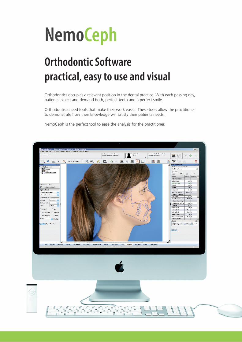
NemoCephOrthodontic Softwarepractical, easy to use and visualOrthodontics occupies a relevant position in the dental practice. With each passing day, patients expect and demand both, perfect teeth and a perfect smile.
Orthodontists need tools that make their work easier. These tools allow the practitioner to demonstrate how their knowledge will satisfy their patients needs.
NemoCeph is the perfect tool to ease the analysis for the practitioner.
NemoCeph
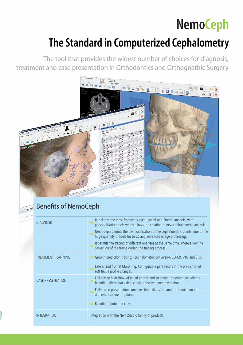
The tool that provides the widest number of choices for diagnosis, treatment and case presentation in Orthodontics and Orthognathic Surgery
The Standard in Computerized Cephalometry
DIAGNOSISItincludesthemostfrequentlyusedLateralandFrontalanalysis,withpersonalizationtoolswhichallowsthecreationofnewcephalometricanalysis.
NemoCephpermitsthebestlocalizationofthecephalometricpoints,duetothehugequantityoftoolsforbasicandadvancedimageprocessing.
Itpermitsthetracingofdifferentanalysesatthesametime.Theseallowthecorrectionoftheframeduringthetracingprocess.
TREATMENTPLANNING Growthpredictiontracings,cephalometricconversionCO-CR,VTOandSTO.
LateralandfrontalMorphing.Configurableparametersinthepredictionofsofttissueprofilechanges.
Benefits of NemoCeph
CASEPRESENTATION
Full-screenpresentationcombinestheinitialstateandthesimulationofthedifferenttreatmentoptions.
Blendingphotoandxray
IntegrationwiththeNemoStudiofamilyofproductsINTEGRATION
Full-screenSlideshowofinitialphotosandtreatmentprogress,includingaBlendingeffectthatvideosimulatethetreatmentevolution.

importing diagnostic records
NemoCeph offers the possibility of importing clinical photographs, xrays and study models in a simple manner with any device, including: digital cameras, radiographic scanner, CD-ROM, digital OPG, CT ( in combination with NemoScan or 3D NemoCeph), etc.
cepHaLometric tracing
In the tracing process, a software wizard will guide you step by step. An intelligent zoom transports you automatically to the area where the cephalometric point will be placed. The image processing tools allow you to clearly visualize and identify bone and dental structures.
When the tracing has been finished, the clinical photo can be superimposed on the xray simply marking two points in both. A window shows you the patient’s position in the cephalostat and the patient’s position in the photo. This window allows the adjustment of the natural head position.
treatment pLanning
You may add as many treatment options or predictions as you wish without having to duplicate your images.
The cephamotetric VTO is combined with a dental VTO, which allows you to make predictions. These predictions may be combinations of mixing the profile and space analysis, assessing the possibility for extractions, stripping, expansions, etc ...
STO allows for surgical, pre-surgical and preorthodontics predictions. These include all typesof osteotomies: BSSO, Vertical Mandibular, Bi-Maxillary, Occlusal Plane Alterations, Segmentary and Genioplasty; resulting in total control of the fulcrums using the chosen osteotomies.
case and progress presentation
With a simple click of the mouse, NemoCeph’s various SlideShows allow you to quickly and clearly present your diagnosis, treatment options and the progress of the treatment.
NemoCeph Work Protocol
NemoCeph 3D
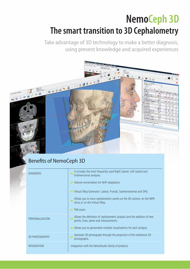
Take advantage of 3D technology to make a better diagnosis,using present knowledge and acquired experiences
The smart transition to 3D Cephalometry
DIAGNOSISItincludesthemostfrequentlyusedRightLateral,LeftLateralandtridimensionalanalyses.
VolumereorientationforNHPadaptation.
VirtualXRayGenerator:Lateral,Frontal,SubmentovertexandOPG
Allowsyoutotracecephalometricpointsonthe3Dvolume,ontheMPRslicesorontheVirtualXRay
TMJexam.
Benefits of NemoCeph 3D
PERSONALIZACIÓN
Allowsyoutopersonalizemultiplevisualizationsforeachanalysis.
Generate3Dphotographthroughtheprojectionofthetraditional2Dphotographs.
IntegrationwiththeNemoStudiofamilyofproducts.INTEGRATION
Allowsthedefinitionofcephalometricanalysisandtheadditionofnewpoints,lines,planeandmeasurements.
3DPHOTOGRAPHY

importing 3d records
Our advice is that teenagers and young people should be referred to radiological centers that have cone beam tomography (Cone Beam CT, CBCT), because the radiation produced is clearly less than traditional CT.
NemoCeph 3D allows you to import your patient’s tomographic exam in DICOM format and to create 3D volume complete with an automatic segmentation of the bone and the soft tissue.
tomograpHy preprocessing
The first step is to reorient the volume in order to obtain the natural head position (NHP). This step is the key for obtaining correct radiographic and cephalometric records. NemoCeph 3D offers simple and intuitive tools to reorient the three spatial planes.
When the volume reorientation has been performed, the following step is the generation of lateral, frontal, panoramic and submentovertex xrays.
If you send your patient to an associated nemo center, you can avoid the import step and preprocess of the tomography. Since there the exam will be preprocessed so that you can receive it via CD-ROM or you can download it directly by the Internet.
cepHaLometric tracing
A software wizard will guide you step by step in the tracing process. You will be able to mark the cephalometric points on the 3D volume, on tomographic sections or on virtual xrays. This feature is unique in the market and it allows you to produce a very precise tracing in a very short time.
3d pHotograpHy
The patient’s frontal photograph is superimposed on the NemoCeph 3D volume and this generates a 3D photograph, which is sufficient in most cases. If you wish a improve its quality, you are able to add the right lateral, left lateral and a submentovertex photographs to obtain the completed 3D photo.
NemoCeph 3D Work Protocol
NemoCeph 3D - OSDigital Orthognathic Surgery
From Planning to the Surgical Stent
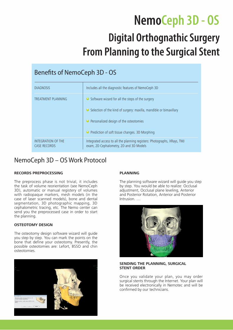
records preprocessing
The preprocess phase is not trivial, it includes the task of volume reorientation (see NemoCeph 3D), automatic or manual registery of volumes with radiopaque markers, mesh models (in the case of laser scanned models), bone and dental segmentation, 3D photographic mapping, 3D cephalometric tracing, etc. The Nemo center can send you the preprocessed case in order to start the planning.
osteotomy design
The osteotomy design software wizard will guide you step by step. You can mark the points on the bone that define your osteotomy. Presently, the possible osteotomies are: Lefort, BSSO and chin osteotomies.
pLanning
The planning software wizard will guide you stepby step. You would be able to realize: Occlusaladjustment, Occlusal plane leveling, Anteriorand Posterior Rotation, Anterior and PosteriorIntrusion. ....
sending tHe pLanning, sUrgicaLstent order
Once you validate your plan, you may order surgical stents through the Internet. Your plan will be received electronically in Nemotec and will be confirmed by our technicians.
NemoCeph 3D – OS Work Protocol
DIAGNOSIS IncludesallthediagnosticfeaturesofNemoCeph3D
TREATMENTPLANNING Softwarewizardforallthestepsofthesurgery
Selectionofthekindofsurgery:maxilla,mandibleorbimaxillary
Benefits of NemoCeph 3D - OS
INTEGRATIONOFTHECASERECORDS
Integratedaccesstoalltheplanningregisters:Photographs,XRays,TMJexam,2DCephalometry,2Dand3DModels
Personalizeddesignoftheosteotomies
Predictionofsofttissuechanges.3DMorphing

HeadquartersMarqués de Riscal, 828010 Madrid (Spain)Tel. +34 902 13 10 43
R&D Center
Juan de la Cierva, 6628939 Arroyomolinos
Madrid - (Spain)Tel. +34 91 668 64 85
Request more information Email / [email protected] / www.nemotec.com
MedifarmCevizli mahallesi Tugay Yolu cad.
No. 13 Pol-Ar İş Merkezi K1-2Maltepe 34846 İstanbul Türkiye
Tel: 0216-3520303 Fax: 0216-3520399
Email: [email protected]