nei vision research, needs, gaps, and opportunities
TRANSCRIPT
Vision ResearchNeeds, Gaps, and Opportunities
NIH…Turning Discovery Into Health®
DIRECTOR’S MESSAGE
I am pleased to present this newest update that provides a comprehensive review of highlights of recent progress in vision research and considers opportunities that lie ahead. This document reflects the intellectual energy of more than 300 individuals who contributed to identifying and distilling a compendium of “Needs, Gaps, and Opportunities in Vision Research.” In majority they represented expertise in biomedical disciplines closely tied to vision research. With so many opportunities created by substantial advances in both new and traditional research areas, this document represents the critical first step in our efforts to identify the broad spectrum of vision research priorities. It will also increase public awareness of the importance of vision research and the accomplishments that the vision research community has delivered in recent years.
With this thorough accounting of the current state of vision research in place, and in consultation with the National Advisory Eye Council (NAEC), the National Eye Institute (NEI) will now embark on the next phase of the planning process and will extend and expand our efforts to include broad and diverse input from academia, industry, private foundations, and other governmental agencies and individuals. The emphasis will be to look beyond the next steps and to the horizons where great opportunities may lie that could be captured through careful investment in the future. Historically, great advances have developed from unlikely sources, and with assistance from NAEC, we are planning a process to solicit novel framing of far-reaching goals in consultation with creative scientists and individuals from disciplines beyond vision science alone. Careful ascertainment and identification of ideas and approaches from across the full spectrum of science and engineering can well be expected to energize our research efforts further. The outcomes of this process will help frame Institute priority setting and resource allocation in support of our mission to reduce the burden of ocular disorders and diseases in this country and worldwide.
I invite you to visit our website to learn about our most current strategic planning efforts to develop and support a national vision research agenda (http://www.nei.nih.gov/strategicplanning).
Paul A. Sieving, M.D., Ph.D.Director, National Eye InstituteAugust 2012
TABLE OF CONTENTS 1
TABLE OF CONTENTS
INTRODUCTION .........................................................................................................................................2
PANEL REPORTS
Retinal Diseases ................................................................................................. 4
Corneal Diseases .............................................................................................. 19
Lens and Cataract ............................................................................................ 30
Glaucoma and Optic Neuropathies ............................................................... 37
Strabismus, Amblyopia, and Visual Processing ........................................... 48
Low Vision and Blindness Rehabilitation ..................................................... 54
APPENDICES
Appendix 1: Framework for Vision Research ............................................... 63
Appendix 2: The Planning Process ................................................................ 66
Appendix 3: Contributors ............................................................................... 68
2 VISION RESEARCH NEEDS, GAPS, AND OPPORTUNITIES
INTRODUCTION
N A T I O N A L E Y E I N S T I T U T E M I S S I O NAs part of the federal government’s National Institutes of Health (NIH), the mission of the National Eye Institute (NEI) is to “conduct and support research, training, health information dissemination, and other programs with respect to blinding eye diseases, visual disorders, mechanisms of visual function, preservation of sight, and the special health problems and requirements of the blind.”
Since its inception more than 40 years ago, the National Eye Institute (NEI) has engaged in strategic planning activities, which have culminated in a series of national plans and workshop reports that identify needs and opportunities in vision research. NEI strives to be inclusive by requesting input from the community of vision researchers as well as professional and patient advocacy organizations. NEI planning activities are conducted under the auspices of the National Advisory Eye Council (NAEC), a committee of clinicians, researchers, patients and stakeholders that advises the Institute on funding decisions, initiatives, and strategic planning.
Planning allows us to assess the current state of vision science by highlighting recent achievements, and helps prepare for the future by identifying gaps in our knowledge, new opportunities that have arisen because of recent
advances in knowledge or technology, and challenges and barriers to our understanding of ocular function in health and disease. The plan is useful to clinicians and scientists who are considering applying for NEI support to identify high-priority research areas. The plan also provides confidence to the vision research community, patients, patient-advocacy organizations, and Congress that NEI stewardship of vision research is well-placed. The national plan is not intended to be a detailed blueprint for research, but an overview that represents the state-of-the-science at the time of publication. NEI recognizes that new ideas and concepts are constantly emerging, and that the main engine for scientific discovery is investigator-initiated research. The most important priority is to support the highest quality research that will help achieve the mission of NEI.
NEI ADMINISTRATIVE PROGRAMS
1. Retinal Diseases2. Corneal Diseases3. Lens and Cataract4. Glaucoma and Optic Neuropathies5. Strabismus, Amblyopia, and Visual Processing6. Low Vision and Blindness Rehabilitation
7. Ocular Genetics 8. Ocular Infection, Inflammation, and Immunology 9. Myopia and Refractive Error10. Oculomotor Systems and Neuro-Ophthalmology11. Ocular Pain12. Collaborative Clinical Research13. Small Business Innovation Research14. Research Training and Career Development15. Research Resources
INTRODUCTION 32 VISION RESEARCH NEEDS, GAPS, AND OPPORTUNITIES
FRAMEWORK FOR VISION RESEARCH
Biomedical research is a highly specialized endeavor, but a number of concepts are common to virtually every biomedical research field. NEI thus established a Framework for Vision Research that consists of the following overarching core principles in the context of vision research: I. Gather comprehensive knowledge of the
molecular basis of ocular health and disease, and use that knowledge to improve diagnosis, treatment, and prevention of eye disease.
II. Understand the systems biology underlying visual function.
III. Accelerate the translation of basic research into clinical studies.
IV. Use clinical, epidemiological, and statistical tools to identify populations at risk of blinding eye diseases and visual disorders, evaluate new therapeutics, and improve functional consequences of visual loss.
V. Strengthen the clinical research of visual disorders.
VI. Strengthen the pool of vision researchers.
These core principles are developed more fully in Appendix 1. They were developed by NEI staff in consultation with a planning advisory panel and NAEC (see Appendix 2).
NEEDS, GAPS, AND OPPORTUNITIES IN VISION RESEARCH – SIX PANEL REPORTS
NEI assembles experts in vision research to identify recent advances and to outline current and future scientific needs and opportunities. Since the early 1980s, panels of experts have been assembled every 5–7 years for each of the first six administrative program areas established by NEI (see #1–6 on the previous page). These program areas are not statements of scientific priorities or disease burden, but primarily reflect a need to partition and manage a large portfolio of grants. Over the past 30 years, new biomedical research areas have emerged, and the NEI budget has increased more than five-fold with a concomitant increase in the number of awarded grants. NEI has kept pace with the changing scientific landscape and increased workload by creating new cross-cutting programs (#7–15 on the previous page).
Although we considered holding separate panel meetings for each of the 15 program areas, a more integrated approach was used that maintained the six traditional program panels. Panel members consisted primarily of experienced, NEI-funded investigators and were selected with careful attention paid to include expertise from the cross-cutting scientific areas (see #7–15 on the previous page) as well as other newer disciplines such as bioengineering, stem cell technology, and nanotechnology. Each panel was charged with highlighting important vision research advances and generating a set of needs, gaps, and opportunities for basic, translational, and clinical research.
Each panel report begins with a short Introduction, which is intended to introduce all audiences to the importance of the research area, followed by Highlights of Recent Progress, and then more detailed and technically oriented Research Needs, Gaps, and Opportunities.
A full description of the NEI planning process is described in Appendix 2 and on the NEI Website (http://www.nei.nih.gov/strategicplanning).
4 VISION RESEARCH NEEDS, GAPS, AND OPPORTUNITIES
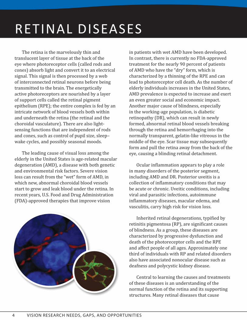
RETINAL DISEASESThe retina is the marvelously thin and
translucent layer of tissue at the back of the eye where photoreceptor cells (called rods and cones) absorb light and convert it to an electrical signal. This signal is then processed by a web of interconnected retinal neurons before being transmitted to the brain. The energetically active photoreceptors are nourished by a layer of support cells called the retinal pigment epithelium (RPE); the entire complex is fed by an intricate network of blood vessels both within and underneath the retina (the retinal and the choroidal vasculature). There are also light-sensing functions that are independent of rods and cones, such as control of pupil size, sleep–wake cycles, and possibly seasonal moods.
The leading cause of visual loss among the elderly in the United States is age-related macular degeneration (AMD), a disease with both genetic and environmental risk factors. Severe vision loss can result from the “wet” form of AMD, in which new, abnormal choroidal blood vessels start to grow and leak blood under the retina. In recent years, U.S. Food and Drug Administration (FDA)-approved therapies that improve vision
in patients with wet AMD have been developed. In contrast, there is currently no FDA-approved treatment for the nearly 90 percent of patients of AMD who have the “dry” form, which is characterized by a thinning of the RPE and can lead to photoreceptor cell death. As the number of elderly individuals increases in the United States, AMD prevalence is expected to increase and exert an even greater social and economic impact. Another major cause of blindness, especially in the working-age population, is diabetic retinopathy (DR), which can result in newly formed, abnormal retinal blood vessels breaking through the retina and hemorrhaging into the normally transparent, gelatin-like vitreous in the middle of the eye. Scar tissue may subsequently form and pull the retina away from the back of the eye, causing a blinding retinal detachment.
Ocular inflammation appears to play a role in many disorders of the posterior segment, including AMD and DR. Posterior uveitis is a collection of inflammatory conditions that may be acute or chronic. Uveitic conditions, including viral and parasitic infections, autoimmune inflammatory diseases, macular edema, and vasculitis, carry high risk for vision loss.
Inherited retinal degenerations, typified by retinitis pigmentosa (RP), are significant causes of blindness. As a group, these diseases are characterized by progressive dysfunction and death of the photoreceptor cells and the RPE and affect people of all ages. Approximately one third of individuals with RP and related disorders also have associated nonocular disease such as deafness and polycystic kidney disease.
Central to learning the causes and treatments of these diseases is an understanding of the normal function of the retina and its supporting structures. Many retinal diseases that cause
RETINAL DISEASES 5
catastrophic vision loss are characterized by abnormalities in the retinal and choroidal blood vessels. Thus, it is critical to understand normal vascular development and events leading to abnormalities in these vessels. Photoreceptors have a unique biology in which the outer segments of the cells contain stacks of membranous discs loaded with the photosensitive pigment proteins (e.g., rhodopsin). When excited by light, pigment proteins initiate a biochemical cascade called phototransduction. The stacked photoreceptor discs are constantly renewed. New discs are added at the base of the outer segment as older discs are displaced, shed, and engulfed by the RPE, so that the visual pigment can be
recycled. The photoreceptors and RPE are closely associated, and an abnormality in one leads to dysfunction in the other.
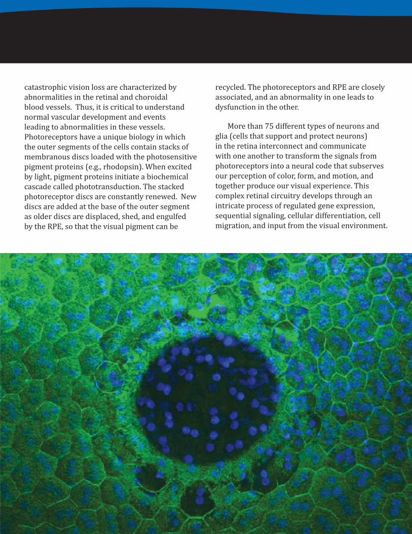
More than 75 different types of neurons and glia (cells that support and protect neurons) in the retina interconnect and communicate with one another to transform the signals from photoreceptors into a neural code that subserves our perception of color, form, and motion, and together produce our visual experience. This complex retinal circuitry develops through an intricate process of regulated gene expression, sequential signaling, cellular differentiation, cell migration, and input from the visual environment.
6 VISION RESEARCH NEEDS, GAPS, AND OPPORTUNITIES
RETINAL DISEASES HIGHLIGHTS OF RECENT PROGRESS
GENETICS AND GENE THERAPY
In the past decade, biomedical science has undergone a paradigm shift as a result of the progress in genetics, DNA sequencing technology, and the mapping of the human genome. Applying these new tools in genetics studies has led to the identification of genes responsible for many Mendelian (single-gene) retinal degenerative diseases as well as complex diseases (involving many genes and environmental influences). Gene therapy is a promising new field of medicine that replaces mutant genes with functioning genes. Pioneering studies in ocular gene therapy were initiated partly because the eye is easily accessible and somewhat immunologically isolated from the rest of the body, which minimizes adverse systemic side effects (see sidebar page 7).
One example of ocular gene therapy’s success has been in developing viral-based strategies to deliver genes to adult tissue. Advances in the understanding of gene expression as well as virology have facilitated the development of cell-specific promoters that can target replacement genes specifically to individual cell types within the retina and RPE. In a major early success, teams of scientists and clinicians used this approach to add a correct copy of the RPE65 gene to the RPE cells of patients with a severe, early-onset retinal degeneration called Leber congenital amaurosis (LCA). After receiving a correct copy of the gene for RPE65, patients performed better on tests of visual function and had improved ability to perform visual tasks when using their treated eye.
Gene therapy requires that the disease-causing mutations already be known. Fortunately, in the past decade, great advances have been made in identifying genes responsible for many
Mendelian retinal degenerative diseases. The number of such genes underlying major diseases has been surprisingly large. For example, LCA can be caused by a mutation in any one of 12 different genes expressed in either the photoreceptors or the RPE. RP also exhibits several patterns of inheritance. At present, 51 genetic loci have been implicated in nonsyndromic autosomal-recessive RP, each accounting for only a few percent of RP cases. An additional 56 loci are associated with syndromic disease, and still, the identified disease genes account for only half of affected patients. Classical genetics approaches have led to the discovery of many RP genes, and in some cases, the combined approaches of human genomic sequencing, animal modeling, and in silico prediction of protein function identified a genetic variation linking RP to a biological pathway.
In the past decade, it has become apparent that some of the most common blinding diseases, like glaucoma and AMD, are complex and may involve the individual’s immune response and environmental factors. In the first successful application of Genome-Wide Association Studies (GWAS) for identifying genes that contribute to common diseases, geneticists discovered a major contribution to AMD from a variant of a gene involved in the innate immune system, complement factor H. Since this discovery, other risk factors in the complement pathway have been associated with AMD. In addition to identifying genes that either protect from or predispose individuals to AMD, epidemiological studies have identified environmental risk factors such as smoking, hormone therapy, and possible dietary factors that may either contribute to or protect from AMD risk. Such factors likely operate both independently and through their interactions with various genes.
RETINAL DISEASES 7
For complex degenerative diseases in which single-gene therapy is insufficient, major advances have been made in animal models by expressing neurotrophic genes that promote cell survival. In cases where either these cell survival strategies are ineffective or photoreceptor degeneration already has occurred, an alternative strategy is to deliver
genes for light-sensitive proteins and channels to other retinal neurons that are normally unable to detect light. Cellular expression of these light-sensitive molecules turns neurons into functional substitutes for the dead photoreceptors. This approach, called optogenetics, is becoming widely used in neuroscience research to manipulate neuronal function.
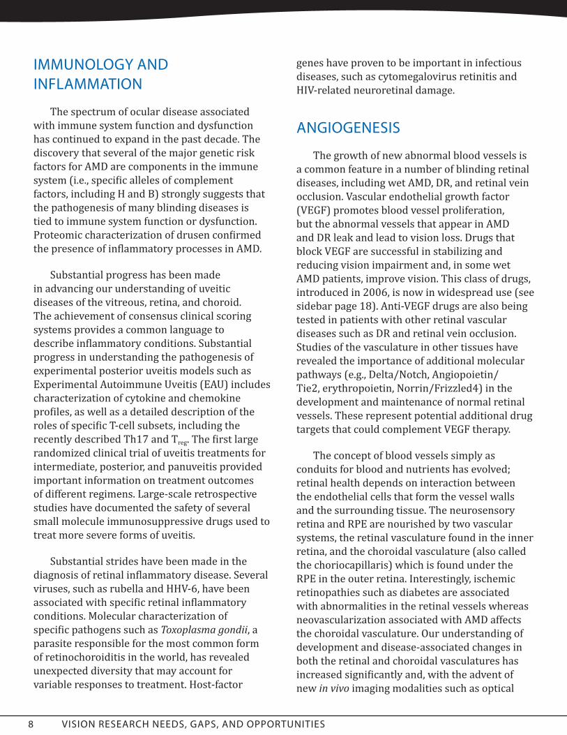
Restoring Sight One Gene at a TimeWe are learning that the most common diseases are genetically complex. That is, many genes may contribute to the course of a disease, and the influence of these genes, termed risk factors, may vary tremendously from patient to patient, depending on other factors such as excess weight or smoking. However, there are thousands of rare diseases, many of which primarily affect the eye, that are caused by a defect, i.e., mutation, of a single gene. Gene therapy consists of replacing a mutated gene with a
healthy gene to cure patients. The eye is particularly well-suited to test gene replacement therapy because the eye is easily accessible and somewhat isolated from the rest of the body. Therefore, injections of relatively small quantities of the replacement gene can be targeted specifically to the diseased tissue without much exposure to other tissues in the body. This helps to minimize side effects.
The first ocular gene therapy success—one of the first successes in any tissue—was achieved in 2009 in a clinical trial for patients with Leber congenital amaurosis, a disease that usually begins in childhood and eventually leads to blindness. Vision improved after a single treatment with a normal gene. This NEI-supported landmark clinical trial validated gene therapy as a viable investigational therapy and paved the way for additional gene therapy trials of other ocular diseases such as retinitis pigmentosa, choroideremia, age-related macular degeneration, and Stargardt’s disease.
To identify individuals with inherited blinding disorders that could potentially benefit from gene therapy, and to facilitate patient recruitment for gene therapy trials, NEI created eyeGENE®, a nationwide partnership of 250 clinical organizations that was formed to broaden accessibility of diagnostic genetic testing. Since it was established in 2006, eyeGENE® has tested more than 3,500 patient samples for 70 mutant genes and created a repository of DNA from patients and their families that is available to researchers for future studies on the genetic causes of eye diseases.
8 VISION RESEARCH NEEDS, GAPS, AND OPPORTUNITIES
IMMUNOLOGY AND INFLAMMATION
The spectrum of ocular disease associated with immune system function and dysfunction has continued to expand in the past decade. The discovery that several of the major genetic risk factors for AMD are components in the immune system (i.e., specific alleles of complement factors, including H and B) strongly suggests that the pathogenesis of many blinding diseases is tied to immune system function or dysfunction. Proteomic characterization of drusen confirmed the presence of inflammatory processes in AMD.
Substantial progress has been made in advancing our understanding of uveitic diseases of the vitreous, retina, and choroid. The achievement of consensus clinical scoring systems provides a common language to describe inflammatory conditions. Substantial progress in understanding the pathogenesis of experimental posterior uveitis models such as Experimental Autoimmune Uveitis (EAU) includes characterization of cytokine and chemokine profiles, as well as a detailed description of the roles of specific T-cell subsets, including the recently described Th17 and Treg. The first large randomized clinical trial of uveitis treatments for intermediate, posterior, and panuveitis provided important information on treatment outcomes of different regimens. Large-scale retrospective studies have documented the safety of several small molecule immunosuppressive drugs used to treat more severe forms of uveitis.
Substantial strides have been made in the diagnosis of retinal inflammatory disease. Several viruses, such as rubella and HHV-6, have been associated with specific retinal inflammatory conditions. Molecular characterization of specific pathogens such as Toxoplasma gondii, a parasite responsible for the most common form of retinochoroiditis in the world, has revealed unexpected diversity that may account for variable responses to treatment. Host-factor
genes have proven to be important in infectious diseases, such as cytomegalovirus retinitis and HIV-related neuroretinal damage.
ANGIOGENESIS
The growth of new abnormal blood vessels is a common feature in a number of blinding retinal diseases, including wet AMD, DR, and retinal vein occlusion. Vascular endothelial growth factor (VEGF) promotes blood vessel proliferation, but the abnormal vessels that appear in AMD and DR leak and lead to vision loss. Drugs that block VEGF are successful in stabilizing and reducing vision impairment and, in some wet AMD patients, improve vision. This class of drugs, introduced in 2006, is now in widespread use (see sidebar page 18). Anti-VEGF drugs are also being tested in patients with other retinal vascular diseases such as DR and retinal vein occlusion. Studies of the vasculature in other tissues have revealed the importance of additional molecular pathways (e.g., Delta/Notch, Angiopoietin/Tie2, erythropoietin, Norrin/Frizzled4) in the development and maintenance of normal retinal vessels. These represent potential additional drug targets that could complement VEGF therapy.
The concept of blood vessels simply as conduits for blood and nutrients has evolved; retinal health depends on interaction between the endothelial cells that form the vessel walls and the surrounding tissue. The neurosensory retina and RPE are nourished by two vascular systems, the retinal vasculature found in the inner retina, and the choroidal vasculature (also called the choriocapillaris) which is found under the RPE in the outer retina. Interestingly, ischemic retinopathies such as diabetes are associated with abnormalities in the retinal vessels whereas neovascularization associated with AMD affects the choroidal vasculature. Our understanding of development and disease-associated changes in both the retinal and choroidal vasculatures has increased significantly and, with the advent of new in vivo imaging modalities such as optical
RETINAL DISEASES 9
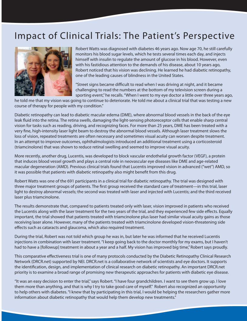
Impact of Clinical Trials: The Patient’s PerspectiveRobert Watts was diagnosed with diabetes 46 years ago. Now age 70, he still carefully monitors his blood sugar levels, which he tests several times each day, and injects himself with insulin to regulate the amount of glucose in his blood. However, even with his fastidious attention to the demands of his disease, about 10 years ago, Robert noticed that his vision was declining. He learned he had diabetic retinopathy, one of the leading causes of blindness in the United States.
“Street signs became difficult to read when I was driving at night, and it became challenging to read the numbers at the bottom of my television screen during a sporting event,” he recalls. “When I went to my eye doctor a little over three years ago,
he told me that my vision was going to continue to deteriorate. He told me about a clinical trial that was testing a new course of therapy for people with my condition.”
Diabetic retinopathy can lead to diabetic macular edema (DME), where abnormal blood vessels in the back of the eye leak fluid into the retina. The retina swells, damaging the light-sensing photoreceptor cells that enable sharp central vision for tasks such as reading, driving, and recognizing faces. For more than 25 years, DME has been treated with a very fine, high-intensity laser light beam to destroy the abnormal blood vessels. Although laser treatment slows the loss of vision, repeated treatments are often necessary and sometimes visual acuity can worsen despite treatment. In an attempt to improve outcomes, ophthalmologists introduced an additional treatment using a corticosteroid (triamcinolone) that was shown to reduce retinal swelling and seemed to improve visual acuity.
More recently, another drug, Lucentis, was developed to block vascular endothelial growth factor (VEGF), a protein that induces blood vessel growth and plays a central role in neovascular eye diseases like DME and age-related macular degeneration (AMD). Previous clinical trials found that Lucentis improved vision in advanced (“wet”) AMD, so it was possible that patients with diabetic retinopathy also might benefit from this drug.
Robert Watts was one of the 691 participants in a clinical trial for diabetic retinopathy. The trial was designed with three major treatment groups of patients. The first group received the standard care of treatment—in this trial, laser light to destroy abnormal vessels; the second was treated with laser and injected with Lucentis; and the third received laser plus triamcinolone.
The results demonstrate that, compared to patients treated only with laser, vision improved in patients who received the Lucentis along with the laser treatment for the two years of the trial, and they experienced few side effects. Equally important, the trial showed that patients treated with triamcinolone plus laser had similar visual acuity gains as those receiving laser alone. However, many of the patients treated with triamcinolone developed vision-threatening side effects such as cataracts and glaucoma, which also required treatment.
During the trial, Robert was not told which group he was in, but later he was informed that he received Lucentis injections in combination with laser treatment. “I keep going back to the doctor monthly for my exams, but I haven’t had to have a [followup] treatment in about a year and a half. My vision has improved big time,” Robert says proudly.
This comparative effectiveness trial is one of many protocols conducted by the Diabetic Retinopathy Clinical Research Network (DRCR.net) supported by NEI. DRCR.net is a collaborative network of scientists and eye doctors. It supports the identification, design, and implementation of clinical research on diabetic retinopathy. An important DRCR.net priority is to examine a broad range of promising new therapeutic approaches for patients with diabetic eye disease.
“It was an easy decision to enter the trial,” says Robert. “I have four grandchildren. I want to see them grow up. I love them more than anything, and that is why I try to take good care of myself.” Robert also recognized an opportunity to help others with diabetes. “I knew that by participating in this trial, I would be helping the researchers gather more information about diabetic retinopathy that would help them develop new treatments.”
10 VISION RESEARCH NEEDS, GAPS, AND OPPORTUNITIES
coherence tomography (OCT), will further expand our understanding of how these vessels respond to disease. The role of the vascular microenvironment under conditions of health and disease, as well as nonvascular cells associated with the vasculature, are critical in maintaining vascular homeostasis.
Large, retina-specific cells that support the neurons, called Müller glia, play an important role in regulating the amount of water and electrical current in the retina through their contacts with both retinal neurons and vascular endothelial cells. Müller cells are damaged in certain diseases and can lead to significant changes in the retinal vasculature. Following hypoxic injury—where the tissue is deprived of oxygen—microglia and circulating white blood cells are “activated” in the retina and facilitate the repair and regrowth of blood vessels. Thus, the importance of cellular cross-talk in development and maintenance of a healthy retinal vasculature is critical to normal retinal function.
PHOTORECEPTOR AND RPE BIOLOGY
The absorption of photons in rod and cone photoreceptors is the first step in a chain of biochemical events known as phototransduction, which initiates electrical signals that lead to visual perception. Photon absorption occurs in the visual pigments, rhodopsin and cone opsins, which consist of protein polypeptides and the vitamin A-derived chromophore, 11-cis retinal. In the last decade, major advances occurred in understanding how 11-cis retinal is provided to rods and cones and in determining the consequences when the chromophore is not synthesized in adequate quantities. The enzymes responsible for chromophore production and regeneration, the visual cycle, have been identified in both photoreceptors and the RPE.
RPE regeneration of 11-cis retinal is not its only source. An alternative recycling pathway, via Müller cells selectively feeding back to cones but not rods, was recently discovered. This pathway may contribute to faster regeneration of visual pigment in cones, which must maintain light sensitivity in much brighter light conditions. Because 11-cis retinal is important for normal folding and trafficking of visual pigments, this new pathway may be important for treating RP and LCA since these diseases often stem from improper folding or trafficking of visual pigment proteins.
Phototransduction is a model system for cellular signaling and is one of the most highly amplifying signaling mechanisms known in biology. Absorption of photons triggers a molecular chain reaction with signal amplification at each step in the chain. The amplification can be modified by feedback systems that alter the sensitivity or activation of signaling molecules. A recent advance is the discovery that the G-protein coupled receptor, rhodopsin, remains active for only a short time after it absorbs a photon. Subsequent G-protein signaling molecules remain active for a longer time, which may have functional consequences for the temporal resolution of vision. Indeed, mutations that slow down G-protein inactivation cause bradyopsia in humans, a disorder characterized by difficulties in perceiving low contrast moving objects and adapting to large changes in light intensity.
Another important area of progress related to photoreceptor biology is the recognition that the outer segments of rods and cones are specialized sensory cilia. It is now evident that primary cilia are present on most vertebrate cell types. These structures are typically sensory organelles, and are involved in many critical aspects of cell biology. Like other cilia, the outer segments contain an axoneme, which begins at the basal bodies, passes through a transition zone (also called the “connecting cilium”) and into the outer segment. An important benefit of recognizing
RETINAL DISEASES 11
photoreceptor outer segments as cilia is that it connects retinal degenerative disorders such as LCA and RP to other cilia disorders such as cystic renal disease, polydactyly, mental retardation, obesity, gonadal malformations, and diabetes.
The ability to adapt to large variations in light intensity is important for human vision. In the past few years, our understanding of adaptation has changed dramatically, owing to the discovery that many phototransduction proteins move in and out of subcellular compartments in response to different levels of illumination. Such large-scale movements reduce the light-generated signals, thereby contributing to the protection and health of photoreceptors and their function in bright light.
Another discovery with both research and therapeutic implications is that the retina contains neurons other than the rods and cones that are directly light responsive. These intrinsically-photosensitive retinal ganglion cells (ipRGCs) use an 11-cis retinal-based visual pigment called melanopsin, and project not only to the midbrain and hypothalamus, but also to the thalamus. As such, ipRGCs are important for signaling overall luminance levels needed for circadian entrainment and the pupillary light reflex and also play a direct role in the general perception of light.
RPE cells are polarized, compartmentalized, and have 22 known functions to date. They play critical roles in maintaining photoreceptor and choroidal function by producing key neural and blood vessel growth factors, such as VEGF and pigment epithelial–derived factor, recycling photoreceptor components, phagocytosing shed photoreceptor outer segments, and regulating subretinal fluid. Recent advances have identified new roles in orchestrating the immune response of the outer retina through complement regulators and secreting proteins and lipoproteins that contribute to drusen formation.
RETINAL STRUCTURE, FUNCTION, AND CIRCUITRY
With only a handful of different neuronal classes (photoreceptors, horizontal cells, bipolar cells, amacrine, cells and ganglion cells), the retina functions across 10 log units of luminance intensity and also performs the initial processing of visual information. For example, within the retinal circuit, salient features of the visual world become encoded, including color, contrast, motion, and direction of visual stimuli. The unique experimental accessibility and anatomy of the circuitry have allowed rigorous investigations of synaptic and circuit function unlike anywhere else in the brain and serves as a model system for understanding the behavior of circuits in the central nervous system (CNS).
In recent years, understanding of the diversity of retinal cell types has greatly expanded due to the ability to identify molecular markers unique to individual classes of cells. This “molecular fingerprinting” has led to an increase in the identification of specific promoters that control which genes get turned on in a given cell type. Using these promoters, researchers can identify specific types of retinal cells much more easily, which has led to a better understanding of cell fate and development, and the role of specific cell classes in circuit function. Knowledge of these cell-specific promoters also has been critical for optogenetics studies to restore light-sensitivity to specific types of cells in nonfunctional retinas in animal models of disease.
Retinal neurons communicate with each other through an interlaced network of branched axons and dendrites. The identification of molecular cues (DSCAM, sidekick, semaphorins, and other proteins) that govern the development of laminar organization of the inner plexiform layer is important for understanding normal retinal development and circuitry.
12 VISION RESEARCH NEEDS, GAPS, AND OPPORTUNITIES
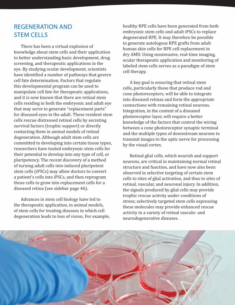
REGENERATION AND STEM CELLS
There has been a virtual explosion of knowledge about stem cells and their application to better understanding basic development, drug screening, and therapeutic applications in the eye. By studying ocular development, scientists have identified a number of pathways that govern cell fate determination. Factors that regulate this developmental program can be used to manipulate cell fate for therapeutic applications, and it is now known that there are retinal stem cells residing in both the embryonic and adult eye that may serve to generate “replacement parts” for diseased eyes in the adult. These resident stem cells rescue distressed retinal cells by secreting survival factors (trophic support) or directly contacting them in animal models of retinal degeneration. Although adult stem cells are committed to developing into certain tissue types, researchers have touted embryonic stem cells for their potential to develop into any type of cell, or pluripotency. The recent discovery of a method of turning adult cells into induced pluripotent stem cells (iPSCs) may allow doctors to convert a patient’s cells into iPSCs, and then reprogram those cells to grow into replacement cells for a diseased retina (see sidebar page 46).
Advances in stem cell biology have led to the therapeutic application, in animal models, of stem cells for treating diseases in which cell degeneration leads to loss of vision. For example,
healthy RPE cells have been generated from both embryonic stem cells and adult iPSCs to replace degenerated RPE. It may therefore be possible to generate autologous RPE grafts from adult human skin cells for RPE cell replacement in dry AMD. Using noninvasive, real-time imaging, ocular therapeutic application and monitoring of labeled stem cells serves as a paradigm of stem cell therapy.
A key goal is ensuring that retinal stem cells, particularly those that produce rod and cone photoreceptors, will be able to integrate into diseased retinas and form the appropriate connections with remaining retinal neurons. Integration, in the context of a diseased photoreceptor layer, will require a better knowledge of the factors that control the wiring between a cone photoreceptor synaptic terminal and the multiple types of downstream neurons to transmit images to the optic nerve for processing by the visual cortex.
Retinal glial cells, which nourish and support neurons, are critical in maintaining normal retinal structure and function, and have now also been observed in selective targeting of certain stem cells to sites of glial activation, and thus to sites of retinal, vascular, and neuronal injury. In addition, the signals produced by glial cells may provide trophic rescue activity under conditions of stress; selectively targeted stem cells expressing these molecules may provide enhanced rescue activity in a variety of retinal vasculo- and neurodegenerative diseases.
RETINAL DISEASES 13
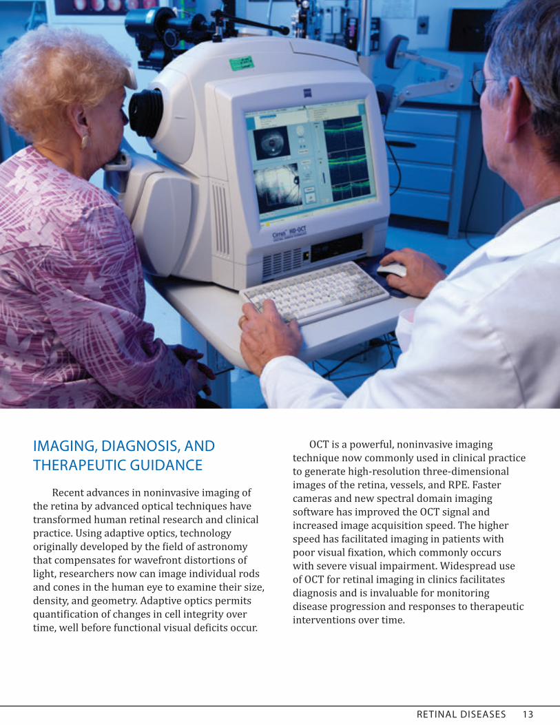
IMAGING, DIAGNOSIS, AND THERAPEUTIC GUIDANCE
Recent advances in noninvasive imaging of the retina by advanced optical techniques have transformed human retinal research and clinical practice. Using adaptive optics, technology originally developed by the field of astronomy that compensates for wavefront distortions of light, researchers now can image individual rods and cones in the human eye to examine their size, density, and geometry. Adaptive optics permits quantification of changes in cell integrity over time, well before functional visual deficits occur.
OCT is a powerful, noninvasive imaging technique now commonly used in clinical practice to generate high-resolution three-dimensional images of the retina, vessels, and RPE. Faster cameras and new spectral domain imaging software has improved the OCT signal and increased image acquisition speed. The higher speed has facilitated imaging in patients with poor visual fixation, which commonly occurs with severe visual impairment. Widespread use of OCT for retinal imaging in clinics facilitates diagnosis and is invaluable for monitoring disease progression and responses to therapeutic interventions over time.
14 VISION RESEARCH NEEDS, GAPS, AND OPPORTUNITIES
RETINAL DISEASES NEEDS, GAPS, AND OPPORTUNITIES
GENETICS AND GENE THERAPY• Develop better gene delivery methods,
including viral-mediated or new technologies, such as nanoparticles, that target specific subclasses of neurons, glia, and RPE.
• Expand, improve, and coordinate shared bioinformatics approaches and resources to analyze and annotate genetics data, including improved predications of potential pathogenicity of identified sequence variants.
• Develop higher throughput experimental methods to assess the functional significance of sequence variants identified by next-generation sequencing (NGS) and GWAS studies in retinal cells.
• Explore the role of non-Mendelian genetics, including epigenetic modifications, microRNA gene regulation, mitochondrial genetics and genetic modifier effects during development, normal aging, and neurodegeneration in order to understand the causes of complex disease and the risk of disease development.
• Understand how individuals’ genetic makeup may predict their response to a treatment, since not all individuals respond the same to standard therapies.
ANGIOGENESIS• Identify and characterize new angiostatic and
antipermeability agents, especially for use in individuals who are unresponsive to anti-VEGF therapy.
• Test the combination therapy of angiostatic, antipermeability, anti-inflammatory, and neuroprotective agents for retinal vasculo- and neurodegenerative diseases.
• Explore the potential use of targeted, cell-based therapies for the treatment of retinal vascular diseases.
• Identify and better understand the potential use of microRNAs, antimicroRNAs, and other RNA-based therapeutics for the treatment of retinal vascular diseases.
• Analyze the role of nonendothelial cells (e.g., astrocytes, Müller glia, microglia, pericytes, macrophages) in the maintenance of normal retinal vasculature and their role during retinal vascular disease.
• Gain a better understanding of mechanisms of choroidal involution and its role in providing nutrition to the RPE and photoreceptors.
PHOTORECEPTOR, RPE, AND GLIAL BIOLOGY• Elucidate the molecular mechanisms that
lead to photoreceptor degeneration, including signal transduction pathways, defects in protein folding, ciliogenesis, functional compartmentalization, and trafficking. Translate these molecular footholds into therapies for Mendelian and complex diseases.
• Understand the molecular mechanisms and pathways in cone photoreceptors that have not been as extensively studied as rods. Primates rely on cone-based color vision (concentrated in the macula), unlike many animal models commonly used for vision research, such as rodents, that do not have a macula.
• Understand the natural neuroprotective mechanisms in photoreceptors, including the regulation of energy metabolism in order to develop therapeutic strategies extending the healthy lifetime of these cells.
RETINAL DISEASES 1514 VISION RESEARCH NEEDS, GAPS, AND OPPORTUNITIES
• Translate research progress in rod and cone retinoid visual cycles into clinical trials with retinoid/retinoid-like compounds. Recent discoveries that several retinal degenerations result from enzyme malfunctions at different points in the retinoid generation cycles provide a therapeutic opportunity to supply missing retinoids.
• Better understand highly differentiated RPE functions and elucidate mechanisms for maximizing RPE health over the lifespan of an individual.
• Better understand the role of various posterior segment extracellular matrices (e.g., Bruch’s membrane, vitreous) in maintaining normal retinal functioning.
• Characterize and better understand the roles for various resident and transient populations of glia present in the retina.
• Characterize the macula and perifoveal regions of the retina to better understand the predilection of the macula for disease.
PHOTORECEPTION AND RETINAL CIRCUITRY • Understand and model the structure, function,
and circuitry of retinal neurons. This research forms the basis for interpreting tests of retinal function such as the electroretinogram and various psychophysical paradigms that are used to detect retinal diseases, monitor the progression of disease, and assess treatments. Also, for stem cell replacement therapies to become a reality, transplanted neurons must functionally integrate and synapse with the existing retina.
• Decode the electrical patterns used by retinal neurons to transmit visual information to the
brain. Opportunities include newly developed techniques of viral-mediated pathway tracing and large-scale multielectrode arrays, genetically directed cell-specific labeling, updated and automated serial EM reconstruction, and single-cell recordings to achieve high-resolution maps of functional retinal circuits. New technologies such as optogenetics and multiphoton microscopy confer an ability to manipulate neuronal activity at the cellular level and visually and functionally dissect circuits by activating or suppressing specific activity in the retina.
• Build on the existing arsenal of tools to develop methods for measuring activity in large groups of neurons during light responses. Neuroscientists have been able to simultaneously study large populations of neurons through optical techniques that track activity with fluorescent markers introduced in groups of cells. However, many of these techniques focus light in the visible spectrum onto the tissue, which limits their use in the retina, in which cells are activated by this light. The light may bleach the photoreceptor pigments. Two-photon technology, which uses infrared light outside the visible spectrum and large microelectrode arrays, will help overcome these barriers.
• Characterize the molecular mechanisms that establish and maintain synapses and define circuits in the inner and outer plexiform layers. This would include determining the extent to which those contacts are structurally and functionally modifiable to predict pathological consequences as well as to predict the best retinal layer at which to aim treatments to restore visual sensitivity.
• Improve understanding of the role of retinal glial cells (Müller glia, astrocytes, microglia)
16 VISION RESEARCH NEEDS, GAPS, AND OPPORTUNITIES
with regard to wound healing (e.g., gliosis), angiogenesis (e.g., neovascularization) and neuronal survival. These cells have been viewed as largely structural, but recent advances have demonstrated a much more dynamic role (paracrine and autocrine) in most of the diseases that lead to vision loss.
IMMUNOLOGY • Continue the process of standardizing
nomenclature of uveitic diseases. The Standardization of Uveitis Nomenclature working group is continuing to validate descriptions of specific syndromes to allow standardization of criteria for clinical research.
• Recognize the heterogeneity of posterior uveitis in clinical trial design such that diverse entities such as birdshot chorioretinopathy, pars planitis, serpiginous choroiditis, sarcoid-associated uveitis, and Behcet’s disease–associated uveitis are treated as unique diagnoses for any assessment of outcome.
• Understand the role of innate immune responses (including the complement, chemokine, and inflammasome system) in retinal degenerative diseases, and determine how the immune system influences survival or death of retinal cells. Develop tools and markers to identify subsets of microglia, dendritic cells, circulating myeloid cells, and progenitors to establish their role in ocular immunity. Identify commonalities across different retinal degenerative diseases and degenerative diseases of the CNS such as Alzheimer’s disease and other dementias.
• Understand the roles of specific adaptive and innate immune mediators in specific inflammatory syndromes. While EAU is relatively well characterized, few other animal models of uveitis have been studied mechanistically. Understanding the functions of specific cytokines and chemokines in a range of models (including rodent models of autoinflammatory diseases as well as
spontaneous uveitis models in animals such as dogs and horses) will inform the application of emerging biologic treatments to specific classes of uveitis.
• Integrate advances in understanding of immune regulatory mechanisms to advancing our understanding of ocular immune privilege. The first human clinical trials of both virally mediated retinal gene therapy and retinal stem cell transplantation are underway, and understanding mechanisms of tolerance induction in the eye will assume increased importance.
• Advance diagnostic tools for ocular inflammatory disease by using new molecular information to inform clinical studies. Improvement in imaging techniques for immune cells in the retina will allow in vivo assessment of specific inflammatory mediators.
• Pursue delivery technologies for sustained treatments of chronic ocular inflammatory diseases, including cell-mediated, virally mediated, and sustained-release technologies for anti-inflammatory biologics and small molecules. The ability to enhance the delivery of drugs locally to the eye should be complemented by a greater understanding of the role of the immune response within the eye as opposed to the systemic immune response.
• Characterize the immune response to intraocular therapies such as monoclonal antibodies or gene therapies so that those treatments can be optimized.
• Test novel therapies including monoclonal antibodies, kinase inhibitors, and resolvins for their potential to control intraocular inflammation.
IMAGING AND DIAGNOSIS• Translate high-resolution retinal imaging
technologies, like adaptive optics, into cost-effective and easy-to-use platforms for routine clinical use.
RETINAL DISEASES 17
• Develop novel, noninvasive imaging techniques for monitoring electrical or metabolic activity of retinal neurons in vivo, ideally at the spatial resolution of photoreceptors or better for early detection of disease and monitoring of therapeutic intervention. This would be a more cost- and time-effective approach to studying retinal function than current biophysical techniques (e.g., single-cell electrical recordings), with the additional benefit of studying cellular activity in its native environment.
THERAPEUTICS • Develop appropriate animal models for
pathological features of complex human diseases, including aging, noninjury-based choroidal neovascularization, proliferative diabetic retinopathy, cone visual transduction in macula, atrophic AMD, and retinal degenerations, including syndromic disorders such as Usher’s disease.
• Explore the impact of cholesterol and lipoproteins for AMD, inflammation, and angiogenesis. Multiple GWAS studies have identified gene variants in the cholesterol and lipid pathway associated with AMD risk. The role of lipids in AMD had been suggested based on their presence in both drusen and Bruch’s membrane in AMD patients. Recent results from the Women’s Health Study, a large prospective epidemiological study, also showed a substantially reduced risk of AMD in individuals with lower lipid intake in their diet.
• Explore the use of stem cells and other cell-based therapy as targeted delivery vehicles for trophic/survival factors to the retina. Neuroprotection in the face of ongoing underlying disease may serve to maintain retinal function.
• Understand how to direct stem cells (adult, embryonic, or iPS cells) down specific cell lineages for targeted cell replacement therapy for diverse retinal neuronal cell types as
well as RPE. Regenerative medicine and cell replacement therapy require further understanding of retinal stem cell niches and key developmental regulators and pathways.
• Propel research on retinal transplantation therapy by investigating requirements for functional integration of photoreceptors within a degenerate retina. The abnormal microenvironments in diseased eyes could affect transplanted cell survival, differentiation, and integration with the host.
IMPROVING PUBLIC HEALTH • Understand why certain at-risk populations
with systemic diseases do not develop ocular manifestations, e.g., diabetes patients who do not develop retinopathy. African Americans appear to develop drusen at the same rate as Caucasians, yet have a lower risk of developing end-stage AMD. The opportunity to identify which factors (molecular, environmental) protect these populations from progression or disease will be informative in understanding the disease progress and designing therapeutic interventions.
• Evaluate implementation of recent clinical discoveries in retinopathy of prematurity (ROP) and their impact on reducing childhood blindness. Clinicians have developed a web-based algorithm based on postnatal weight gain, IGF-1, and omega-3 fatty acids, which accurately predicts those infants who will develop ROP an average of three weeks after birth. These treatment decision tools, coupled with results of trials evaluating telemedicine for ROP, can be used to concentrate care on vulnerable patients.
• Expand efforts in telemedicine to manage retinal diseases like diabetic retinopathy and AMD via web-based networks, increasing access to specialists for populations in rural and/or underserved areas. Determine how these tools can be adapted to improve vision in different health delivery environments, such
18 VISION RESEARCH NEEDS, GAPS, AND OPPORTUNITIES
as in developing countries where diabetes in particular is becoming epidemic.
• Understand the connections between obesity and metabolic syndrome and vision loss, especially in aging eye diseases.
• Evaluate the long-term use of therapeutics in patients with chronic retinal disease (including retinal degenerative disease, atrophic AMD, aging retina, chronic rejection of transplants) to minimize chronic side-effects and manage adherence issues.
• Translate progress in research into best clinical practices to reduce preventable blindness or reduce the functional consequences of visual impairment. Evaluate disparities in optimal treatment by determining how research findings are
implemented by eye care providers and barriers which prevent optimal treatment. Further understanding why patients stop effective therapies (such as glucose control in diabetes, or VEGF injections in AMD) and what cost-effective practices could be implemented to identify and bring to early diagnosis and treatment patients who present with vision loss due to preventable causes.
• Explore potential similarities between AMD and other diseases of aging that affect the CNS (e.g., amyloid and Alzheimer’s).
• Expand the use of “omics” (e.g., transcriptomics, proteomics, metabolomics) to characterize ocular and systemic fluids from patients with various diseases and stages of various diseases.
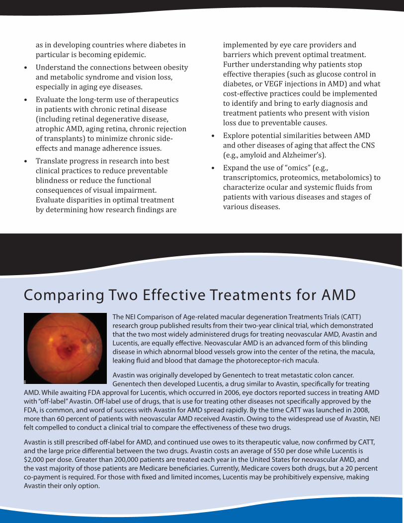
Comparing Two Effective Treatments for AMDThe NEI Comparison of Age-related macular degeneration Treatments Trials (CATT) research group published results from their two-year clinical trial, which demonstrated that the two most widely administered drugs for treating neovascular AMD, Avastin and Lucentis, are equally effective. Neovascular AMD is an advanced form of this blinding disease in which abnormal blood vessels grow into the center of the retina, the macula, leaking fluid and blood that damage the photoreceptor-rich macula.
Avastin was originally developed by Genentech to treat metastatic colon cancer. Genentech then developed Lucentis, a drug similar to Avastin, specifically for treating
AMD. While awaiting FDA approval for Lucentis, which occurred in 2006, eye doctors reported success in treating AMD with “off-label” Avastin. Off-label use of drugs, that is use for treating other diseases not specifically approved by the FDA, is common, and word of success with Avastin for AMD spread rapidly. By the time CATT was launched in 2008, more than 60 percent of patients with neovascular AMD received Avastin. Owing to the widespread use of Avastin, NEI felt compelled to conduct a clinical trial to compare the effectiveness of these two drugs.
Avastin is still prescribed off-label for AMD, and continued use owes to its therapeutic value, now confirmed by CATT, and the large price differential between the two drugs. Avastin costs an average of $50 per dose while Lucentis is $2,000 per dose. Greater than 200,000 patients are treated each year in the United States for neovascular AMD, and the vast majority of those patients are Medicare beneficiaries. Currently, Medicare covers both drugs, but a 20 percent co-payment is required. For those with fixed and limited incomes, Lucentis may be prohibitively expensive, making Avastin their only option.
CORNEAL DISEASES 19
CORNEAL DISEASES
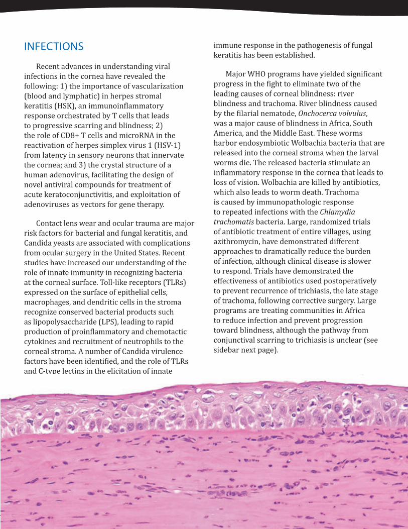
The normal cornea appears to be a simple crystal-clear structure; however, it is an elegant, complex living tissue that prevents infectious agents and debris from entering the eye. The cornea and tear film together form the primary refractive (light bending) element in the optical path, and are essential to precisely focus light on the retina. These characteristics are possible due to unique functional properties of the three primary corneal tissue layers (outward-facing epithelium, central connective tissue or stroma, and inner corneal endothelium), the resident immune cells, and the sensory nerve cells. In fact, the corneal epithelium has one of the highest densities of sensory nerve endings in the body, rendering an extremely painful sensation when even a tiny particle becomes trapped under the eyelid. Ocular surface tissues uniquely remain healthy and nourished despite the fact that the cornea has no blood vessels. Understanding the normal and diseased cornea and tear-secreting glands remains a critical need in order to reduce the burden of visual disorders worldwide.
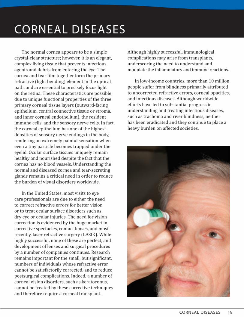
In the United States, most visits to eye care professionals are due to either the need to correct refractive errors for better vision or to treat ocular surface disorders such as dry eye or ocular injuries. The need for vision correction is evidenced by the huge market in corrective spectacles, contact lenses, and most recently, laser refractive surgery (LASIK). While highly successful, none of these are perfect, and development of lenses and surgical procedures by a number of companies continues. Research remains important for the small, but significant, numbers of individuals whose refractive error cannot be satisfactorily corrected, and to reduce postsurgical complications. Indeed, a number of corneal vision disorders, such as keratoconus, cannot be treated by these corrective techniques and therefore require a corneal transplant.
Although highly successful, immunological complications may arise from transplants, underscoring the need to understand and modulate the inflammatory and immune reactions.
In low-income countries, more than 10 million people suffer from blindness primarily attributed to uncorrected refractive errors, corneal opacities, and infectious diseases. Although worldwide efforts have led to substantial progress in understanding and treating infectious diseases, such as trachoma and river blindness, neither has been eradicated and they continue to place a heavy burden on affected societies.
20 VISION RESEARCH NEEDS, GAPS, AND OPPORTUNITIES
CORNEAL DISEASES HIGHLIGHTS OF RECENT PROGRESS
ANGIOGENESIS AND LYMPHANGIOGENESIS
Corneal avascularity is the result of a fine balance between angiogenic (vessel-forming) and anti-angiogenic factors. Infection, injury, and inflammation can tip the balance in favor of blood vessel formation (hemangiogenesis) and lymph vessel formation (lymphangiogenesis). Recent studies have shown that many regulators, including VEGF, integrins, and semaphorins, play a central role in regulating corneal angiogenesis. Epithelial-derived soluble VEGF receptor-1 (sflt-1) maintains corneal avascularity by acting as a decoy receptor, suppressing the major angiogenic factor, VEGF-A. Similarly, epithelial-derived soluble VEGF receptor-2 and cell surface-bound VEGF receptor-3 serve as decoys for VEGF-C/-D, thereby inhibiting lymphangiogenesis.
TRANSPLANTATION
Corneal transplantation is the most common solid organ transplant performed in the United States. Researchers have recently found that blood and lymphatic vessels have a role in initiating and promoting immune rejection of corneal grafts. Recent findings implicating molecular attractants (chemokines) and their receptors may lead to the development of therapeutic compounds to prevent corneal graft rejection. Specifically, the addition of interleukin 17 to the list of known Th1 cytokine mediators of corneal graft rejection broadens the armamentarium of potential anti-rejection drugs. Similarly, recent evidence suggests that immune cells (e.g., Treg
cells) and molecules (e.g., PD-1/PD-L1) that help prevent our immune system from attacking our own tissue can be exploited to prevent rejection of corneal grafts. Finally, improved imaging
technology has facilitated real-time observation of the innate and adaptive immune cells that infiltrate the corneal graft, greatly expanding our understanding of the interplay of these cells in rejecting the graft or promoting tolerance and graft survival.
Methods to surgically replace abnormal corneal tissue have advanced considerably. Endothelial keratoplasty techniques for the endothelial dystrophies have largely supplanted the use of full-thickness-penetrating keratoplasty, resulting in faster recovery times and fewer side effects.
The limited availability of donor tissue, especially worldwide, as well as the possible rejection of donor corneas, makes the development of artificial, bioengineered corneas highly desirable. Artificial corneas engineered from cross-linked collagen have undergone extensive in vitro testing and are currently being tested in both animals and humans. Based on results from early studies, tissue-engineered corneas have been shown to maintain clarity, strength, biocompatibility, and are well-integrated in the eyes of patients, but further study is necessary.
REGENERATIVE MEDICINE
The ability to isolate and expand limbal-derived stem cells to generate confluent sheets of tissue on a support structure, such as decellularized amniotic membrane or other carriers, and successfully resurface the corneal epithelium represents an honorable accomplishment for regenerative medicine. This breakthrough resulted from a combination of basic research on the biology of epithelial stem cells and enhanced clinical understanding of anterior surface diseases, yielding one of the first therapeutic uses of stem cells.
CORNEAL DISEASES 21
WOUND HEALING
Corneal epithelial injury resulting from mechanical or chemical trauma, as well as elective refractive surgeries (e.g., LASIK or PRK), involves a loss of nerves and keratocytes at the site of injury. Delayed wound healing is associated with diseases such as diabetes. Recent studies using animal models of corneal injury show the accompanying inflammatory response proceeds as a cascade of events that supports epithelial, nerve, and keratocyte recovery. Recruitment of inflammatory leukocytes to the injured cornea is orchestrated by specific cell-adhesion molecules, chemokines and cytokines. Several components of the inflammatory cascade that are critical to the beneficial healing effect have been identified at the limbus and include epithelial γδ T cells, stromal neutrophils, and platelets. Growth factors such as VEGF are critical for nerve regeneration and show increased expression in the wounded cornea that depends on recruited neutrophils and platelets.
TEARS/DRY EYE DISORDERS
Improved understanding of the composition and multiple functions of tears includes new understanding of anti-bacterial defensins; functional characterization of a tear-specific protein, lacritin; understanding the structure and function of lipocalin; and identification of meibomian gland lipids. A desiccating stress animal model has led to new information on the role of inflammation in dry eye as it relates to environmental stress. A model of spontaneous ocular surface inflammation demonstrated the genetic susceptibility to cholinergic impairment of lacrimal gland and goblet cell secretion that leads to a chronic disease involving proinflammatory
Th17 T cells. Another important finding is the association of dry eye disease with an increase in corneal lymphangiogenesis driven by VEGF receptor 3. Important advances have also been made in understanding the regulation of goblet cell mucin secretion and the role of membrane-spanning mucins in protecting the ocular surface.
A large multicountry study has recently developed strict diagnostic criteria for Sjögren’s syndrome, a disease in which immune cells attack and destroy the exocrine glands that produce tears and saliva, allowing future studies to focus on the particular syndrome rather than a group of unrelated disorders. Epidemiological studies on dry eyes have documented the incidence, dietary and associated drug-related influences, and impact on the quality of life.
BIOMECHANICS, IMAGING, AND REFRACTIVE ERROR CORRECTION
Effective treatments for diseases of the cornea require early and definitive disease diagnosis, well-defined staging, and clinically relevant endpoints. The location and transparency of the cornea lends itself to noninvasive assays and monitoring. Recent developments in imaging technology have advanced our understanding of corneal structure, function, and dysfunction in corneal diseases. Advances in confocal and multiphoton microscopy have enabled direct imaging and real-time characterization of the cornea at the cellular scale. Dramatic improvements in OCT technology now allow for rapid, volumetric imaging of the cornea and anterior segment visual pathway, with the promise of enabling routine biometric and biomechanical measurements in normal as well as surgically altered corneas.
22 VISION RESEARCH NEEDS, GAPS, AND OPPORTUNITIES
Fundamental recent advances in theory and modeling of corneal optics and biomechanics are laying the groundwork for optimizing corneal surgical interventions and bioengineered implants. Improved schematic and personalized ray-traced modeling of corneal optics have been introduced, coupled with new methods for measuring high-order and dynamic aberrations. Optimized finite-element numerical modeling techniques have been developed, which promise to improve predictions of corneal structural response to refractive surgery.
New wavefront-sensing techniques are being used to identify higher order aberrations of the eye, which can then be corrected during refractive surgery, making glare and haloes less common and improving visual acuity. Femtosecond lasers have moved rapidly into widespread use in LASIK refractive surgery and increasingly in corneal transplantation. Femtosecond lasers have improved the safety of these procedures while hastening the recovery time after surgery.
Advances have been made in our understanding of the progressive loss of corneal biomechanical stability found both in keratoconus and sometimes after laser vision correction surgery. New nonsurgical techniques for
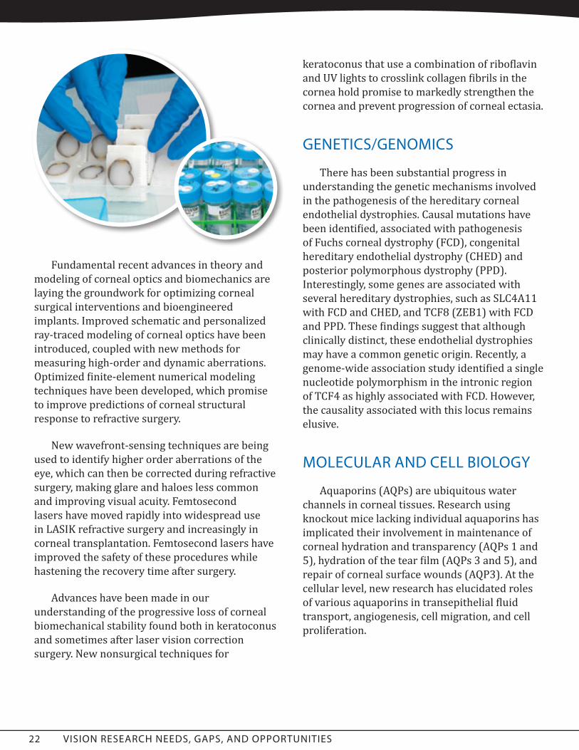
keratoconus that use a combination of riboflavin and UV lights to crosslink collagen fibrils in the cornea hold promise to markedly strengthen the cornea and prevent progression of corneal ectasia.
GENETICS/GENOMICS
There has been substantial progress in understanding the genetic mechanisms involved in the pathogenesis of the hereditary corneal endothelial dystrophies. Causal mutations have been identified, associated with pathogenesis of Fuchs corneal dystrophy (FCD), congenital hereditary endothelial dystrophy (CHED) and posterior polymorphous dystrophy (PPD). Interestingly, some genes are associated with several hereditary dystrophies, such as SLC4A11 with FCD and CHED, and TCF8 (ZEB1) with FCD and PPD. These findings suggest that although clinically distinct, these endothelial dystrophies may have a common genetic origin. Recently, a genome-wide association study identified a single nucleotide polymorphism in the intronic region of TCF4 as highly associated with FCD. However, the causality associated with this locus remains elusive.
MOLECULAR AND CELL BIOLOGY
Aquaporins (AQPs) are ubiquitous water channels in corneal tissues. Research using knockout mice lacking individual aquaporins has implicated their involvement in maintenance of corneal hydration and transparency (AQPs 1 and 5), hydration of the tear film (AQPs 3 and 5), and repair of corneal surface wounds (AQP3). At the cellular level, new research has elucidated roles of various aquaporins in transepithelial fluid transport, angiogenesis, cell migration, and cell proliferation.
CORNEAL DISEASES 23
INFECTIONS
Recent advances in understanding viral infections in the cornea have revealed the following: 1) the importance of vascularization (blood and lymphatic) in herpes stromal keratitis (HSK), an immunoinflammatory response orchestrated by T cells that leads to progressive scarring and blindness; 2) the role of CD8+ T cells and microRNA in the reactivation of herpes simplex virus 1 (HSV-1) from latency in sensory neurons that innervate the cornea; and 3) the crystal structure of a human adenovirus, facilitating the design of novel antiviral compounds for treatment of acute keratoconjunctivitis, and exploitation of adenoviruses as vectors for gene therapy.
Contact lens wear and ocular trauma are major risk factors for bacterial and fungal keratitis, and Candida yeasts are associated with complications from ocular surgery in the United States. Recent studies have increased our understanding of the role of innate immunity in recognizing bacteria at the corneal surface. Toll-like receptors (TLRs) expressed on the surface of epithelial cells, macrophages, and dendritic cells in the stroma recognize conserved bacterial products such as lipopolysaccharide (LPS), leading to rapid production of proinflammatory and chemotactic cytokines and recruitment of neutrophils to the corneal stroma. A number of Candida virulence factors have been identified, and the role of TLRs and C-type lectins in the elicitation of innate
immune response in the pathogenesis of fungal keratitis has been established.
Major WHO programs have yielded significant progress in the fight to eliminate two of the leading causes of corneal blindness: river blindness and trachoma. River blindness caused by the filarial nematode, Onchocerca volvulus, was a major cause of blindness in Africa, South America, and the Middle East. These worms harbor endosymbiotic Wolbachia bacteria that are released into the corneal stroma when the larval worms die. The released bacteria stimulate an inflammatory response in the cornea that leads to loss of vision. Wolbachia are killed by antibiotics, which also leads to worm death. Trachoma is caused by immunopathologic response to repeated infections with the Chlamydia trachomatis bacteria. Large, randomized trials of antibiotic treatment of entire villages, using azithromycin, have demonstrated different approaches to dramatically reduce the burden of infection, although clinical disease is slower to respond. Trials have demonstrated the effectiveness of antibiotics used postoperatively to prevent recurrence of trichiasis, the late stage of trachoma, following corrective surgery. Large programs are treating communities in Africa to reduce infection and prevent progression toward blindness, although the pathway from conjunctival scarring to trichiasis is unclear (see sidebar next page).
24 VISION RESEARCH NEEDS, GAPS, AND OPPORTUNITIES
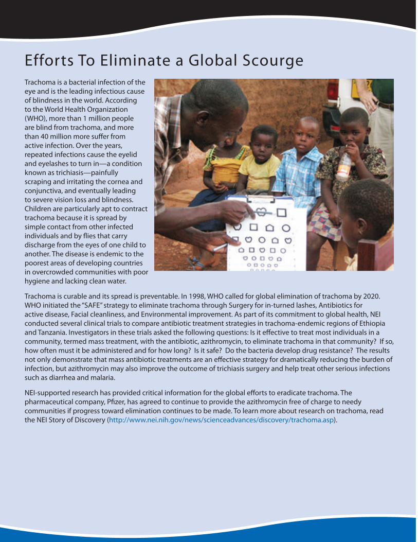
Efforts To Eliminate a Global ScourgeTrachoma is a bacterial infection of the eye and is the leading infectious cause of blindness in the world. According to the World Health Organization (WHO), more than 1 million people are blind from trachoma, and more than 40 million more suffer from active infection. Over the years, repeated infections cause the eyelid and eyelashes to turn in—a condition known as trichiasis—painfully scraping and irritating the cornea and conjunctiva, and eventually leading to severe vision loss and blindness. Children are particularly apt to contract trachoma because it is spread by simple contact from other infected individuals and by flies that carry discharge from the eyes of one child to another. The disease is endemic to the poorest areas of developing countries in overcrowded communities with poor hygiene and lacking clean water.
Trachoma is curable and its spread is preventable. In 1998, WHO called for global elimination of trachoma by 2020. WHO initiated the “SAFE” strategy to eliminate trachoma through Surgery for in-turned lashes, Antibiotics for active disease, Facial cleanliness, and Environmental improvement. As part of its commitment to global health, NEI conducted several clinical trials to compare antibiotic treatment strategies in trachoma-endemic regions of Ethiopia and Tanzania. Investigators in these trials asked the following questions: Is it effective to treat most individuals in a community, termed mass treatment, with the antibiotic, azithromycin, to eliminate trachoma in that community? If so, how often must it be administered and for how long? Is it safe? Do the bacteria develop drug resistance? The results not only demonstrate that mass antibiotic treatments are an effective strategy for dramatically reducing the burden of infection, but azithromycin may also improve the outcome of trichiasis surgery and help treat other serious infections such as diarrhea and malaria.
NEI-supported research has provided critical information for the global efforts to eradicate trachoma. The pharmaceutical company, Pfizer, has agreed to continue to provide the azithromycin free of charge to needy communities if progress toward elimination continues to be made. To learn more about research on trachoma, read the NEI Story of Discovery (http://www.nei.nih.gov/news/scienceadvances/discovery/trachoma.asp).
CORNEAL DISEASES 25
CORNEAL DISEASES NEEDS, GAPS AND OPPORTUNITIES
ANGIOGENESIS AND LYMPHANGIOGENESIS• Examine the long-term effects of VEGF
inhibition, particularly since corneal nerve regeneration and wound healing are compromised by VEGF blockade. Elucidate the interplay between various angiogenic and anti-angiogenic factors and their resultant signal transduction pathways.
• Identify regulators of lymphangiogenesis in addition to VEGF (e.g., integrins, members of a family of cell surface receptors whose ligands are extracellular matrix proteins and immunoglobulin superfamily molecules, and macrophages, either as progenitor cells or as a source of prolymphangiogenic growth factors and proteases) to facilitate the development of therapeutic strategies for minimizing corneal transplant rejection. The mouse corneal micropocket assay, as well as development of a specific lymphatic marker (LYVE-1), offer new possibilities for studying hemangiogenesis and lymphangiogenesis.
• Develop new anti-angiogenic drugs besides the monoclonal antibody bevacizumab given that more than 20 different angiogenic factors exist in addition to VEGF-A, which bevacizumab targets. Conventional therapy for corneal neovascularization involves steroid therapy, but there are potential negative treatment side effects such as glaucoma and cataract.
TRANSPLANTATION• Determine how dendritic cells are modified by
the ocular microenvironment, as corneal graft rejection occurs almost exclusively by indirect allo-recognition.
• Understand the interaction of recruited innate and adaptive immune cells at the site of transplantation and corneal graft destruction.
• Improve understanding of host responses in high-risk corneal transplantation, which involves an incredibly complex array of interactions that may act at different times or contemporaneously, with either additive or opposing effects on the cells central to transplant tolerance or graft rejection. Find a balance between stimulation and co-stimulation and induction of tolerance to alloantigens in anti-rejection therapy.
REGENERATIVE MEDICINE• Resolve major questions of limbal epithelial
stem cell biology, such as the following: o What keeps stem cells in their quiescent
state yet enables these cells to periodically divide to give rise to their progeny? A better understanding of the “stem cell niche” will provide answers to this gap in our knowledge.
o How are the conjunctival, limbal, and corneal epithelial boundaries established and maintained? Unraveling the complexities of inter-cell communication will help fill this void.
o How do limbal epithelial stem cells and their progeny regulate their metabolism?
• Improve the ability to culture epithelial cells using safe media components, develop an efficient system to ensure safety and reproducibility, and generate better carrier components to improve the success of transplantation of cultured epithelial cells.
26 VISION RESEARCH NEEDS, GAPS, AND OPPORTUNITIES
• Define the ability of corneal endothelial cells to regenerate and improve techniques to cultivate endothelial cells, whether from the patient or from allogenic sources. Develop new carrier substrates with improved handling characteristics to allow diffusion and optical clarity for use in transplantations.
• Identify new materials that provide a better transparency and possess biomechanical strength that are similar to the native cornea, and are biocompatible. Develop new biodegradable constructs that allow replacement by normal corneal or native corneal tissue and cells over time. Develop other approaches such as synthetic stromal equivalents that could obviate the need for functional endothelium.
• Translate findings on defining and characterizing corneal keratocyte progenitor cells and their utility in restoring corneal transparency in the mouse model into the human.
• Develop an understanding of the epigenetic regulation of wound healing and develop methods to accelerate wound closure and control epithelial cell proliferation to prevent hyperplasia.
INFLAMMATION AND IMMUNITY• Increase our understanding of leukocyte
and epithelial cell trafficking in the cornea, and develop strategies geared toward the enhancement of the wound-healing process. Elucidate the beneficial roles played by macrophages, dendritic cells, Langerhans cells, and other lymphocyte subsets in corneal inflammation. Investigate the role of hypoxia and the chemokine/cytokine network driving inflammatory cell recruitment.
• Develop new prostheses and interventions that can effectively treat severe scarring diseases, including trachoma and onchocerciasis, and ocular surface inflammation.
• Generate models of acute or chronic ocular allergy that closely mimic human disease and identify new targets in order to develop more effective therapies to treat extreme pain and discomfort caused by this condition.
• Develop an understanding of the underlying mechanism behind why atopic patients have a higher incidence of, and are more susceptible to, dry eye disease and to DNA viruses, including the human papillomavirus and herpes simplex viruses.
TEARS/DRY EYE DISORDERS• Develop an understanding of the regulation of
tear production and, in particular, the role of innervation and other factors (e.g., hormones, inflammatory mediators) in controlling meibomian gland secretion.
• Characterize the normal regulation of lacrimal gland, meibomian gland, goblet cell, and conjunctival stratified squamous cell secretion to identify the dysfunctions occurring in dry eye.
• Understand the causes and mechanisms of dry eye pathology and inflammation, including the role of the different types of mucins, lipids, toll-like receptors, the effect of sex-related hormones, and the role of autoimmunity.
• Understand the effect of aging on tear production and on corneal and conjunctival function, and characterize the effect of diabetes on ocular surface disease.
• Develop new animal models of dry eye disease and meibomian gland dysfunction, and identify transcription factors and promoters that can be used to develop transgenic animals with tissue specific over-expression of genes.
• Develop novel ocular therapeutics based on pathophysiology of different dry eye subtypes, for example, modulation of AQP expression and function. Increasing AQP expression, for example, may have utility for treatment of dry eye syndromes and corneal injury.
CORNEAL DISEASES 27
• Facilitate the development of effective treatments by identifying new diagnostics to define dry eye conditions, including biomarkers or paradigms that use multiple diagnostic tests, and correlate the impact on quality of life and patient symptoms with new diagnostics or paradigms.
• Understand the impact of contact lens wear in dry eye conditions, including meibomian gland dysfunction.
OCULAR PAIN AND SENSITIVITY• Develop a better classification of ocular pain
syndromes. A common nomenclature and classification scheme would enhance future research in both the laboratory and the clinic. See the report of the NEI Workshop on Ocular Pain and Sensitivity.1
• Correlate the molecular and structural composition of corneal nerves with their function. Elucidate the peripheral and central mechanisms for long-term ocular pain syndromes, using approaches that include, but are not limited to, electrophysiological analysis and advanced esthesiometry.
• Develop topical and/or systemic anesthetics or analgesics that are targeted for ocular pain, as well as novel agents capable of stimulating appropriate corneal nerve regeneration.
BIOMECHANICS AND IMAGING• Develop noninvasive methods to “interrogate”
the cornea for the presence of biomechanical instability prior to the onset of clinical disease, which may prevent the development of corneal ectasia secondary to keratoconus or postrefractive surgery.
• Increase understanding of biomechanical properties of the cornea and how they change during aging and in patients with glaucoma.
1 http://www.nei.nih.gov/strategicplanning/pain_workshop.asp
• Develop high-resolution imaging techniques to diagnose and guide treatment of common corneal disorders, including inflammation, dry eye, corneal ectasia, scarring, infection, and neuropathic conditions.
• Increase our understanding of the cornea as the portal to the entire optical system of the eye, using personalized systems (e.g., retinal photography, scanning laser ophthalmoscopy, and OCT). Develop new technologies for surface-resolved aberrometry that could facilitate wavefront-optimized corneal and/or lens transplants.
• Advance the analysis of nerve morphology to allow for diagnosis of clinical syndromes based upon corneal nerve imaging. Develop new automated instruments capable of higher resolution images with better registration coupled with advanced software.
GENETICS/GENOMICS• Identify the major genetic factors that are
involved in FCD, which is responsible for the majority of corneal transplantations performed each year in the United States. Many loci have been identified for FCD with large intervals, but the relation of genetic factors to the pathophysiology of disease remains elusive. Better defined regions could be obtained using newer techniques such as next-generation sequencing.
• Improve the identification of the phenotypic variations in the severity and progression of the endothelial dystrophies, including longitudinal studies of the disease phenotype in subjects with known genotypes, especially when therapeutic interventions are contemplated.
• Identify novel common causal variants of corneal endothelial dystrophies to help our understanding of the genetic interaction between “corneal dystrophies” genes and how they modulate the expressivity of the
28 VISION RESEARCH NEEDS, GAPS, AND OPPORTUNITIES
phenotype. Although clinically distinct, corneal endothelial dystrophies may be allelic variants of the same disease.
• Link recent data identifying both gene expression profiles and regulatory pathways in the tissues of the anterior eye during development and disease to 1) predict who is at high risk of developing corneal diseases and 2) define disease initiation, progression, and staging requisite for the design and implementation of appropriately targeted preventions and therapies.
• Identify the mechanisms of regulation of cellular gene expression in corneal health and disease. Use epigenetic chromatin markers to connect the environment and the genome, possibly revealing key mechanisms for how experience can feed back into cellular genetic identity.
INFECTIONS• Resolve major questions of viral infection,
such as the following:o Generate new animal models for
studying varicella zoster virus (VZV) corneal infections and latency as well as postherpetic neuralgia.
o Characterize mechanisms and regulation of the innate immune response that clears HSV-1, VZV, and adenovirus from the cornea.
o Develop therapeutic and prophylactic vaccines aimed at augmenting T cell control of HSV-1 latency in sensory ganglia and replication in the cornea.
o Develop novel and specific antiviral compounds for treating adenovirus corneal infections.
o Characterize the immunopathology mechanisms associated with HSV-1, VZV, and adenovirus keratitis; and
translate this knowledge of mechanisms of immunopathology into improved clinical management.
• Further characterize the pathogenesis of fungal, acanthamoebal, and bacterial keratitis, including mediators of innate and adaptive immunity, and microbial virulence factors that have potential as targets for novel therapies.
• Elucidate the epidemiology and pathophysiology of emerging pathogens in contact-lens related keratitis, including mechanisms of resistance.
• Develop novel and sustainable delivery methods for the prevention and treatment of ocular surface infection.
CLINICAL RESEARCH• Apply new in vivo corneal biomechanical
measurement and modeling techniques for larger clinical studies of corneal response to injury, ectatic disease, and the aging cornea.
• Characterize the extra-ocular anomalies associated with corneal dystrophies, including those with extra-ocular phenotypes such as the Harboyan syndrome.
• Investigate application of nanoparticles for use in drug delivery in inflammation and to control immunity in corneal transplantation.
• Develop nanotechnology to improve imaging of the various ocular structures.
• Conduct treatment studies, preferably definitive clinical trials, which are critical to establishing an evidence-based approach to treatments for dry eye and meibomian gland dysfunction; endothelial replacement surgeries; treatment of fungal, viral, bacterial, and acanthamoeba keratitis; laser and lens replacement approaches to treating moderate to high myopia; and use of corneal crosslinking for keratoconus.
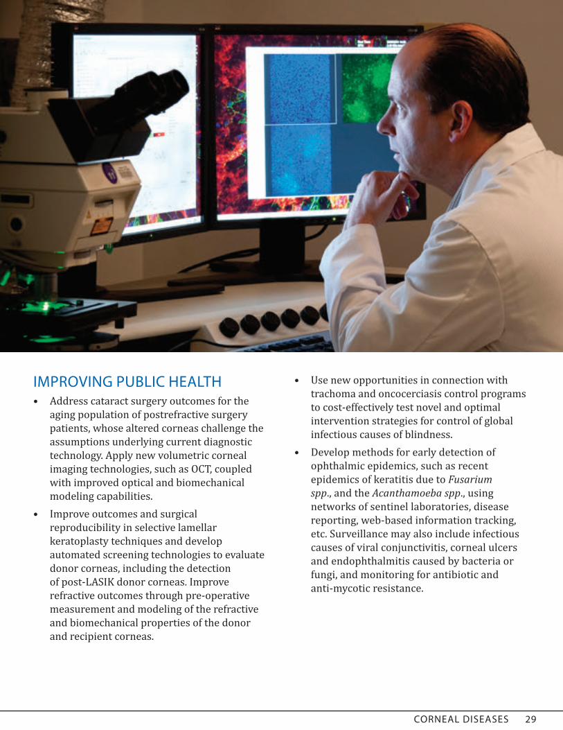
CORNEAL DISEASES 29
IMPROVING PUBLIC HEALTH• Address cataract surgery outcomes for the
aging population of postrefractive surgery patients, whose altered corneas challenge the assumptions underlying current diagnostic technology. Apply new volumetric corneal imaging technologies, such as OCT, coupled with improved optical and biomechanical modeling capabilities.
• Improve outcomes and surgical reproducibility in selective lamellar keratoplasty techniques and develop automated screening technologies to evaluate donor corneas, including the detection of post-LASIK donor corneas. Improve refractive outcomes through pre-operative measurement and modeling of the refractive and biomechanical properties of the donor and recipient corneas.
• Use new opportunities in connection with trachoma and oncocerciasis control programs to cost-effectively test novel and optimal intervention strategies for control of global infectious causes of blindness.
• Develop methods for early detection of ophthalmic epidemics, such as recent epidemics of keratitis due to Fusarium spp., and the Acanthamoeba spp., using networks of sentinel laboratories, disease reporting, web-based information tracking, etc. Surveillance may also include infectious causes of viral conjunctivitis, corneal ulcers and endophthalmitis caused by bacteria or fungi, and monitoring for antibiotic and anti-mycotic resistance.
30 VISION RESEARCH NEEDS, GAPS, AND OPPORTUNITIES
LENS AND CATARACT
The healthy lens is optically clear and flexible. Loss of lens transparency (cataract) and/or reduced ability to focus on near objects with age (presbyopia) are visual impairments that afflict a large portion of the world’s population. Although cataract surgery is an effective procedure, it is not available in many less developed areas of the world. According to WHO, 48 percent of global blindness (corresponding to 18 million people) is attributable to age-related cataract.2 The largest single risk factor for cataract is age, although it can result from genetic factors or environmental stresses. Cataract afflicts more than 50 percent of people over the age of 80. Even in developed countries, cataract remains a significant cause of blindness and low vision, especially for individuals who have limited financial resources, or rare but potentially blinding surgical complications. When the lens loses its flexibility, the ability to adjust the curvature of the lens to focus on objects at different distances (accommodation) is impaired. Presbyopia, the age-related loss of accommodative ability, usually manifests in midlife, causing difficulty when reading or doing other near work. Although not a blinding condition, presbyopia is a nearly universal visual impairment that requires eyeglasses or other corrective devices.
Understanding the unique physical and developmental attributes of the lens has advanced our knowledge of fundamental biological processes. For example, the lens mainly consists of densely packed, transparent fiber cells that differentiate from a single layer of epithelial cells in the periphery. The fiber cells do not turn over, which means they must last for a lifetime. Consequently, investigators have used the lens in studies of cellular and molecular longevity, proliferation, and death.
2 Resnikoff, S. et al. (2004). Bulletin of the World Health Organization, 82, 844-851.
Insights into the molecular aging of lens proteins (crystallins) serve as a paradigm for understanding aging of structural molecules. Another common physiological property of living tissues is communication between neighboring cells. In the avascular lens, intercellular communication plays a key role in delivering nutrients and removing waste products. The connections between communicating cells are termed gap junctions, and the major proteins (connexins) in gap junctions are expressed at extraordinarily high levels in fiber cells, causing a tight coupling of fiber cell behavior. The abundance of gap junctions and the relative ease with which connexin genes can be manipulated in the lens have contributed greatly to a general understanding of cell–cell communication.
Finally, studies of the lens may provide a window into understanding other diseases. Recently, it was discovered that amyloid-β protein accumulates in the lenses of patients with Alzheimer’s disease. The formation of amyloid- β plaques in the brain is a well-known characteristic of Alzheimer’s patients, but detecting the plaques early in a noninvasive manner is challenging. Now, there is promise that a new, simple optical technique that measures light scattering by clumped proteins in the lens may serve as a tool for screening and early detection of Alzheimer’s and other neurodegenerative diseases. In sum, discoveries from lens research are providing new insights not only into lens function (refraction of light and accommodation) and dysfunction (cataract and presbyopia), but also into the biology and pathobiology of other tissues and organ systems.
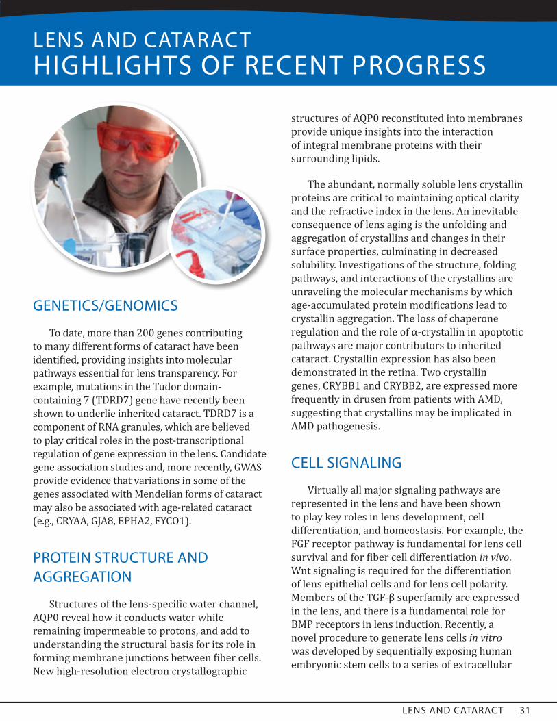
LENS AND CATARACT 31
LENS AND CATARACT HIGHLIGHTS OF RECENT PROGRESS
GENETICS/GENOMICS
To date, more than 200 genes contributing to many different forms of cataract have been identified, providing insights into molecular pathways essential for lens transparency. For example, mutations in the Tudor domain-containing 7 (TDRD7) gene have recently been shown to underlie inherited cataract. TDRD7 is a component of RNA granules, which are believed to play critical roles in the post-transcriptional regulation of gene expression in the lens. Candidate gene association studies and, more recently, GWAS provide evidence that variations in some of the genes associated with Mendelian forms of cataract may also be associated with age-related cataract (e.g., CRYAA, GJA8, EPHA2, FYCO1).
PROTEIN STRUCTURE AND AGGREGATION
Structures of the lens-specific water channel, AQP0 reveal how it conducts water while remaining impermeable to protons, and add to understanding the structural basis for its role in forming membrane junctions between fiber cells. New high-resolution electron crystallographic
structures of AQP0 reconstituted into membranes provide unique insights into the interaction of integral membrane proteins with their surrounding lipids.
The abundant, normally soluble lens crystallin proteins are critical to maintaining optical clarity and the refractive index in the lens. An inevitable consequence of lens aging is the unfolding and aggregation of crystallins and changes in their surface properties, culminating in decreased solubility. Investigations of the structure, folding pathways, and interactions of the crystallins are unraveling the molecular mechanisms by which age-accumulated protein modifications lead to crystallin aggregation. The loss of chaperone regulation and the role of α-crystallin in apoptotic pathways are major contributors to inherited cataract. Crystallin expression has also been demonstrated in the retina. Two crystallin genes, CRYBB1 and CRYBB2, are expressed more frequently in drusen from patients with AMD, suggesting that crystallins may be implicated in AMD pathogenesis.
CELL SIGNALING
Virtually all major signaling pathways are represented in the lens and have been shown to play key roles in lens development, cell differentiation, and homeostasis. For example, the FGF receptor pathway is fundamental for lens cell survival and for fiber cell differentiation in vivo. Wnt signaling is required for the differentiation of lens epithelial cells and for lens cell polarity. Members of the TGF-β superfamily are expressed in the lens, and there is a fundamental role for BMP receptors in lens induction. Recently, a novel procedure to generate lens cells in vitro was developed by sequentially exposing human embryonic stem cells to a series of extracellular
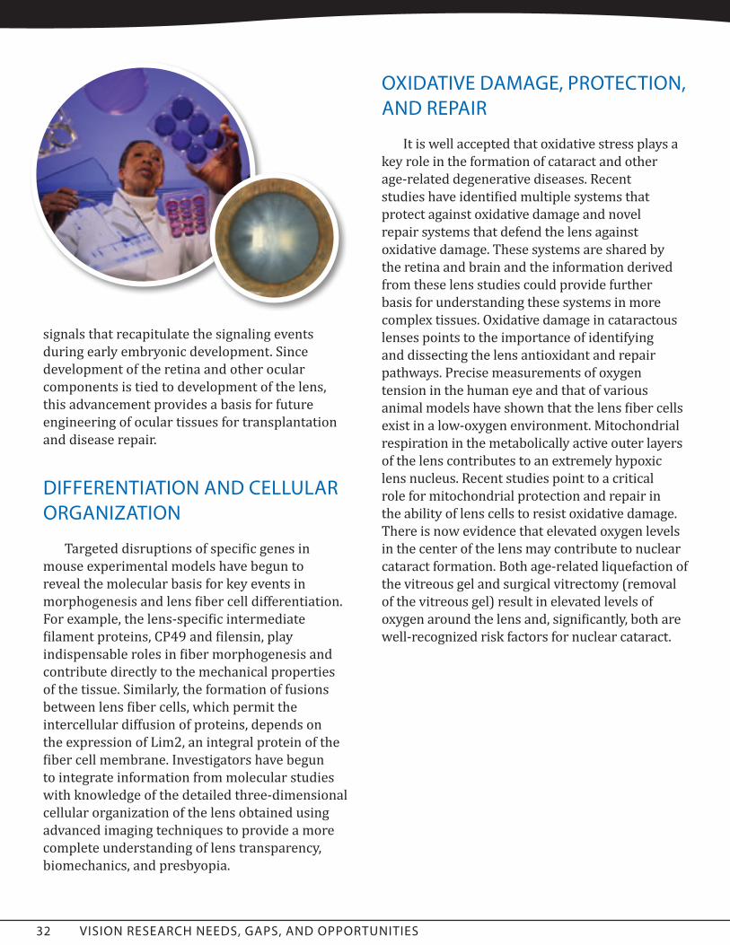
32 VISION RESEARCH NEEDS, GAPS, AND OPPORTUNITIES
signals that recapitulate the signaling events during early embryonic development. Since development of the retina and other ocular components is tied to development of the lens, this advancement provides a basis for future engineering of ocular tissues for transplantation and disease repair.
DIFFERENTIATION AND CELLULAR ORGANIZATION
Targeted disruptions of specific genes in mouse experimental models have begun to reveal the molecular basis for key events in morphogenesis and lens fiber cell differentiation. For example, the lens-specific intermediate filament proteins, CP49 and filensin, play indispensable roles in fiber morphogenesis and contribute directly to the mechanical properties of the tissue. Similarly, the formation of fusions between lens fiber cells, which permit the intercellular diffusion of proteins, depends on the expression of Lim2, an integral protein of the fiber cell membrane. Investigators have begun to integrate information from molecular studies with knowledge of the detailed three-dimensional cellular organization of the lens obtained using advanced imaging techniques to provide a more complete understanding of lens transparency, biomechanics, and presbyopia.
OXIDATIVE DAMAGE, PROTECTION, AND REPAIR
It is well accepted that oxidative stress plays a key role in the formation of cataract and other age-related degenerative diseases. Recent studies have identified multiple systems that protect against oxidative damage and novel repair systems that defend the lens against oxidative damage. These systems are shared by the retina and brain and the information derived from these lens studies could provide further basis for understanding these systems in more complex tissues. Oxidative damage in cataractous lenses points to the importance of identifying and dissecting the lens antioxidant and repair pathways. Precise measurements of oxygen tension in the human eye and that of various animal models have shown that the lens fiber cells exist in a low-oxygen environment. Mitochondrial respiration in the metabolically active outer layers of the lens contributes to an extremely hypoxic lens nucleus. Recent studies point to a critical role for mitochondrial protection and repair in the ability of lens cells to resist oxidative damage. There is now evidence that elevated oxygen levels in the center of the lens may contribute to nuclear cataract formation. Both age-related liquefaction of the vitreous gel and surgical vitrectomy (removal of the vitreous gel) result in elevated levels of oxygen around the lens and, significantly, both are well-recognized risk factors for nuclear cataract.
LENS AND CATARACT 33
LENS AND CATARACT NEEDS, GAPS AND OPPORTUNITIESPROTEIN STRUCTURE AND FUNCTION• Develop advanced optical and mass
spectroscopic imaging techniques and introduce novel model systems to help guide the development of effective anti-cataract strategies. Recent biochemical studies have provided a detailed mechanistic understanding of crystallin stability, interactions, and chaperone function, and in vitro studies will continue to provide critical data on crystallin structure/function relationships in the future. There needs to be an emphasis on testing the physiological validity and implications of biochemical data obtained from in vitro experiments.
• Unravel the noncanonical functions of lens proteins. Originally believed to function exclusively in refraction, it is now clear that lens crystallins have multiple roles in the lens, retina, and elsewhere that represent an important area of future study. The multiplicity of roles played by crystallins might be the rule rather than the exception for lens proteins. For example, AQP0 acts as a water channel in the lens, but likely also serves an important adhesive function. Similarly, connexin50, a channel-forming protein, also interacts with multiple signaling pathways to help regulate epithelial cell proliferation and lens size.
• Further investigate the various posttranslational modifications (PTMs) to lens proteins, particularly those that correlate with lens aging or lens opacity. The functional impact of PTMs and proteolysis is probably best studied in situ, or in a milieu that replicates the highly concentrated protein solution of the fiber cell cytoplasm.
SYSTEMS BIOLOGY AND LENS PHYSIOLOGY• Apply systems biology approaches to lens
physiology and pathophysiology. Consisting of only two differentiated cell types and isolated from the blood supply, the lens represents a unique opportunity to define the specific physiological events and factors that govern cell differentiation and development. For instance, understanding the mechanisms that orchestrate the specific degradation of individual organelles during lens fiber cell maturation is critical. Recent advances such as the identification of DNAseIIb as a component of nuclear degradation in lens fibers and the possible role of autophagy in lens cell maintenance and differentiation should be further explored. Gene transcription profiles from lens cells at various developmental stages need to be integrated with lens epigenetic and post-transcriptional regulatory mechanisms. Shot-gun proteomic and lipidomic approaches and MALDI imaging promise to provide an unprecedented view of lens cell biology and the causes and consequences of cellular aging. Lens cell membranes are unique in their lipid composition and investigating changes with cataract and aging presents an opportunity for needed insights into protein-lipid interactions. Improving spatial resolution of imaging techniques will uncover the molecular fate of cells in specific regions of the lens, from embryonic epithelial cells to aged fiber cells.
34 VISION RESEARCH NEEDS, GAPS, AND OPPORTUNITIES
MODEL SYSTEMS• Develop robust, quantitative methods to
accurately measure lens aberrations in small animal models. The size and shape of the lens are fundamental to its refractive properties and the genetic determinants of lens refractive function are beginning to emerge. Although considerable insights into lens morphogenesis and physiology have been achieved using avian, rodent and, more recently, fish systems, modeling lens pathology has proved more challenging. Because no animal model fully recapitulates the growth kinetics of the human lens, the time course of presbyopia, or the development of age-related cataract, new physiologically relevant models are needed.
CELL SIGNALING• Continue studies of key developmental and cell
signaling pathways. The lens is an exceptional system for studies of signaling pathways that control cell proliferation, differentiation, fiber cell migration, polarity, and tissue organization. New reagents allow lens gene expression to be temporally modulated in vivo in a cell-type specific fashion. This, coupled with the geometric organization of the tissue and the availability of optical probes to visualize the consequence of genetic manipulations, ensures that studies on the lens will continue to make important contributions to the broad field of cell signaling.
• Study the mechanism of TGF-β-mediated lens fibrosis in order to develop effective means of preventing posterior capsule opacification (PCO), the most common long-term complication of cataract surgery. The fibrotic process that underlies PCO is an example of aberrant cell signaling. TGF-β has emerged as a central mediator of lens fibrosis and is believed to induce epithelial/mesenchymal transdifferentiation, an early step in the fibrotic response leading to PCO.
DEVELOPMENT AND REGENERATIVE MEDICINE• Understand the regulation of epithelial cell
proliferation. Specifically, identify the factors that promote cell division in the germinative zone of the epithelium and suppress mitosis in the central zone. The optical role of the lens mandates that its size and shape must be regulated within very narrow tolerances and yet little is known about the regulatory mechanisms that control epithelial proliferation and the number of cells in the tissue.
• Settle the issue of whether there is a stem cell population in the lens epithelium. The lens grows throughout life and this is often taken as a priori evidence for the presence of a stem cell population in the lens epithelium. However, the location of lens stem cells, if such exist, has remained a matter of debate for many years.
• Gain an in-depth understanding of the temporal and morphogenetic stages of fiber cell differentiation to determine whether it may be possible to produce human lenses in vitro for transplantation. Growing mechanically functional transparent tissue in vitro will also require the identification of biocompatible scaffolds. Delineating the influence of the lens on the differentiation of surrounding ocular tissues and structures is very important for understanding overall ocular development and function.
• Investigate the consequence of lacking a natural lens in the adult eye for long time periods. Early in development, the lens plays a central role in ocular physiology and development, but little is known about its contribution to these processes in the aging eye.
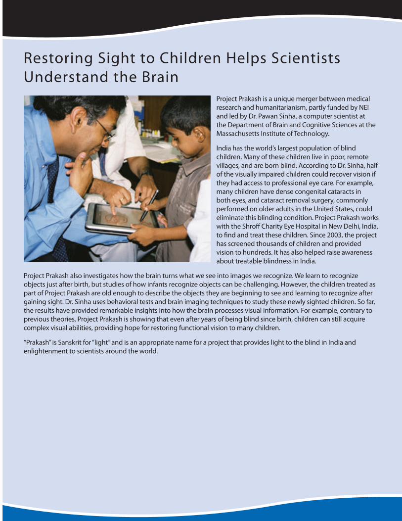
LENS AND CATARACT 35
Restoring Sight to Children Helps Scientists Understand the Brain
Project Prakash is a unique merger between medical research and humanitarianism, partly funded by NEI and led by Dr. Pawan Sinha, a computer scientist at the Department of Brain and Cognitive Sciences at the Massachusetts Institute of Technology.
India has the world’s largest population of blind children. Many of these children live in poor, remote villages, and are born blind. According to Dr. Sinha, half of the visually impaired children could recover vision if they had access to professional eye care. For example, many children have dense congenital cataracts in both eyes, and cataract removal surgery, commonly performed on older adults in the United States, could eliminate this blinding condition. Project Prakash works with the Shroff Charity Eye Hospital in New Delhi, India, to find and treat these children. Since 2003, the project has screened thousands of children and provided vision to hundreds. It has also helped raise awareness about treatable blindness in India.
Project Prakash also investigates how the brain turns what we see into images we recognize. We learn to recognize objects just after birth, but studies of how infants recognize objects can be challenging. However, the children treated as part of Project Prakash are old enough to describe the objects they are beginning to see and learning to recognize after gaining sight. Dr. Sinha uses behavioral tests and brain imaging techniques to study these newly sighted children. So far, the results have provided remarkable insights into how the brain processes visual information. For example, contrary to previous theories, Project Prakash is showing that even after years of being blind since birth, children can still acquire complex visual abilities, providing hope for restoring functional vision to many children.
“Prakash” is Sanskrit for “light” and is an appropriate name for a project that provides light to the blind in India and enlightenment to scientists around the world.
36 VISION RESEARCH NEEDS, GAPS, AND OPPORTUNITIES
BIOMECHANICS AND IMAGING• Develop high-resolution optical imaging
technologies and new fluorescent reporter molecules to allow the visualization of individual cells and molecular species in vivo and in organ-cultured lenses. In model organisms, imaging will provide answers to fundamental cell biological questions and insights into the cellular basis of accommodation and presbyopia.
• Study the microscopic changes that accompany accommodative changes in lens shape. A more complete understanding of the biomechanics of accommodation is needed to form an integrated model describing cellular and molecular parameters. Determine how cytoskeletal elements within the lens fiber cells influence the mechanical parameters of the tissue, and how structural forces generated during accommodation impact lens physiology. The effects of aging on the underlying structural elements need to be determined.
MECHANISMS OF CATARACT FORMATION, CATARACT PREVENTION, AND PUBLIC HEALTH• Conduct comprehensive, long-term,
epidemiological studies to identify genetic determinants and environmental factors that contribute to cataract formation. Furthermore, well-designed studies to identify genes and mutations associated with syndromic, nonsyndromic and particularly age-related cataract, and the interaction with known environmental cataractogenic agents, are warranted.
• Evaluate the effects of elevated oxygen levels and altered redox states on lens function and cataract formation. Identify the proteins, lens organelles, and signaling pathways that are altered in response to altered oxygen levels and redox conditions. Chronic exposure of fiber cells in the center of the lens to elevated levels of oxygen may contribute to age-related nuclear cataract, and acute oxygen exposure could be the cause of postvitrectomy cataract. Implementing new strategies to prevent oxygen exposure or mitigate the effects of exposure may therefore prevent certain types of cataract.
• Determine the efficacy of noninvasive optical techniques used in lens research, such as dynamic light scattering, and take advantage of cross-disciplinary opportunities for use in measuring amyloid-β accumulation/aggregation, and evaluate its use as a prognostic indicator for neurological diseases.
• Investigate the etiology of diabetic cataract, which is likely to increase in frequency as obesity rates continue to rise in the United States. Understanding the etiology of diabetic cataract is important toward developing preventative strategies for this condition that will aid in the management of other diabetic conditions, including diabetic retinopathy.
• Determine reasons for disparities in access and outcomes of cataract surgery across populations, and evaluate cost-efficiencies in approaches to cataract surgery. Such research has applicability to populations worldwide and would address this leading cause of blindness.
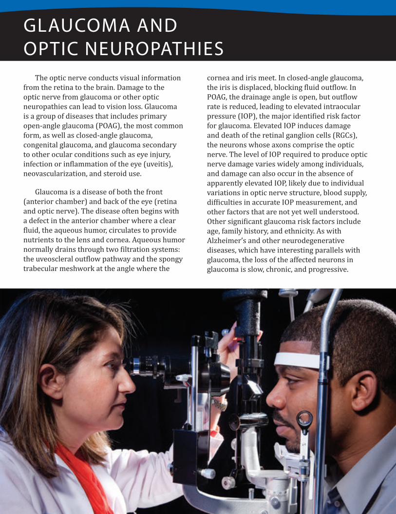
GLAUCOMA AND OPTIC NEUROPATHIES 37
GLAUCOMA AND OPTIC NEUROPATHIES
The optic nerve conducts visual information from the retina to the brain. Damage to the optic nerve from glaucoma or other optic neuropathies can lead to vision loss. Glaucoma is a group of diseases that includes primary open-angle glaucoma (POAG), the most common form, as well as closed-angle glaucoma, congenital glaucoma, and glaucoma secondary to other ocular conditions such as eye injury, infection or inflammation of the eye (uveitis), neovascularization, and steroid use.
Glaucoma is a disease of both the front (anterior chamber) and back of the eye (retina and optic nerve). The disease often begins with a defect in the anterior chamber where a clear fluid, the aqueous humor, circulates to provide nutrients to the lens and cornea. Aqueous humor normally drains through two filtration systems: the uveoscleral outflow pathway and the spongy trabecular meshwork at the angle where the
cornea and iris meet. In closed-angle glaucoma, the iris is displaced, blocking fluid outflow. In POAG, the drainage angle is open, but outflow rate is reduced, leading to elevated intraocular pressure (IOP), the major identified risk factor for glaucoma. Elevated IOP induces damage and death of the retinal ganglion cells (RGCs), the neurons whose axons comprise the optic nerve. The level of IOP required to produce optic nerve damage varies widely among individuals, and damage can also occur in the absence of apparently elevated IOP, likely due to individual variations in optic nerve structure, blood supply, difficulties in accurate IOP measurement, and other factors that are not yet well understood. Other significant glaucoma risk factors include age, family history, and ethnicity. As with Alzheimer’s and other neurodegenerative diseases, which have interesting parallels with glaucoma, the loss of the affected neurons in glaucoma is slow, chronic, and progressive.
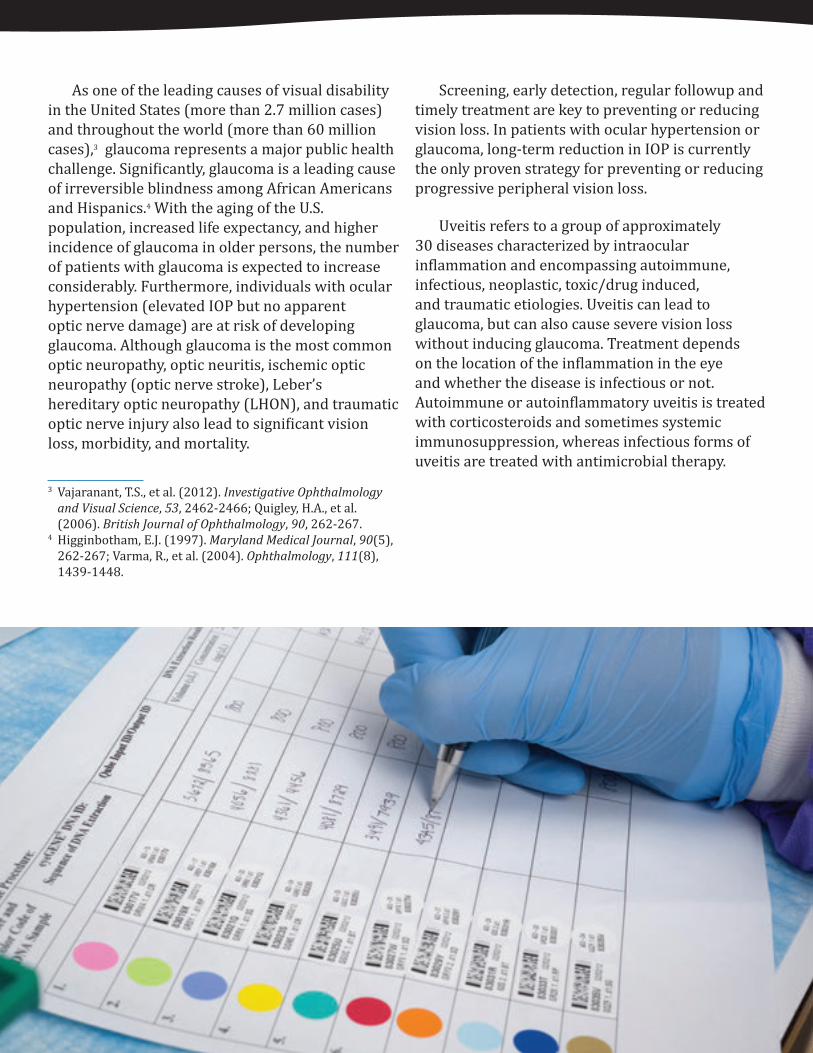
38 VISION RESEARCH NEEDS, GAPS, AND OPPORTUNITIES
As one of the leading causes of visual disability in the United States (more than 2.7 million cases) and throughout the world (more than 60 million cases),3 glaucoma represents a major public health challenge. Significantly, glaucoma is a leading cause of irreversible blindness among African Americans and Hispanics.4 With the aging of the U.S. population, increased life expectancy, and higher incidence of glaucoma in older persons, the number of patients with glaucoma is expected to increase considerably. Furthermore, individuals with ocular hypertension (elevated IOP but no apparent optic nerve damage) are at risk of developing glaucoma. Although glaucoma is the most common optic neuropathy, optic neuritis, ischemic optic neuropathy (optic nerve stroke), Leber’s hereditary optic neuropathy (LHON), and traumatic optic nerve injury also lead to significant vision loss, morbidity, and mortality.
3 Vajaranant, T.S., et al. (2012). Investigative Ophthalmology and Visual Science, 53, 2462-2466; Quigley, H.A., et al. (2006). British Journal of Ophthalmology, 90, 262-267.
4 Higginbotham, E.J. (1997). Maryland Medical Journal, 90(5), 262-267; Varma, R., et al. (2004). Ophthalmology, 111(8), 1439-1448.
Screening, early detection, regular followup and timely treatment are key to preventing or reducing vision loss. In patients with ocular hypertension or glaucoma, long-term reduction in IOP is currently the only proven strategy for preventing or reducing progressive peripheral vision loss.
Uveitis refers to a group of approximately 30 diseases characterized by intraocular inflammation and encompassing autoimmune, infectious, neoplastic, toxic/drug induced, and traumatic etiologies. Uveitis can lead to glaucoma, but can also cause severe vision loss without inducing glaucoma. Treatment depends on the location of the inflammation in the eye and whether the disease is infectious or not. Autoimmune or autoinflammatory uveitis is treated with corticosteroids and sometimes systemic immunosuppression, whereas infectious forms of uveitis are treated with antimicrobial therapy.
GLAUCOMA AND OPTIC NEUROPATHIES 39
GLAUCOMA AND OPTIC NEUROPATHIES HIGHLIGHTS OF RECENT PROGRESS
PHYSIOLOGY AND MOLECULAR AND CELL BIOLOGY
Family history and ethnic background are strong predictors of glaucoma risk, and yet, genes involved in POAG have remained elusive for years. Large-scale GWAS, such as the NEI Glaucoma Human Genetics Collaboration, have identified new genes that contribute to increased glaucoma risk in both rare and common complex forms. Among the genetic regions that have been identified are the CAV1/CAV2 locus, the CDKN2B/2A locus, and LOXL1 (exfoliation syndrome and glaucoma). These findings will help doctors predict disease risk in patients, and will point to new molecular pathways—and potential therapeutic targets—in glaucoma. These studies have validated the importance of using POAG-related quantitative endophenotypes such as central corneal thickness and cup-to-disc ratio for disease gene discovery.
Improved animal models for glaucoma have facilitated their use in studies to understand cellular mechanisms of optic nerve damage from elevated IOP. An anterior chamber hypertonic saline perfusion model has become widely accepted. New murine genetic models for glaucoma have been developed that complement existing models, such as the DBA/2 mouse. In a particularly useful model developed in recent years, aqueous outflow is inhibited by injecting microbeads into the anterior chamber of mice. Models of anterior ischemic optic neuropathy (AION) and posterior ION, optic neuritis, and mitochondrial optic neuropathies have also been characterized, and are providing new testing grounds for diagnostic and therapeutic investigations.
As increased IOP remains the major identified glaucoma risk factor, considerable effort has been devoted to studying various pathways through which aqueous humor drains. Studies focused on contributions of the juxtacanalicular extracellular matrix, juxtacanalicular trabecular meshwork cells, Schlemm’s canal inner wall endothelium, and uveoscleral outflow have slowly shifted the paradigm for understanding outflow resistance to provide a more comprehensive view of aqueous outflow.
The initial site of RGC damage in glaucoma remains unclear, but increasing evidence suggests the principal site of insult may be the optic nerve head (ONH), the location where RGC axons exit the eye to form the optic nerve. At this location, these unmyelinated axons are subjected to mechanical stress and are particularly sensitive to changes in IOP. The biomechanics of the ONH in turn contribute to the health of the RGCs with respect to their structural, nutritional, and cellular homeostasis. Structural biomechanics determined by both the 3D anatomy and the material stiffness of the load-bearing tissues are likely to be different among patient populations, which may underlie the differential susceptibilities to glaucoma. RGCs die through the process of apoptosis, but the molecular details are just now being understood. A key recent finding is that mechanisms of damage to RGCs at the optic nerve head, within axons, may be separate from those affecting retinal ganglion cell bodies. Purified cell populations, obtained by laser capture microdissection and other methods paired with microarray studies, which can provide a profile of gene transcripts expressed in a single cell type (transcriptome), have identified some of the gene expression changes and pathways associated with RGC development, injury, and death. Microarray
40 VISION RESEARCH NEEDS, GAPS, AND OPPORTUNITIES
studies of the ONH have also identified gene changes that may reflect initial responses to elevated IOP and initiating events in RGC axonal injury. Additionally, there have been advances in high-resolution imaging of RGC in animal models, making possible the definition of dendritic trees and capillary beds in situ and their response to elevated IOP.
Other risk factors for glaucoma are being identified, such as properties of the cornea (central corneal thickness, corneal hysteresis). Additionally, the role of glial cells in RGC health and disease and in regulating degenerative and regenerative processes has recently been recognized. Inflammatory mediators and signals derived from these cells influence the life and death of RGCs in glaucoma and other retinal and optic nerve pathologies. This includes research identifying TNF-α as a causative agent in a mouse model of glaucoma, and complement factors that may play a role in the mechanisms of RGC injury.
BIOMARKERS AND IMAGING FOR GLAUCOMA DIAGNOSIS AND PROGRESSION
Advances in imaging of the optic nerve and retina have aided in understanding disease processes and in the developing novel diagnostic technologies. Recent advances include confocal laser scanning ophthalmoscopy, scanning laser polarimetry, Fourier-domain OCT, and adaptive optics imaging of the retina. Longitudinal studies have shown that these tools accurately detect early abnormalities and small changes in the retina and the optic nerve in patients who ultimately experienced glaucomatous degeneration in their visual field. Studies applying new tools like laser Doppler flowmetry, color Doppler ultrasound, and Doppler OCT have documented a relationship between glaucomatous damage and decreased blood flow in the optic nerve head and retina.
Animal studies using continuous IOP sensors suggest that previously undetected variations in IOP may be significant for progressive visual field deterioration, even when average IOP is within a normal range. In addition, cerebral spinal fluid pressure and perfusion pressure (defined as the pressure difference between arterial blood pressure in the eye and IOP) interact with IOP and may have a role in glaucomatous optic nerve damage.
Newer phenotypic definitions for patients with narrow angles, patients at risk of developing angle closure, and those with closed-angle glaucoma have provided insights into the mechanisms of acute and chronic angle closure. In addition to the role of anatomical abnormalities, dynamic changes in the iris and choroid have been implicated in the development of angle closure. Newer, OCT-based methods of imaging the anterior chamber and the angle have provided a better understanding of the mechanisms underlying angle closure. They have also helped identify novel anatomical risk factors for the development of angle closure.
GLAUCOMA AND OPTIC NEUROPATHY TREATMENT
Important advances have been made in the development of a variety of neuroprotective molecules that may promote the survival of RGCs, even in the presence of increased IOP and other potentially harmful stressors. Neuroprotection therapy has the potential to complement current glaucoma therapies that target ocular hypertension. Some molecules/approaches are in clinical trials for ION (optic nerve stroke). Gene therapy approaches for LHON, which results from a mitochondrial gene dysfunction, have also been developed and are moving toward clinical trials.
Injured RGC axons can degenerate from the site of the insult to the cell body, leading to cell death. Recently, researchers found
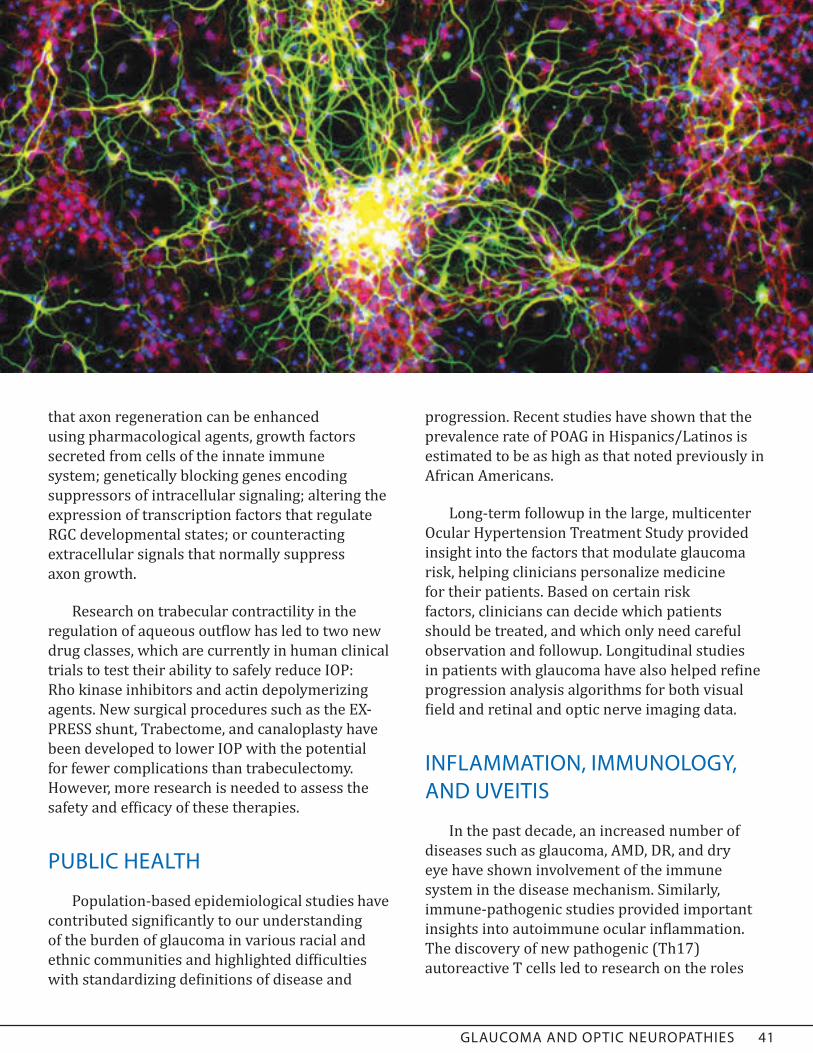
GLAUCOMA AND OPTIC NEUROPATHIES 41
that axon regeneration can be enhanced using pharmacological agents, growth factors secreted from cells of the innate immune system; genetically blocking genes encoding suppressors of intracellular signaling; altering the expression of transcription factors that regulate RGC developmental states; or counteracting extracellular signals that normally suppress axon growth.
Research on trabecular contractility in the regulation of aqueous outflow has led to two new drug classes, which are currently in human clinical trials to test their ability to safely reduce IOP: Rho kinase inhibitors and actin depolymerizing agents. New surgical procedures such as the EX-PRESS shunt, Trabectome, and canaloplasty have been developed to lower IOP with the potential for fewer complications than trabeculectomy. However, more research is needed to assess the safety and efficacy of these therapies.
PUBLIC HEALTH
Population-based epidemiological studies have contributed significantly to our understanding of the burden of glaucoma in various racial and ethnic communities and highlighted difficulties with standardizing definitions of disease and
progression. Recent studies have shown that the prevalence rate of POAG in Hispanics/Latinos is estimated to be as high as that noted previously in African Americans.
Long-term followup in the large, multicenter Ocular Hypertension Treatment Study provided insight into the factors that modulate glaucoma risk, helping clinicians personalize medicine for their patients. Based on certain risk factors, clinicians can decide which patients should be treated, and which only need careful observation and followup. Longitudinal studies in patients with glaucoma have also helped refine progression analysis algorithms for both visual field and retinal and optic nerve imaging data.
INFLAMMATION, IMMUNOLOGY, AND UVEITIS
In the past decade, an increased number of diseases such as glaucoma, AMD, DR, and dry eye have shown involvement of the immune system in the disease mechanism. Similarly, immune-pathogenic studies provided important insights into autoimmune ocular inflammation. The discovery of new pathogenic (Th17) autoreactive T cells led to research on the roles
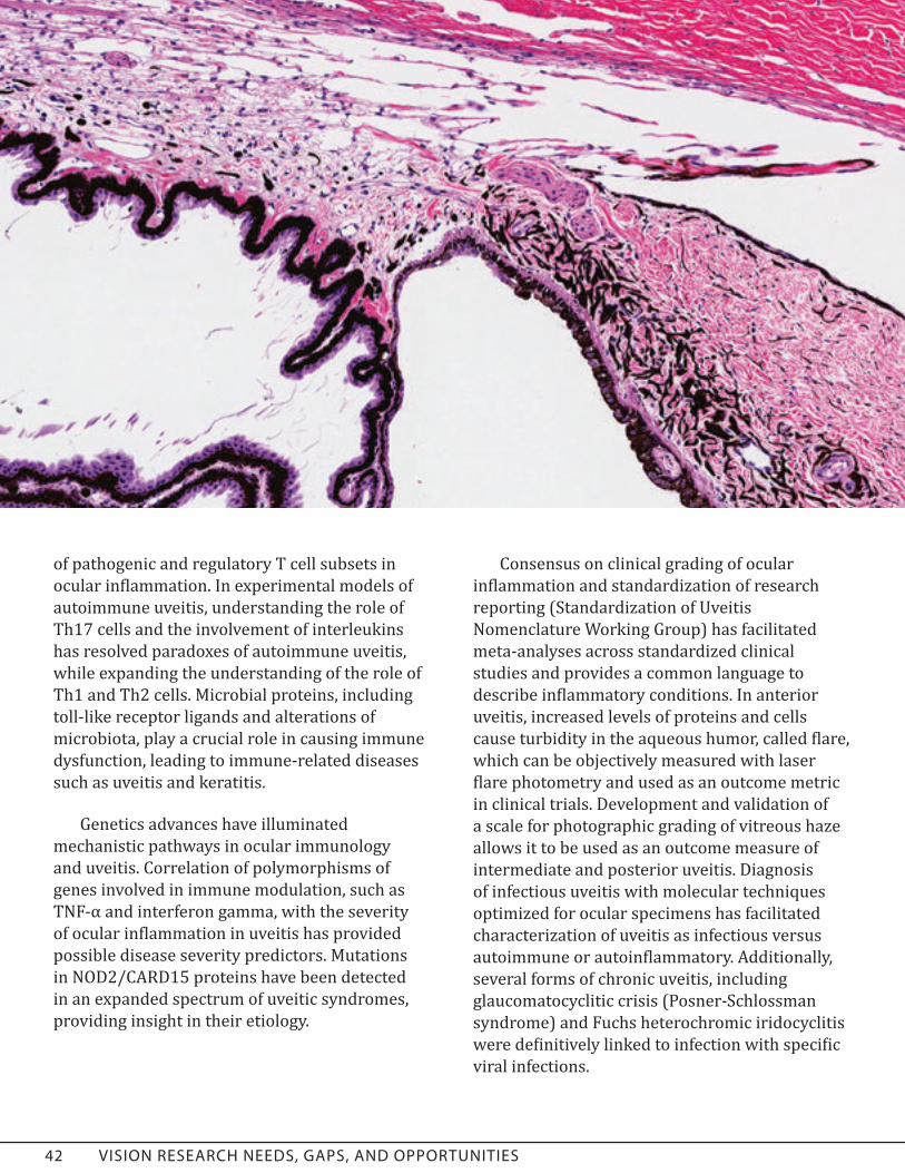
42 VISION RESEARCH NEEDS, GAPS, AND OPPORTUNITIES
of pathogenic and regulatory T cell subsets in ocular inflammation. In experimental models of autoimmune uveitis, understanding the role of Th17 cells and the involvement of interleukins has resolved paradoxes of autoimmune uveitis, while expanding the understanding of the role of Th1 and Th2 cells. Microbial proteins, including toll-like receptor ligands and alterations of microbiota, play a crucial role in causing immune dysfunction, leading to immune-related diseases such as uveitis and keratitis.
Genetics advances have illuminated mechanistic pathways in ocular immunology and uveitis. Correlation of polymorphisms of genes involved in immune modulation, such as TNF-α and interferon gamma, with the severity of ocular inflammation in uveitis has provided possible disease severity predictors. Mutations in NOD2/CARD15 proteins have been detected in an expanded spectrum of uveitic syndromes, providing insight in their etiology.
Consensus on clinical grading of ocular inflammation and standardization of research reporting (Standardization of Uveitis Nomenclature Working Group) has facilitated meta-analyses across standardized clinical studies and provides a common language to describe inflammatory conditions. In anterior uveitis, increased levels of proteins and cells cause turbidity in the aqueous humor, called flare, which can be objectively measured with laser flare photometry and used as an outcome metric in clinical trials. Development and validation of a scale for photographic grading of vitreous haze allows it to be used as an outcome measure of intermediate and posterior uveitis. Diagnosis of infectious uveitis with molecular techniques optimized for ocular specimens has facilitated characterization of uveitis as infectious versus autoimmune or autoinflammatory. Additionally, several forms of chronic uveitis, including glaucomatocyclitic crisis (Posner-Schlossman syndrome) and Fuchs heterochromic iridocyclitis were definitively linked to infection with specific viral infections.
GLAUCOMA AND OPTIC NEUROPATHIES 43
GLAUCOMA AND OPTIC NEUROPATHIES NEEDS, GAPS AND OPPORTUNITIES
BASIC BIOLOGY• Define the complete genetic architecture of
glaucoma. Glaucoma is a family of complex diseases with potentially many genetic and nongenetic factors; identifying specific genetic variants has been a difficult challenge. Genetics not only provides direct potential targets for therapy, it might point to novel glaucoma-related pathways. Risk alleles could be used to screen individuals before irreversible blindness develops and to help establish individual prognostic expectations and treatment responses, making a personalized approach to glaucoma therapy possible. Increasingly powerful genetic techniques, such as next-generation sequencing, offer the opportunity to identify the presumed genetic factors that modulate glaucoma, but have thus far eluded researchers.
• Develop new cell and animal models that better approximate human glaucoma and other optic neuropathies. In particular, models should be developed that can be used in a laboratory setting, with events occurring over days to months, to provide a better understanding of the chronic injury processes that occur in glaucoma patients over years and decades. Another research gap is to find a model suitable for testing RGC vulnerability to different genetic and environmental factors. Another need is to develop approaches to predict whether therapies developed in animal models will translate into safe and efficacious treatments for humans.
• Determine the mechanisms by which risk factors, such as age and prior glaucomatous injury, influence susceptibility of remaining RGC axons to elevated IOP.
• Characterize pathological changes of RGCs caused by elevation of IOP. A better understanding of the condition of injured RGCs and axons in chronic glaucoma could lead to neuroprotective interventions that preserve these cells while their connections through the ONH to brain targets still exist. Protecting injured RGCs has fewer technical challenges than regenerating new RGCs and getting them to grow appropriate connections.
• Resolve the relative contributions to aqueous outflow resistance by various components. This entails understanding the molecular nature and regulation of the cellular cytoskeleton, extracellular matrix components, the endothelium in the inner wall of Schlemm’s canal, and uveoscleral outflow pathway. Combining novel molecular, genetic, proteomic, ultra-high resolution (sub-cellular) real-time optical imaging and bioengineering methods provide an untapped opportunity in this field.
• Apply molecular biology techniques to RGC neuroscience to dissect factors important for survival, axon regeneration, and physiology. Determine the overlap and differences between molecular pathways that promote RGC survival versus those that control neuronal function and axon regeneration. This will help answer the question of whether the functionality and survival of injured cells can be improved. While progress has been made in therapies that promote axon regeneration in the optic nerve, more research is needed to understand if regenerated axons lead to the formation of appropriate, functional connections in the brain. Appropriate connections in regenerated axons would need to recapitulate the topographic maps
44 VISION RESEARCH NEEDS, GAPS, AND OPPORTUNITIES
formed in normal development and ultimately lead to recovery of vision. Establish whether particular classes of retinal ganglion cells are especially capable of restoring their connections, and if so, identify molecular features, such as transcription factors or microRNA, that may underlie this capability.
• Explore biomechanical responses of cell populations in the ONH to better understand the remodeling response of the ONH to chronic IOP elevations. Cell populations such as glia, lamina cribrosa cells, and scleral fibroblasts in the ONH control damage responses and remodeling in glaucoma, so it is important to understand their biomechanical responses to strain (stretch, compression, and shear), as well as the effect these remodeling responses have on axonal injury and altered perfusion. Biomarkers reflecting initial response to RGC injury may be present as early molecular and signaling events.
• Understand “collateral” injury to retinal cells (horizontal and bipolar cells) that may result from loss or dysfunction of RGC from glaucoma. Understand how these events can contribute to other, less well understood but clinically significant aspects of altered visual function in glaucoma, such as reduced contrast sensitivity and glare.
DIAGNOSIS, IMAGING, AND BIOMARKERS• Establish a consensus definition of POAG
and standardize tools to assess its various phenotypes. Absence of a highly sensitive and specific definition of POAG remains a major impediment to sharing data and gaining further insights into genes, risk factors, diagnosis, and treatment. Common instruments and collection methods need to be developed to standardize disease definitions across various studies; improved statistical methods are required for determining progression; novel bioengineering methods
are needed for diagnostic tools that do not rely solely on visual function to better characterize the phenotypes.
• Develop and deploy continuous monitors for IOP, blood pressure, and cerebrospinal fluid pressure (CSFp) that are reliable in various human body postures and positions. Elevated IOP is an important risk factor for glaucoma, but new evidence suggests ONH biomechanics are also influenced by CSFp and ocular perfusion pressure (a function of blood pressure and IOP). The role of variations in IOP needs to be better characterized over time; body posture can have a big impact on various internal fluid pressures.
• Engineer and apply new imaging methods and image processing techniques to study the aqueous outflow system and blood flow in the retina and the optic nerve. These methods will provide a better understanding of pathophysiology, development of glaucomatous damage, and response to treatment. Imaging the dynamic behavior of the iris and choroid should be assessed as potential biomarkers that denote risk of closed-angle glaucoma.
• Model the ONH and scleral biomechanics using experimental and computational inputs from genetic, molecular, proteomic, histologic, and imaging modalities to determine the cellular and extracellular architectures in the normal and diseased sclera and lamina cribrosa. Studies coupling in vivo imaging and biomechanics have the potential to identify markers for increased individual susceptibility to disease onset and progression.
TREATMENT STRATEGIES• Explore neuroprotection as an approach for
prolonging RGC function and survival. This involves improving our understanding of endogenous retinal protection mechanisms, identifying additional potent neuroprotective molecules, demonstrating their application
GLAUCOMA AND OPTIC NEUROPATHIES 45
in animal models, and translating this research into clinical trials. Leveraging progress in other areas of neuroscience, such as mechanisms in other neurodegenerative diseases, will inform glaucoma research. Develop long-term drug delivery systems and devices to enhance diagnostics and therapeutics. Recent identification of molecular targets in the aqueous outflow system and RGC survival provides an opportunity to specifically target drugs for glaucoma and other optic neuropathies.
• Expand research on differentiating pluripotent stem cells into retinal cells (see sidebar page 46). Although more progress has been made in differentiating pluripotent cells into photoreceptor and RPE-like cells, creation of RGCs could help restore vision in glaucoma and other optic neuropathies.
INFLAMMATION, IMMUNOLOGY, AND UVEITIS• Characterize the role of inflammation and
immune responses in the development and progression of glaucomatous damage, with attention to the role of the complement system in response to RGC injury, and to the dual role of glial cells in supporting retinal cells and mediating their injury.
• Understand the interaction between innate and adaptive immunity in ocular inflammation, including molecular, cellular, and physiological mechanisms of inflammation; uveitis; and immunological regulation. Evaluate the role of innate immunity in uveitis, including the role of complement, toll-like receptors, macrophages, natural killer cells, gamma delta T cells, natural antibodies, and T cell subsets (e.g., Th17 cells, Treg cells), the role of cell adhesion molecules, and immune cell entry into the eye. Evaluate the mechanism by which immune privilege is lost (and restored) in the eye, resulting in spontaneous autoimmune uveitis.
Identifying key immune factors will advance research not only on uveitis, but also glaucoma, AMD, DR, and dry eye.
• Identify the specific antigenic targets of the aberrant immune response in uveitis and scleritis and determine the role of antigens derived from apoptotic cells in disease. Emerging evidence supports the role of both genetic and environmental triggers in many autoimmune diseases. Compared to the progression in other autoimmune diseases, such as rheumatic diseases, understanding of the pathogenesis of the uveitides has lagged.
• Characterize the molecular effectors of specific uveitic diseases and their sequelae by gene expression, multiplex cytokine, and proteomic approaches. For example, these tools can help identify risk factors and pathogenic mechanisms leading to noninfectious uveitis and to its remission. Once specific mechanisms of uveitis are identified, the next step is to develop additional animal models, including autoinflammatory models and animal models of spondyloarthropathies, as well as animal models permitting noninvasive grading of inflammation and response to therapy.
• Improve diagnosis, prognosis, and treatment of uveitic syndromes by better identifying clinical phenotypes. Identify factors predictive of clinical response to or intolerance of conventional immunosuppressive drugs. Clarify why there is natural resolution of acute, but not chronic, autoimmune uveitis, and determine why inflammation recurs in some, but not all, patients. Evaluate the safety and comparative effectiveness of various treatment algorithms for autoimmune uveitis and its complications. Evaluate treatments for uveitic complications, such as macular edema, which is one of the most common causes of visual impairment in patients with uveitis. Identify the long-term consequences of chronic uveitis, which may be a life-long disease, and of its treatments.
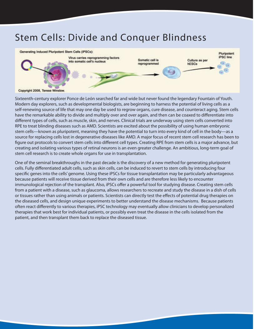
46 VISION RESEARCH NEEDS, GAPS, AND OPPORTUNITIES
Stem Cells: Divide and Conquer Blindness
Sixteenth-century explorer Ponce de León searched far and wide but never found the legendary Fountain of Youth. Modern day explorers, such as developmental biologists, are beginning to harness the potential of living cells as a self-renewing source of life that may one day be used to regrow organs, cure disease, and counteract aging. Stem cells have the remarkable ability to divide and multiply over and over again, and then can be coaxed to differentiate into different types of cells, such as muscle, skin, and nerves. Clinical trials are underway using stem cells converted into RPE to treat blinding diseases such as AMD. Scientists are excited about the possibility of using human embryonic stem cells—known as pluripotent, meaning they have the potential to turn into every kind of cell in the body—as a source for replacing cells lost in degenerative diseases like AMD. A major focus of recent stem cell research has been to figure out protocols to convert stem cells into different cell types. Creating RPE from stem cells is a major advance, but creating and isolating various types of retinal neurons is an even greater challenge. An ambitious, long-term goal of stem cell research is to create whole organs for use in transplantation.
One of the seminal breakthroughs in the past decade is the discovery of a new method for generating pluripotent cells. Fully differentiated adult cells, such as skin cells, can be induced to revert to stem cells by introducing four specific genes into the cells’ genome. Using these iPSCs for tissue transplantation may be particularly advantageous because patients will receive tissue derived from their own cells and are therefore less likely to encounter immunological rejection of the transplant. Also, iPSCs offer a powerful tool for studying disease. Creating stem cells from a patient with a disease, such as glaucoma, allows researchers to recreate and study the disease in a dish of cells or tissues rather than using animals or patients. Scientists can directly test the effects of potential drug therapies on the diseased cells, and design unique experiments to better understand the disease mechanisms. Because patients often react differently to various therapies, iPSC technology may eventually allow clinicians to develop personalized therapies that work best for individual patients, or possibly even treat the disease in the cells isolated from the patient, and then transplant them back to replace the diseased tissue.
GLAUCOMA AND OPTIC NEUROPATHIES 47
IMPROVING PUBLIC HEALTH• Validate new biomarkers for early diagnosis
and prediction of progression. Clinicians need tools to differentiate between small physiologic age-related change and small pathologic glaucomatous change. Biomarkers currently used in glaucoma trials focus on IOP reduction, but there is a research need for biomarkers of neuroprotection. Biomarkers, especially those related to optic nerve injury, would aid greatly in the design and execution of future cost-efficient and meaningful clinical trials. Investigation is needed to address whether such endpoints could be served by current technologies, including structural measures like retinal nerve fiber layer thickness, functional measures from improved electrophysiological testing or visual field interpretation, or the interplay between these two.
• Find surrogates or alternatives for visual field testing, and to determine over time the correspondence to performance of daily activities and quality of life.
• Track glaucoma burden across the increasingly diversifying U.S. population. Glaucoma represents a significant health disparity for some minority groups (Hispanic/Latino and African American populations have high prevalence of POAG; East Asian populations have a high prevalence of angle closure). POAG has been underdiagnosed, historically, and current tools used to assess risk in patients need to be kept in perspective with their recognized limitations. Up-to-date epidemiology data, including diagnosis, monitoring, and treatment are essential in detecting trends that may highlight health disparities and inform public health officials on how to address these disparities.
• Establish registries and bioinformatics networks that integrate genetic, ocular, systemic, and environmental factors to enhance personalized medicine in ocular disease. Harnessing the potential of bioinformatics would leverage existing and future medical record data so that clinicians could select appropriate followup intervals, and optimize medications and dosages with minimal side effects with improved outcomes at reduced costs.
• Assess the comparative effectiveness of treatments for different forms of glaucoma in diverse populations and communities. Such studies include cost-effectiveness and risk–benefit ratios of various diagnostic tests such as imaging and visual field testing, as well as interval assessment, particularly in populations at high risk of developing or having glaucoma (African Americans and Hispanics/Latinos). It is particularly important to determine the cost-effectiveness of monitoring persons with different rates of progression at different intervals. Closed-angle glaucoma is associated with a higher rate of irreversible blindness than open-angle glaucoma, but little is known about the effectiveness of various interventions for acute angle-closure crisis, primary angle closure, and closed-angle glaucoma.
• Determine how to improve patient compliance with glaucoma therapy. There are many reasons for noncompliance, including cultural and economic factors. Some glaucoma drugs cause ocular surface discomfort, affecting compliance; drugs that increase experience of dry eye limit patient quality of life, despite preserving visual function. Studies from bioengineering approaches to drug delivery to social determinants of medication compliance are needed. Reduction in glaucomatous blindness depends on addressing causes of noncompliance.
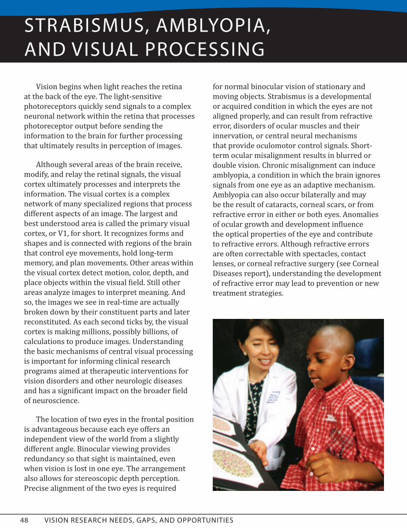
48 VISION RESEARCH NEEDS, GAPS, AND OPPORTUNITIES
STRABISMUS, AMBLYOPIA, AND VISUAL PROCESSING
Vision begins when light reaches the retina at the back of the eye. The light-sensitive photoreceptors quickly send signals to a complex neuronal network within the retina that processes photoreceptor output before sending the information to the brain for further processing that ultimately results in perception of images.
Although several areas of the brain receive, modify, and relay the retinal signals, the visual cortex ultimately processes and interprets the information. The visual cortex is a complex network of many specialized regions that process different aspects of an image. The largest and best understood area is called the primary visual cortex, or V1, for short. It recognizes forms and shapes and is connected with regions of the brain that control eye movements, hold long-term memory, and plan movements. Other areas within the visual cortex detect motion, color, depth, and place objects within the visual field. Still other areas analyze images to interpret meaning. And so, the images we see in real-time are actually broken down by their constituent parts and later reconstituted. As each second ticks by, the visual cortex is making millions, possibly billions, of calculations to produce images. Understanding the basic mechanisms of central visual processing is important for informing clinical research programs aimed at therapeutic interventions for vision disorders and other neurologic diseases and has a significant impact on the broader field of neuroscience.
The location of two eyes in the frontal position is advantageous because each eye offers an independent view of the world from a slightly different angle. Binocular viewing provides redundancy so that sight is maintained, even when vision is lost in one eye. The arrangement also allows for stereoscopic depth perception. Precise alignment of the two eyes is required
for normal binocular vision of stationary and moving objects. Strabismus is a developmental or acquired condition in which the eyes are not aligned properly, and can result from refractive error, disorders of ocular muscles and their innervation, or central neural mechanisms that provide oculomotor control signals. Short-term ocular misalignment results in blurred or double vision. Chronic misalignment can induce amblyopia, a condition in which the brain ignores signals from one eye as an adaptive mechanism. Amblyopia can also occur bilaterally and may be the result of cataracts, corneal scars, or from refractive error in either or both eyes. Anomalies of ocular growth and development influence the optical properties of the eye and contribute to refractive errors. Although refractive errors are often correctable with spectacles, contact lenses, or corneal refractive surgery (see Corneal Diseases report), understanding the development of refractive error may lead to prevention or new treatment strategies.
STRABISMUS, AMBLYOPIA, AND VISUAL PROCESSING 49
STRABISMUS, AMBLYOPIA, AND VISUAL PROCESSINGHIGHLIGHTS OF RECENT PROGRESS
VISUAL SYSTEM CIRCUITRY
Significant advances in technology and analytical approaches have greatly improved our understanding of how visual information is processed by individual neurons, local circuits, and larger networks within the CNS. The high spatial and temporal resolution of two-photon calcium imaging allows real-time monitoring of activity in many cells, which has significantly advanced our knowledge of the interactions among neurons within local circuits. Improvements in multiple microelectrode and multiple-site recording technology, and the development of advanced decoding algorithms provide a new understanding of how visual information is contained in the activity of large and distributed populations of neurons.
Studying populations and networks of neurons provides a better understanding of how multiple areas of the brain interact to produce visual perceptions and visually guided motor and oculomotor responses. Multivariate pattern analysis, computational modeling, and methods for combining electrophysiology, functional imaging, and psychophysical measures of behavior have yielded a more mechanistic understanding of vision. Brain–computer interfaces have been developed for electrical stimulation of neural tissue in those deprived of vision, and are used to produce higher-order signals in the visual system for the control of assistive devices for individuals lacking control of their limbs.
MULTISENSORY INTEGRATION AND HIGHER-ORDER FUNCTIONS
There is a high degree of homology between the human and nonhuman primate visual systems, and studies in both have led to a better mechanistic understanding of visual function. Studies using functional brain imaging in humans and the combination of functional imaging and single-unit recording in non-human primates have identified specific cortical areas that are specialized for a variety of visual functions, including the recognition of faces. These discoveries are complemented by the development of Bayesian models and decoding algorithms that have aided our understanding of the representation of visual information at the neural and behavioral levels. A better understanding of the perception of faces and recognition of objects is the result of studies in multiple areas of the brain, from the occipital to frontal cortex. Advances in this field provide important insights into neurodegenerative diseases and developmental disorders such as prosopagnosia, the inability to recognize faces, in which these perceptual functions are impaired.
Human and animal studies have also demonstrated a number of multimodal cortical areas where visuospatial, vestibular, and eye position information are integrated along with information from other senses to produce a representation of where one is in relation to one’s surroundings and signals for navigating within that environment. This information provides a framework for understanding spatial perception and deficits like those observed in spatial neglect syndromes. Neural correlates have also been identified for abstract visual concepts such as object category and numerosity.
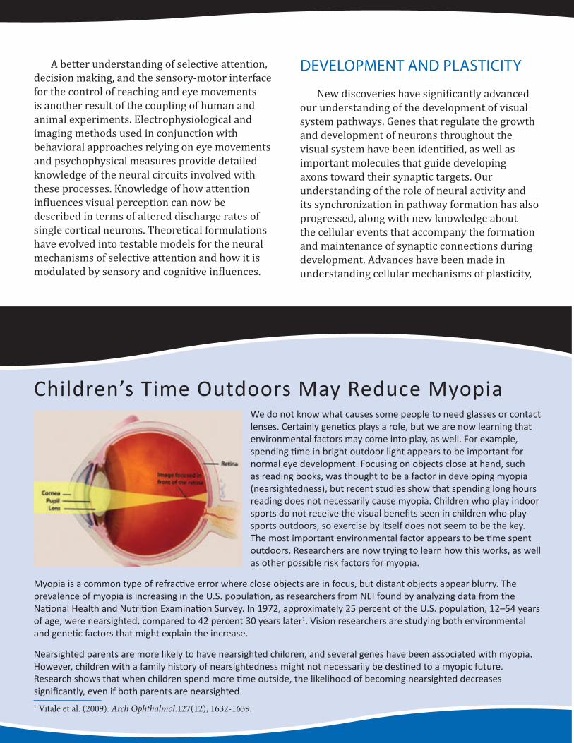
50 VISION RESEARCH NEEDS, GAPS, AND OPPORTUNITIES
A better understanding of selective attention, decision making, and the sensory-motor interface for the control of reaching and eye movements is another result of the coupling of human and animal experiments. Electrophysiological and imaging methods used in conjunction with behavioral approaches relying on eye movements and psychophysical measures provide detailed knowledge of the neural circuits involved with these processes. Knowledge of how attention influences visual perception can now be described in terms of altered discharge rates of single cortical neurons. Theoretical formulations have evolved into testable models for the neural mechanisms of selective attention and how it is modulated by sensory and cognitive influences.
DEVELOPMENT AND PLASTICITY
New discoveries have significantly advanced our understanding of the development of visual system pathways. Genes that regulate the growth and development of neurons throughout the visual system have been identified, as well as important molecules that guide developing axons toward their synaptic targets. Our understanding of the role of neural activity and its synchronization in pathway formation has also progressed, along with new knowledge about the cellular events that accompany the formation and maintenance of synaptic connections during development. Advances have been made in understanding cellular mechanisms of plasticity,
Children’s Time Outdoors May Reduce MyopiaWe do not know what causes some people to need glasses or contact lenses. Certainly genetics plays a role, but we are now learning that environmental factors may come into play, as well. For example, spending time in bright outdoor light appears to be important for normal eye development. Focusing on objects close at hand, such as reading books, was thought to be a factor in developing myopia (nearsightedness), but recent studies show that spending long hours reading does not necessarily cause myopia. Children who play indoor sports do not receive the visual benefits seen in children who play sports outdoors, so exercise by itself does not seem to be the key. The most important environmental factor appears to be time spent outdoors. Researchers are now trying to learn how this works, as well as other possible risk factors for myopia.
Myopia is a common type of refractive error where close objects are in focus, but distant objects appear blurry. The prevalence of myopia is increasing in the U.S. population, as researchers from NEI found by analyzing data from the National Health and Nutrition Examination Survey. In 1972, approximately 25 percent of the U.S. population, 12–54 years of age, were nearsighted, compared to 42 percent 30 years later1. Vision researchers are studying both environmental and genetic factors that might explain the increase.
Nearsighted parents are more likely to have nearsighted children, and several genes have been associated with myopia. However, children with a family history of nearsightedness might not necessarily be destined to a myopic future. Research shows that when children spend more time outside, the likelihood of becoming nearsighted decreases significantly, even if both parents are nearsighted. 1 Vitale et al. (2009). Arch Ophthalmol.127(12), 1632-1639.
STRABISMUS, AMBLYOPIA, AND VISUAL PROCESSING 51
including knowledge of events leading to the critical period. The understanding of plasticity in adults is the result of new experimental paradigms and models of perceptual learning. These studies have important implications for clinical rehabilitation for a variety of visual system disorders.
EYE MOVEMENTS
New discoveries have shed light on how eye muscles are structured and controlled in health and disease. Each eye muscle has its own pulley-like structure that plays a role in precisely aligning and moving the eyes toward visual targets. Eye muscles have anatomical compartments with specialized patterns of innervation that may be targeted differentially by disease processes. A newly described signal from eye muscles provides the brain with information about current eye position that is important for directing the gaze to the objects of interest. Eye muscles have a complement of genes and proteins that differ from normal skeletal muscle, which may explain why some diseases target eye muscles preferentially, whereas other muscle disorders spare the oculomotor system. These new discoveries regarding the control of eye muscles and their unique properties provide avenues for the creation of novel treatments for infantile and acquired disorders that can affect the accuracy of eye movements and the ability to maintain stable gaze.
CLINICAL ADVANCES
Studies in large numbers of patients have revealed the genetic basis of some forms of strabismus and regions of chromosomes that are important in the development and progression of myopia. New discoveries demonstrate how genes and the visual environment interact to influence the development and progression of refractive
error. These studies show that spending more time outdoors significantly lowers the chances of children becoming myopic (see sidebar page 50). Animal experiments also indicate that higher ambient light levels such as those that occur outdoors alter eye growth signals and have a strong protective influence, slowing myopia development. Recent work in both animals and humans indicates that these growth signals can come from the entire retina and not just the central portion. These discoveries are stimulating new approaches in myopia treatment, from changing children’s visual environment to sophisticated optical design in glasses or contact lenses that optimizes focus for the entire eye.
New treatments show promise for the treatment of amblyopia, the loss of vision in one eye that often results from strabismus or severe defocus during early childhood. Clinical trials demonstrate that atropine eye drop therapy has advantages over patching the dominant eye in some situations. Advanced behavioral methods using perceptual learning have also been shown to improve vision in these patients, even at ages that are beyond the critical period in development.
EMERGING TECHNOLOGY
Optogenetics is a newly developed technology that allows for targeted neuronal populations to be activated or inhibited by the presentation of light. Genetic methods are used to insert light-sensitive ion channels into specifically targeted cells. This provides a method of cellular control not possible with existing microelectrode technology. The method has been applied to tissue slices and whole animal models and offers a significant new advance for the field of visual prostheses as well as an important research tool for dissecting the functions of retinal and central neural circuits.
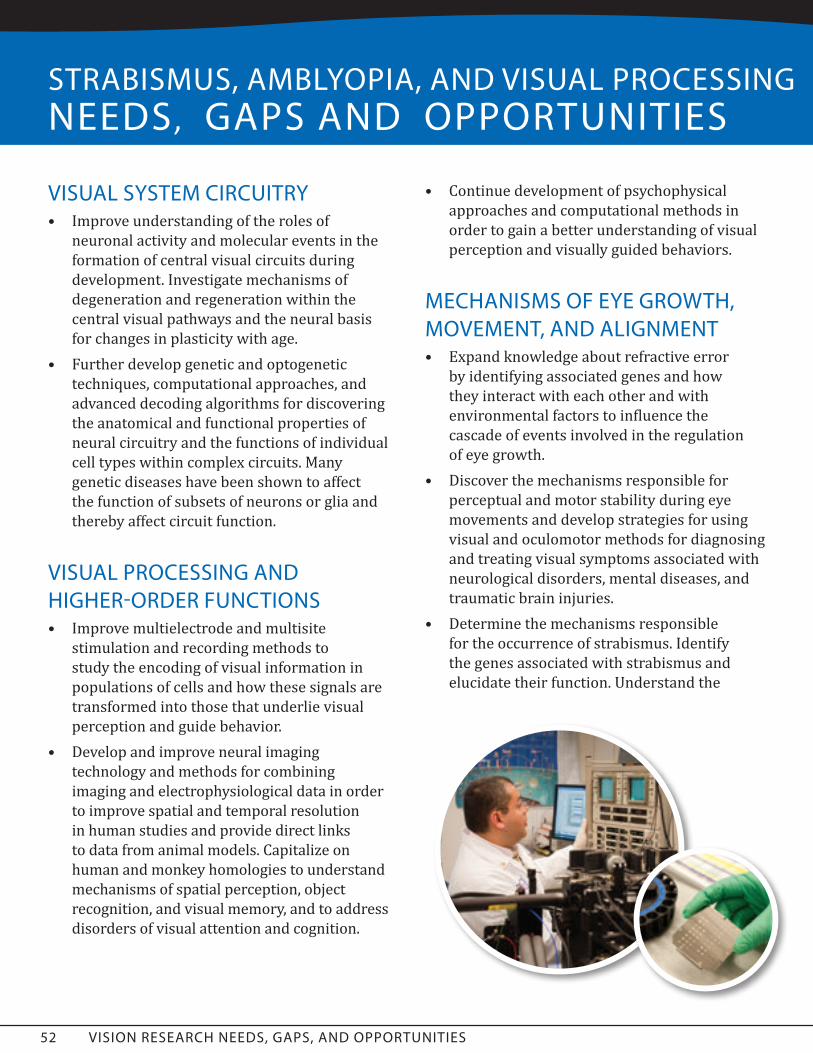
52 VISION RESEARCH NEEDS, GAPS, AND OPPORTUNITIES
STRABISMUS, AMBLYOPIA, AND VISUAL PROCESSINGNEEDS, GAPS AND OPPORTUNITIES
VISUAL SYSTEM CIRCUITRY• Improve understanding of the roles of
neuronal activity and molecular events in the formation of central visual circuits during development. Investigate mechanisms of degeneration and regeneration within the central visual pathways and the neural basis for changes in plasticity with age.
• Further develop genetic and optogenetic techniques, computational approaches, and advanced decoding algorithms for discovering the anatomical and functional properties of neural circuitry and the functions of individual cell types within complex circuits. Many genetic diseases have been shown to affect the function of subsets of neurons or glia and thereby affect circuit function.
VISUAL PROCESSING AND HIGHER-ORDER FUNCTIONS• Improve multielectrode and multisite
stimulation and recording methods to study the encoding of visual information in populations of cells and how these signals are transformed into those that underlie visual perception and guide behavior.
• Develop and improve neural imaging technology and methods for combining imaging and electrophysiological data in order to improve spatial and temporal resolution in human studies and provide direct links to data from animal models. Capitalize on human and monkey homologies to understand mechanisms of spatial perception, object recognition, and visual memory, and to address disorders of visual attention and cognition.
• Continue development of psychophysical approaches and computational methods in order to gain a better understanding of visual perception and visually guided behaviors.
MECHANISMS OF EYE GROWTH, MOVEMENT, AND ALIGNMENT• Expand knowledge about refractive error
by identifying associated genes and how they interact with each other and with environmental factors to influence the cascade of events involved in the regulation of eye growth.
• Discover the mechanisms responsible for perceptual and motor stability during eye movements and develop strategies for using visual and oculomotor methods for diagnosing and treating visual symptoms associated with neurological disorders, mental diseases, and traumatic brain injuries.
• Determine the mechanisms responsible for the occurrence of strabismus. Identify the genes associated with strabismus and elucidate their function. Understand the
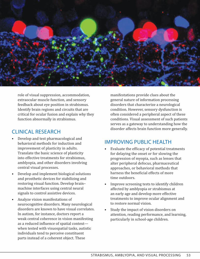
STRABISMUS, AMBLYOPIA, AND VISUAL PROCESSING 53
role of visual suppression, accommodation, extraocular muscle function, and sensory feedback about eye position in strabismus. Identify brain regions and circuits that are critical for ocular fusion and explain why they function abnormally in strabismus.
CLINICAL RESEARCH• Develop and test pharmacological and
behavioral methods for induction and improvement of plasticity in adults. Translate the basic science of plasticity into effective treatments for strabismus, amblyopia, and other disorders involving central visual processes.
• Develop and implement biological solutions and prosthetic devices for stabilizing and restoring visual function. Develop brain–machine interfaces using central neural signals to control assistive devices.
• Analyze vision manifestations of neurocognitive disorders. Many neurological disorders are known to have visual correlates. In autism, for instance, doctors report a weak central coherence in vision manifesting as a reduced influence of spatial context—when tested with visuospatial tasks, autistic individuals tend to perceive constituent parts instead of a coherent object. These
manifestations provide clues about the general nature of information processing disorders that characterize a neurological condition. However, sensory dysfunction is often considered a peripheral aspect of these conditions. Visual assessment of such patients serves as a gateway to understanding how the disorder affects brain function more generally.
IMPROVING PUBLIC HEALTH• Evaluate the efficacy of potential treatments
for delaying the onset or for slowing the progression of myopia, such as lenses that alter peripheral defocus, pharmaceutical approaches, or behavioral methods that harness the beneficial effects of more time outdoors.
• Improve screening tests to identify children affected by amblyopia or strabismus at an early age and develop more effective treatments to improve ocular alignment and to restore normal vision.
• Study the impact of vision disorders on attention, reading performance, and learning, particularly in school-age children.
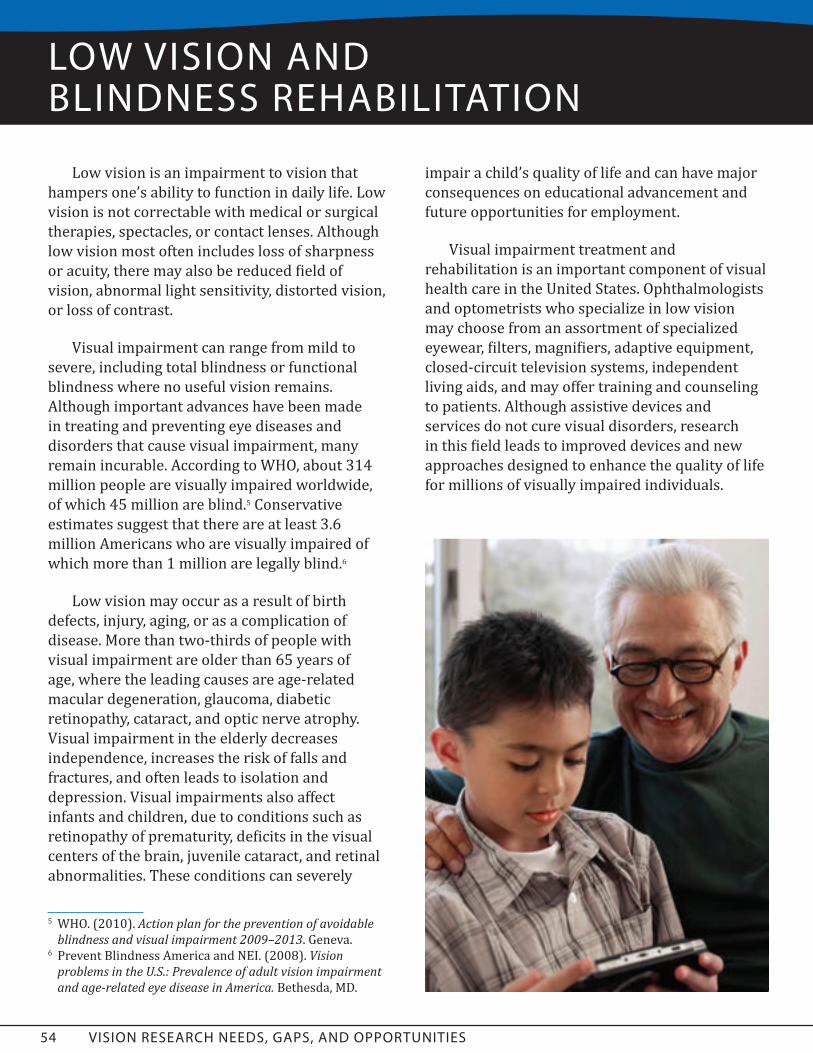
54 VISION RESEARCH NEEDS, GAPS, AND OPPORTUNITIES
LOW VISION AND BLINDNESS REHABILITATION
Low vision is an impairment to vision that hampers one’s ability to function in daily life. Low vision is not correctable with medical or surgical therapies, spectacles, or contact lenses. Although low vision most often includes loss of sharpness or acuity, there may also be reduced field of vision, abnormal light sensitivity, distorted vision, or loss of contrast.
Visual impairment can range from mild to severe, including total blindness or functional blindness where no useful vision remains. Although important advances have been made in treating and preventing eye diseases and disorders that cause visual impairment, many remain incurable. According to WHO, about 314 million people are visually impaired worldwide, of which 45 million are blind.5 Conservative estimates suggest that there are at least 3.6 million Americans who are visually impaired of which more than 1 million are legally blind.6
Low vision may occur as a result of birth defects, injury, aging, or as a complication of disease. More than two-thirds of people with visual impairment are older than 65 years of age, where the leading causes are age-related macular degeneration, glaucoma, diabetic retinopathy, cataract, and optic nerve atrophy. Visual impairment in the elderly decreases independence, increases the risk of falls and fractures, and often leads to isolation and depression. Visual impairments also affect infants and children, due to conditions such as retinopathy of prematurity, deficits in the visual centers of the brain, juvenile cataract, and retinal abnormalities. These conditions can severely 5 WHO. (2010). Action plan for the prevention of avoidable
blindness and visual impairment 2009–2013. Geneva.6 Prevent Blindness America and NEI. (2008). Vision
problems in the U.S.: Prevalence of adult vision impairment and age-related eye disease in America. Bethesda, MD.
impair a child’s quality of life and can have major consequences on educational advancement and future opportunities for employment.
Visual impairment treatment and rehabilitation is an important component of visual health care in the United States. Ophthalmologists and optometrists who specialize in low vision may choose from an assortment of specialized eyewear, filters, magnifiers, adaptive equipment, closed-circuit television systems, independent living aids, and may offer training and counseling to patients. Although assistive devices and services do not cure visual disorders, research in this field leads to improved devices and new approaches designed to enhance the quality of life for millions of visually impaired individuals.
LOW VISION AND BLINDNESS REHABILITATION 55
LOW VISION AND BLINDNESS REHABILITATIONHIGHLIGHTS OF RECENT PROGRESS
ASSISTIVE TECHNOLOGY
Because most content on the Internet is displayed visually, several new applications are geared to interpret this content for visually impaired users such as dynamic pin displays for online Braille and tactile graphics. Recent advances in computing power and image-processing have revolutionized applications for the visually impaired. Screen-magnifying and screen-reading software are widely deployed so that computers and smartphones are now accessible to low-vision and blind users, without the need for costly third-party software accessories.
The latest generation of smartphones has stimulated development of innovative products to aid the visually impaired. These include flexible image magnifiers, mobile optical character recognition, bar code readers, and indoor wayfinding aids. In the past five years, research and development of global positioning system-based navigation aids for visually impaired people has refined commercially available products.
BEHAVIORAL AND NEUROSCIENCE BASIC RESEARCH
When we move around our environment, in our mind’s eye, we construct a picture of the world around us. This picture, or cognitive map, is attributed to a neural network thought to be located in the medial temporal brain area. The parietal area of the brain is closely tied to accrual of perceptual information. The interaction between the parietal and temporal regions may explain how ongoing perceptual input leads to the formation of cognitive maps and has implications for understanding how visually impaired individuals learn different strategies for orienting and navigating.
Following pathological insult to retina or brain, perceptual training techniques have been examined using videogames. Behavioral studies have demonstrated that extensive practice can improve visual sensitivity. Infants with congenital cataracts or other treatable eye conditions experience visual deprivation during a period of robust visual development. Studies of these children are finding that once vision is restored, substantial functional and organizational changes in the visual cortex occur; whether the same holds true later in life is an actively debated topic. This neuroplasticity can now be studied noninvasively with functional magnetic resonance imaging. These findings regarding plasticity hold promise for informing vision rehabilitation efforts (see sidebar page 35).
Different areas of the brain respond to different types of perceptual stimuli (vision, touch, sound). Perception depends on integrating this multisensory information. Research shows that normally sighted, as well as visually impaired individuals recruit the visual cortex when interpreting tactile or auditory information. The functional roles of such cross-modal activations are not well understood, but are hot topics of current research and may provide the neural basis for sensory substitution, where perceptual processing for one sensory modality is largely replaced for another.
IMPLICATIONS OF VISION LOSS
With an increased understanding of the interdependence of physical and mental health, vision loss has been shown to be an independent predictor of depression. Depression (both major depressive disorders and subthreshold depression) affects roughly one-third of older adults with vision loss, which is similar to rates
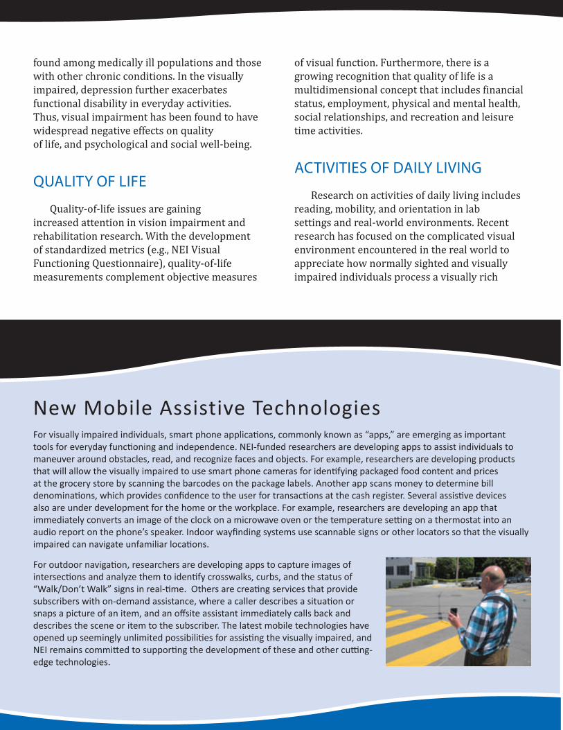
56 VISION RESEARCH NEEDS, GAPS, AND OPPORTUNITIES
found among medically ill populations and those with other chronic conditions. In the visually impaired, depression further exacerbates functional disability in everyday activities. Thus, visual impairment has been found to have widespread negative effects on quality of life, and psychological and social well-being.
QUALITY OF LIFE
Quality-of-life issues are gaining increased attention in vision impairment and rehabilitation research. With the development of standardized metrics (e.g., NEI Visual Functioning Questionnaire), quality-of-life measurements complement objective measures
of visual function. Furthermore, there is a growing recognition that quality of life is a multidimensional concept that includes financial status, employment, physical and mental health, social relationships, and recreation and leisure time activities.
ACTIVITIES OF DAILY LIVING
Research on activities of daily living includes reading, mobility, and orientation in lab settings and real-world environments. Recent research has focused on the complicated visual environment encountered in the real world to appreciate how normally sighted and visually impaired individuals process a visually rich
New Mobile Assistive TechnologiesFor visually impaired individuals, smart phone applications, commonly known as “apps,” are emerging as important tools for everyday functioning and independence. NEI-funded researchers are developing apps to assist individuals to maneuver around obstacles, read, and recognize faces and objects. For example, researchers are developing products that will allow the visually impaired to use smart phone cameras for identifying packaged food content and prices at the grocery store by scanning the barcodes on the package labels. Another app scans money to determine bill denominations, which provides confidence to the user for transactions at the cash register. Several assistive devices also are under development for the home or the workplace. For example, researchers are developing an app that immediately converts an image of the clock on a microwave oven or the temperature setting on a thermostat into an audio report on the phone’s speaker. Indoor wayfinding systems use scannable signs or other locators so that the visually impaired can navigate unfamiliar locations.
For outdoor navigation, researchers are developing apps to capture images of intersections and analyze them to identify crosswalks, curbs, and the status of “Walk/Don’t Walk” signs in real-time. Others are creating services that provide subscribers with on-demand assistance, where a caller describes a situation or snaps a picture of an item, and an offsite assistant immediately calls back and describes the scene or item to the subscriber. The latest mobile technologies have opened up seemingly unlimited possibilities for assisting the visually impaired, and NEI remains committed to supporting the development of these and other cutting-edge technologies.
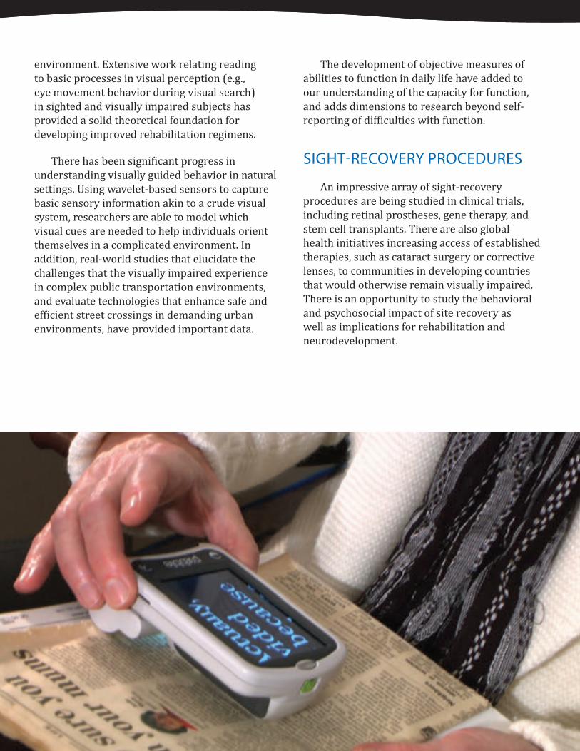
LOW VISION AND BLINDNESS REHABILITATION 57
environment. Extensive work relating reading to basic processes in visual perception (e.g., eye movement behavior during visual search) in sighted and visually impaired subjects has provided a solid theoretical foundation for developing improved rehabilitation regimens.
There has been significant progress in understanding visually guided behavior in natural settings. Using wavelet-based sensors to capture basic sensory information akin to a crude visual system, researchers are able to model which visual cues are needed to help individuals orient themselves in a complicated environment. In addition, real-world studies that elucidate the challenges that the visually impaired experience in complex public transportation environments, and evaluate technologies that enhance safe and efficient street crossings in demanding urban environments, have provided important data.
The development of objective measures of abilities to function in daily life have added to our understanding of the capacity for function, and adds dimensions to research beyond self-reporting of difficulties with function.
SIGHT-RECOVERY PROCEDURES
An impressive array of sight-recovery procedures are being studied in clinical trials, including retinal prostheses, gene therapy, and stem cell transplants. There are also global health initiatives increasing access of established therapies, such as cataract surgery or corrective lenses, to communities in developing countries that would otherwise remain visually impaired. There is an opportunity to study the behavioral and psychosocial impact of site recovery as well as implications for rehabilitation and neurodevelopment.
58 VISION RESEARCH NEEDS, GAPS, AND OPPORTUNITIES
LOW VISION AND BLINDNESS REHABILITATIONNEEDS, GAPS AND OPPORTUNITIES
UNDERSTANDING VISUAL IMPAIRMENT• Investigate multisensory processes and
cross-modal plasticity. Determine whether cross-modal plasticity associated with visual deprivation differs from normal multisensory interactions. Determine the organizing principles and limits that exist for cross-modal plasticity. Determine the informational requirements of a task that affect whether a remaining sensory modality (e.g., touch) can substitute for the absence of sight. Further research using neural network approaches, behavioral, and neuropsychological methods are necessary to inform effective sensory substitution and improve rehabilitation efforts.
• Characterize variation in spatial cognition, important for many tasks such as navigation. Spatial cognitive abilities vary widely. Normally sighted individuals with poor spatial cognition may be able to compensate through parallel processing using visual areas of the brain as well as perspective-free cognitive maps. In severe visual impairment, such compensation is limited or impossible, so the consequences of poor spatial cognition (whether innate or through lack of spatial cognitive experience) could be much more profound for the visually impaired than for the normally sighted.
• Determine spatial cognitive abilities of visually impaired individuals to determine the extent to which nonvisual cues contribute to spatial cognition and how the contribution of nonvisual cues can be enhanced to improve spatial cognition under conditions of visual impairment.
• Understand the use of multisensory spatial representations, or cognitive maps, in learning
strategies and whether learning strategies change with cross-modal plasticity. Does visual impairment result in fundamentally different learning strategies and does this differ by age?
SCREENING AND TESTING • Design and validate tests of visual perception.
Early detection of visual deficits will improve outcomes, but such tests can be used to benchmark outcomes of rehabilitative treatments. Sensitive, efficient testing is important to refine rehabilitation therapies and reduce burden on patients and clinicians.
• Create and validate vision tests relevant for the tasks of daily living. Eye charts that test letter acuity are not sufficient to characterize functional vision for people with visual impairment. Eye charts of high-contrast patterns are not useful or predictive for a majority of the visual tasks of daily living, which involve dynamic inputs of objects and scenes with varying color and contrast.
• Develop and standardize tests for evaluating more complex visually intensive behavior, such as reading, face recognition, mobility, and driving. Such tests will be useful in determining disability and progress toward rehabilitative goals.
• Develop testing specific to various patient populations. Nonverbal methods for testing vision (e.g., visual fields, retinal function) in special populations (children, neurological patients) are an important, yet unaddressed, area. In addition, developmental testing of visually impaired children, particularly pre- or nonverbal children, is limited, as current tests require vision to accomplish some or many of the tasks (e.g., stack blocks; match figures).
LOW VISION AND BLINDNESS REHABILITATION 59
Even motor milestones are affected by vision impairment, but details of how this occurs are unknown, and standards for normal development for visually impaired children are unknown.
ASSISTIVE DEVICE TECHNOLOGY• Develop new technology to improve access to
Internet, print, graphic display, and navigation resources. Some areas require specialized technology innovation (such as online Braille and tactile graphics, specialized embossers, and Braille software), but there are also opportunities for developing new assistive devices and products by modifying devices such as mobile phones, global-positioning system, accelerometers, and speech engines. Success in the visually impaired community depends on optimizing format and delivery methods, particularly for elderly, cognitively impaired, or technologically naïve individuals.
• Use models of eccentric vision (using peripheral retina when central field is dysfunctional) to translate technology developed for sighted users to visually impaired users. Additionally, basic science on navigation, limited vision use, and multimodal perception can inform new assistive device development. For instance, a fundamental question concerns the limits of sensory substitution: Conveying visual information via auditory and tactile means has proven difficult. Normally, visual recognition of an embossed line drawing is trivial, but using either auditory or tactile means is challenging. Even when sensory substitution succeeds, only fairly simple visual information can be conveyed via these other modalities. A better understanding of the key aspects of
visual information that are difficult to convey via other modalities could clearly inform the development of assistive technologies. Furthermore, studies of the effectiveness of these technologies are needed.
VISUAL PROSTHESES • Determine critical parameters for continued
improvement of prostheses. The visual prostheses are now being tested in patients. Different approaches include obtaining visual information (detected by a camera) to electrically stimulate the visual system (retina, or the visual cortex) (see sidebar page 60). Another relies on sensory substitution (e.g., encoding visual information into sensations on the tongue). In using a prosthesis, certain questions need to be resolved, for example, given a person’s state of vision and what tasks can be done. If synchrony between modalities is required, how is one modality affected when the other input is degraded?
• Understand the level of acceptable visual enhancement using prosthetics. This can be determined using models of low vision, such as low-quality images, to determine how much information is necessary for particular visual tasks. ‘Low-quality’ indicates low-resolution, reduced fields, poor color contrast and, more generally, any other image quality decrement that can be associated with low vision. Although normally sighted humans use high visual acuity to perform tasks such as face recognition, experiments with highly degraded images suggest that many of these skills are exceptionally robust in spite of acuity reductions. Such results could inform prosthesis design engineering and serve as feasibility criteria—if a planned neural
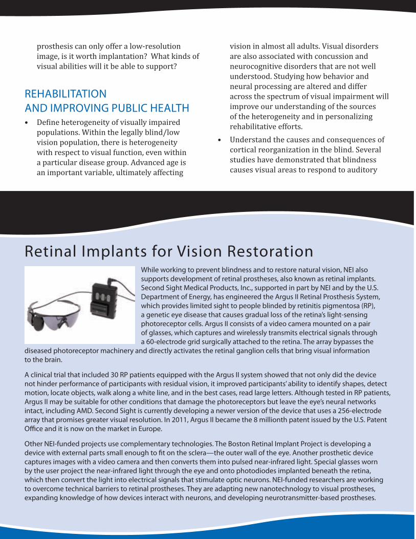
60 VISION RESEARCH NEEDS, GAPS, AND OPPORTUNITIES
prosthesis can only offer a low-resolution image, is it worth implantation? What kinds of visual abilities will it be able to support?
REHABILITATION AND IMPROVING PUBLIC HEALTH• Define heterogeneity of visually impaired
populations. Within the legally blind/low vision population, there is heterogeneity with respect to visual function, even within a particular disease group. Advanced age is an important variable, ultimately affecting
vision in almost all adults. Visual disorders are also associated with concussion and neurocognitive disorders that are not well understood. Studying how behavior and neural processing are altered and differ across the spectrum of visual impairment will improve our understanding of the sources of the heterogeneity and in personalizing rehabilitative efforts.
• Understand the causes and consequences of cortical reorganization in the blind. Several studies have demonstrated that blindness causes visual areas to respond to auditory
Retinal Implants for Vision RestorationWhile working to prevent blindness and to restore natural vision, NEI also supports development of retinal prostheses, also known as retinal implants. Second Sight Medical Products, Inc., supported in part by NEI and by the U.S. Department of Energy, has engineered the Argus II Retinal Prosthesis System, which provides limited sight to people blinded by retinitis pigmentosa (RP), a genetic eye disease that causes gradual loss of the retina’s light-sensing photoreceptor cells. Argus II consists of a video camera mounted on a pair of glasses, which captures and wirelessly transmits electrical signals through a 60-electrode grid surgically attached to the retina. The array bypasses the
diseased photoreceptor machinery and directly activates the retinal ganglion cells that bring visual information to the brain.
A clinical trial that included 30 RP patients equipped with the Argus II system showed that not only did the device not hinder performance of participants with residual vision, it improved participants’ ability to identify shapes, detect motion, locate objects, walk along a white line, and in the best cases, read large letters. Although tested in RP patients, Argus II may be suitable for other conditions that damage the photoreceptors but leave the eye’s neural networks intact, including AMD. Second Sight is currently developing a newer version of the device that uses a 256-electrode array that promises greater visual resolution. In 2011, Argus II became the 8 millionth patent issued by the U.S. Patent Office and it is now on the market in Europe.
Other NEI-funded projects use complementary technologies. The Boston Retinal Implant Project is developing a device with external parts small enough to fit on the sclera—the outer wall of the eye. Another prosthetic device captures images with a video camera and then converts them into pulsed near-infrared light. Special glasses worn by the user project the near-infrared light through the eye and onto photodiodes implanted beneath the retina, which then convert the light into electrical signals that stimulate optic neurons. NEI-funded researchers are working to overcome technical barriers to retinal prostheses. They are adapting new nanotechnology to visual prostheses, expanding knowledge of how devices interact with neurons, and developing neurotransmitter-based prostheses.
LOW VISION AND BLINDNESS REHABILITATION 61
and tactile stimuli. However, the behavioral consequences of the plasticity are not yet clear. Determine if rehabilitative processes can improve functional plasticity. Determine whether such reorganization affects visual learning if sight is restored. Recent studies on establishing sight in previously blind individuals provide opportunities to investigate neuroplasticity and how the brain adapts to new visual input (anatomically and functionally). Determining which visual skills can be acquired, as well as how these changes correlate with age and extent of visual deprivation, will inform the prospects for recovery.
• Develop and test rehabilitation models and training paradigms. Low vision and blindness rehabilitation models continue to evolve. Interventions that are multimodal, multidisciplinary, and address functional and emotional aspects uniquely related to low vision, blindness, loss of vision, and vision restoration hold particular promise. These include findings from basic psychophysical research (e.g., training visual skills to develop a preferred retinal locus in central vision loss or for the use of a visual prosthetic), research on psychosocial implications of vision impairment (e.g., the mechanisms by which visual impairment leads to reduced quality of life), and developing training for
prevention and adaptation strategies (e.g., lifestyle changes).
• Create and standardize performance-based outcome assessments as well as quality-of-life or other self-reported measures for low-vision interventions, whether they are prosthetic devices, assistive devices, or multidisciplinary rehabilitative strategies.
• Compare the effectiveness of rehabilitation approaches using randomized, controlled clinical trials. Rehabilitation currently lacks standardized methods and outcome assessments. Agreement on protocol and instrumentation will enable large-scale, multisite clinical trials.
• Identify comorbidities that interact with vision impairment and their influence on rehabilitation outcomes, and integrate visual rehabilitation models into subacute rehabilitation inpatient units. Poor vision hampers rehabilitation in a variety of age-related conditions (e.g., stroke, falls, hip fracture). Specialized rehabilitation needs for the visually impaired as they age (e.g., arthritis for life-long cane users), as well as for individuals with age-related vision loss, are not understood. Needs can vary depending on environment (e.g., nursing home residents) and comorbidities (e.g., wheelchair user; hearing-impaired individuals).

62 VISION RESEARCH NEEDS, GAPS, AND OPPORTUNITIES
APPENDICES
APPENDIX 1 63
APPENDIX 1:FRAMEWORK FOR VISION RESEARCH
Biomedical research is a highly specialized endeavor. Fundamental, overarching core principles drive the pursuit to understand, treat, and prevent ocular disease. A deep knowledge of the underlying biology of vision is critical to understand and treat the pathological mechanisms of visual disorders and diseases. In addition, an appreciation for the interaction of host factors with environmental factors from the cellular to the population level is needed to fully treat and prevent vision loss. The following provides a framework for considering these fundamental properties and core principles in the context of eye and vision research.
The framework was provided to each of the program panels as a reference to help the panel members focus on the needs and opportunities of their specific programs rather than the global concepts described here. New general concepts that arise from planning activities may yield important information that could help further shape the framework in the future.
I. GATHER COMPREHENSIVE KNOWLEDGE OF THE MOLECULAR BASIS OF OCULAR HEALTH AND DISEASE, AND USE THAT KNOWLEDGE TO IMPROVE DIAGNOSIS, TREATMENT, AND PREVENTION OF EYE DISEASE
a. Develop a detailed, comprehensive understanding of the molecular, cellular, and physiological mechanisms that promote and maintain ocular health.
b. Identify key signaling and transcriptional pathways in normal and pathological states.c. Determine key genetic and epigenetic variants that prevent or cause Mendelian as well as complex
ocular diseases.d. Characterize the changes in molecular processes affected by nutritional, behavioral, and other
environmental factors that impact ocular health and disease.e. Understand the regulation of cellular oxidation, energetics, and mitochondrial function in
maintaining healthy ocular tissues.f. Develop a better understanding of protein–protein interactions, regulation of protein folding, and
diseases that result from mis-folded proteins.g. Develop the ability to specifically modulate molecular pathways, systems, and intracellular
complexes to treat and cure ocular injury and disease.h. Develop tools (e.g., genomic markers, gene and/or protein expression profiles, and other new
measurable indicators) to assess an individual’s risk for particular ocular disorders.
II. UNDERSTAND THE SYSTEMS BIOLOGY UNDERLYING VISUAL FUNCTION
a. Determine the role of inflammation, immunity, infection, blood pressure, circadian rhythms, and other systemic processes in complex eye diseases.
b. Understand the integration of structural, vascular, and neural processes that constitute the unique physiology of the ocular system.
c. Understand visual processing in normal and pathological states.d. Characterize the developmental stages of the visual system from its precursor cells.
64 VISION RESEARCH NEEDS, GAPS, AND OPPORTUNITIES
e. Gain a deep understanding of the physiology of aging. Understand the contribution of the aging process to adult onset disease.
f. Develop models to understand cellular pathways, systems, and interactions of systems in healthy and pathological ocular tissues.
III. ACCELERATE THE TRANSLATION OF BASIC RESEARCH INTO CLINICAL STUDIES
a. Develop, refine, and set standards for new technologies (e.g., molecular imaging, telemedicine, small molecules) for clinical research and clinical trial application.
b. Develop and validate biomarkers that are useful in diagnosing and stratifying patients, measuring disease progression, and gauging therapeutic outcomes.
c. Foster multidisciplinary teams to develop new ocular diagnostic and therapeutic approaches (e.g., nanotechnology, prosthetics, cell-based therapies, molecular engineering, and synthetic biology).
d. Develop appropriate animal models and cell cultures to study disease mechanisms and evaluate investigational therapies.
e. Develop and evaluate assays and high-throughput screens to further vision research and clinical application.
f. Improve gene delivery techniques that yield precise targeting and regulation of gene expression.g. Integrate advances in regenerative medicine and tailor them to develop clinical applications in
specific ocular tissues.h. Develop new targeted therapies and combine them as appropriate to maximize treatment response. i. Make research tools widely available, including easy access to novel compounds, reagents, animal
models, cell lines, etc.j. Develop an infrastructure and distribute strategies for the collection and rapid dissemination of
genomic, proteomic, and other molecular library data to the vision research community.
IV. USE CLINICAL, EPIDEMIOLOGICAL, AND STATISTICAL TOOLS TO IDENTIFY POPULATIONS AT RISK OF BLINDING EYE DISEASES AND VISUAL DISORDERS, TO EVALUATE NEW THERAPEUTICS, AND TO IMPROVE FUNCTIONAL CONSEQUENCES OF VISUAL LOSS
a. Develop strategies and/or infrastructure to foster early, well-designed pilot clinical trials to rapidly assess the potential of translational therapies or candidate predictive markers.
b. Conduct clinical trials for improved prevention and treatment strategies for ocular diseases and disorders.
c. Improve the understanding of the burden of eye diseases and their visual outcomes at the societal and individual level through epidemiological and social sciences research. Identify population disparities and use these data to inform and enhance translational and clinical trials.
d. Develop and apply new methodologies for epidemiological research and strategies for the screening and detection of visual disorders.
e. Apply epidemiological methods and clinical trial interventions to blinding global eye diseases.
APPENDIX 1 65
V: STRENGTHEN CLINICAL RESEARCH OF VISUAL DISORDERS
a. Utilize developments in biotechnology and genomics to prioritize the testing of important molecular targets.
b. Develop new disease classification schemes and predictive models that incorporate genetic and molecular profiling.
c. Develop strategies and infrastructure to enable and encourage patients with ocular conditions to participate in clinical trials with special emphasis on recruiting minority populations.
d. Develop improved methods of clinical trial design that provide the capacity to evaluate therapeutic agents among patient subpopulations.
e. Develop strategies to improve efficiency in the clinical research enterprise such as common data elements, harmonized electronic data-capture systems, standardized training, and coordinated specimen management.
f. Complement bench discoveries and clinical trial results with focused behavioral and social science research, particularly in low-vision populations.
g. Integrate molecular and cellular knowledge with epidemiological and clinical information to inform clinical research priorities.
h. Promote the development and implementation of evidence-based guidelines for prevention, diagnosis, and treatment of visual disorders by conducting comparative effectiveness research.
VI: STRENGTHEN THE POOL OF VISION RESEARCHERS
a. Increase recruitment of clinician-scientists to vision research. b. Encourage early-stage vision researchers to participate in team science. Develop multidisciplinary
teams with expertise in fields such as bioinformatics, nanotechnology, engineering, and physics. c. Increase the participation of minorities in vision research. d. Recruit early-stage investigators from other disciplines to apply their knowledge and techniques to
study disorders of the visual system.
66 VISION RESEARCH NEEDS, GAPS, AND OPPORTUNITIES
APPENDIX 2:THE NEI PLANNING PROCESS
In 2011, NEI funded more than 1,400 research and training awards to extramural7 organizations such as universities and small businesses. The awards, primarily grants and cooperative agreements, are administered by scientists in the NEI Division of Extramural Research and are grouped into administrative portfolios (also referred to as programs; see box on page 67). NEI continues its tradition of identifying scientific priorities with a transparent and participatory process to assess progress, identify gaps and opportunities, and set realistic goals and objectives for the future. The program directors and the staff of the NEI Office of Program Planning and Analysis (OPPA) developed and implemented the planning process under the auspices of National Advisory Eye Council (NAEC), which represents the community of vision researchers and other stakeholders in eye research and ocular health.
INITIAL STEPS
We began with a retrospective examination of the NEI strategic planning process by convening a planning oversight group of current and former NAEC members and other leaders of major vision research organizations. The goal was to create a meaningful process to help NEI identify scientific opportunities, fund the best science, properly direct resources, and attract and train the best and the brightest investigators to vision research.
The oversight group endorsed the NEI philosophy that the bulk of extramural research should be driven by investigator-initiated projects, and NEI should continue to have a balance of basic, translational, and clinical research aims. Support was expressed for developing a series of important scientific goals 7 Extramural refers to work done outside NIH, mostly at
universities and other research organizations.
and priorities for all areas of vision research. As a first step, the group encouraged NEI to create an overarching document of general concepts that are common to most biomedical disciplines to help focus the efforts of planning panels on the problems and challenges specific to vision research. The NEI Framework for Vision Research (see Appendix 1) was created by NEI staff, was vetted by the oversight group, and then reviewed and approved by NAEC.
There also was recognition that an iterative, ongoing NEI planning process was necessary to keep pace with rapid advances in knowledge. The oversight group suggested proceeding with a series of workshops to identify new opportunities as they arose rather than in intervals of five or more years. Finally, it was agreed that the plan should identify a wide range of opportunities in all areas of vision research, but the NEI effort to implement the plan would depend on continual attention to program and resource management, new technologies, and a vibrant intramural8 program in basic science, translational, and clinical research.
INPUT TO THE NEI
In December 2010, NEI solicited input from the public and scientific community by issuing a Request for Information (RFI). NEI widely publicized the RFI through grantee and interest group e-mail lists and professional organizations. The RFI invited the scientific vision research community, health professionals, patient advocates, professional societies, and the general public to provide input on two broad questions:
1. What are the most significant scientific discoveries in vision research that have occurred since 2004?
8 Intramural refers to research by NEI scientists and clini-cians at the NEI facilities in Bethesda, MD.
APPENDIX 2 67
NEI ADMINISTRATIVE PROGRAMS
1. Retinal Diseases2. Corneal Diseases3. Lens and Cataract4. Glaucoma and Optic Neuropathies5. Strabismus, Amblyopia, and Visual Processing6. Low Vision and Blindness Rehabilitation
7. Ocular Genetics 8. Ocular Infection, Inflammation, and Immunology 9. Myopia and Refractive Error10. Oculomotor Systems and Neuro-Ophthalmology11. Ocular Pain12. Collaborative Clinical Research13. Small Business Innovation Research14. Research Training and Career Development15. Research Resources
2. What are the most significant scientific research needs, gaps, and opportunities that should be addressed by NEI?
Responses were collected through March 2011 from individual research scientists, small businesses, patient-advocacy groups, clinical trial networks, and professional societies. In particular, NEI is indebted to the Association for Research in Vision and Ophthalmology (ARVO)—an international organization of more than 12,500 individuals—for engaging its board of trustees, leaders of its scientific sections, and more than 300 members who generated a detailed response to the RFI.
PANEL MEETINGS (SPRING 2011)
Central to the current planning effort was the creation of planning panels with appropriate vision research expertise. Panels for all 15 administrative programs would have been too fragmented, and similar to previous planning efforts, we convened six panels. NEI program directors selected two internationally recognized co-chairs with broad expertise for each panel. In December 2010, all the co-chairs met with the NEI program directors and planning staff to discuss the scope and format of the plan and to finalize panel member selection. Expertise for each panel was selected to ensure that all areas of vision research were well represented.
To prepare for the spring panel meetings, each panel member submitted answers to the RFI questions. Panelists received a copy of the most recent National Plan (2004), recent workshop reports, and RFI responses submitted by the other panelists, the public, and ARVO. NEI encouraged panelists to think broadly, beyond the scope of their own work and specific field of vision research and carefully consider all input received. Premeeting teleconferences were held so that cochairs, panelists, and NEI staff understood the expectations and goals of the planning process. The panels were instructed to focus on the science and resources (clinical needs, animal models) rather than NEI-specific infrastructure such as funding mechanisms, training, and peer review.
Following the panel meetings, panel cochairs, in consultation with full panel membership and NEI staff drafted the panel reports found in this document, which were submitted to NAEC for comment in December 2011, prior to posting on the web for public comment.
The program panel reports in this document are the essential first step in developing a thoughtful strategic plan for NEI. With a thorough accounting of the state of vision research in place, NEI must begin to prioritize critical research to ensure that progress continues at a rapid pace. In the next phase of the planning process, NEI will re-engage the community to help prioritize the most pressing opportunities to develop ever more effective treatments for eye disease.
68 VISION RESEARCH NEEDS, GAPS, AND OPPORTUNITIES
APPENDIX 3:LIST OF CONTRIBUTORS
Ted Acott, Ph.D.Oregon Health and Science UniversityPortland, OR
Rando L. Allikmets, Ph.D.Columbia UniversityNew York, NY
Richard Andersen, Ph.D. (co-chair, Strabismus, Amblyopia, and Visual Processing)California Institute of TechnologyPasadena, CA
Vadim Y. Arshavsky, Ph.D.Duke UniversityDurham, NC
Steven Bassnett, Ph.D. (co-chair, Lens and Cataract)Washington University School of Medicine St. Louis, MO
Michele Basso, Ph.D.University of Wisconsin, MadisonMadison, WI
Larry Benowitz, Ph.D.Harvard School of MedicineChildren’s HospitalBoston, MA
Peter Bex, Ph.D.Schepens Eye Research Institute Boston, MA
Alan R. Burns, Ph.D.University of HoustonHouston, TX
Marie E. Burns, Ph.D. (co-chair, Retinal Diseases)University of California, DavisDavis, CA
Edward Callaway, Ph.D.Salk Institute for Biological StudiesLa Jolla, CA
Barbara Chapman, Ph.D.University of California, DavisDavis, CA
John I. Clark, Ph.D.University of Washington Seattle, WA
Anne Coleman, M.D., Ph.D.University of California, Los AngelesLos Angeles, CA
C. Edward Connor, Ph.D.Johns Hopkins UniversityKrieger Mind/Brain InstituteBaltimore, MD
Rosalie K. Crouch, Ph.D.Medical University of South Carolina Charleston, SC
Darlene A. Dartt, Ph.D.Harvard Medical School Boston, MA
Steven H. DeVries, M.D., Ph.D.Northwestern University Feinberg School of MedicineChicago, IL
APPENDIX 3 69
Elizabeth Engle, M.D.Harvard School of MedicineChildren’s HospitalBoston, MA
Velia M. Fowler, Ph.D.The Scripps Research InstituteLa Jolla, CA
Martin Friedlander, M.D., Ph.D. (co-chair, Retinal Diseases)Scripps Research InstituteLa Jolla, CA
John A. Gardner, Ph.D. ViewPlus Technologies, Inc.Corvallis, OR
Charles Gilbert, M.D., Ph.D. (co-chair, Planning Oversight Committee)Rockefeller UniversityNew York, NY
Michael Gilmore, Ph.D.Massachusetts Eye and Ear InfirmaryBoston, MA
Adrian Glasser, Ph.D.University of Houston Houston, TX
Jeff Goldberg, M.D., Ph.D.University of Miami School of Medicine Miami, FL
William V. Good, M.D. (co-chair, Low Vision and Blindness Rehabilitation)Smith-Kettlewell Eye Research InstituteSan Francisco, CA
Dale S. Gregerson, Ph.D.University of MinnesotaMinneapolis, MN
Jonathan Haines, Ph.D.Vanderbilt University School of MedicineNashville, TN
Robert L. Hendricks, Ph.D.University of PittsburghPittsburgh, PA
Amy Horowitz, D.S.W. Fordham UniversityNew York, NY Jonathan Horton, M.D., Ph.D.University of California, San FranciscoSan Francisco, CA
David Huang, M.D., Ph.D.Oregon Health and Science UniversityPortland, OR
Joseph A. Izatt, Ph.D.Duke UniversityDurham, NC
David Kaufman, D.O.Michigan State UniversityEast Lansing, MI
Daniel Kersten, Ph.D.University of MinnesotaMinneapolis, MN
Robert M. Lavker, Ph.D.Northwestern UniversityChicago, IL
Gordon Legge, Ph.D.University of MinnesotaMinneapolis, MN
Dennis Levi, O.D., Ph.D.University of California, BerkeleyBerkeley, CA
70 VISION RESEARCH NEEDS, GAPS, AND OPPORTUNITIES
Tom Lietman, M.D.University of California, San FranciscoSan Francisco, CA
Richard G. Long, Ph.D., COMSWestern Michigan UniversityKalamazoo, MI
Jack M. Loomis, Ph.D.University of California Santa Barbara, CA
Todd Margolis, M.D., Ph.D.University of California, San FranciscoF.I. Proctor FoundationSan Francisco, CA
M. Christine McGahan, Ph.D. (co-chair, Lens and Cataract)North Carolina State University Raleigh, NC
Hassane S. Mchaourab, Ph.D.Vanderbilt University Medical Center Nashville, TN
David C. Musch, Ph.D., M.P.H.The University of MichiganAnn Arbor, MI
Michael Mustari, Ph.D.University Of WashingtonSeattle, WA
Donald Mutti, O.D., Ph.D.Ohio State UniversityColumbus, OH
Jeremy Nathans, M.D., Ph.D.Johns Hopkins UniversityBaltimore, MD
Eric Pearlman, Ph.D. (co-chair, Corneal Diseases)Case Western Reserve UniversityCleveland, OH
Victor L. Perez, M.D. (co-chair, Corneal Diseases)Bascom Palmer Eye InstituteMiami, FL
Margaret A. Pericak-Vance, Ph.D.University of MiamiCoral Gables, FL
Joram Piatigorsky, Ph.D.National Eye InstituteBethesda, MD
Michael L. Robinson, Ph.D.Miami University Oxford, OH
James Rosenbaum, M.D.Oregon Health and Science UniversityPortland, OR
Mark Rosenblatt, M.D., Ph.D.Weill Cornell Medical CollegeNew York, NY
Krishnankutty Sathian, M.D., Ph.D. (co-chair, Low Vision and Blindness Rehabilitation)Emory UniversityAtlanta, GA
Val Sheffield, M.D., Ph.D.University of IowaIowa City, IA
D.J. Sidjanin, Ph.D. Medical College of Wisconsin Milwaukee, WI
Pawan Sinha, Ph.D.Massachusetts Institute of TechnologyCambridge, MA
APPENDIX 3 71
Earl Smith, O.D., Ph.D. (co-chair, Strabismus, Amblyopia, and Visual Processing)University of HoustonHouston, TX
Lois E. H. Smith, M.D., Ph.D.Harvard School of MedicineChildren’s HospitalBoston, MA
Alfred Sommer, M.D.Johns Hopkins UniversityBaltimore, MD
Dwight Stambolian, M.D., Ph.D.University of HoustonHouston, TX
Paul Sternberg Jr., M.D.Vanderbilt UniversityNashville, TN
Richard A. Stone, M.D. (co-chair, Planning Oversight Committee)University of PennsylvaniaPhiladelphia, PA
Stephen P. Sugrue, Ph.D.University of Florida College of MedicineGainesville, FL
Deming Sun, M.D.Doheny Eye InstituteLos Angeles, CA
Mriganka Sur, Ph.D.Massachusetts Institute of TechnologyCambridge, MA
Bosco Tjan, Ph.D.University of Southern CaliforniaLos Angeles, CA
Frank Tong, Ph.D.Vanderbilt UniversityNashville, TN
Russell Van Gelder, M.D., Ph.D.University of WashingtonSeattle, WA
Rohit Varma, M.D., M.P.H. (co-chair, Glaucoma and Optic Neuropathies)Keck School of MedicineLos Angeles, CA
Alan S. Verkman, M.D., Ph.D.University of California, San FranciscoSan Francisco, CA
Monica L. Vetter, Ph.D.University of UtahSalt Lake City, UT
Thomas Walz, Ph.D. Harvard Medical School Boston, MA
Robert Weinreb, M.D.University of California, San DiegoLa Jolla, CA
Sheila West, Ph.D.Johns Hopkins UniversityBaltimore, MD
Scott Whitcup, M.D.Allergan, Inc.Irvine, CA
Janey Wiggs, M.D., Ph.D.Massachusetts Eye and Ear InfirmaryBoston, MA
Donald Zack, M.D., Ph.D. (co-chair, Glaucoma and Optic Neuropathies)Johns Hopkins University School of MedicineBaltimore, MD
72 VISION RESEARCH NEEDS, GAPS, AND OPPORTUNITIES
CONTRIBUTORSNATIONAL EYE INSTITUTE STAFF
OFFICE OF THE DIRECTOR
Director, NEIPaul A. Sieving, M.D., Ph.D.
Deputy Director, NEIDeborah Carper, Ph.D.
EXTRAMURAL PROGRAM STAFF
Director, Division of Extramural ResearchLoré Anne McNicol, Ph.D.
Extramural Policy OfficerEllen S. Liberman, Ph.D., M.B.A.
Fundamental Retinal ProcessesAndrew P. Mariani, Ph.D.
Retinal DiseasesLisa A. Neuhold, Ph.D.
Retinal NeuroscienceThomas N. Greenwell, Ph.D.
Corneal DiseasesOcular Infection, Inflammation, and ImmunologyGrace L. Shen, Ph.D.
Lens and Cataract;Oculomotor Systems and Neuro-Ophthalmology; Ocular PainHoumam Araj, Ph.D.
Glaucoma and Optic NeuropathiesNeeraj Agarwal, Ph.D.
Strabismus, Amblyopia, & Visual ProcessingMichael A. Steinmetz, Ph.D.
Ocular GeneticsHemin R. Chin, Ph.D.
Myopia and Refractive ErrorJerome R. Wujek, Ph.D.
Perception & Psychophysics andLow Vision and Blindness RehabilitationCheri Wiggs, Ph.D.
Collaborative Clinical ResearchDonald F. Everett, M.A.Natalie Kurinij, Ph.D.Steven Oversby, Ph.D., R.N.Maryann Redford, D.D.S., M.P.H.Eleanor B. Schron, Ph.D., R.N. F.A.A.N.
OFFICE OF PROGRAM PLANNING AND ANALYSIS
Richard S. Fisher, Ph.D., Director
Shefa Gordon, Ph.D.
Thomas G. Hoglund, B.A.
Jennifer E. Mehren, Ph.D.

Photo CreditsFront Cover: Left image courtesy of Drs. Robert Fariss and Mercedes Campos. Bottom background image courtesy of Drs. Kent Sheridan and Michael Redmond.
Page 4: Left circle image courtesy of Dr. Wei Li; Right circle image courtesy of Drs. Jen-Yue Tsai and Robert Fariss.
Page 9: Image courtesy of Robert Watts.
Page 24: Image courtesy of Dr. Sheila West.
Page 35: Image courtesy of Dr. Pawan Sinha.
Page 41: Image courtesy of Drs. Thomas Johnson, Naoki Nakaya and Stanislav Tomarev.
Page 46: Image copywright by Terese Winslow.
Page 53: Image courtesy of Dr. Wei Li.
Page 56: Image courtesy of Dr. James Coughlan.
Page 60: Image courtesy of Second Sight.
Unless noted above, all other images are from the NEI image library.

U.S. Department of Health and Human ServicesNational Institutes of HealthNational Eye Institutehttp://www.nei.nih.govAugust 2012