near-infrared scintillation of liquid argon - fermilab |...
TRANSCRIPT

FERMILAB-CONF-15-511-E-ND
ACCEPTED
Near-infrared scintillation of liquid argon
T. Alexander,a C.O. Escobar,a,b,1 W.H. Lippincotta and P. Rubinova
aFermi National Accelerator Laboratory,
Kirk Rd and Pine St., Batavia, IL 60510, U.S.A. b Instituto de Física da Universidade Estadual de Campinas,
rua Sergio Buarque de Holanda 777, 1308-859, Campinas, SP, Brazil
E-mail: [email protected]
Abstract: Since the 1970s it has been known that noble gases scintillate in the near infrared (NIR)
region of the spectrum (0.7 µm < λ < 1.5 µm). More controversial has been the question of the NIR
light yield for condensed noble gases. We first present the motivation for using the NIR scintillation
in liquid argon detectors, then briefly review early as well as more recent efforts and finally show
encouraging preliminary results of a test performed at Fermilab.
Keywords: Scintillators, scintillation and light emission processes (solid, gas and liquid scintilla-
tors); Noble liquid detectors (scintillation, ionization, double-phase)
1Corresponding author.
Operated by Fermi Research Alliance, LLC under Contract No. De-AC02-07CH11359 with the United States Department of Energy.

– 1 –
Contents
1 Introduction 1
2 Brief historical review 2
3 Results from a preliminary test 4
4 Conclusions 7
1 Introduction Motivation for using NIR light. It is well-known that the detection of both charge and light
signals is a desirable feature in many experiments using liquid noble gases (LNG) [1], ranging from
dark matter (DM) searches [2] to large liquid argon (LAr) time projection chambers (TPC) used in
neutrino physics [3]. For the latter the scintillation signal is important in providing the start time
t0 for non-accelerator related physics, while for DM experiments the detection of the light signal
provides background rejection by separating nuclear recoils from electronic ones.
So far the vast majority of the current or planned experiments focus on the detection of the
vacuum-ultraviolet (VUV) scintillation light from noble gases either in gaseous or condensed states
with wavelengths as short as 78 nm for liquid neon up to 175 nm in liquid xenon. The detection of the
VUV light presents many experimental challenges starting with the lack of availability of reasonably
priced (cost per unit sensitive area) cryogenic VUV sensitive photodetectors. Most argon detectors
use wavelength shifters (WLS) such as tetra phenyl butadiene (TPB) [4], for example in the light-
guide bars designed for the DUNE experiment [5] or directly coating a visible light sensitive pho-
todetector as for the ArDM experiment [6]. The use of TPB raises concerns about long term stability
and extreme care in the storage and handling of coated surfaces due to the degradation of TPB when
exposed to UV light or by its oxidation in air or hydration when kept in non-dry atmospheres [7]. An-
other challenge, significant for very large TPC with long drift distances, is the presence of Rayleigh
scattering which introduces pernicious effects such as a two to three-fold decrease in the number of
collected photons coming from distances at or beyond 2.5 m. Poor reflectivity of VUV light from
most materials make reflective surfaces, unless coated with TPB, almost useless and finally, Rayleigh
scattering smears the time resolution [8]. These negative effects introduced by Rayleigh scattering
are brought to the fore if a recent estimate of the Rayleigh scattering length [9] placing it at 55cm is
taken into account. Lastly it should be mentioned that the recently observed delayed light emission
from TPB makes the detection of the triplet light harder, since it occurs on a longer time scale
than the singlet for LAr [10]. All of the above difficulties cease to exist if LNGs have a significant
emission in the NIR and that light is used either as a replacement or in addition to the VUV signal.
One further advantage of using the NIR scintillation as the light signal in large LAr TPCs is the
possibility for doping LAr with a photosensitive chemical such as tetra-methyl-germanium (TMG)

– 2 –
Figure 1. Spectra of transient absorption of Ar. (a) solid; (b) liquid and (c) gas. From ref. [14].
to help recover the charge lost by recombination, as proposed by the ICARUS collaboration [11].
TMG due to its large photo-absorption cross-section significantly reduces the amount of useful VUV
light while any NIR light would travel without any attenuation, enhanced charge and light collection
would go hand in hand, violating the complementarity [1] between charge and light signals.
2 Brief historical review
Early results. Near-infrared emission from noble gases has been known since the late 1940s
with emission lines around 1,300 nm [12]. Using the technique of transient optical absorption
spectroscopy via electron excitation, NIR transitions were observed in noble gases both in the
gaseous state [13] and in condensed form [14]. The similarity of the spectral features in gas and in
liquid or solid in the particular case of argon, as seen in figure 1, from reference [14], has led to the
interpretation that these features are due to the formation of self-trapped excitons.
In all three phases the location of the peak is around 1.27 eV which corresponds to a wavelength
of 976 nm. In the gas phase one further peak is observed around 1 eV (1,240 nm).
The interest on the NIR emission from noble gases resurfaced with work of Lindblom and
Solin [15] who observed atomic lines in the NIR under excitation by a low-energy (3–5 MeV)
proton beam. This was followed by a systematic investigation of NIR emission from liquid and
gaseous Ar and Xe by Bressi, Carugno and co-workers [16], with the goal of developing new particle
detectors. Their results showed that both xenon and argon scintillate in the NIR, but the results were
inconclusive for the liquid state of both noble gases, as the light yield was poorly estimated [17].

– 3 –
Figure 2. Results from the Munich group [20]: emission spectra of LAr at 85K. The peak at 557 nm is
identified as due to oxygen impurity. For our purposes notice the spectral feature at 970 nm (close to the
cut-off wavelength of their spectrometer).
Recent results. In the last five years, two groups have pursued the investigation of NIR scintillation
in LAr, one based in Novosibirsk, Russia [18] and the other at the Technical University of Munich,
Germany [19]. Both groups use table-top setups, with volumes of a few cubic centimeters of LAr
excited by very intense, low-energy beams: 12 keV electrons (pulsed or continuous) for the Munich
experiments and pulsed X-rays with energies between 30 and 40 keV for the Novosibirsk setup. We
shall now briefly describe the techniques used by each group and summarize their results.
The Munich group studied the scintillation of LAr over a wide range of wavelengths, from the
VUV to the NIR, using a monochromator and a VUV sensitive photomultiplier for their study of
the second and third continua (up to 320 nm) and a spectrometer to study the region from 250 nm
to 1000 nm. No absolute light yield is provided but several spectral features were associated with
traces impurities. Figure 2 shows a light spectrum from one of their earliest publications [20].
Later, we will return to the more recent results from the Munich group but now we would like
to summarize the results obtained by Bondar et al. [18, 21, 22], which are revelant for our own
preliminary test, which we will report in the next section.
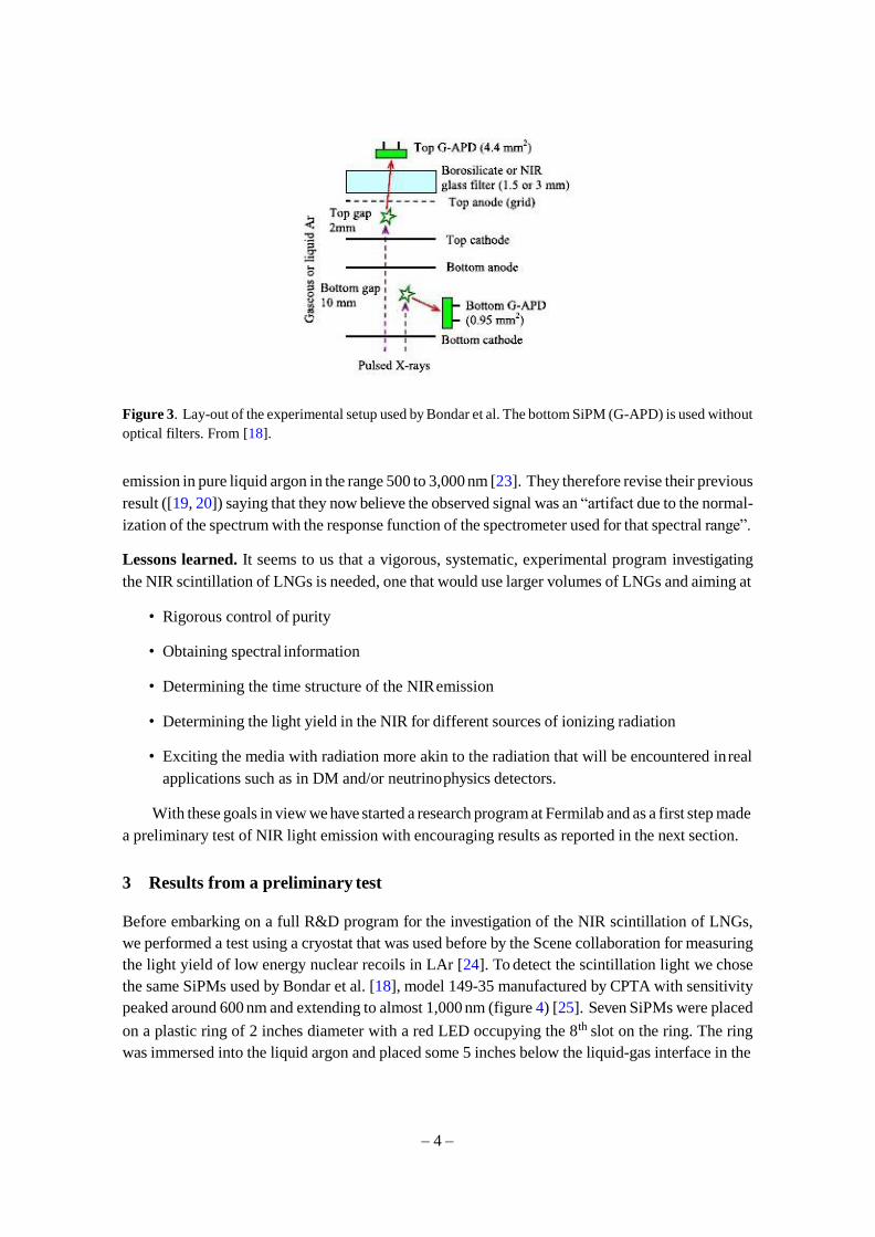
The Novosibirsk group employed silicon photo multipliers (SiPM) with sensitivity peaked
around 600 nm, extending to 1,000 nm. As they do not use a spectrometer, in contrast to the Munich
group, they obtain an integrated scintillation yield by convoluting the photon detection efficiency of
their SiPMs with the spectrum obtained by the Munich group and the geometrical acceptance of their
apparatus (shown in figure 3), obtained by Monte Carlo simulation. The normalization of the scin-
tillation spectrum is thus obtained by matching the number of counted photoelectrons in the SiPMs.
Bondar et al. obtained a light yield of 1.7x10⁴ photons/MeV in gaseous Ar in the range 690–
1,000 nm and of 5.1x10² photons/MeV in LAr in the range 400–1,000 nm. It is important to
emphasize that Bondar et al. use in their analysis the unnormalized spectrum obtained by Heindl et
al. [19, 20] as this leads us to review a recent result from the Munich group that points out to the
difficulties intrinsic to this kind of measurement and its interpretation.
More recently, the Munich group has published new results of their on-going studies of the scin-
tillation of pure liquid argon and liquid Ar-Xe mixtures, stating that they no longer see any relevant
– 4 –
Figure 3. Lay-out of the experimental setup used by Bondar et al. The bottom SiPM (G-APD) is used without
optical filters. From [18].
emission in pure liquid argon in the range 500 to 3,000 nm [23]. They therefore revise their previous
result ([19, 20]) saying that they now believe the observed signal was an “artifact due to the normal-
ization of the spectrum with the response function of the spectrometer used for that spectral range”.
Lessons learned. It seems to us that a vigorous, systematic, experimental program investigating
the NIR scintillation of LNGs is needed, one that would use larger volumes of LNGs and aiming at
• Rigorous control of purity
• Obtaining spectral information
• Determining the time structure of the NIR emission
• Determining the light yield in the NIR for different sources of ionizing radiation
• Exciting the media with radiation more akin to the radiation that will be encountered in real
applications such as in DM and/or neutrino physics detectors.
With these goals in view we have started a research program at Fermilab and as a first step made
a preliminary test of NIR light emission with encouraging results as reported in the next section.
3 Results from a preliminary test
Before embarking on a full R&D program for the investigation of the NIR scintillation of LNGs,
we performed a test using a cryostat that was used before by the Scene collaboration for measuring
the light yield of low energy nuclear recoils in LAr [24]. To detect the scintillation light we chose
the same SiPMs used by Bondar et al. [18], model 149-35 manufactured by CPTA with sensitivity
peaked around 600 nm and extending to almost 1,000 nm (figure 4) [25]. Seven SiPMs were placed
on a plastic ring of 2 inches diameter with a red LED occupying the 8th slot on the ring. The ring
was immersed into the liquid argon and placed some 5 inches below the liquid-gas interface in the

– 5 –
Figure 4. Photo detection efficiency of the CPTA 149-35 SiPM [25].
Figure 5. Configuration of the experiment showing tagging detector (PMT).
inner cryostat vessel. A 1µCi 22Na positron source was used to produce two positron annihilation
gamma rays (0.511 MeV) with opposite momenta. We tag the backward-going gamma ray using a
liquid scintillator counter (EJ301 liquid scintillator from Eljen) read-out by a photomultiplier tube.
The schematic lay-out of the setup is shown in figure 5.
The readout chain for the experiment consists entirely of warm electronics positioned outside
the cryostat. The connection from the cryostat is carried on 50 ohm RG58 coaxial cables, with
the bias and signal on the inner conductor and reference ground on the shield. The main amplifier
used, the Photonique model AMP-0611 was packaged in a custom NIM module, supporting up to
8 amplifiers, bias distribution and filter circuits as well as bias adjusting trim pots. The bias voltage
was generated by a Keithley 2400 SourceMeter. The power from the amplifiers was supplied by
a standard NIM crate. The outputs from the NIM module were routed to a Tektronix DPO3054
digital oscilloscope which was used as the main digitizer. The readout was accomplished via a
small module within Excel using the USB interface of the scope. The scope was used to trigger
on the signals and could be used in single channel or “Logic OR” mode for up to 4 channels. In
addition, the external trigger output of the DPO3054 was also routed to a HP53131A counter timer
unit (read out via GPIB) to allow for measurement of signal rates.
With the above configuration the only degrees of freedom available for introducing variation in
the setup were the height of the source and counter with respect to the ring with the SiPMs (or the
gas-liquid interface) and the distance between the radioactive source and the scintillation counter

– 6 –
Figure 6. Scope traces from the SiPM (bottom) and PMT (top) showing predominance of early time PMT
signals as expected if the SiPM is triggering on scintillation light.
which defines the solid angle covered by the annihilation gamma-rays. The rates are expected to
be low due to the low source intensity, small geometrical acceptance and small energy loss of the
gammas in the LAr (mostly to Compton electrons). We ran the SiPMs at a bias voltage of 44 V.
To avoid excessive noise that arose when using multiple SiPMs in coincidence, we trigger on the
best SiPM at a fraction of a single p.e., looking for the EJ301 PMT pulse inside a time window that
spans negative (PMT fires before the trigger) and positive times (PMT signal after the SiPM). If
the SiPM is triggering on scintillation light we expect to have more PMT signals occurring earlier
(negative times) than later (positive times). Figure 6 shows a typical collection of scope traces from
the SiPM and the scintillation counter (the time axis has the origin translated so as to have only
positive times). A cut is placed on the pulse height of the scintillator PMT traces so as to select the
0.511 MeV gamma, eliminating the 1.275 MeV gamma that also comes from the decay of 22Na. As
a consistency check we verified that the latter signal is evenly spread in early and late time regions,
as it should since it is not correlated with the scintillation signal. These results confirm that ionizing
radiation in LAr produces light with wavelength between 600 and 1000 nm. The light yield in this
region has not been determined due to the low geometrical acceptance of our experimental setup
and the small energy loss in the corresponding volume of LAr.
Figure 7 shows a typical time distribution from the PMT signals, which we fit with a double
exponential. The results show a fast component with a time constant of less than 200 ns and a
slow one with 2.72 µs. This result is consistent with the time distribution obtained by Bondar
et al. [21, 22] who also obtained short and slow components, with the latter remaining without
explanation regarding its origin.

– 7 –
Figure 7. Time distribution of the scintillation counter signal. Notice the presence of a fast and a slow
component.
4 Conclusions NIR scintillation from liquid noble gases is a promising alternative to VUV light for determining
the start time of an event (t0 ), helping with particle identification and the reconstruction of very
low-energy events, which is important for the detection of supernova neutrinos and search for
proton decay [26]. The use of NIR light would by-pass problems and difficulties associated with
the use of VUV scintillation and opens the door for simultaneously improving charge collection
in Lar TPC’s through doping, avoiding the quenching of the scintillation light. The simultaneous
detection of the VUV and NIR signals in DM experiments could help separate nuclear recoils
from the electromagnetic background if, as expected on theoretical grounds, the NIR light is more
abundant for denser ionization tracks.
In order to fulfil these goals we plan to continue investigating the NIR scintillation in LNGs
addressing the challenges outlined in section 2.3.
Acknowledgments
We would like to thank Michael Reid for a GEANT4 simulation that helped us check the soundness
of our results. The technical staff at the Fermilab Proton Assembly Building is thanked for their
support. Fermilab is Operated by Fermi Research Alliance, LLC under Contract No. De-AC02-
07CH11359 with the United States Department of Energy.
References
[1] E. Aprile, A.E. Boltnikov, A.I. Bolozdynya and T. Doke, Noble gas detectors, Wiley-VCH, Weinheim
Germany (2006).
[2] V. Chepel and H. Araujo, Liquid noble gas detectors for low energy particle physics, 2013 JINST 8
R04001 [arXiv:1207.2292].
[3] A. Marchionni, Status and new ideas regarding liquid argon detectors, Ann. Rev. Nucl. Part. Sci. 63
(2013) 269 [arXiv:1307.6918].
[4] W.M. Burton and B.A. Powell, Fluorescence of tetraphenyl-butadiene in the vacuum ultraviolet, Appl.
Opts. 12 (1973) 87.
– 8 –
[5] D. Whittington, Photon detection system designs for the Deep Underground Neutrino Experiment,
presented at LIDINE, U.S.A. (2015) [arXiv:1511.06345].
[6] A. Badertscher et al., ArDM: first results from underground commissioning, presented at LIDINE,
U.S.A. (2015) [2013 JINST 8 C09005] [arXiv:1309.3992].
[7] C.S. Chiu et al., Environmental effects on TPB wavelength-shifting coatings, 2012 JINST 7 P07007
[arXiv:1204.5762].
[8] C. Thorn, Consequences of Rayleigh scattering in Lar, LBNE-doc-6302-v2, August 2012.
[9] E. Grace and J.A. Nikkel, Index of refraction, Rayleigh scattering length and Sellmeier coefficients in
solid and liquid argon and xenon, arXiv:1502.04213.
[10] E. Segreto, Evidence of delayed light emission of tetraphenyl butadiene excited by liquid argon
scintillation light, Phys. Rev. C 91 (2015) 035503 [arXiv:1411.4524].
[11] P. Cennini et al., Improving the performance of the liquid argon TPC by doping with tetramethyl
germanium, Nucl. Instrum. Meth. A 355 (1995) 660.
[12] C.J. Humphreys and E.K. Plyler, Infrared emission spectra of krypton and argon, research paper
RP1790, J. Res. NBS 38 (1947) 499.
[13] S. Arai et al., Near infrared abosrptions of neon, argon, krypton and xenon excited diatomic
molecules, J. Chem. Phys. 68 (1978) 4595.
[14] T. Suemoto et al., Observation of relaxed exciton states in condensed argon, Phys. Lett. A 61 (1977)
131.
[15] P. Lindblom and G. Solin, Atomic near-infrared noble gas scintillation I: optical spectra, Nucl.
Instrum. Meth. A 268 (1998) 204.
[16] G. Carugno, Infrared emission in gaseous media induced by ionizing particles and by drifting
electrons, Nucl. Instrum. Meth. A 419 (1998) 617.
[17] G. Bressi et al., Infrared scintillation in liquid Ar and Xe, Nucl. Instrum. Meth. A 440 (2000) 254.
[18] A. Buzulutskov, A. Bondar and A. Grebenuk, Infrared scintillation yield in gaseous and liquid argon,
Europhys. Lett. 94 (2011) 52001 [arXiv:1102.1825].
[19] T. Heindl et al., Table-top setup for investigating the scintillation properties of liquid argon, 2011
JINST 6 P02011 [arXiv:1511.07720].
[20] T. Heindl et al., The scintillation of liquid argon, Europhys. Lett. 91 (2010) 62002
[arXiv:1511.07718].
[21] A. Bondar, A. Buzulutskov, A. Dolgov, A. Grebenuk, E. Shemyakina and A. Sokolov, Study of
infrared scintillations in gaseous and liquid argon — part I: methodology and time measurements,
2012 JINST 7 P06015 [arXiv:1204.0180].
[22] A. Bondar et al., Study of infrared scintillations in gaseous and liquid argon — part II: light yield and
possible applications, 2012 JINST 7 P06014 [arXiv:1204.0580].
[23] A. Neumeier et al., Intense infrared scintillation of liquid Ar-Xe mixtures, Europhys. Lett. 106 (2014)
32001 [arXiv:1511.07722].
[24] SCENE collaboration, T. Alexander et al., Observation of the dependence on drift field of scintillation
from nuclear recoils in liquid argon, Phys. Rev. D 88 (2013) 092006 [arXiv:1306.5675].
[25] CPTA (Centre of Perspective Technology and Apparatus) webpage, http://www.cpta-apd.ru.
[26] P. Cennini et al., Detection of scintillation light in coincidence with ionizing tracks in a liquid argon
time projection chamber, Nucl. Instrum. Meth. A 432 (1999) 240.