national cancer institute · pdf filenci immunotherapy agent workshop proceedings national...
TRANSCRIPT

Immunotherapy Agent WorkshopJuly 12, 2007
Natio
nal C
ance
r Ins
titut
e
U.S. Department of HealtH anD HUman ServiceS
national institutes of Health
NCI Immunotherapy Agent Workshop Proceedings
CONTENTS
Executive Summary ........................................................................................................................ 3
Final Rankings of Agents with High Potential for Use in Treating Cancer ................................... 5
Opening Remarks............................................................................................................................ 6
Details of the Proceedings .............................................................................................................. 9
(1) Adjuvants ............................................................................................................................ 9
(2) T-cell Growth Factors ....................................................................................................... 28
(3) Anti-Checkpoint and Varied Agents................................................................................. 36
(4) Co-Stimulatory and Varied Agents................................................................................... 49
Online Discussions Leading to Final Rankings ............................................................................ 65
Final Rankings .............................................................................................................................. 67
Participant List .............................................................................................................................. 71
Appendix A: PowerPoint Template for Presentations .................................................................. 77
Note: The presenters’ PowerPoint slides and an electronic version of this report may be accessed and downloaded from the Website of the Biological Resources Branch (BRB): http://web.ncifcrf.gov/research/brb/workshops.asp
1

NCI Immunotherapy Agent Workshop Proceedings
2

NCI Immunotherapy Agent Workshop Proceedings
NATIONAL CANCER INSTITUTE IMMUNOTHERAPY AGENT WORKSHOP
JULY 12TH, 2007
EXECUTIVE SUMMARY There is an ongoing explosion of knowledge in the immunological sciences with the discovery of many agents that have the potential to serve as immunotherapeutic drugs. For a variety of reasons, few of these are being tested in humans. The workshop developed a ranked list of agents with high potential for use in treating cancer. Despite substantial demonstrated immunological efficacy, these agents are not broadly available for testing in patients with cancer. The ranking by workshop participants was based on the likelihood for efficacy in cancer therapy and was exceedingly well-vetted, with broad and substantial input. The exceedingly broad nature of the consensus behind this list will facilitate subsequent NCI discussions on the availability of clinical grade immunotherapeutic drugs for human trials and will inform other governmental agencies, nongovernmental funding agencies, industry, and individual investigators that these agents have broad appeal to the immunotherapy community and, by consensus, hold particular promise for use in cancer therapy. Twenty agents are presented on the list, presented in rank order. However, all are considered to have substantial potential for cancer therapy. Criteria essential for inclusion on the list included:
• Potential for use in cancer therapy.
• Perceived need by multiple, independent clinical investigators.
• Potential use in more than one clinical setting (i.e., against different tumor types or as part of multiple therapy regimens).
• Not broadly available for testing in patients.
• Not commercially available or likely to be approved for commercial use in the near future.
The 20 agents were selected from a list of 124 agents suggested to an NCI Web site asking for suggestions and advice about “agents with known substantial immunologic or physiologic activity that have not been tested or have been inadequately tested in cancer patients.” The Web site was publicized widely by the NCI with requests for advice sent to grantees with immunology or immunotherapy grants and to prior recipients of RAID awards, as well as to intramural scientists involved in immunology or immunotherapy. The Web site was further publicized to the membership of the major scientific societies involved in immunology, immunotherapy and cancer research, namely the American Association for Cancer Research (AACR), American Association of Immunologists (AAI), American Society of Oncology (ASCO), American Society of Hematology (ASH), the Cancer Vaccine Consortium (CVC), and the International Society of Biological Therapy of Cancer (iSBTc).
3

NCI Immunotherapy Agent Workshop Proceedings
Web respondents expressed particular interest in vaccine adjuvants; T-cell growth factors; agents to inhibit immune checkpoint blockade; functional antibodies, cytokines, ligands, and receptors; including agents “left on the shelf” by drug companies as well as suggestions for specific antigens for vaccines and antigen-specific antibodies. The organizing committee winnowed the list of agents to the top 30 for presentation and ranking by the Workshop. The committee focused on agents with the greatest potential for broad usage in multiple types of regimens, thereby excluding specific antigens for vaccines and antigen-specific antibodies desired by individual investigators and groups of investigators, regardless of their attractiveness or potential utility. The workshop participants were selected from suggestions by the AACR, AAI, ASCO, ASH, CVC, and iSBTc, and by the NCI intramural and extramural programs. The participants broadly represented academia, industry, and the NCI. The workshop was open to the public. Observers from industry, the NCI, and the FDA were invited and asked to comment during the proceedings. The final ranked list derived from discussions of each agent. Agents at the top of the list were considered the most desirable based on current evidence. It was well recognized by the participants that many agents with less data, including agents not currently on the list, may ultimately prove to be more important than those at the top of the list. Although the ranking is well vetted and based on the cumulative knowledge of the broad immunotherapy and cancer research communities, the choice and desirability of individual agents will undoubtedly change with new knowledge. Because the priorities are based on incomplete knowledge, the process should be a dynamic, ongoing one that can be revised as more data appear. A common suggestion was that a mechanism should be developed to continually update the list. Possible positive outcomes of having a well-vetted ranked list based on a broad consensus of the immunology and immunotherapy community should include encouragement of (1) RAID applications for manufacture, (2) NCI distribution of company-manufactured agents, and (3) reinvigoration of pharma/biotech efforts to develop them. Future availability of these agents for broad testing and development will provide a benchmark for the strength and resolve of the national cancer therapy development enterprise.
4
NCI Immunotherapy Agent Workshop Proceedings
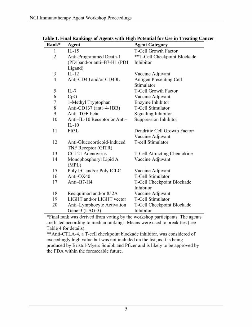
Table 1. Final Rankings of Agents with High Potential for Use in Treating Cancer
Rank* Agent Agent Category 1 IL-15 T-Cell Growth Factor 2 Anti-Programmed Death-1
(PD1)and/or anti–B7-H1 (PD1 Ligand)
**T-Cell Checkpoint Blockade Inhibitor
3 IL-12 Vaccine Adjuvant 4 Anti-CD40 and/or CD40L Antigen Presenting Cell
Stimulator 5 IL-7 T-Cell Growth Factor 6 CpG Vaccine Adjuvant 7 1-Methyl Tryptophan Enzyme Inhibitor 8 Anti-CD137 (anti–4-1BB) T-Cell Stimulator 9 Anti–TGF-beta Signaling Inhibitor 10 Anti–IL-10 Receptor or Anti–
IL-10 Suppression Inhibitor
11 Flt3L Dendritic Cell Growth Factor/ Vaccine Adjuvant
12 Anti-Glucocorticoid-Induced TNF Receptor (GITR)
T-cell Stimulator
13 CCL21 Adenovirus T-Cell Attracting Chemokine 14 Monophosphoryl Lipid A
(MPL) Vaccine Adjuvant
15 Poly I:C and/or Poly ICLC Vaccine Adjuvant 16 Anti-OX40 T-Cell Stimulator 17 Anti–B7-H4 T-Cell Checkpoint Blockade
Inhibitor 18 Resiquimod and/or 852A Vaccine Adjuvant 19 LIGHT and/or LIGHT vector T-Cell Stimulator 20 Anti–Lymphocyte Activation
Gene-3 (LAG-3) T-Cell Checkpoint Blockade Inhibitor
*Final rank was derived from voting by the workshop participants. The agents are listed according to median rankings. Means were used to break ties (see Table 4 for details). **Anti-CTLA-4, a T-cell checkpoint blockade inhibitor, was considered of exceedingly high value but was not included on the list, as it is being produced by Bristol-Myers Squibb and Pfizer and is likely to be approved by the FDA within the foreseeable future.
5

NCI Immunotherapy Agent Workshop Proceedings
NATIONAL CANCER INSTITUTE IMMUNOTHERAPY AGENT WORKSHOP PROCEEDINGS
OPENING REMARKS
Martin A. “Mac” Cheever, M.D., and Stephen Creekmore, M.D., Ph.D., the workshop co-chairs, welcomed and thanked the participants, including several who participated via teleconference. The goal of the meeting is to develop a recommended prioritized list of agents that have the potential to become immunotherapeutic drugs1 for treating cancer. The purpose of the list is to recommend certain agents that hold particular promise to the National Cancer Institute (NCI), nongovernmental funding agencies, industry, and individual investigators. Possible positive outcomes could include encouragement of (1) Rapid Access to Interventional Development (RAID) applications for the manufacture or (2) distribution of company-manufactured agents through RAID or the Cancer Therapy Evaluation Program (CTEP), (3) reinvigoration of their development by companies with such agents on the shelf or licensing them to other companies for development, or (4) investment by venture capitalists in new development. This rank-setting exercise could also serve as a report card: if a year or two goes by and the list remains substantially unchanged, it would be a signal that the current system for developing immunotherapeutic agents is not working optimally. Dr. Creekmore emphasized the importance of the workshop’s priority list to the RAID program, the Division of Cancer Treatment and Diagnosis (DCTD), and the National Cancer Advisory Board (NCAB), as well as to the Special Emphasis Panel that guides the progress of promising agents through RAID. He also speculated that some participants might wish to offer opinions or input after this workshop. Dr. Creekmore emphasized that the recommendations generated are not binding, although the outcome will be of great interest to NCI at multiple levels within the Clinical Center Research (CCR) group and the Developmental Therapeutics Program (DTP). The deliberations, opinions, and rankings will be taken very seriously. Dr. Cheever highlighted the evolution of the prioritization process, which started with a Web site designed to elicit input from various parties about agents with known substantial immunologic or physiologic activity that have not been tested or have been inadequately tested in cancer patients. The Web site was broadly publicized by the NCI through e-mail contacts with intramural immunologists and immunotherapists, extramural holders of immunology and immunotherapy grants, and with past RAID investigators and reviewers, as well as notification via the NCI Cancer Bulletin. The Web site was also broadly publicized through journal ads and newsletter notices by the most relevant scientific societies including the AACR, AAI, ASCO, ASH, CVC, and iSBTc.
1 “Immunotherapeutic drug,” for the purpose of this workshop, was defined as an agent that requires participation of or modifies the host immune system for efficacy; for example, cells, antibodies or other specific cell-targeting agents, and vaccines, cytokines, and pathogen-associated molecular pattern (PAMP) agonists. Many are expected to work in synergy with or by an additive effect with other immunotherapeutic or small molecule drugs. Some are likely to be very effective in activating or otherwise substantially modifying immune responses with little expectation that they can be efficacious when used as monotherapy, that is, without other agents.
6
NCI Immunotherapy Agent Workshop Proceedings
In all, 124 agents were suggested via the Web site. Respondents expressed particular interest in vaccine adjuvants; T-cell growth factors; agents to inhibit immune checkpoint blockade; functional antibodies, cytokines, ligands, and receptors; and agents “left on the shelf” by drug companies, as well as suggestions for specific antigens for vaccines and antigen-specific antibodies. The organizing committee2 winnowed the list of 124 agents down to 30. The committee’s focus was on agents with the greatest potential for multiple uses by multiple investigators supporting the development of multiple types of regimens, thereby excluding specific antigens for vaccines and antigen-specific antibodies desired by individual groups, regardless of their attractiveness or potential utility. The organizing committee established the following criteria for the workshop participants to use as they assigned priorities to the agents under consideration:
• Potential for use in cancer therapy. • Perceived need by multiple, independent clinical investigators. • Potential use in more than one clinical setting (i.e., against different tumor types or as
part of multiple therapy regimens). • Not broadly available for testing in patients. • Not commercially available or likely to be approved for commercial use in the near
future. Criteria that should not be used for priority ranking included:
• Prior failed attempts to commercialize an agent and ownership of an agent. • Intellectual property. Ownership status is subject to change.
For ease of discussion, the candidates were organized loosely into four groups: (1) Adjuvants (2) T-cell growth factors (3) Anti-checkpoint blockade and varied agents (4) Co-stimulatory and varied agents Each one of the 30 agents was presented by a workshop participant as a primary reviewer, followed by comments by secondary and tertiary reviewers. In advance of the meeting, the primary presenters submitted PowerPoint slides based on a standard template (Appendix A). Although these slides were not projected during the meeting, they served as outlines for the presentations and were printed in a workshop book. The PowerPoint slides can be accessed and downloaded from: http://web.ncifcrf.gov/research/brb/site/home.asp.
2 The organizing committee included members of the Joint American Association of Immunologists/American Association for Cancer Research Extramural Immunology Expert Steering Committee (James Allison, Mac Cheever, Olja Finn, Ira Mellman, Drew Pardoll, Ralph Steinman, and Louis Weiner) and NCI scientists from the Division of Cancer Biology (Kevin Howcroft, Susan McCarthy, and Alan Mufson) and the Division of Cancer Treatment and Diagnosis (Richard Camalier, Jerry Collins, Stephen Creekmore, Toby Hecht, Jill Johnson, Howard Streicher, and James Zwiebel).
7

NCI Immunotherapy Agent Workshop Proceedings
At the end of each presentation, the participants conferred about the pros and cons of all agents presented to that point in the session and, by consensus, ranked them according to the established criteria. At the end of each of the four sessions, the participants ranked all agents in that category by consensus. After all the presentations, the participants generated a preliminary ranking of the top 20 agents across all four categories by verbal acclamation. The preliminary ranking was used as the basis for subsequent exchanges and balloting by e-mail. The final ranking was determined by e-mail ballots from the workshop participants (see Table 4 for a listing of votes). The workshop participants were selected by the organizing committee from suggestions submitted by the AACR, AAI, ASCO, ASH, CVC, and iSBTc, as well as from the leadership of the NCI Center for Cancer Research, the Division of Cancer Biology, and the Division of Cancer Diagnosis and Therapy. Members of the RAID SEP, including academic and industry, representatives were also included. Representatives from industry and the FDA were invited to observe and comment during the proceedings. The final ranking is presented in Table 1 above. Details of the proceedings follow. Each agent is presented in the order presented in the workshop.
8
NCI Immunotherapy Agent Workshop Proceedings
DETAILS OF THE PROCEEDINGS
(1) ADJUVANTS Monophosphoryl Lipid A (TLR4 Agonist) Presenter: Mac Cheever, M.D. Monophosphoryl lipid A (MPL or MPLA) is a component of lipopolysaccharide (LPS), or endotoxin, the first identified agonist to Toll-like receptor 4 (TLR4). LPS functions as a vaccine adjuvant but is considered too toxic for clinical use. However, purifying MPL from Salmonella minnesota endotoxin yields an excellent, low-toxicity adjuvant capable of activating macrophages and especially dendritic cells (DCs). It has been shown in animal models to elicit responses to antigens of low immunogenic potential such as malarial sporozoites. It has been administered by various routes and used in multiple formulations, including in combination with other adjuvants, and has been proposed for use as monotherapy to prevent viral, bacterial, and fungal disease. In this capacity, it may have a role in biodefense. More than 120,000 doses have been administered to more than 50,000 human subjects. Already approved as a component of an HBV vaccine in the European Union, it is a safe adjuvant with a side-effect profile equivalent to that of alum. The “standard” HBV vaccine includes hepatitis B surface protein plus alum as adjuvant. Addition of MPL to the standard vaccine formulation stimulates a greater antibody response than alum alone. The standard HBV vaccine requires three doses to achieve protective responses in almost all patients. The addition of MPL provides protective antibody responses in almost all patients after two vaccinations. GlaxoSmithKline has presented similar data with a human papillomavirus (HPV) vaccine formulation with MPL as an adjuvant. Dr. Cheever reported on two cancer vaccine trials that used MPL in combination with QS21. One involved the MAGE-A2 protein for melanoma and the other the HER2 protein in combination with QS21 and CpG against breast cancer. MPL is available as a purified biologic consisting of several closely related molecules, although a pure synthetic TLR4 agonist, glucopranosyl lipid (GLA), is also available. The Infection Disease Research Institute in Seattle has expressed an interest in collaborating with investigators and a willingness to supply MPL at cost. The Institute’s intention is to make it available for use as an adjuvant for vaccines in developing countries. Dr. Cheever proposed using MPL as an adjuvant in combination with various antigens, noting that it is the “workhorse” of GlaxoSmithKline—the largest world-wide manufacturer of vaccines. MPL could be useful in the context of cancer vaccines. Discussion The other reviewers agreed that there has been a great deal of experience with this agent and that is was an effective and non-toxic adjuvant. MPL will probably not be approved as monotherapy, but vaccines that contain MPL such as HBV and HPV vaccines will be approved. There is such a
9
NCI Immunotherapy Agent Workshop Proceedings
desperate need by academic researchers for cancer vaccines that once infectious disease vaccines containing MPL are approved, the infectious disease vaccines will be added to cancer vaccine regimens. Currently, GM-CSF is commonly used as a cancer vaccine adjuvant because it’s available as a GMP agent, albeit for another purpose. It is highly likely that HBV and HPV vaccines containing MPL will likewise be used as components of academic cancer vaccines. The synthetic version may be available from IDRI for research. It is not clear if it is currently being used in investigator-initiated trials or whether there is human data. One participant asked whether a drug master file for infectious diseases could be cross-referenced by cancer vaccine researchers. MPL is an older agent and is off patent. Drew Pardoll, M.D., Ph.D., referred to a recent article in Science [Mata-Haro et al., The vaccine adjuvant monophosphoryl lipid A as a TRIF-biased agonist of TLR4. Science, 316(5831):1628-32, 2007] reporting that the low toxicity of MPLA, as compared to the parent compound LPS, is likely caused by the active suppression of proinflammatory activity. Karolina Palucka, M.D., Ph.D., posited that MPL would be of strong interest to investigators studying DC vaccines. Jeffrey Weber, M.D., Ph.D., said not much evidence is available that MPL alone stimulates T-cell activity. Not until CpG was added to the AS15 adjuvant combination were significant clinical and immunologic reactions seen. Elizabeth Jaffee, M.D., referred to preclinical data indicating that TLR4 can affect DC activation. Several participants brought up points related to TRIF and MyD88 signaling. TLR9 is very limited in the human and not expressed to a significant extent on conventional DCs. MPL is very interesting in the context of prophylactic cancer vaccines (e.g., MAGE and HER2). Most participants agreed that MPL would most likely be part of a regimen consisting of multiple agents. Louis Weiner, M.D., emphasized the importance of having agents available that could be used to demonstrate important biologic consequences of manipulating signals in certain ways. MPL would be useful because of its restricted mechanism of action. Most agreed that lipopolysaccharide (LPS) is the best activator of DCs and would be interesting to include in a comparison or control arm. It is available from Dr. Anthony Suffredini’s laboratory for research purposes. It was mentioned that MPL really refers to two agents: the synthetic form and the natural form. Most information is available on the natural form. The purification procedure is reputed to be challenging. References
• Gay NJ, Gangloff M. Structure and function of toll receptors and their ligands. Annu Rev Biochem, 76:141-165, 2007.
10

NCI Immunotherapy Agent Workshop Proceedings
• Zanin-Zhorov A, Tal-Lapidot G, Cahalon L, Cohen-Sfady M, Pevsner-Fischer M, Lider O, Cohen IR. Cutting edge: T cells respond to lipopolysaccharide innately via TLR4 signaling. J Immunol, 179(1):41-44, 2007.
• Mata-Haro V, Cekic C, Martin M, Chilton PM, Casella CR, Mitchell TC. The vaccine adjuvant monophosphoryl lipid A as a TRIF-biased agonist of TLR4. Science, 316:1628-1632, 2007.
• Elamanchili P, Lutsiak CM, Hamdy S, Diwan M, Samuel JJ. “Pathogen-mimicking” nanoparticles for vaccine delivery to dendritic cells. Immunother, 4:378-395, 2007.
• Nevens F, Zuckerman JN, Burroughs AK, Jung MC, Bayas JM, Kallinowski B, Rivas EF, Duvoux C, Neuhaus P, Saliba F, Buti M, Zarski JP, Pons F, Vanlemmens C, Hamtiaux V, Stoffel M. Immunogenicity and safety of an experimental adjuvanted hepatitis B candidate vaccine in liver transplant patients. Liver Transpl, 10:1489-1495, 2006.
• Giannini SL, Hanon E, Moris P, Van Mechelen M, Morel S, Dessy F, Fourneau MA, Colau B, Suzich J, Losonsky G, Martin MT, Dubin G, Wettendorff MA. Enhanced humoral and memory B cellular immunity using HPV16/18 L1 VLP vaccine formulated with the MPL/aluminum salt combination (AS04) compared to aluminium salt only. Vaccine, 24(33-34):5937-5949, 2006.
• Baldridge JR, McGowan P, Evans JT, Cluff C, Mossman S, Johnson D, Persing D. Taking a Toll on human disease: Toll-like receptor 4 agonists as vaccine adjuvants and monotherapeutic agents. Review. Expert Opin Biol Ther, 4:1129-1138, 2004.
• Atanackovic D, Altorki NK, Stockert E, Williamson B, Jungbluth AA, Ritter E, Santiago D, Ferrara CA, Matsuo M, Selvakumar A, Dupont B, Chen YT, Hoffman EW, Ritter G, Old LJ, Gnjatic S. Vaccine-induced CD4+ T cell responses to MAGE-3 protein in lung cancer patients. J Immunol, 172(5):3289-3296, 2004.
• Ismaili J, Rennesson J, Aksoy E, Vekemans J, Vincart B, Amraoui Z, Van Laethem F, Goldman M, and Dubois PM. Monophosphoryl lipid A activates both human dendritic cells and T cells. J Immunol, 168:926-932, 2002.
• Sondak VK, Liu PY, Tuthill RJ, Kempf RA, Unger JM, Sosman JA, Thompson JA, Weiss GR, Redman BG, Jakowatz JG, Noyes RD, Flaherty LE. Adjuvant immunotherapy of resected, intermediate-thickness, node-negative melanoma with an allogeneic tumor vaccine: overall results of a randomized trial of the Southwest Oncology Group. J Clin Oncol, 20(8):2058-2066, 2002.
• Baldrick P, Richardson D, Elliott G, Wheeler AW. Safety evaluation of monophosphoryl lipid A (MPL): an immunostimulatory adjuvant. Regul Toxicol Pharmacol, 35:398-413, 2002.
• Childers NK, Miller KL, Tong G, Llarena JC, Greenway T, Ulrich JT, Michalek SM. Adjuvant activity of monophosphoryl lipid A for nasal and oral immunization with soluble or liposome-associated antigen. Infect Immun, 68(10):5509-5516, 2000.
• Chase, JJ, Kubey W, Dulek MH, Holmes CJ, Salit MG, Pearson FC, 3rd, Ribi E. Effect of monophosphoryl lipid A on host resistance to bacterial infection. Infect Immun, 53:711-712, 1986.
11

NCI Immunotherapy Agent Workshop Proceedings
CpG (TLR9 Agonist) Presenter: Ellis Reinherz, M.D. CpG belongs to a category of drugs called immunomodulators. The nature of the agents is well defined in the literature. GMP-grade synthesis and purification are simple and economical. The distribution of the receptor is quite distinct. In humans, it is expressed on B cells and plasmacytoid dendritic cells (DCs). In the mouse, it is expressed on B cells, monocytes, and all DCs. These species-based differences make it a bit difficult when discussing preclinical data. The biology is straightforward. The pathway activates through MyD88. Interaction of the agent with the target, toll-like receptor 9 (TLR9), leads to B-cell proliferation and differentiation, maturation of plasmacytoid DCs, and activation of natural killer (NK) cells. Proinflammatory cytokine release and Treg generation are problematic, however, because they counteract many of the desirable effects. In preclinical studies, TLR9 agonist as monotherapy seems to work best when injected into or around small tumors. It has been used in various combination therapies, all of which showed a greater effect than CpG-ODN (oligodeoxynucleotides) given alone. Toxicology studies in rats showed the presence of mononuclear cell infiltrates in liver, kidney, spleen, and bone marrow. Cytokine storms and proinflammatory cytokine increases in serum were seen at higher doses. Autoimmunity has not been reported, but CpG reportedly increases autoimmunity observed in lupus, multiple sclerosis, colitis, and arthritis mouse models. The agent has been studied in phase I and II trials as monotherapy, in combinations, and as a vaccine adjuvant. Results vary, depending on the CpG studied. (“Not all CpGs are created equal.”) In humans, CpG has demonstrated activity with few adverse events (AEs). Most reported AEs were tolerable local effects at the injection site. Several phase 3 trials are getting under way: 1. Randomized trial of gemcitabine/cisplatin + PF-3512676 vs. gemcitabine/cisplatin alone in
patients with advanced non−small-cell lung cancer (NSCLC) (Pfizer/Coley).
2. Randomized trial of paclitaxel/carboplatin + PF-3512676 vs. paclitaxel/carboplatin alone in patients with advanced NSCLC (Pfizer/Coley).
3. Adjuvant therapy with recombinant MAGE-A3 protein + CPG7909 in MAGE-A3–positive patients with early stage, completely resected stage IB, II, or IIIA NSCLC (GlaxoSmithKline/Coley).
However, with regard to 1 and 2 above, both trials have been discontinued for NSCLC, as reported by Jesus Gomez-Navarro at this meeting. More specifically, the scheduled interim analysis of the phase 3 clinical trials by an independent Data Safety Monitoring Committee (DSMC) found no evidence that PF-3512676 produced additional clinical efficacy over that
12

NCI Immunotherapy Agent Workshop Proceedings
achieved with the standard cytotoxic chemotherapy regimen alone. The DSMC concluded that the risk-benefit profile did not justify continuation of the trials. According to Dr. Reinherz, this agent seems to be readily producible in a synthetic form. It is largely tolerable with minor side effects. An important limitation is its activation of Tregs, a phenomenon that counteracts some desired effects. It might be possible to combine CpG with other agents to counteract this. The other reviewers pointed out that CpG has not been evaluated in breast or prostate cancer trials. They agreed that if this agent is to move forward, it would have to be used with agents that inhibit Tregs. Despite the research activity involving CpG, it is not generally available. Dr. Weiner suggested that CpG might not meet milestones used for most oncology agents. He suggested thinking about ways to incorporate such activators in vaccine studies. Dr. Weber recalled that several small phase 2 studies have involved CpG. He mentioned Prof. Pedro Romero’s study comparing peptide/IFA, and CpG as adjuvants. T-cell and tetramer responses were boosted with CpG. Near the mean toxic dose (MTD), no antitumor activity was observed when given intravenously. As monotherapy, it does not appear very promising although it may be useful in combination treatments. Jay Berzofsky, M.D., Ph.D., mentioned that suppressor-type CpGs could inhibit Tregs. Any type of immunization induces some counterbalancing Treg activity. It is not clear whether CpG induces Tregs more than other vaccines do. One participant observed that TLRs are also present on tumor cells. What is the effect of these agonists on tumor cells? Are there data showing that solid tumors express TLR9? Theresa Whiteside, Ph.D., referred to her own data involving squamous cell carcinoma. Dr. Palucka emphasized that such products could have tremendous value as adjuvants. This CpG has been studied extensively. Nora Disis, M.D., said that local injection of CpGs is relatively unexplored and might be more efficacious than systemic delivery. She mentioned that one group observed interesting results with intranodal injection for lymphoma. Several participants mentioned the importance of testing immunotherapies based on biologically relevant end points. Trying to reach end points in very ill patients is probably not going to show promising results. CpG is backed with sound science, but attempts to develop it with commercial intent led to the agent’s becoming unavailable to those working on proof of concept. Many people remain interested in learning how such agents work. Having it available for studies that capitalize on its biologic strengths would be very useful. Others recommended focusing on local rather than systemic administration of CpG and similar agents. Crystal Mackall, M.D., asked how to select the most promising of the three CpG classes. All agreed that this is an important question. It was suggested that Dr. Klinman of the National
13

NCI Immunotherapy Agent Workshop Proceedings
Cancer Institute could advise on this point. Jay Berzofsky, M.D., Ph.D., observed that Dr. Klinman uses a different nomenclature. After completing discussion of each agent, the participants discussed the relative ranking of agents discussed to that point in the workshop and gave a relative rank by general consensus and acclamation. The general consensus was that CpG should rank higher than MPL in the priority list of adjuvants. References • Krieg, A. M. Development of TLR9 agonists for cancer therapy. J Clin Invest, 117:1184-
1194, 2007.
• Kanzler, H., Barrat, F. J., Hessel, E. M., and Coffman, R. L. Therapeutic targeting of innate immunity with Toll-like receptor agonists and antagonists. Nat Med, 13:552-559, 2007.
• Molenkamp, B. G., van Leeuwen, P. A., Meijer, S., Sluijter, B. J., Wijnands, P. G., Baars, A., van den Eertwegh, A. J., Scheper, R. J., and de Gruijl, T. D. Intradermal CpG-B activates both plasmacytoid and myeloid dendritic cells in the sentinel lymph node of melanoma patients. Clin Cancer Res, 13:2961-2969, 2007.
• Valmori, D., Souleimanian, N. E., Tosello, V., Bhardwaj, N., Adams, S., O’Neill, D., Pavlick, A., Escalon, J. B., Cruz, C. M., Angiulli, A., Angiulli, F., Mears, G., Vogel, S. M., Pan, L., Jungbluth, A. A., Hoffmann, E. W., Venhaus, R., Ritter, G., Old, L. J., and Ayyoub, M. Vaccination with NY-ESO-1 protein and CpG in Montanide induces integrated antibody/Th1 responses and CD8 T cells through cross-priming. Proc Natl Acad Sci U S A, 104:8947-8952, 2007.
• Krieg, A. M. Therapeutic potential of Toll-like receptor 9 activation. Nat Rev Drug Discov, 5:471-484, 2006.
• CpG 7909: PF 3512676, PF-3512676. Drugs R D, 7:312-316, 2006.
• Pashenkov, M., Goess, G., Wagner, C., Hormann, M., Jandl, T., Moser, A., Britten, C. M., Smolle, J., Koller, S., Mauch, C., Tantcheva-Poor, I., Grabbe, S., Loquai, C., Esser, S., Franckson, T., Schneeberger, A., Haarmann, C., Krieg, A. M., Stingl, G., and Wagner, S. N. Phase II trial of a toll-like receptor 9-activating oligonucleotide in patients with metastatic melanoma. J Clin Oncol, 24:5716-5724, 2006.
• Link, B. K., Ballas, Z. K., Weisdorf, D., Wooldridge, J. E., Bossler, A. D., Shannon, M., Rasmussen, W. L., Krieg, A. M., and Weiner, G. J. Oligodeoxynucleotide CpG 7909 delivered as intravenous infusion demonstrates immunologic modulation in patients with previously treated non-Hodgkin lymphoma. J Immunother (1997), 29:558-568, 2006.
• Speiser, D. E., Lienard, D., Rufer, N., Rubio-Godoy, V., Rimoldi, D., Lejeune, F., Krieg, A. M., Cerottini, J. C., and Romero, P. Rapid and strong human CD8+ T cell responses to vaccination with peptide, IFA, and CpG oligodeoxynucleotide 7909. J Clin Invest, 115:739-746, 2005.
• Friedberg, J. W., Kim, H., McCauley, M., Hessel, E. M., Sims, P., Fisher, D. C., Nadler, L. M., Coffman, R. L., and Freedman, A. S. Combination immunotherapy with a CpG
14
NCI Immunotherapy Agent Workshop Proceedings
oligonucleotide (1018 ISS) and rituximab in patients with non-Hodgkin lymphoma: increased interferon-alpha/beta-inducible gene expression, without significant toxicity. Blood, 105:489-495, 2005.
Resiquimod and 852A Presenter: Louis M. Weiner, M.D. The imidazoquinolinamines resiquimod and 852A are TLR7/8 agonists, which induce innate and adaptive immune responses. Their biology is similar to that of imiquimod (TLR7 agonist), which is currently FDA approved as a topical medication for basal cell skin cancer. Anecdotal reports have indicated that imiquimod is useful for managing some cases of melanoma with cutaneous metastases. Significantly, TLR7 distribution is similar to that of TLR9. Imiquimod also acts on TLR8 to a small extent, but not at achievable doses. Resiquimod induces production of interferon-alpha; Interleukins 6, 8, and 12; and TNF-alpha from DCs, monocytes, and macrophages. Activation stimulates the innate immune response and leads to subsequent Th1 cell-mediated immune responses. Among the contemplated uses of resiquimod is as monotherapy for immune activation. This does not appear to be useful as a systemic approach because topical administration is required. It might also be used in combination with other chemotherapy agents or with antigen-specific antibodies. Another possibility would be use as a vaccine adjuvant. Based on information provided by 3M, resiquimod could be formulated for oral administration, although it is not clear that this would provide any advantage in a vaccine adjuvant setting. A recent presentation at the American Society for Clinical Oncology meeting indicated that cytokine storm–type toxicities occur, but clinical responses have been observed in a variety of tumor types. This type of reaction could possibly be a harbinger of immunologic benefit, but more information would be required. Dr. Weiner opined that in an ideal world, either resiquimod or imiquimod would be developed as a means of exploring biologic activity, but how they compare with other agents is unknown at this point. The Coley Pharmaceutical Group has taken over the TLR program from 3M. Modeling with CpGs is difficult because animals do not have the same TLR distribution. Another TLR7 agonist is 852A, which stimulates plasmacytoid DCs and is administered as an intravenous solution. Scant data are available on 852A, although indications are that it may be more potent than resiquimod. Dudek et al. reported that clinical responses have been seen in carcinoid tumor, melanoma, and breast cancer. Both resiquimod and 852A are relatively easy to manufacture and potentially available in various formulations. In sum, Dr. Weiner said that having TLR7 agonists available would add to vaccine adjuvant options. Having topical and systemic formulations could also be useful. Resiquimod, however, might not be sufficiently distinct from imiquimod to warrant development unless a parenteral formulation is possible. Because of its potent immune activation and a demonstration of having
15

NCI Immunotherapy Agent Workshop Proceedings
some activity in a phase I trial, 852A merits consideration for future clinical development. Such agents are being studied as a means of stimulating antigen-presenting cells and generating large numbers of T cells in the setting of adoptive T-cell therapy. Discussion George Prendergast, Ph.D., commented that TLR7 or TLR8 agonists are important components of current thinking; therefore, a role exists for CpG ligands and associated regulatory mechanisms. The imiquimods can also tamp down desirable responses. The participants discussed the dearth of publications on some promising agents, for example, 852A. Much research goes unpublished. Several participants commented on the potential diversity of studies that could be done with these agents. The entire TLR program is in the hands of Coley Pharmaceutical Group, which has been cooperative about providing agents for small pilot trials and exchanging information. It might be possible to obtain additional information. One participant asked whether any investigators have looked into injecting imiquimod into tumors, noting that this agent is approved for treating basal cell carcinoma topically and it induces major inflammatory responses. The notion of using these agents in a local fashion as opposed to systemically is very under-explored. Several people emphasized the importance of moving away from “drug” studies because they probably will not be useful for most immune therapies. Mixed TLR 7/8 agonists would be very interesting used locally. A robust series of studies is needed. Dr. Pardoll cited the experience of Stengall, who used imiquimod topically (Aldara) over GVAX vaccination sites; the effects were dramatic. Type 1 interferons and other inflammatory cytokines increased, and biopsy of the vaccination site showed an inflammatory infiltrate. Additional data are being analyzed to learn whether Aldara enhanced the vaccine response. It was suggested that the priority ranking should incorporate some flexibility so that as more is learned, priorities may be modified. Dr. Creekmore said it might be possible to obtain resiquimod/852A for the repository to make it more widely available through CTEP or DTP. The group was very interested in gaining access to this drug, although it was not clear that it would be ranked highly. All agreed that more information—unpublished data, in particular—is needed. Perhaps a confidentiality agreement could be executed to gain access to such data. The participants ranked resiquimod/852A below CpG and MPL at this point. References • Mark KE, Corey L, Meng T-C, Magaret AS, Huang M-L, Selke S, Slade HB, Tyring SK,
Warren T, Sacks SL, Leone P, Bergland VA, Wald A. Topical resiquimod 0.01% gel decreases herpes simplex virus type 2 genital shedding: a randomized, controlled trial. J. Inf. Dis, 195:1324–1331, 2007.
• Wu JJ, Huang DB, Tyring SK. Resiquimod: a new immune response modifier with potential as a vaccine adjuvant for Th1 immune responses. Antiviral Res, 64:79-83, 2004.
16

NCI Immunotherapy Agent Workshop Proceedings
• Jones T. Resiquimod 3M. Curr Opin Investig Drugs, 4:214-218, 2003.
• Hengge UR, Benninghoff B, Ruzicka T, Goos M. Topical immunomodulators—progress towards treating inflammation, infection, and cancer. Lancet Infect Dis, 1:189-198, 2001.
• Trinchieri G, Sher A. Cooperation of Toll-like receptor signals in innate immune defence. Nat Rev Immunol, 7:179-190, 2007.
• Akira S, Takeda K. Toll-like receptor signalling. Nat Rev Immunol, 4:499-511, 2004.
• Dudek AZ, Yunis C, Kumar S, Harrison LI, Hawkinson RW, Miller JS. ASCO Annual Meeting Proceedings. J Clin Oncol, 23(16S, Part I of II June 1 Supplement):2515, 2005.
Flt3 Ligand Presenter: Drew Pardoll, M.D., Ph.D. Dr. Pardoll reported that much information is available on the Flt3 ligand, a hematopoietic growth factor that binds to the Flk2/Flt3 receptor tyrosine kinase in the c-kit/fms family. It demonstrates broad activity, but is notable for inducing the expansion and differentiation of all DC progenitors, especially interferon-producing killer and plasmacytoid DCs. Such discoveries have led to a slew of preclinical models in which it has been used systemically as a single agent, a vaccine adjuvant, or in conjunction with DC activators such as CpGs and anti-CD40. It is very clear that systemic administration of Flt3 ligand increases DC numbers in blood, secondary lymphoid tissues, and tumors. Some investigators have reported that it also increases DC numbers in the tumor but others have not been able to replicate this finding. A great deal of preclinical and a small amount of clinical data are available. Scattered phase I/II reports have presented results of using Flt3 ligand alone, with peptide vaccines, as DC stimulators, and after bone marrow transplant. Giving the agent as an adjuvant with DC vaccines would be a basis for very interesting studies. Using Flt3 ligand with two peptides bumped up numbers of interferon-gamma–producing T cells. Flt3 ligand appears to be reasonably well tolerated. Development of Sjögren’s–type syndrome in one patient was reported in one study. Immunex, which has merged with Amgen, terminated studies after trying several “drug-type” approaches to evaluating its efficacy as a single agent or with soluble CD40 ligand. Dr. Pardoll was not sure about the agent’s current status. It appears that it has not been tested in a more biologically logical way, such as in conjunction with a DC activator and an antigen. Small studies in academic centers would be appropriate for some interesting immunologic studies such as local administration at the tumor site.
17

NCI Immunotherapy Agent Workshop Proceedings
Discussion Dr. Weber commented on the pattern of developing potential adjuvants as stand alone drugs and then terminating the studies when they do not show typical “drug” efficacy in a few clinical studies. Flt3 ligand is an interesting agent that merits more study based on its performance in early studies, but it is no longer available. Another participant noted that developers of dendritic cell vaccines were interested in Flt3 ligand’s capacity to mobilize DCs that could then be collected and manipulated ex vivo. Flt3 ligand would serve as a good base to which other agents could be added. Frank Calzone, Ph.D., clarified that Amgen has made the agent available for preclinical studies. Clinical trials are a very expensive undertaking. The results of efficacy testing have not been encouraging to date. Most participants agreed that if Flt3 ligand would be a very interesting agent to pursue, particularly in combination therapies. One participant observed that when treating patients with proteins that have endogenous counterparts, one must consider immune responses to the proteins and resultant autoimmune response against important normal proteins. For an end-stage cancer patient, the risk might be acceptable. Another person noted that Flt3 ligand is a very potent activator of thymic function and dramatically increases CD4+ T cells. This aspect of Flt3 ligand is underappreciated, but could be interesting for treating patients after bone marrow transplantation. The group discussed the priority rankings of the adjuvants presented thus far. Flt 3 ligand is similar to CpG in the sense that it has profound and interesting activity, but clinical trials to date have used it in the wrong way and have not taken maximum account of its intrinsic biology. By voice acclamation, the agents were ranked thus: CpG, Flt3 ligand, MPL, resiquimod/852A. However, each agent was considered quite important. References • Matthews W, Jordan CT, Wiegand GW, Pardoll D, Lemischka IR. A receptor tyrosine kinase
specific to hematopoietic stem and progenitor cell-enriched populations. Cell, 65(7):1143-1152, 1991.
• Lynch DH, Andreasen A, Maraskovsky E, Whitmore J, Miller RE, Schuh JC. Flt3 ligand induces tumor regression and antitumor immune responses in vivo. Nat Med, 3(6):625-631, 1997.
• Marroquin CE, Westwood JA, Lapointe R, Mixon A, Wunderlich JR, Caron D, Rosenberg SA, Hwu P. Mobilization of dendritic cell precursors in patients with cancer by flt3 ligand allows the generation of higher yields of cultured dendritic cells. J Immunother, 25(3):278-288, 2002.
18

NCI Immunotherapy Agent Workshop Proceedings
• Cui Y, Kelleher E, Straley E, Fuchs E, Gorski K, Levitsky H, Borrello I, Civin CI, Schoenberger SP, Cheng L, Pardoll DM, Whartenby KA. Immunotherapy of established tumors using bone marrow transplantation with antigen gene–modified hematopoietic stem cells. Nat Med, 9(7):952-958, 2003.
• Furumoto K, Soares L, Engleman EG, Merad M. Induction of potent antitumor immunity by in situ targeting of intratumoral DCs. J Clin Invest, 113(5):774-783, 2004.
• Chen W, Chan AS, Dawson AJ, Liang X, Blazar BR, Miller JS. FLT3 ligand administration after hematopoietic cell transplantation increases circulating dendritic cell precursors that can be activated by CpG oligodeoxynucleotides to enhance T cell and natural killer cell function. Biol Blood Marrow Transplant, 11(1):23-34, 2005.
• Davis ID, Chen Q, Morris L, Quirk J, Stanley M, Tavarnesi ML, Parente P, Cavicchiolo T, Hopkins W, Jackson H, Dimopoulos N, Tai TY, MacGregor D, Browning J, Svobodova S, Caron D, Maraskovsky E, Old LJ, Chen W, Cebon J. Blood dendritic cells generated with Flt3 ligand and CD40 ligand prime CD8+ T cells efficiently in cancer patients. J Immunother, 29(5):499-511, 2006.
Poly I:C and Poly-ICLC Presenter: Anna Karolina Palucka, M.D., Ph.D. Dr. Palucka explained that poly I:C is double-stranded polyinosinic:polycytidylic acid. When stabilized with poly-L-lysine and carboxymethylcellulose, it is known as poly-ICLC, which is more stable and, in that regard has greater activity. The target for the agents is TLR-3. In vivo preclinical studies have demonstrated that they activate human DCs, improve antigen presentation, and enhance Th1 polarization. In animal models, they exert an adjuvant effect when administered with cancer or infectious disease vaccines. They also improve cross-priming and activate natural killer cells. In humans, they are strong activators of Th1 responses, CD8 T cells, and natural killer cells. Dr. Palucka highlighted clinical experience, stating that monotherapy has not been very effective. Recently, Ampligen (polyI:polyC12U) was tested for activity against viral infections, including HIV, SARS, HPV, and HCV, because of its demonstrated antiviral activity and its ability to stimulate production of type 1 interferon and activate RNase-L (antiviral). Clinicaltrials.gov lists trials accruing HIV and chronic fatigue syndrome patients for study. Ongoing phase I/II trials of Hiltonol (poly-ICLC) involve patients with malignant gliomas. The agent is also being tested in prostate cancer patients for adjuvant effect with a MUC1 100-mer peptide vaccine. In all likelihood, poly I:C and poly-ICLC would be of limited utility as systemic agents for monotherapy, but they might be useful adjuvants for cancer vaccines based on ex vivo DCs or in vivo as an adjuvant, although this remains to be seen. More work should also be done to investigate the efficacy of immunotherapy administered within or around the tumor site.
19

NCI Immunotherapy Agent Workshop Proceedings
According to Dr. Palucka, both agents might be available for use in clinical trials. She cautioned that TLR4 and TLR3 agonists are not always beneficial in humans; therefore, a great deal of thought needs to go into understanding the rationale for combining different biologics, as well as dosing and kinetics. Discussion Theresa Whiteside, Ph.D., raised a point about the interaction between DCs and up-regulation of Tregs. Dr. Ho reiterated that these agents have been around for some time. Newer versions are more stable. Some trials are studying their use in chronic fatigue syndrome. Dr. Weber noted that using CD40 agonist with poly I:C gives good clinical effect and immunologic responses. According to Dr. Cheever, poly I:C was discovered and used clinically before TLRs were defined at the molecular level. Dr. Berzofsky pointed out that poly I:C and poly-ICLC are among the few TLR ligands that work exclusively on one receptor type (i.e., TLR3 that acts through TRIF rather than MyD88 as the other TLRs do). Therefore, it does not duplicate the other TLR ligands on the list of agents under consideration; it would be complementary. The participants discussed the ranking of adjuvants considered thus far. Dr. Cheever suggested that if the company is making an agent broadly available, it should be lower on the priority list. Even if the agent is exceedingly valuable for study it does not need the attention of this group. Dr. Palucka opined that, from the standpoint of vaccine efficacy and clinical utility, she would place it above CpG in the rankings, but because it seems to be more broadly available, it probably does not merit that position on the priority list. By voice acclamation, the agents were ranked thus: CpG, Flt3 ligand, poly I:C or poly-ICLC, MPL, resiquimod/852A. References
• Adams M, Navabi H, Jasani B, Man S, Fiander A, Evans AS, Donninger C, Mason M. Dendritic cell (DC) based therapy for cervical cancer: use of DC pulsed with tumour lysate and matured with a novel synthetic clinically non-toxic double stranded RNA analogue poly [I]:poly [C(12)U] (Ampligen R). Vaccine, 30;21(7-8):787-790, 2003.
• Choe J, Kelker MS, Wilson IA. Crystal structure of human toll-like receptor 3 (TLR3) ectodomain. Science, 22;309(5734):581-585, 2005.
• Ewel CH, Urba WJ, Kopp WC, Smith JW, Steis RG, Rossio JL, Longo DL, Jones MJ, Alvord WG, Pinsky CM, et al. Polyinosinic-polycytidylic acid complexed with poly-L-lysine and carboxymethylcellulose in combination with interleukin 2 in patients with cancer: clinical and immunological effects. Cancer Res, 52(11):3005-3010, 1992.
20

NCI Immunotherapy Agent Workshop Proceedings
• Giantonio BJ, Hochster H, Blum R, Wiernik PH, Hudes GR, Kirkwood J, Trump D, Oken MM. Toxicity and response evaluation of the interferon inducer poly ICLC administered at low dose in advanced renal carcinoma and relapsed or refractory lymphoma: a report of two clinical trials of the Eastern Cooperative Oncology Group. Invest New Drugs, 19(1):89-92, 2001.
• Gitlin L, Barchet W, Gilfillan S, Cella M, Beutler B, Flavell RA, Diamond MS, Colonna M. Essential role of mda-5 in type I IFN responses to polyriboinosinic:polyribocytidylic acid and encephalomyocarditis picornavirus. Proc Natl Acad Sci U S A, 30;103(22):8459-8464, 2006.
• Kawai T, Akira S. Innate immune recognition of viral infection. Nat Immunol, 7(2):131-137, 2006.
• Mailliard RB, Wankowicz-Kalinska A, Cai Q, Wesa A, Hilkens CM, Kapsenberg ML, Kirkwood JM, Storkus WJ, Kalinski P. Alpha-type-1 polarized dendritic cells: a novel immunization tool with optimized CTL-inducing activity. Cancer Res, 64(17):5934-5937, 2004.
• Uematsu S, Akira S. Toll-like receptors and Type I interferons. J Biol Chem, 282(21):15319-15323, 2007.
• Yoneyama M, Kikuchi M, Natsukawa T, Shinobu N, Imaizumi T, Miyagishi M, Taira K, Akira S, Fujita T. The RNA helicase RIG-I has an essential function in double-stranded RNA-induced innate antiviral responses. Nat Immunol, 5(7):730-737, 2004.
Interleukin-12 (IL-12) Presenter: Jeffrey Weber, M.D., Ph.D. Interleukin-12 is a cytokine that binds to IL-12 receptor on natural killer cells, T cells, DCs, and macrophages. It promotes interferon-gamma release and induces Th1 polarization and proliferation of interferon-gamma–expressing T cells. It has anti-angiogenic activity and, according to recent reports, a role in autoimmunity, although it is likely that IL-23 is the more important factor. IL-12 plays a central role in resistance to mycobacterial and intracellular pathogens (e.g., parasites). It also plays an important part in anticancer development and immunity in animal systems. Nevertheless, it has not demonstrated sufficient clinical activity as a stand-alone drug to warrant further development according to the standard oncology paradigm. It was originally developed as a systemic cytokine, but it proved challenging to administer safely. This agent is an exceedingly potent immune adjuvant. It can be incorporated into vaccines or added at the local site. A handful of phase I and II studies have suggested that IL-12 used alone has modest efficacy in melanoma and renal cell carcinoma. Benefit might have been associated with elevated interferon-gamma levels. Reported adverse events included hepatitis, fevers, and cytokine storm. One septic death occurred. Several trials were halted prematurely because no supply of IL-12 was available, although the investigators very much wanted to continue the work because of interesting results.
21

NCI Immunotherapy Agent Workshop Proceedings
Based on murine and human data, IL-12 appears to have excellent potential as either adjunctive cytokine therapy or as an adjuvant in a vaccine approach. It could be delivered locally via viral or other plasmid vectors. Its use as an adjuvant could both polarize Th1 responses and augment CD8 responses in any antigen-specific strategy. No phase III data are available. Discussion One meeting participant said, “It is among the most interesting vaccine adjuvants I’ve ever tested.” Dr. Weiner concurred, stating that the whole research community has wanted access to this protein for a long time. Dr. Weber said that giving IL-12 at the vaccination site can cause systemic effects. Dr. Pardoll noted concerns about whether the half-life of IL-12 is sufficiently long to garner an effect when administered locally. Dr. Weber responded that admixing IL-12 with alum prolongs the half-life and augments clinical response in murine models. Dr. Creekmore said that CTEP has a small amount of IL-12. Steve Hermann, Ph.D., pointed out that all the agents discussed thus far are toxic if administered intravenously and quite toxic if administered subcutaneously. Nora Disis, M.D., reported on a study using IL-12 delivered intraperitoneally. Another participant asked if any trials have been planned for local delivery in bladder cancer. Because the drug is no longer available, no trials are planned. Dr. Hermann said that Wyeth plans to donate its remaining vials of IL-12 to the National Cancer Institute (NCI). Dr. Creekmore confirmed that NCI has received 4,000 vials and is expecting more, plus a supply of placebo. He reported on the status of processing and recertification of this supply of IL-12. He cautioned that after distributing the agent to finish the prematurely terminated studies, the amount left will not be large. A manufacturing agreement might be in the works. The participants discussed toxicities associated with systemic administration of IL-12, including a recent report of central nervous system effects when given in low doses to patients with Kaposi’s syndrome. Toxicities are dependent on dose and route of administration. Among the topics covered were possible paths forward based on local administration, vector delivery with adenovirus or avipox, or combining it with other agents, including IL-2. One participant cautioned that vector work is quite risky. Giving IL-12 as a cancer vaccine adjuvant would allow use of IL-12 concentrations that would not be highly toxic. Kimberly Benton, Ph.D., said that IL-12 is a complicated molecule that has not been studied in the right way. She exhorted the group to consider strategies to learn more about it. Another participant mentioned Seeger’s work in neuroblastoma and ways to achieve prolonged release with local injection. One person spoke about slow release of IL-12 via microspheres in a mouse model.
22

NCI Immunotherapy Agent Workshop Proceedings
Dr. Weiner summed up, saying this agent has generated enormous enthusiasm in the investigator community. Industry has had trouble understanding its value because the developmental path is not clear. Dr. Creekmore estimated that some 9,000 or 10,000 vials will be available, but the supply will probably run out in a few years. As was previously done with IL-7, the NCI might be able to manufacture a pilot lot of IL-12, although this would be very expensive. The best approach, he suggested, might be to work with the company for manufacture. Dr. Weiner agreed that a significant, pent-up demand exists for this agent; the existing supply will likely be depleted in short order. Dr. Jamie Zwiebel of CTEP said that once the quantity of IL-12 available is known, it might be possible to solicit studies and then prioritize them. Dr. Weiner said that a small firm is interested in producing GMP-grade IL-12 but would like some idea of how much demand would exist. Dr. Walter Urba requested more information about the studies that will be receiving IL-12. It would be important to confirm that these studies are properly designed to capitalize on the strengths of immunotherapeutic agents. For example, it would not be appropriate to study the agent in patients with advanced disease. By voice acclamation, the priority ranking of adjuvants was determined to be IL-12, CpG, Flt3 ligand, poly I:C or poly-ICLC, MPL, resiquimod/852A. References • Fallarino, F., Uyttenhove, C., Boon, T. & Gajewski, T. F. Endogenous IL-12 is necessary for
rejection of P815 tumor variants in vivo. J. Immunol, 156:1095-1100, 1996.
• Smyth, M. J., et al. Differential tumor surveillance by natural killer (NK) and NKT cells. J. Exp. Med, 191:661-668, 2000.
• Brunda, M. J., et al. Antitumor and antimetastatic activity of interleukin-12 against murine tumors. J. Exp. Med, 178:1223-1230, 1993.
• Noguchi, Y., Jungbluth, A., Richards, E. C. & Old, L. J. Effect of interleukin-12 on tumor induction by 3-methylcholanthrene. Proc Natl Acad Sci U S A, 93:11798-11801, 1996.
• Nanni, P., et al. Combined allogeneic tumor-cell vaccination and systemic interleukin-12 prevents mammary carcinogenesis in HER-2/neu transgenic mice. J Exp Med, 194: 1195-1205, 2001.
• Colombo, M P, Trinchieri G. Interleukin-12 in anti-tumor immunity and immunotherapy. Cytokine Growth Factor Rev, 13:155-168, 2002.
• Voest EE, et al. Inhibition of angiogenesis in vivo by interleukin-12. J Natl Cancer Inst, 87:581-586, 1995.
• Cebon J, Jager E, Shackleton MJ, Gibbs P, Davis ID, Hopkins W, Gibbs S, Chen Q, Karbach J, Jackson H, MacGregor DP, Sturrock S, Vaughan H, Maraskovsky E, Neumann A, Hoffman E, Sherman ML, Knuth A. Two phase I studies of low dose recombinant human IL-12 with Melan-A and influenza peptides in subjects with advanced malignant melanoma. Cancer Immun, 3:7, 2003.
23
NCI Immunotherapy Agent Workshop Proceedings
• Peterson AC, Harlin H, Gajewski TF. Immunization with Melan-A peptide-pulsed peripheral blood mononuclear cells plus recombinant human interleukin-12 induces clinical activity and T cell responses in advanced melanoma. J Clin Oncol, 21(12):2342-2348, 2003.
• Hamid O, Solomon JC, Scotland R, Garcia M, Sian S, Ye W, Groshen SL, Weber JS. Alum with interleukin-12 augments immunity to a melanoma peptide vaccine: correlation with time to relapse in patients with resected high-risk disease. Clin Cancer Res, 13(1):215-222, 2007.
• Gollob JA, Mier JW, Veenstra K, McDermott DF, Clancy D, Clancy M, Atkins MB. Phase I trial of twice-weekly intravenous interleukin 12 in patients with metastatic renal cell cancer or malignant melanoma: ability to maintain IFN-gamma induction is associated with clinical response. Clin Cancer Res, 6(5):1678-1692, 2000.
Interleukin-4 (IL-4) Presenter: Theresa Whiteside, Ph.D., ABMLI Interleukin-4 (IL-4) structurally resembles GM-CSF (granulocyte-macrophage colony-stimulating factor) and has 20% homology with IL-13. It targets a broad variety of cells that express IL-4 receptor, including B cells, T cells, natural killer cells, monocytes, and various tissue cells. It exerts a broad range of biologic effects, including allergic-type inflammation, especially of the eye, by causing mast cells to release histamine. This cytokine signals through the IL-4 receptor, of which there are two types. The classical type I receptor, expressed on hematopoietic cells, consists of an IL-4 receptor alpha chain and a gamma chain. Type II receptor, expressed on cancer cells, consists of the IL-4 receptor alpha chain plus an IL-13 receptor alpha chain; therefore type II IL-4 receptor also binds IL-13. In vitro studies have demonstrated that IL-4 suppresses growth of some IL-4 receptor–expressing tumor cells but promotes growth in others (e.g., head and neck squamous cell carcinoma). Dr. Whiteside summarized the cumulative preclinical experience with the agent, which is an important cytokine for differentiation and maturation of T cells and DCs. The toxicity profile is well defined. The maximum tolerated dose (MTD) has been defined. When given in small doses, it appears to be safe and well tolerated. Only phase I and II clinical studies have been done. It has been given as monotherapy to more than 300 patients with advanced malignancies and showed no antitumor clinical efficacy. When given in combination with GM-CSF to patients with metastatic disease, however, it demonstrated some efficacy: one partial response, eight stable disease (8.5 mo), and 12 progressive disease. Hepatotoxicity has been reported rarely. It has also been used in vectored studies, yielding immunologic responses in some patients; one glioma patient had a transient response and survived for 10 months. IL4 conjugated to diphtheria or Pseudomonas toxin has also been studied. Such fusion proteins are highly toxic to tumor cells. No objective clinical responses were observed per the literature. This cytokine appears to have some other interesting effects. For example, in murine models, it can protect T cells from suppression by Tregs, presumably by up-regulating BCL2. When used
24
NCI Immunotherapy Agent Workshop Proceedings
in autoimmune diseases such as systemic lupus, it exhibits paradoxical effects by promoting Th2 responses (autoantibody) while exerting a T cell–suppressive effect. Dr. Whiteside speculated that IL-4 could potentially be used as an adjuvant for cancer vaccines, perhaps in combination with other cytokines, to increase the number and activity of antigen-presenting cells. In hematopoietic cell transplant, it could be used to ameliorate graft-versus-host disease and to augment antitumor Th1/Th2 responses. Another potential use would be in chronic inflammatory conditions, for modulating Th1/Th2 balance, as a way to explore the agent’s anti-inflammatory activities. It is critical for many research groups in ex vivo culture regimens of myeloid DCs or IL-4 polarized CD4+ T cells. Discussion Dr. Ho reported that the most likely application of this cytokine would be for local delivery or in vitro use. He noted that it is available. Dr. Palucka reported that although several investigators are moving away from using IL-4 to generate DCs, in favor of interferon, many studies are still ongoing. Nevertheless, because clinical grade IL-4 is available, it should have lower priority than other agents discussed during the meeting. Most agreed that its potential for in vivo use as a cancer adjuvant was limited. It is primarily useful as a T-cell growth factor. IL-4 has been around almost 20 years, but researchers do not really understand its effects on different subsets of cells. Dr. Berzofsky mentioned its usefulness for studying autoimmunity and skewing the immune response away from Th1. By voice acclamation, the view was that IL-4 is interesting and potentially quite valuable, but consensus was to place IL-4 at the bottom of the list of adjuvants in priority. References • Leach MW, Rybak, ME, Rosenblum IY. Safety Evaluation of Recombinant Human
Interleukin-4 II. Clinical Studies. Clin Immunol Immunopath, 83:12-14, 1997.
• Whitehead RP, Lew D, Flanigan RC, Weiss GR, Roy V, Glode ML, Dakhil SR, Crawford ED. Phase II trial of recombinant human interleukin-4 in patients with advanced renal cell carcinoma: a Southwest Oncology Group Study. J Immunother, 25(4):352-358, 2002.
• Gitlitz, BJ, Figlin RA, Kiertscher SM, Moldawer N, Rosen F, Roth MD. Phase I trial of granulocyte macrophage-colony stimulating factor and interleukin-4 as a combined immunotherapy for patients with cancer. J Immunother, 26(2):171-178, 2003.
• Okada H, Lieberman FS, Edington HD, Witham TF, Wargo MJ, Cai Q, Elder EH, Whiteside TL, Schold Jr CS, Pollack IF. Autologous glioma cell vaccine admixed with interleukin-4 gene transfected fibroblasts in the treatment of recurrent glioblastoma: preliminary observations in a patient with a favorable response to therapy. J Neurooncol, 64:13-20, 2003.
• Garland L, Gitlitz B, Ebbinghaus S, Pan H, de Haan H, Puri RK, Von Hoff D, Figlin R. Phase I trial of intravenous IL-4 Pseudomonas exotoxin protein (nbi-3001) in patients with advanced solid tumors that express the il-4 receptor. J Immunother, 28(4):376-381, 2005.
25
NCI Immunotherapy Agent Workshop Proceedings
• Fowler DH, Odom J, Steinberg SM, and 17 others. Phase I clinical trial of costimulated, IL-4 polarized donor CD4+ T cells as augmentation of allogeneic hematopoietic cell transplantation. Biol Blood Marrow Transplant, 12:1150-1160, 2006.
• Pace L, Rizzo S, Palombi C, Brombacher F, Doria G. Cutting edge: IL-4-induced protection of CD4+CD25- Th cells from CD4+CD25+ regulatory T cell-mediated suppression. J Immunol, 176:3900-3904, 2005.
Discussion of Adjuvant Prioritization By voice acclamation, the priority ranking of all the adjuvants discussed was determined to be:
1. IL-12. 2. CpG Flt3 ligand. 3. poly I:C and/or poly-ICLC. 4. MPL. 5. resiquimod/852A. 6. IL-4. Dr. Pardoll expressed some concern about relying on an “Iowa Caucus” approach because even the agents at the bottom of the list are very interesting and have potential application in particular settings. Any agents that merit discussion at this meeting are of potentially great value. The final priority ranking should be a means of reflecting both value and availability. Because the priorities are based on incomplete knowledge, the process should be a dynamic, ongoing one that can be revised as more data appear. The prioritization is not intended to reflect the overall potential of these agents; rather, the priorities should be deemed a recommendation to NCI about agents that should be made available for wider study. For example, if a very exciting agent is broadly available, it should receive a lower priority rank. It was agreed that cost should not be a factor when assessing availability. Purchasing an agent, even at great cost, is likely to be less expensive than manufacturing it. As a possible outcome of this meeting, NCI might be convinced to produce or obtain an agent, or industry might be stimulated to reinvigorate or refocus its efforts. The group questioned the ranking of poly I:C. The ranking reflected a perception that the agent is potentially broadly available. Several suggested that poly I:C should be ranked below MPL, which is not commercially available. MPL seems to be the workhorse of GSK’s vaccines going forward. It is nontoxic and can be combined with virtually every other adjuvant. “Academics should have access to it like water,” stated one participant. Dr. Pardoll emphasized the importance of establishing an ongoing process to priority setting. Dr. Cheever expressed a hope that the group could be involved in subsequent workshops, but no commitment has been made for additional meetings. The prioritization focus should be on drugs needed in the clinic now rather than on a common desire to conduct further preclinical work. The participants briefly discussed phase 0 studies.
26
NCI Immunotherapy Agent Workshop Proceedings
Despite its interesting biology, 852A has not made it to the clinic because the commercial entity no longer wants to develop it. Sufficient quantities of IL-4 are available to sustain existing programs. There was consensus that IL-4 is of lower priority than the other adjuvants. IL-12 is also an antiangiogenic compound. As such, it could follow a different development pathway. Dr. Raj Puri said that the FDA sees many trials that use IL-4 and other cytokines to activate DCs. Dr. Berzofsky said that for DC generation, IL-15 and certain interferons might be better than IL-4. However, until IL-15 becomes available, IL-4 is the gold standard and will be needed for a long time to come. Dr. Weiner said that MPL is a potentially useful adjuvant that would be of broad interest. More people would want access to MPL than to poly I:C for their vaccine studies. He recommended a higher priority for MPL. Other participants agreed that MPL is a useful agent but it does not have the intellectual interest of some other agents. Dr. Urba suggested, since it is considered to be more broadly available, that poly I:C should appear below resiquimod on the list. Several participants recommended creating a scientific list informed by scientific priorities. It must reflect the needs of general immunotherapy community as well as limitations of availability. Ultimately the priority rankings for adjuvants were not changed at the workshop.
27

NCI Immunotherapy Agent Workshop Proceedings
(2) T-CELL GROWTH FACTORS Interleukin-15 (IL-15) Presenter: Jay Berzofsky, M.D., Ph.D. Interleukin-15 (IL-15) is a four-helix-bundle cytokine similar to IL-2. It is made by DCs, macrophages, and stromal cells, but not by T cells. It acts on CD8+ T cells, CD4+ T cells, natural killer cells, and mast cells. It binds to a unique IL-15 receptor alpha chain; whether this is presented in cis or trans configuration affects how IL-15 functions. IL-15 inhibits antigen-induced cell death (AICD) of T cells, in contrast to IL-2, which promotes AICD. In vaccines, it promotes induction of longer-lived and higher-avidity CD8+ T cells that kill tumor cells very effectively. It is not just a matter of maintaining T-cell memory. IL-15 selects for a different population of cells. Greenberg’s group showed that IL-15 can reverse T-cell anergy. Dr. Palucka demonstrated that IL-15 promotes in vitro differentiation of monocyte-derived DCs that are potent inducers of CD8+ T cells. Unpublished data from Dr. Berzofsky’s lab indicate that IL-15 can overcome lack of CD4 help in CTL induction. Data from Dr. Khleif and Drs. Pavlakis and Felber show that IL-15−expressing plasmids can induce tumor regression in mice via intratumoral injection or hydrodynamic delivery. No clinical data are available. As a vaccine adjuvant, it might be used to induce longer-lived, higher avidity, more efficacious CD8+ T cells. As a single agent, it could potentially be used to overcome T-cell anergy and could be used in place of IL-2 as a T-cell growth factor to sustain adoptively transferred T cells. It would be useful also for in vitro differentiation of dendritic cells to use as cellular vaccines. The risk-benefit profile would have to be taken into account if it were to be used as a systemic agent due to its side-effect profile (e.g., cytokine storm). IL-15 could have an important role for cancer vaccines and adoptive immunotherapy, as well as for direct therapy in vivo and for DC differentiation in vitro for DC vaccine therapy. At least 10 investigators are working to obtain GMP-grade IL-15 for clinical use. Discussion Dr. Mackall agreed that IL-15 could have an important role in many areas of interest, including adoptive T-cell therapy. Paul Sondel, M.D., Ph.D., said that IL-15 might also be a potent activator of NK cells without up-regulating Tregs. The agent has been the subject of preclinical investigations for quite some time. Investigators have encountered a number of barriers when attempting to initiate trials. Several participants discussed the lack of IL-15 availability for conducting clinical trials. According to Dr. Calzone, Amgen has released several such molecules for preclinical studies, but clinical trials are costly and entail a great deal of work. Dr. Weber emphasized the importance of a first-in-human trial to garner some toxicity and safety data as a critical first step before commencing other trials.
28

NCI Immunotherapy Agent Workshop Proceedings
Jeffrey Schlom, Ph.D., brought up the topic of vector-driven agents and emphasized the importance of keeping them under consideration by this group. Pharmaceutical firms might possibly be more willing to go down those paths. Recently, several groups have shown that IL-15 is more potent and stable when given in combination with IL-15 receptor alpha. IL-15 is a very interesting cytokine, but more than one form exists. References • Burton JD, Bamford RN, Peters C, Grant AJ, Kurys G, Goldman CK, Brennan J, Roessler E,
Waldmann TA. A lymphokine, provisionally designated interleukin T and produced by a human adult T cell leukemia line, stimulates T cell proliferation and the induction of lymphokine-activated killer cells. Proc Natl Acad Sci U S A, 91:4935-4939, 1994.
• Dubois S, Mariner J, Waldmann TA, Tagaya Y. IL-15 Ralpha recycles and presents IL-15 in trans to neighboring cells. Immunity, 17:537-547, 2002.
• Mohamadzadeh M, Berard F, Essert G, Chalouni C, Pulendran B, Davoust J, Bridges G, Palucka AK, Banchereau J. Interleukin 15 skews monocyte differentiation into dendritic cells with features of Langerhans cells. J Exp Med, 194:1013-1020, 2001.
• Tan JT, Ernst B, Kieper WC, LeRoy E, Sprent J, Surh CD. Interleukin (IL)-15 and IL-7 jointly regulate homeostatic proliferation of memory phenotype CD8+ cells but are not required for memory phenotype CD4+ cells. J Exp Med, 195:1523-1532, 2002.
• Becker TC, Wherry EJ, Boone D, Murali-Krishna K, Antia R, Ma A, Ahmed R. Interleukin 15 is required for proliferative renewal of virus-specific memory CD8 T cells. J Exp Med, 195:1541-1548, 2002.
• Koka R, Burkett PR, Chien M, Chai S, Chan F, Lodolce JP, Boone DL, Ma A. Interleukin (IL)-15R[alpha]-deficient natural killer cells survive in normal but not IL-15R[alpha]-deficient mice. J Exp Med, 197:977-984, 2003.
• Burkett PR, Koka R, Chien M, Chai S, Boone DL, Ma A. Coordinate expression and trans presentation of interleukin (IL)-15R alpha and IL-15 supports natural killer cell and memory CD8+ T cell homeostasis. J Exp Med, 200:825-834, 2004.
• Kobayashi H, Dubois S, Sato N, Sabzevari H, Sakai Y, Waldmann TA, Tagaya Y. Role of trans-cellular IL-15 presentation in the activation of NK cell-mediated killing, which leads to enhanced tumor immunosurveillance. Blood, 105:721-727, 2005.
• Oh S, Berzofsky JA, Burke DS, Waldmann TA, Perera L.P. Coadministration of HIV vaccine vectors with vaccinia viruses expressing IL-15 but not IL-2 induces long-lasting cellular immunity. Proc Natl Acad Sci U S A, 100:3392-3397, 2003.
• Oh S, Perera LP, Burke DS, Waldmann TA, Berzofsky JA. IL-15/IL-15R alpha-mediated avidity maturation of memory CD8+ T cells. Proc Natl Acad Sci U S A, 101:15154-15159, 2004.
29
NCI Immunotherapy Agent Workshop Proceedings
• Klebanoff CA, Finkelstein SE, Surman DR, Lichtman MK, Gattinoni L, Theoret MR, Grewal N, Spiess PJ, Antony PA, Palmer DC, Tagaya Y, Rosenberg SA, Waldmann TA, Restifo NP. IL-15 enhances the in vivo antitumor activity of tumor-reactive CD8+ T cells. Proc Natl Acad Sci U S A, 101:1969-1974, 2004.
• Klebanoff CA, Gattinoni L, Torabi-Parizi P, Kerstann K, Cardones AR, Finkelstein SE, Palmer DC, Antony PA, Hwang ST, Rosenberg SA, Waldmann TA, Restifo NP. Central memory self/tumor-reactive CD8+ T cells confer superior antitumor immunity compared with effector memory T cells. Proc Natl Acad Sci U S A, 102:9571-9576, 2005.
• Zeng R, Spolski R, Finkelstein SE, Oh S, Kovanen PE, Hinrichs, C.S., Pise-Masison CA, Radonovich MF, Brady JN, Restifo NP, Berzofsky JA, Leonard WJ. Synergy of IL-21 and IL-15 in Regulating CD8+ T cell Expansion and Function. J Exp Med, 201:139-148, 2005.
• Kutzler MA, Robinson TM, Chattergoon MA, Choo DK, Choo AY, Choe PY, Ramanathan MP, Parkinson R, Kudchodkar S, Tamura Y, Sidhu M, Roopchand V, Kim JJ, Pavlakis GN, Felber BK, Waldmann TA, Boyer JD, Weiner DB. Coimmunization with an optimized IL-15 plasmid results in enhanced function and longevity of CD8 T cells that are partially independent of CD4 T cell help. J Immunol, 175:112-123, 2005.
• Teague RM, Sather BD, Sacks JA, Huang MZ, Dossett ML, Morimoto J, Tan X, Sutton SE, Cooke MP, Ohlen C, Greenberg PD. Interleukin-15 rescues tolerant CD8+ T cells for use in adoptive immunotherapy of established tumors. Nat Med, 12:335-341, 2006.
Interleukin-7 (IL-7) Presenter: Crystal Mackall, M.D. Interleukin-7 is required for T-cell development in humans and for naive T-cell survival in the periphery. IL-7 signaling or T-cell activation results in receptor down-regulation—an effect opposite to that of IL-2 and IL-15. IL-7 signaling on mature T cells leads to homeostatic expansion of naïve cells during lymphopenia. The IL-7 receptor is present throughout T-cell development but not on effector or senescent cells. Receptor expression marks cells destined to become memory T cells during the evolution of the immune response. Preclinical studies have established IL-7’s usefulness as a vaccine adjuvant. The agent enhances CD4+ and CD8+ effector and CD8 memory populations but does not have much of an effect on myeloid or B cells. The most dramatic effects occur on the subdominant responses. Proof-of-principle has been established in phase I trials conducted with cancer patients. Dramatic increases in total body CD4+ and CD8+ T cells, as well as modest increases in natural killer cells have been observed. No selective increase in Tregs occurs. No significant toxicities have been reported. This agent could be potentially useful as a means to restore T cells after bone marrow transplantation. Improving immune reconstitution in this setting may diminish leukemia relapse. Also, combining IL-7 with Treg depletion has shown therapeutic benefit in the setting of
30

NCI Immunotherapy Agent Workshop Proceedings
adoptive immunotherapy for B16 melanoma. However, whether IL-7 will be in adoptive cell therapy remains untested because many cells will not express the receptor. Alternatively, patients who experienced a beneficial effect from adoptive cell therapy in studies conducted by Steve Rosenberg have a modest increase in IL7 receptor expression compared to those in whom adoptive cell therapy is not effective. Therefore it remains possible that selective expansion of IL7R-expressing T cells could improve the effectiveness of adoptive cell therapy and studies are needed to assess this. IL-7 administration preferentially expands the pool of naïve T cells. Given that older people are the ones more likely to get cancer, it is remarkable how many of them develop naïve cells when given IL-7. In response to a question from a participant, Dr. Mackall said that she was not sure what percentage of T cells generated after a peripheral stem cell transplant express low levels of IL-7 receptor. Naïve cells from the thymus would expand in this setting. IL-7 promotes low-avidity T-cell responses. Other questions dealt with the risk of autoimmune disease occurring in cancer patients treated with IL-7. Neutralizing antibodies merit close attention because IL-7 is such a good adjuvant. Discussion Dr. Ho said that IL-7 really should be in its own category because of its profound effect on naïve cells. An ongoing trial is seeking answers to this research question in the context of adoptive immunotherapy, which entails immunodepletion beforehand. Dr. Whitehead said that she thinks of IL-7 as a survival cytokine. It might be a very good addition to antitumor vaccines. Dr. Berzofsky said that if the induced effector cells do not have IL-7 receptor, it is not clear that the agent would have a survival effect on those cells. A discussion ensued about using IL-7 and IL-15 in combination because, at least in theory, IL-15 would be effective after the expansion effect. At least one study found, however, that giving the two cytokines together showed no additive effect. Some investigators have access to IL-7, and NCI has a repository available. The Institute was able to provide the agent for toxicity studies and gave manufacturing guidance to a biotech company, Cytheris, which is now producing it and sponsoring a couple of trials in areas of HIV treatment and bone marrow transplantation. Production problems appear to have been resolved. An Italian group is studying the agent’s use in children with IL-7 receptor alpha deficiency. Mouse knockouts are more susceptible to carcinogenesis because they resemble mice with severe combined immunodeficiency (SCID). Dr. Amy Rosenberg inquired whether solid tumor cells express IL-7 receptor. Apparently, it is expressed in early B-cell lymphoid cancers and lung cancer.
31

NCI Immunotherapy Agent Workshop Proceedings
By voice acclamation, the participants determined the priority ranking of the T cell growth factors to be IL-15, IL-7. According to the participants, both agents are very interesting, and they hoped that both could be made available. References
• Schluns KS, Kieper WC, Jameson SC, Lefrancois L. Interleukin-7 mediates the homeostasis of naive and memory CD8 T cells in vivo. Nat Immunol, 1:426-432, 2000.
• Fry TJ, Mackall CL. Interleukin-7: master regulator of peripheral T cell homeostasis? Trends Immunol, 22:564-571, 2001.
• Geiselhart LA, Humphries CA, Gregorio TA, Mou S, Subleski J, Komschlies KL. IL-7 administration alters the CD4:CD8 ratio, increases T cell numbers, and increases T cell function in the absence of activation. J Immunol, 166:3019-3027, 2001.
• Fry TJ, Moniuszko M, Creekmore S, Donohue SJ, Douek DC, Giardina S, Hecht TT, Hill BJ, Komschlies K, Tomaszewski J, Franchini G, Mackall CL. IL-7 therapy dramatically alters peripheral T cell homeostasis in normal and SIV-infected nonhuman primates. Blood, 101:2294-2299, 2003.
• Storek J, Gillespy T, 3rd, Lu H, Joseph A, Dawson MA, Gough M, Morris J, Hackman RC, Horn PA, Sale GE, Andrews RG, Maloney DG, Kiem HP. Interleukin-7 improves CD4 T cell reconstitution after autologous CD34 cell transplantation in monkeys. Blood, 101:4209-4218, 2003.
• Fry TJ, Mackall CL. The many faces of IL-7: from lymphopoiesis to peripheral T cell maintenance. J Immunol, 174:6571-6576, 2005.
• Gattinoni L, Finkelstein SE, Klebanoff CA, Antony PA, Palmer DC, Spiess PJ, Hwang LN, Yu Z, Wrzesinski C, Heimann DM, Surh CD, Rosenberg SA, Restifo NP. Removal of homeostatic cytokine sinks by lymphodepletion enhances the efficacy of adoptively transferred tumor-specific CD8+ T cells. J Exp Med, 202:907-912, 2005.
• Melchionda F, Fry TJ, Milliron MJ, McKirdy MA, Tagaya Y, Mackall CL. Adjuvant IL-7 or IL-15 overcomes immunodominance and improves survival of the CD8+ memory cell pool. J Clin Invest, 115:1177-1187, 2005.
• Wang LX, Li R, Yang G, Lim M, O’Hara A, Chu Y, Fox BA, Restifo NP, Urba WJ, Hu HM. Interleukin-7-dependent expansion and persistence of melanoma-specific T cells in lymphodepleted mice lead to tumor regression and editing. Cancer Res, 65:10569-10577, 2005.
• Rosenberg SA, Sportes C, Ahmadzadeh M, Fry TJ, Ngo LT, Schwarz SL, Stetler Stevenson M, Morton KE, Mavroukakis SA, Morre M, Buffet R, Mackall CL, Gress RE. IL-7 administration to humans leads to expansion of CD8+ and CD4+ cells but a relative decrease of CD4+ T-regulatory cells. J Immunother, 29:313-319, 2006.
32
NCI Immunotherapy Agent Workshop Proceedings
Interleukin-21 (IL-21) Presenter: William Ho, M.D., Ph.D. Dr. Ho emphasized that he is not speaking for Genentech; rather, he is offering his personal point of view. IL-21 is not a Genentech product. A member of the common gamma-chain family of cytokines, IL-21 induces and preserves CD28+ T cells. It also has been found to improve the degree of expansion and affinity of antigen-specific CTL clones generated in vitro. It is produced primarily by CD4+ T cells. The receptor is expressed by T cells, B cells, natural killer cells, DC/myeloid cells, and non-immune cells. Recent work has demonstrated IL-21’s involvement in inducing differentiation of pro-inflammatory murine CD4+ TH17 cells. It can also induce apoptosis in natural killer cells. Perhaps counterintuitive to its immunostimulatory function with CD8+ T cells, it can inhibit maturation, activation, and differentiation of DCs, producing an immunosuppressive phenotype. It also causes apoptosis in naïve or incompletely activated B cells. In vitro studies have shown that IL-21 can promote apoptosis in B-CLL cells but has also been shown to induce proliferation, and it inhibits apoptosis in some acute T-cell leukemia and multiple myeloma cell lines. Investigations of in vivo models have indicated that IL-21 has activity in multiple tumor types, causing tumor rejection, preventing metastases, and enhancing immune memory. Several phase I trials of IL-21 in metastatic melanoma and renal cell carcinoma have been conducted. Objective response rates of < 10% were seen (one partial remission in renal cell carcinoma, one complete remission in melanoma; the majority experienced stable disease). A phase IIa trial in melanoma is under way. Other phase I or II studies, either planned or ongoing, include IL-21 in combination with rituximab (anti-CD20), sorafenib, or cetuximab (anti-epidermal growth factor receptor). IL-21 might be used in the clinic as a systemic immunomodulator in monotherapy or in combination with antibody-dependent cell-mediated cytotoxic (ADCC) agents. It is also of interest as a cancer vaccine adjuvant and for cultivating CD8+ cells or clones for adoptive T-cell transfer. Discussion Kim Margolin, M.D., observed that IL-21 went quickly into human trials from preclinical work. She speculated that demand might not be as great for this agent as many others because of the lack of preclinical data demonstrating its potential. The company that holds the intellectual property is doing a good job of collecting biomarkers. IL-21 is in active clinical development, in contrast to IL-15. Dr. Urba said that IL-21 may have an effect on memory cells and differential effects—features that make study of this cytokine important. Expansion of memory cells is the important factor.
33

NCI Immunotherapy Agent Workshop Proceedings
Dr. Berzofsky reported observing some synergy with IL-15 in a collaborative study with Warren Leonard’s lab (See Zeng et al. below). References • Alves NL, Arosa FA, van Lier RA. IL-21 sustains CD28 expression on IL-15-activated
human naive CD8+ T cells. J Immunol, 175(2):755-762, 2005.
• Brandt K, Singh PB, Bulfone-Paus S, Ruckert R. Interleukin-21: A new modulator of immunity, infection, and cancer. Cytokine Growth Factor Rev, 18(3-4):223-232, 2007.
• di Carlo E, de Totero D, Piazza T, Fabbi M, Ferrini S. Role of IL-21 in immune-regulation and tumor immunotherapy. Cancer Immunol Immunother, (Apr 20, DOI: 10:1007/s00262-007-0326-z) 2007.
• Korn T, Bettelli E, Gao W, Awasthi A, Jager A, Strom TG, Oukka M, Kuchroo VK. IL-21 initiates an alternative pathway to induce proinflammatory TH17 cells. Nature, (doi:10.1038/nature05970) 2007.
• Leonard WJ, Spolski R. Interleukin-21: a modulator of lymphoid proliferation, apoptosis and differentiation. Nat Rev Immunol, 5(9):688-698, 2005.
• Li Y, Bleakley M, Yee C. IL-21 influences the frequency, phenotype, and affinity of the antigen-specific CD8 T cell response. J Immunol, 175(4):2261-2269, 2005.
• Nurieva R, Yang XO, Martinez G, Zhang Y, Panopoulos AD, Ma L, Schluns K, Tian Q, Watowich SS, Jetten AM, Dong C. Essential autocrine regulation by IL-21 in the generation of inflammatory T cells. Nature, (DOI: 10.1038/nature05969) 2007.
• Ozaki, K, Kikly K, Michalovich D, Young PR, and Leonard WJ. Cloning of a type I cytokine receptor most related to the IL-2 receptor beta chain. Proc Natl Acad Sci U S A, 97(21):11439-11444, 2000.
• Parrish-Novak J, Dillon SR, Nelson A, Hammond A, Sprecher C, Gross JA, Johnston J, Madden K, Xu W, West J, Schrader S, Burkhead S, Heipel M, Brandt C, Kuijper JL, Kramer J, Conklin D, Presnell SR, Berry J, Shiota F, Bort S, Hambly K, Mudri S, Clegg C, Moore M, Grant FJ, Lofton-Day C, Gilbert T, Rayond F, Ching A, Yao L, Smith D, Webster P, Whitmore T, Maurer M, Kaushansky K, Holly RD, Foster D. Interleukin 21 and its receptor are involved in NK cell expansion and regulation of lymphocyte function. Nature, 408(6808):57-63, 2000.
• Takaki R, Hayakawa Y, Nelson A, Sivakumar PV, Hughes S, Smyth MJ, and Lanier LL. IL-21 enhances tumor rejection through a NKG2D-dependent mechanism. J Immunol, 175(4):2167-2173, 2005.
• Zeng R, Spolski R, Finkelstein SE, Oh S, Kovanen PE, Hinrichs CS, Pise-Masison CA, Radonovich MF, Brady JN, Restifo NP, Berzofsky JA, Leonard WJ. Synergy of IL-21 and IL-15 in regulating CD8+ T cell expansion and function. J Exp Med, 201(1):139-148, 2005.
34

NCI Immunotherapy Agent Workshop Proceedings
Discussion of T-cell Growth Factor Prioritization By voice acclamation, the priority ranking of all the T-cell growth factors discussed was determined to be:
1. IL15 2. IL7 3. IL21
35

NCI Immunotherapy Agent Workshop Proceedings
(3) ANTI-CHECKPOINT AND VARIED AGENTS Agonist Anti-GITR Ligand and Monoclonal Antibody Alan Houghton, M.D. Glucocorticoid-induced TNF (tumor necrosis factor) receptor (GITR) family−related protein is constitutively expressed at high levels by Tregs and minimally by naïve CD4+ and CD8+ T cells. It is up-regulated following T-cell activation. Signaling through GITR abrogates Treg suppressive activity in vitro and is co-stimulatory for effector CD4+ and CD8+ T cells. GITR signaling enhances tumor immunity and rejects tumors. GITR ligation promotes immune responses to cancer antigens by suppressing Tregs and co-stimulating effector T cells. It directly induces cancer immunity and synergizes with anti− CTLA-4 blockade therapy. Additionally, anti-GITR agonists can augment cancer immunity in combination with vaccines against cancer antigens. These agents offer some potential for development because the preclinical data show some efficacy. The direct tumor effect of the antibody or the ligand also synergizes CD4+ blockade. Studies in animal models have shown that the agonist can exacerbate autoimmunity, e.g., colitis, arthritis, vitiligo, and atopy. Dr. Houghton envisions that the agent(s) could be used as systemic therapy alone or in combinations, and might have application across multiple tumor types. They might also be used with vaccines, CTLA-4 blockade, or chemotherapy. Discussion Dr. Pardoll commented on the interesting point of how much of anti-GITR action is directed toward Tregs and how much toward the effector cells to make them resistant to Treg inhibition. This agent might help elicit information about the importance of Tregs in blunting antitumor activity. Dr. Pardoll also mentioned denileukin diftitox, wondering why it kills CD25+ cells very effectively in vitro but not in vivo. Dr. Mackall explained that a progenitor population of CD25− cells refills the niche within 10 days or so; therefore the drug does not eliminate this cell subpopulation. Dr. Schlom agreed that anti-GITR is an interesting agent, although no clinical data are available. One Boston firm is developing an anti-GITR antibody. Academic investigators are developing the ligand, and others may be developing the antibody. Dr. Pardoll observed that this agent has not been used in human trials at all, although reasonable evidence in mice indicates that it enhances immune responses. It is not clear how much of the effect is due to Treg inhibition and how much is action on effector cells. Others commented on the difficulty of killing Tregs and a possible role for agonist anti-GITR as a means of priming Tregs for death. Dr. Houghton said that he has unpublished data from mouse studies showing that both mechanisms are operative. Dr. Berzofsky asked about which cells become resistant to Treg suppressive activity in response to the agonist. Dr. Houghton said both
36
NCI Immunotherapy Agent Workshop Proceedings
CD4+ and CD8+ T cells are affected. Dr. Schlom added that this was demonstrated in Dr. Sakaguchi’s lab. Dr. Houghton noted that two potential agents exist: agonist anti-GITR ligand and the monoclonal antibody. Developmental work on fusion constructs is ongoing in Japan and Australia. These agents could be very interesting, according to Dr. Schlom, because of their Treg inhibition effect. Agents that can inhibit Tregs should have high priority. One participant observed that the agent would have to be given almost continuously. Another opined that giving it with chemotherapy or anti−CTLA-4 would be intriguing avenues of research. Dr. Disis said that her group has done a great deal of work with immunotoxins. It would be significant to have an agent that interferes with Treg suppression. Having to give chronic antibody would not constitute a barrier so long as the effect is maintained and the treatment is of low toxicity. References • Shimizu J, Yamazaki S, Takahashi T, Ishida Y, Sakaguchi S. Stimulation of CD25(+)CD4(+)
regulatory T cells through GITR breaks immunological self-tolerance. Nat Immunol, 3:135-142, 2002.
• Turk MJ, Guevara-Patino JA, Rizzuto GA, Engelhorn ME, Sakaguchi S, Houghton AN. Concomitant tumor immunity to a poorly immunogenic melanoma is prevented by regulatory T cells. J Exp Med, 200:771-782, 2004.
• Muriglan SJ, Ramirez-Montagut T, Alpdogan O, Van Huystee TW, Eng JM, Hubbard VM, Kochman AA, Tjoe KH, Riccardi C, Pandolfi PP, Sakaguchi S, Houghton AN, Van Den Brink MR. GITR activation induces an opposite effect on alloreactive CD4(+) and CD8(+) T cells in graft-versus-host disease. J Exp Med, 200:149-157, 2004.
• Ronchetti S, Zollo O, Bruscoli S, Agostini M, Bianchini R, Nocentini G, Ayroldi E, Riccardi C. GITR, a member of the TNF receptor superfamily, is costimulatory to mouse T lymphocyte subpopulations. Eur J Immunol, 34:613-622, 2004.
• Ko K, Yamazaki S, Nakamura K, Nishioka T, Hirota K, Yamaguchi T, Shimizu J, Nomura T, Chiba T, Sakaguchi S. Treatment of advanced tumors with agonistic anti-GITR mAb and its effects on tumor-infiltrating Foxp3+CD25+CD4+ regulatory T cells. J Exp Med, 202:885-891, 2005.
• Nocentini G, Riccardi C. GITR: a multifaceted regulator of immunity belonging to the tumor necrosis factor receptor superfamily. Eur J Immunol, 35:1016-1022, 2005.
• Cohen AD, Diab A, Perales MA, Wolchok JD, Rizzuto G, Merghoub T, Huggins D, Liu C, Turk MJ, Restifo NP, Sakaguchi S, Houghton AN. Agonist anti-GITR antibody enhances vaccine-induced CD8(+) T cell responses and tumor immunity. Cancer Res, 66:4904-4912, 2006.
37
NCI Immunotherapy Agent Workshop Proceedings
• Ramirez-Montagut T, Chow A, Hirschhorn-Cymerman D, Terwey TH, Kochman AA, Lu S, Miles RC, Sakaguchi S, Houghton AN, van den Brink MR. Glucocorticoid-induced TNF receptor family related gene activation overcomes tolerance/ignorance to melanoma differentiation antigens and enhances antitumor immunity. J Immunol, 176:6434-6442, 2006.
• Nocentini G, Ronchetti S, Cuzzocrea S, Riccardi C. GITR/GITRL: more than an effector T cell co-stimulatory system. Eur J Immunol, 37:1165-1169, 2007.
• Shevach EM, Stephens GL. The GITR-GITRL interaction: co-stimulation or contrasuppression of regulatory activity? Nat Rev Immunol, 6:613-618, 2006.
Anti-OX40 Ligand and Monoclonal Antibody Presenter: Alan Houghton, M.D. OX40 (CD134) is a co-stimulatory receptor for CD4+ and CD8+ T cells. It is involved in signaling for T-cell survival, generation of memory T cells, and reactivation of memory T-cell responses. One interesting property of OX40 is that its signaling seems to inhibit Tregs in vitro. Preclinical work raised the safety issues of autoimmune sequelae and exacerbation of atopy. A study in rhesus macaques showed that the agent was generally well tolerated. Enlarged lymph nodes (gut) and splenomegaly resolved over 28 days. Increased antibody titers and T-cell responses against simian immunodeficiency virus gp130 were observed after immunization. Clinical development is in early phases. A phase I study of a mouse monoclonal antibody is ongoing at the Providence Cancer Center, Portland, Oregon. Elizabeth Jaffee, M.D., said her group has studied the agent in combination with GVAX. It seems to prolong the survival of CD8+ T cells, but does not enhance the non-immunodominant epitope. This would be one of multiple combinations that could act in synergy, but anti-OX40 alone does not have much activity. A human antibody was being developed by a company in the United Kingdom, but the intellectual property is currently owned by a holding company in Bermuda. Dr. Houghton suggested that giving the agent after chemotherapy might be a useful approach. Activity was observed in a mouse model using such a regimen. Discussion Dr. Palucka cautioned that because OX40 is in the Th2 pathway, it would be important to look for late-onset events. Dr. Urba informed the group that his institution is involved in the clinical trial of the monoclonal antibody. Private funds were raised to make a murine GMP antibody. Human monoclonal antibodies are being stored by the company that owns the intellectual property, but they are not being released to allow investigator-initiated research. The murine antibody has been well tolerated. Three dose levels are being tested. He mentioned skewing of Th1/Th2 responses. The investigators have seen evidence of both CD4+ and CD8+ T cells in the peripheral blood. It appears to have a survival-enhancing effect on both types. No subjects have yet met the criteria for partial response. The mouse antibody disappears quite rapidly; to be useful in the long run,
38
NCI Immunotherapy Agent Workshop Proceedings
the product would have to be a humanized antibody. Several agonistic antibodies are available that are fully human. Dr. Urba said that his group had no success trying to procure the clone in order to produce it. By voice acclamation, the participants ranked the first two anti-checkpoint agents thus:
1. Anti-GITR 2. Anti-OX40
References • Croft, M. Co-stimulatory members of the TNFR family: keys to effective T cell immunity?
Nat Rev Immunol, 3:609-620, 2003.
• Sugamura K, Ishii N, Weinberg AD. Therapeutic targeting of the effector T cell co-stimulatory molecule OX40. Nat Rev Immunol, 4:420-431, 2004.
• Takeda I, Ine S, Killeen N, Ndhlovu LC, Murata K, Satomi S, Sugamura K, Ishii N. Distinct roles for the OX40-OX40 ligand interaction in regulatory and nonregulatory T cells. J Immunol, 172:3580-3589, 2004.
• Valzasina B, Guiducci C, Dislich H, Killeen N, Weinberg AD, Colombo MP. Triggering of OX40 (CD134) on CD4(+)CD25+ T cells blocks their inhibitory activity: a novel regulatory role for OX40 and its comparison with GITR. Blood, 105:2845-2851, 2005.
• Weinberg AD, Thalhofer C, Morris N, Walker JM, Seiss D, Wong S, Axthelm MK, Picker LJ, Urba WJ. Anti-OX40 (CD134) administration to nonhuman primates: immunostimulatory effects and toxicokinetic study. J Immunother, (1997) 29:575-585, 2006.
• Morris NP, Peters C, Montler R, Hu HM, Curti BD, Urba WJ, Weinberg AD. Development and characterization of recombinant human Fc:OX40L fusion protein linked via a coiled-coil trimerization domain. Mol Immunol, 44:3112-3121, 2007.
• Ito T, Wang YH, Duramad O, Hanabuchi S, Perng OA, Gilliet M, Qin FX, Liu YJ. OX40 ligand shuts down IL-10-producing regulatory T cells. Proc Natl Acad Sci U S A, 103:13138-13143, 2006.
• Vu MD, Xiao X, Gao W, Degauque N, Chen M, Kroemer A, Killeen N, Ishii N, Li XC. OX40 costimulation turns off Foxp3+ TREGS. Blood, 2007.
• Weinberg AD. OX40: targeted immunotherapy--implications for tempering autoimmunity and enhancing vaccines. Trends Immunol, 23:102-109, 2002.
Anti-Cytotoxic T Lymphocyte−Associated Antigen-4 (CTLA-4, CD152) Presenter: Steve Rosenberg, M.D., Ph.D. CTLA-4, according to Dr. Rosenberg, is an inducible receptor that is engaged by the B7 family of ligands and inhibits CD4+ and CD8+ T-cell activation. By blocking the negative signals of CTLA-4, the antibody can augment and prolong T-cell immune responses. In animal models, anti−CTLA-4 antibody can induce tumor rejection in immunogenic tumors, and in combination
39

NCI Immunotherapy Agent Workshop Proceedings
with antitumor vaccination, can induce rejection of minimally immunogenic tumors. Knockout mice lacking CTLA-4 develop lymphoproliferative disease. Preclinical studies have shown that combinations of anti−CTLA-4 and vaccines are more effective in tumor prevention than they are in models of more advanced disease, although they can slow tumor growth. No evidence of autoimmunity has been found in monkeys given ipilimumab. Dr. Rosenberg highlighted the clinical experience with this agent. A clinical trial of anti− CTLA-4 in metastatic melanoma patients achieved an objective response rate of 17% by RECIST or WHO criteria. The responses were highly durable; some complete responses have gone beyond 4 years with regression at nearly every metastatic site, including the brain. However, 36% of subjects experienced grade III/IV autoimmune toxicity (colitis, 17%; hypophysitis, 9%). The objective response rate was highly correlated with autoimmunity. Most of the significant autoimmune events could be effectively treated, but hypophysitis would likely limit the use of anti−CTLA-4 as a first-line drug because it would require lifelong treatment with steroids. Steroid treatment, however, did not appear to reverse the antitumor effect; those patients had the same durability of response. Interestingly, prior therapy with interferon alpha-2b was associated with decreased survival (12.4 vs. 18.2 months). Only three immunotherapies have been shown to effectively lead to tumor regression by RECIST/WHO criteria; anti−CTLA-4 is one of them. Dr. Rosenberg posited that this is a very active and valuable agent that holds promise for patients with metastatic melanoma. Anti−CTLA-4 is being produced by Bristol-Myers Squibb and Pfizer. It is likely to be approved by the FDA. Discussion Dr. Weber noted that he will serve as principal investigator on a 121-patient phase II trial of this agent. The spectrum of toxicity for anti−CTLA-4 varies with tumor type. With sarcoma, for example, unusual late responses have been observed. It offers great potential for combination therapies. Dr. Rosenberg referred to a paper in PNAS by Dranoff. No evidence has been seen to suggest that the response rate to anti−CTLA-4 was greater when given with a peptide vaccine than without. It appears, therefore, that it does not act as an effective adjuvant. One participant noted that studies have been limited to metastatic melanoma and renal cell carcinoma. Some anecdotal evidence suggests possible action in prostate cancer, but the agent might not have activity in other cancers. Another person asked if this gap is attributable to a lack of data or publications. Dr. Pardoll noted that anti−CTLA-4 will probably be approved for melanoma, but he speculated that it might be interesting to study in combinations or in other tumors. Superb preclinical data have been published. Unpublished data show evidence of synergy in animals using anti-PD1 and
40
NCI Immunotherapy Agent Workshop Proceedings
anti−CTLA-4 antibodies. Other unpublished data showed that among 25 prostate cancer patients treated with anti−CTLA-4, clinical responses were observed in 2 or 3, whereas when it was given with GVAX, clinical responses were seen in 5 or 6 of 25. He would like to see more anti−CTLA-4 available for such studies. The reality is that Bristol-Myers Squibb is working to get the drug approved. Off-label use might interfere with that process. On the question of assigning priorities, Dr. Cheever said this is a valuable agent being used broadly. More than 1,700 patients have been treated with the antibody. Anti−CTLA-4 appears to be on the path to approval. When approved, the only barrier to inhibit its use in studies would be its cost. Thus, despite substantial interest in the agent by workshop participants, it will not be ranked on the priority list. It is being presented primarily because it has shown immunologic and therapeutic effectiveness and if approved, will be “first in class” for immunologic checkpoint antibodies. Dr. Jesus Gomez-Navarro said that kinetic parameters are very important because they help investigators find ways to use anticancer agents in better ways. He advocated placing anti−CTLA-4 in its own special category. Dr. Calzone said that anti−CTLA-4 is “a toehold for therapy” and suggested that anti-PD1 might enhance its effect. No references were provided. Anti-Programmed Death-1 (PD-1) Presenter: Jeffrey Weber, M.D., Ph.D. Structurally related to CTLA-4 and CD28, PD-1 is a receptor that is a member of the immunoglobulin superfamily and that binds to its ligands, PDL1 and PDL2. PD-1 is up-regulated on activated T and B cells and monocytes. It binds to PDL1 on T and B cells, macrophages, and DCs, as well as on parenchymal and tumor cells. PDL2 is present only on DCs and macrophages. PD-1 is a negative regulator of T-cell function and is implicated in tolerance induction in mice. PDL1 expression by tumors appears to protect them from immune attack by cytotoxic T lymphocytes (CTLs); therefore, PDL1 expression on many human tumors is associated with a poor prognosis. This is not true for PDL2, however. Blockade of PDL1 and PD-1 in murine tumor models leads to long-lasting tumor regression. Abrogation of PD-1 in humans increases the numbers of functional cytokine-secreting CTLs. Hamanishi (2007) published a study showing that ovarian cancer patients who had greater levels of PD ligands (especially PDL1) had better survival rates than those who expressed little or no PD ligand. Other data presented by Dr. Weber demonstrated that treatment with anti−PD-1 antibody increased the number of melanoma-specific CTLs. He noted that the effect was not one of diminished apoptosis, but rather, of increased proliferation.
41

NCI Immunotherapy Agent Workshop Proceedings
A phase I trial (first-in-human) is under way in colon cancer patients that will continue to MTD. No significant or dose-limiting toxicities have been observed thus far. A phase II study will commence after the MTD is defined and toxicities are assessed. Preclinical data suggest that squamous esophageal, colon, lung, and ovarian cancers, as well as melanoma, because they express high levels of PDL1, could be targets for interruption of the PD-1/PDL1 axis. Promising avenues of research include use of anti−PD-1 alone or in combination with a vaccine or anti−CTLA-4. Based on experimental data, the combination of anti−PD-1 and anti−CTLA-4 might be a way to generate T cells for promoting an antitumor effect. If PD-1 is shown to be as common on activated tumor-specific T cells as is suspected, then T-cell “exhaustion” (Ahmed, 2006) might be a common immunosuppression mechanism in melanoma and other cancers. PD-1 abrogation could prove to be an important way to dis-inhibit antitumor T-cell immunity. Anti−PDL1 antibody with blockade at the tumor site might be a useful approach, although the antibody would have to be able to penetrate the tumor to a great extent. However, anti−PDL-1 could possibly alter parenchymal tissue and increase its recognition, leading to autoimmunity. Discussion Dr. Urba pointed out that this agent is quite promising, and he reiterated that phase I trials are taking place at Detroit, Henry Ford, Johns Hopkins, and a site in North Carolina. Twelve subjects with five cancer types have been accrued. No adverse events have been reported yet. Preliminary findings were reported at the Special Programs of Research Excellence (SPORE) meeting. Dr. Rosenberg commented that the effectiveness of IL-2 and other nonspecific kinds of immunotherapies would depend on the ability to unmask native antitumor responses to the cancers being treated. It is not clear that such mechanisms exist outside of melanoma or renal cell carcinoma. References • Dong H, Strome SE, Salomao DR, Tamura H, Hirano F, Flies DB, Roche PC, Lu J, Zhu G,
Tamada K, Lennon VA, Celis E, Chen L. Tumor-associated B7-H1 promotes T cell apoptosis: a potential mechanism of immune evasion. Nat Med, 8(8):793-800, 2002. Epub 2002 Jun 24. Erratum in: Nat Med, 8(9):1039, 2002.
• Iwai Y, Ishida M, Tanaka Y, Okazaki T, Honjo T, Minato N. Involvement of PD-L1 on tumor cells in the escape from host immune system and tumor immunotherapy by PD-L1 blockade. Proc Natl Acad Sci U S A, 99(19):12293-12297, 2002. Epub 2002 Sep 6.
• Hirano F, Kaneko K, Tamura H, Dong H, Wang S, Ichikawa M, Rietz C, Flies DB, Lau JS, Zhu G, Tamada K, Chen L. Blockade of B7-H1 and PD-1 by monoclonal antibodies potentiates cancer therapeutic immunity. Cancer Res, 65(3):1089-1096, 2005.
• Konishi J, Yamazaki K, Azuma M, Kinoshita I, Dosaka-Akita H, Nishimura M. B7-H1 expression on non-small cell lung cancer cells and its relationship with tumor-infiltrating lymphocytes and their PD-1 expression. Clin Cancer Res, 10(15):5094-5100, 2004.
42
NCI Immunotherapy Agent Workshop Proceedings
• Liu X, Gao JX, Wen J, Yin L, Li O, Zuo T, Gajewski TF, Fu YX, Zheng P, Liu Y. B7DC/PDL2 promotes tumor immunity by a PD-1-independent mechanism. J Exp Med, 197(12):1721-1730, 2003.
• Barber DL, Wherry EJ, Masopust D, Zhu B, Allison JP, Sharpe AH, Freeman GJ, Ahmed R. Restoring function in exhausted CD8 T cells during chronic viral infection. Nature, 439(7077):682-687, 2006.
• Hamanishi J, Mandai M, Iwasaki M, Okazaki T, Tanaka Y, Yamaguchi K, Higuchi T, Yagi H, Takakura K, Minato N, Honjo T, Fujii S. Programmed cell death 1 ligand 1 and tumor-infiltrating CD8+ T lymphocytes are prognostic factors of human ovarian cancer. Proc Natl Acad Sci U S A, 104:3360–3365, 2007.
B7-H1 Antagonist Presenter: Walter Urba, M.D., Ph.D. This anti-checkpoint agent is an antibody to B7-H1, a PD-1 ligand. PD-1 is expressed on activated CD4+ and CD8+ T cells, as well as natural killer cells and monocytes. The more B7-H1 expressed, the worse the prognosis. Normally it is a negative regulator in that it inhibits T-cell proliferation and cytokine production. B7-H1 expression is increased by interferon-gamma. Blockade of B7-H1/PD-1 enhances T-cell immunity. Blockade with anti−PD-1 is not exactly the same as blockade of B7-H1. Preclinical studies indicate that blockade enhances autoimmunity in models of diabetes mellitus, colitis, and experimental autoimmune encephalomyelitis. Blockade also disrupts fetal-maternal tolerance, resulting in an increased abortion rate. Minimal effects are seen in murine tumor models when anti−B7-H1 is administered alone; it is most active when combined with other immunotherapy (e.g., anti-CD137). One interesting area is T-cell exhaustion. Endogenous responses might be exhausted, but immunotherapy might be able to resurrect a response that is present, albeit limited. B7-H1 antagonist might be useful ex vivo to develop active T cells for adoptive therapy. Also, it might have activity as a single agent or in combination with vaccines or other immunomodulators. The antagonist appears to also have potential as a prognostic or predictive tool. Anti−B7-H1 would likely be useful in several different areas of research, especially in comparison with anti−PD-1, which blocks the other end of the B7-H1 pathway. Discussion Dr. Pardoll commented on the nonequivalence of anti−PD-1 and anti−B7-H1 and offered several possible explanations. He mentioned several investigators’ work in the area, including Chen and Freeman. The anti−B7-H4 enhances responses more than anti−PD-1 antibodies. Lieping did a comparison in knockout mice and found greater enhancement of immunization-induced responses in the B7-H4 knockouts. The cardiac toxicity reported with troponin has not been
43

NCI Immunotherapy Agent Workshop Proceedings
reproduced in PD-1 knockout mice. The Medarex antibodies’ optimal blocking in in vitro assays is similar. Anti−PD-1 and anti−B7-H4 are interesting, but not equivalent, antibodies. Medarex is interested in marketing the antibody, but it is not in active development because of the company’s involvement in the anti−PD-1 trial. One participant commented that NCI might not be able to intervene to procure this agent because it would go against NIH policy. It is not clear that the barrier could be surmounted with these Medarex products. The chances of obtaining anti−PD-1 seem slim because of intellectual property issues. Dr. Jaffee agreed that the preclinical data are very impressive. The target is expressed on some tumors. For the purpose of priority ranking, the participants decided to consider anti−PD-1 and anti− B7-H1 as a single entity because they are similar. By voice acclamation, the participants determined the priority ranking of the anti-checkpoint agents to be anti−PD1 and/or anti−B7-H1, anti-GITR, anti-OX40. References • Blank C, Gajewski TF, Mackensen A. Interaction of PD-L1 on tumor cells with PD-1 on
tumor-specific T cells as a mechanism of immune evasion: implications for tumor immunotherapy. Cancer Immunol Immunother, 54:307-314, 2005.
• Curiel TJ, Wei S, Dong H, Alvarez X, Cheng P, Mottram P, Krzysiek R, Knutson KL, Daniel B, Zimmermann MC, David O, Burow M, Gordon A, Dhurandhar N, Myers L, Berggren R, Hemminki A, Alvarez RD, Emilie D, Curiel DT, Chen L, Zou W. Blockade of B7-H1 improves myeloid dendritic cell-mediated antitumor immunity. Nat Med, 9(9):562-567, 2003.
• Dong H, Zhu G, Tamada K, Chen L. B7-H1, a third member of the B7 family, co-stimulates T cell proliferation and interleukin-10 secretion. Nat Med, 5(12):1365-1369, 1999.
• Flies DB, Chen L. The new B7s: playing a pivotal role in tumor immunity. J Immunother, 30(3):251-260, 2007.
• Freeman GJ, Wherry EJ, Ahmed R, Sharpe AH. Reinvigorating exhausted HIV-specific T cells via PD-1-PD-1 ligand blockade. J Exp Med, 203(10):2223-2227, 2006.
• Greenwald RJ, Freeman GJ, Sharpe AH. The B7 Family Revisited. Annu Rev Immunol, 23:515-548, 2005.
• Hirano F, Kaneko K, Tamura H, Dong H, Wang S, Ichikawa M, Rietz C, Flies DB, Lau JS, Zhu G, Tamada K, Chen L. Blockade of B7-H1 and PD-1 by monoclonal antibodies potentiates cancer therapeutic immunity. Cancer Res, 65(3):1089-1096, 2005.
• Martin-Orozco N, Dong C. New battlefields for costimulation. J Exp Med, 203(4):817-820, 2006.
44
NCI Immunotherapy Agent Workshop Proceedings
• Okazaki T, Honjo T. The PD-1-PD-L pathway in immunological tolerance. Trends in Immunol, 27(4):195-201, 2006.
B7-H4 Antagonist Presenter: Walter Urba, M.D., Ph.D. Dr. Urba explained that this anti-checkpoint agent is an antibody to B7-H4. Its target (B7-H4) has a structure similar to B7-1,2, but it lacks binding sequences for CTLA-4 or CD28. It is expressed on multiple non-lymphoid tissues and is highly expressed in a variety of cancers. It is also expressed on activated T cells, B cells, DCs, monocytes, and particularly on tumor-associated macrophages. B7-H4 binds to an unknown receptor borne on activated but not naïve T cells, thereby negatively regulating T-cell immunity in peripheral tissues. Antibody blockade increases allogenic CTL activity. Some interesting preclinical work has been done. Tregs enable antigen-presenting cell-suppressive activity by increasing B7-H4 expression—a process that is IL-10 dependent. When B7-H4 is depleted, the suppressive activity of Treg-conditioned antigen-presenting cells is reduced. B7-H4 blockade increases T-cell proliferation and reduced tumor volumes in vivo. Human anti−B7-H4 has been produced by Medarex, but no clinical data are available. The company’s development plan is unclear. The agent could have broad applicability in various cancer types. It might be used as a single agent or in combination with vaccines or other immunomodulatory agents. It would likely be useful for multiple investigators. Dr. Urba said that both B7-H1 and B7-H4 antagonists would be potentially beneficial. B7-H1 blockade has more supporting preclinical data, but B7-H4 blockade offers the benefit of possibly interfering with Treg function. Dr. Pardoll pointed out that in contrast to other B7 agents, B7-H4 is inhibitory in all systems. Eugene Kwan published data on a set of patients with renal cell carcinomas. Those with higher H4 expression had worse prognoses and those with high expression of both H1 and H4 had the worst prognoses, suggesting a possible synergistic effect. Might it be possible to try using a knockout as a surrogate for H4 suppression and the antibody for H1 blockade? It was noted that Dr. Lieping Chen voted by proxy for anti−B7-H1 to have a high priority in the rankings. Dr. Cheever noted the lack of data supporting its potential value. By voice acclamation, the participants determined the priority ranking of the anti-checkpoint agents to be anti-PD1 and/or anti−B7-H1, anti-GITR, anti-OX40, anti−B7-H4.
45

NCI Immunotherapy Agent Workshop Proceedings
References • Greenwald R, Freeman G, Sharpe, A. The B7 family revisited. Annu Rev Immunol, 23:515-
548, 2005.
• Flies D, Chen L. The new B7s: playing a pivotal role in tumor immunity. J Immunother, 30(3):251-260, 2007.
• Kryczek I, Wei S, Zou L, Zhu G, Mottram P, Xu H, Chen L, Zou W. Cutting edge: induction of B7-H4 on APCs through IL-10: novel suppressive mode for regulatory T cells. J Immunol, 177:40-44, 2006.
• Kryczek I, Zou L, Rodriguez P, Zhu G, Wei S, Mottram P, Brumlik M, Cheng P, Curiel T, Myers L, Lackner A, Alvarez X, Ochoa A, Chen L, Zou W. B7-H4 expression identifies a novel suppressive macrophage population in human ovarian carcinoma. J Exp Med, 203(4):871-881, 2006.
• Martin-Orozco N, Dong C. New battlefields for costimulation. J Exp Med, 203(4):817-820, 2006.
• Sica G, Choi I, Zhu G, Tamada K, Wang S, Tamura H, Chapoval A, Flies D, Bajorath J, Chen L. B7-H4, a molecule of the B7 family, negatively regulates T cell immunity. Immunity, 18:849-861, 2003.
Human Agonistic Anti−4-1BB (Anti-CD137) Antibody Kim Margolin, M.D. This agent is a non-blocking functional monoclonal antibody to CD137/4-1BB. Its actions are co-stimulatory, anti-apoptotic, and proliferative. Its target, CD137, is a member of the TNF superfamily of receptors and is present on activated T cells, natural killer cells, and natural killer T cells. The receptor is not present on tumors. Interaction of the agent with its target enhances activation. Interferon-gamma plays an essential role. In vitro preclinical studies have shown co-stimulation of T cells suboptimally stimulated with anti-CD3. Also, when given with simian immunodeficiency virus vaccine (gag DNA), anti−4-1BB enhanced the cellular response. Antitumor effects have been observed with murine anti−4-1BB in various in vivo tumor models. Preclinical toxicity studies in mice suggested a predominance of natural killer T cells in the liver (e.g., hepatic necrosis, elevated transaminases). In monkey models, occasional mild colitis was observed, possibly related to the high number of activated lymphocytes in intestinal mucosa. A phase I, first-in-human study is ongoing and is being expanded to a phase II trial involving a single agent and exposure to four doses. The subjects are advanced cancer patients with a variety of tumors. Toxicities have consisted of faint skin rash, mild neutropenia, and hepatoxicity (likely to be dose limiting).
46
NCI Immunotherapy Agent Workshop Proceedings
A phase I trial of anti−4-1BB in combination with paclitaxel and carboplatin is accruing, and another one involving radiotherapy or chemoradiotherapy is being planned. Other future possibilities include using anti−4-1BB as part of antigen-specific strategies, combinations with cytokines, and screening for a possible role in autoimmune modulation, perhaps in combination with checkpoint blockade (e.g., anti−CTLA-4A). Dr. Urba noted that anti−4-1BB is an interesting agent for which a significant body of data exists, based on human studies. It appears that the manufacturer is gearing up for demand via letters of intent. The phase I studies will provide the necessary data on multi-dosing and effects in different histologies. Anti−4-1BB is a promising antibody without severe toxicities. At the recent SPORE meeting, it was reported that the antibody could either co-stimulate or deplete Tregs, depending on the model used. The participants discussed a company called GTC, which is making a chimeric antibody with one of Lieping Chen’s monoclonal antibodies. GTC makes transgenic goats that secrete antibodies in their milk. References • DeBenedette MA, Shahinian A, Mak TW, Watts TH. Costimulation of CD28- T lymphocytes
by 4-1BB ligand. J Immunol, 158:551-559, 1997.
• Hurtado JC, Kim YJ, Kwon BS. Signals through 4-1BB are costimulatory to previously activated splenic T cells and inhibit activation-induced cell death. J Immunol, 158:2600-2609, 1997.
• Melero I, Shuford WW, Newby SA, Aruffo A, Ledbetter JA, Hellstrom KE, Mittler RS, Chen L. Monoclonal antibodies against the 4-1BB T cell activation molecule eradicate established tumors. Nat Med, 3:682-685,1997.
• Shuford WW, Klussman K, Tritchler DD, Loo DT, Chalupny J, Siadak AW, Brown TJ, Emswiler J, Raecho H, Larsen CP, Pearson TC, Ledbetter JA, Aruffo A, Mittler RS. 4-1BB costimulatory signals preferentially induce CD8+ T cell proliferation and lead to the amplification in vivo of cytotoxic T cell responses. J Exp Med, 186:47-55, 1997.
• Takahashi C, Mittler RS, Vella AT. Cutting edge: 4-1BB is a bona fide CD8 T cell survival signal. J Immunol, 162:5037-5040, 1999.
• Wilcox RA, Flies DB, Zhu G, Johnson AJ, Tamada K, Chapoval AI, Strome SE, Pease LR, Chen L. Provision of antigen and CD137 signaling breaks immunological ignorance, promoting regression of poorly immunogenic tumors. J Clin Invest, 109:651-659, 2002.
• Wilcox RA, Chapoval AI, Gorski KS, Otsuji M, Shin T, Flies DB, Tamada K, Mittler RS, Tsuchiya H, Pardoll DM, Chen L. Cutting edge: Expression of functional CD137 receptor by dendritic cells. J Immunol, 168:4262-4267, 2002.
• May KF Jr, Chen L, Zheng P, Liu Y. Anti-4-1BB monoclonal antibody enhances rejection of large tumor burden by promoting survival but not clonal expansion of tumor-specific CD8+ T cells. Cancer Res, 62:3459-3465, 2002.
47
NCI Immunotherapy Agent Workshop Proceedings
• Lee HW, Park SJ, Choi BK, Kim HH, Nam KO, Kwon BS. 4-1BB promotes the survival of CD8+ T lymphocytes by increasing expression of Bcl-xL and Bfl-11. J Immunol, 169:4882-4888, 2002.
• Zhu Y, Zhu G, Luo L, Flies AS, Chen L. CD137 stimulation delivers an antigen-independent growth signal for T lymphocytes with memory phenotype. Blood, 109:4882-4889, 2007.
Discussion of Anti-Checkpoint and Varied Agent Prioritization The participants speculated that some sources of anti−4-1BB are likely to become available soon to the investigator community, whereas anti-GITR is unlikely to. By voice acclamation, the priority ranking of all the anti-checkpoint agents and varied agents discussed in this group was determined to be:
1. Anti−PD-1 and/or anti−B7-H1 2. Anti−4-1BB 3. Anti-GITR 4. Anti-OX40 5. Anti−B7-H4
* Anti−CTLA-4 was of high interest, but was not ranked because registration/approval is likely to occur in the near term.
48

NCI Immunotherapy Agent Workshop Proceedings
(4) CO-STIMULATORY AND VARIED AGENTS Anti-Interleukin-10 Presenter: Theresa Whiteside, Ph.D. According to Dr. Whiteside, much is known about IL-10, and antibodies to IL-10 are already used to treat systemic lupus erythematosus (SLE) and rheumatoid arthritis. Nevertheless, only preclinical data are available regarding its effects in cancer. The potential clinical use of IL-10 antibodies in cancer treatment would be based on neutralization of IL-10, which is known to exert direct growth-inhibitory effects on tumor cells in vitro and in vivo, to serve as a growth factor for B lymphoma and melanoma cells, and to both stimulate and suppress immune cells. IL-10 is produced by tumor cells, B-cells, tumor-associated macrophages, tumor-infiltrating lymphocytes, and Tregs in tumors or the blood of cancer patients. This cytokine is pluripotent, signaling through STAT1 and STAT3 in most cells, but also involving other pathways. In vitro, antibodies to IL-10 sensitize tumors to chemotherapeutic drugs. IL-10 may be anti-apoptotic, perhaps by modulating BCL2. In a murine lupus model, constant IL-10 antibody administration protected the animals from autoimmune effects and prolonged survival, whereas IL-10 accelerated the onset of autoimmunity. Dr. Whiteside summarized clinical experience with anti−IL-10 antibodies. In a pilot study, murine antibodies were given to six steroid-dependent SLE patients for 21 days. No serious adverse events were reported, and clinical improvement was observed in all patients. Monoclonal antibody levels remained higher during treatment than levels of IL-10, suggesting that endogenous IL-10 was being neutralized. Although the patient IL-10 levels remained higher after therapy than those of normal subjects, they were lower than at baseline. The potential for humanized, clinical-grade anti−IL-10 could involve many different settings and tumor types. Such antibodies could be used in multiple therapy regimens. Many independent clinical investigators would likely be interested in having access to them. It might first be necessary to separate anti−IL-10 immunosuppressive effects from its immunostimulatory activities before contemplating the use of antagonists. Theoretically, anti−IL-10 could be used to sensitize resistant tumors to chemotherapeutic drugs. Other potential uses include elimination of Tregs (which produce a great deal of IL-10), direct inhibition of tumor proliferation, up-regulation of antigen process in APCs, down-regulation of tumor-associated inflammation, and elimination of tumor escape. Dr. Whiteside noted that DCs produce a great deal of IL-10 and they might contribute to the development of Tregs. The use of antibodies might defuse the activity of the IL-10−producing DCs.
49
NCI Immunotherapy Agent Workshop Proceedings
Discussion Dr. Berzofsky pointed out that one of the important functions of IL-10 is to block IL-12 production by dendritic cells, so blockade of IL-10 would be expected to increase IL-12 and interferon-gamma production and thus the stimulation of Th1 cells. Anne O’Garra has described a type of Tregs that make and also respond to IL-10. She and Giorgio Trinchieri have found that anti−IL-10R is effective at potentiating a vaccine. Dr. Berzofsky also mentioned that he had observed an ability of IL-10 in vitro to stimulate CTLs. Dr. Pardoll said that this is an interesting but complex agent, and he asked if anyone has investigated the role of IL-10 in Treg suppression of antitumor activity. IL-10 blockade diminishes the Treg effect. Dr. Whiteside said that this question has been studied in vitro but not in vivo. Several participants asked whether anyone has looked at the IL-10 message in Tregs in, for example, ovarian cancer. Dr. Palucka was particularly interested to know if such studies have been done with antigen-specific Tregs. No one was aware of any such studies. Dr. Whiteside spoke about expression of IL-10 by tumor-infiltrating lymphocytes from human tumors. Dr. Pardoll said that anti−IL-10 has some potential but more investigation is needed. Dr. Amy Rosenberg said that anti−IL-10, at least in the pilot study, appears to decrease autoimmunity; however, in a cancer-therapy setting, an autoimmune response would be desirable. She asked why this agent would be worth pursuing. She mentioned that a STAT3 knockout in CD4+ cells abrogates autoimmunity in the EAE model. Dr. Pardoll said that just because the antibody abrogates autoimmunity does not necessary imply that it will eliminate antitumor activity, but it does raise questions. Dr. Berzofsky asked why anti−IL-10 receptor is not on the list. It might be better to block the receptor. Dr. Cheever said that it was not submitted as a candidate to the Web site. Nevertheless, this might be a pathway worth investigating. Dr. Disis said it appears that the candidate agents fall into two categories: those with interesting but scant data and those with a sizeable amount of preclinical and clinical data. Anti−IL-10 falls into the former group. References • Vicari AP, Trinchieri G. Interleukin 10 in viral diseases and cancer: exiting the labyrinth?
Immunol Rev, 202:223-236, 2004.
• Llorrente L, Richaud-Patin Y, Garcia-Padilla C, et al. Clinical and biologic effects of anti-interleukin-10 monoclonal antibody administration in systemic lupus erythematosus. Arthritis Rheum, 43(8): 1790-1800, 2000.
• Ishida H, Muchamuel S, Sakaguchi S, Andrade S, Menon S, Howard M. Continuous administration of anti-interleukin 10 antibodies delays onset of autoimmunity in NZB/W F1 mice. J Exp Med, 179:305-310, 1994.
50

NCI Immunotherapy Agent Workshop Proceedings
• Taylor A, Verhagen J, Blaser K, Akdis M, Akdis CA. Mechanisms of immune suppression by interleukin-10 and transforming growth factor-β: the role of T regulatory cells. Immunology, 117:433-442, 2006.
Anti−LAG-3 and sLAG-3 Presenter: Elizabeth Jaffee, M.D. Lymphocyte activation gene-3 (LAG-3 or CD223) is a negative regulator of activated T cells. Little is known about anti−LAG-3 or soluble LAG-3 fragment (sLAG), although they are very interesting agents. Only a few groups have been studying them. A colleague of Dr. Jaffee’s at Johns Hopkins has shown that the agent has cell-intrinsic function and seems to signal through erk. LAG-3 is expressed on activated natural killer and T cells, but not on resting lymphocytes. It is selectively up-regulated on Tregs and is involved in mediating Treg function in murine models. sLAG-3 is released by activated T cells and is found in serum. Rat anti-mouse LAG-3 blocks LAG-3 function without interfering with its ability to bind to MHC class II molecules in vitro. It blocks Treg activity in vitro and enhances T-cell expansion in vivo. It has a potential role as a check inhibitor by blocking Tregs. Anti−LAG-3 has been shown in two tumor models to block Treg activity. sLAG-3 has a role in T-cell migration. It has been used in two phase I studies. Because it induces secretion of certain chemokines and Th1 cytokines needed for DC migration to secondary lymphoid organs, it could be a candidate adjuvant for cancer vaccines. Two phase I studies have assessed safety and T-cell responses using sLAG-3 (IMP321) as an adjuvant to influenza or hepatitis B vaccines. In the influenza vaccine study, 40 normal volunteers were randomly assigned to receive flu vaccine in one of three doses of sLAG as adjuvant or a saline control. No differences were seen in post-vaccination humoral responses measured at day 29 or 57. The subjects who received the sLAG adjuvant had higher levels of Th1-type flu-specific CD4+ T-cell responses, however. sLAG-3 was well tolerated and is currently being evaluated in a phase I trials in metastatic renal cell carcinoma, breast carcinoma, and disease-free melanoma patients. sLAG-3 is being produced by a company in France. It might have some potential as a cancer vaccine adjuvant for priming the immune response. Anti−LAG-3 has shown some activity in preclinical models as a checkpoint inhibitor, but would probably be better used in combination with a vaccine. Anti−LAG-3 appears to be more interesting but it has not been tested in cancer models. More data are needed about this molecule. Discussion Dr. Disis said that the lack of difference between the groups in the influenza vaccine study seems to indicate that sLAG does not hold a great deal of interest. She suggested eliminating sLAG from consideration but retaining the antibody. Dr. Pardoll indicated that another group did not find any evidence that LAG-3 can activate DCs.
51

NCI Immunotherapy Agent Workshop Proceedings
Most agreed that LAG-3 seems to be at a “more primitive level.” Others mentioned the negative prognostic value of elevated IL-10 and receptor blockade. By voice acclamation, the participants determined the priority ranking of the varied agents to be anti−IL-10 and/or IL-10 receptor, anti−LAG-3, sLAG-3. References • Huang CT, Workman CJ, et al. Role of LAG-3 in regulatory T cells. Immunity, 21(4):503-
513, 2004.
• Andreae S, Piras F, et al. Maturation and activation of dendritic cells induced by lymphocyte activation gene-3 (CD223). J Immunol, 168(8):3874-3880, 2002.
• Triebel F. LAG-3: a regulator of T cell and DC responses and its use in therapeutic vaccination. Trends Immunol, 24(12):619-622, 2003.
• Workman CJ, Vignali DA. Negative regulation of T cell homeostasis by lymphocyte activation gene-3 (CD223). J Immunol, 174(2):688-695, 2005.
• Brignone C, Grygar C, et al. IMP321 (sLAG-3) safety and T cell response potentiation using an influenza vaccine as a model antigen: A single-blind phase I study. Vaccine, 25(24):4641-4650, 2007.
• Fougeray S, Brignone C, et al. A soluble LAG-3 protein as an immunopotentiator for therapeutic vaccines: Preclinical evaluation of IMP321. Vaccine 24(26):5426-5433, 2006.
• Casati C, Camisaschi C, et al. Soluble human LAG-3 molecule amplifies the in vitro generation of type 1 tumor-specific immunity. Cancer Res, 66(8):4450-4460, 2006.
• Brignone C, Grygar C, et al. IMP321 (sLAG-3), an immunopotentiator for T cell responses against a HBsAg antigen in healthy adults: a single blind randomised controlled phase I study. J Immune Based Ther Vaccines, 5:5, 2007.
• Liyanage UK, Moore TT, et al. Prevalence of regulatory T cells is increased in peripheral blood and tumor microenvironment of patients with pancreas or breast adenocarcinoma. J Immunol, 169(5):2756-2761, 2002.
• Terabe M, Berzofsky JA. Immunoregulatory T cells in tumor immunity. Curr Opin Immunol, 16(2):157-162, 2004.
Anti–Transforming Growth Factor (TGF)-beta Presenter: Frank Calzone, Ph.D. According to Dr. Calzone, SMAD-dependent TGF-beta signaling is well understood, although alternative signaling is not. Any antibody or TGF receptor II−based therapeutic should neutralize TGF-beta without cross-reacting with latent ligand. Dr. Calzone provided a list of various TGF-beta−targeted inhibitors and described preclinical experience with using them as cancer immunotherapy or as direct antitumor agents.
52

NCI Immunotherapy Agent Workshop Proceedings
Such inhibitors, however, pose some cancer risks. Inhibiting the SMAD pathway could increase risk of carcinomas that might become apparent long after drug approval and wide clinical acceptance. As evidence, Dr. Calzone pointed out that TGF-beta receptor-I and -II, as well as SMAD4, are frequently inactivated by mutation in human pancreatic and biliary cancers. Also, experimentally, TGF-beta is a potent, negative regulator of epithelial cell proliferation (normal cells and non-aggressive cancers). A number of antibodies have been raised against TGF-beta. Dr. Calzone pointed out several reasons why selecting an antibody would be preferable to the huFc receptor-II. Most importantly, process development for an antibody is well-defined with high yields (1 g/L) readily achievable. Antibodies have a better pharmacokinetic profile than the receptor drugs. Safety events associated with TBR immune recognition are rare but potentially significant. A phase I cancer study of the antibody (GC-1008 manufactured by Genzyme/AstraZeneca) is under way, whereas no human data are available on the huFc receptor-II. No results from the study have been published yet. The trial has the objective of assessing MTD and safety in patients with locally advanced metastatic renal cell carcinoma or malignant melanoma. Another phase I study by AstraZeneca has been completed, enrolling 45 patients with early stage, diffuse, cutaneous systemic sclerosis. More serious adverse events were reported in the treatment group, but the antibody was generally well tolerated, and the adverse events were manageable. No efficacy was shown. Among the contemplated uses of anti−TGF-beta would be as a single agent to amplify or unmask natural immunosurveillance, as an agent to enhance T-cell adoptive immunotherapy in cancer, or to amplify the efficacy of an anticancer vaccine aimed at inducing CTL-mediated tumor regression. A clinical study of TGF beta blockade would require special expertise because this treatment mode could have multiple effects on tumors (stroma, tumor, Tregs). The situation would be very complicated. Dr. Calzone suggested that pan-specific TGF-beta neutralizing offers more opportunity to demonstrate efficacy, and this seems more critical than safety given the available clinical data. Any trial should generate detailed information on the response of T-cell subsets to make the connection between TGF blockade and tumor immunobiology versus direct antitumor activity or stroma-mediated tumor inhibition. Discussion The participants discussed which agents are in development and their proposed uses. Some discussion ensued about Genentech’s activities in this area and the focus on using the agent for various aspects of fibrosis, e.g., to prevent scarring or collagen deposition. Dr. Berzofsky reported that some preclinical work was done in his lab on the immunoregulatory pathway in which natural killer T cells (NKT) induce myeloid cells to make TGF-beta that inhibited CTL-mediated tumor immunosurveillance. In at least three tumor models, his group was able to reduce or eliminate metastases or tumor recurrence. The participants agreed that
53

NCI Immunotherapy Agent Workshop Proceedings
having an agent to target both the NKT pathway and the Treg pathway would be very exciting. Dr. Berzofsky is running the first-in-human trial together with Dr. John Morris of the Metabolism Branch, NCI, in melanoma or renal cell carcinoma patients. The study has four sites, with NCI as the lead site. It is a dose-escalation trial; several dose cohorts are already completed. The investigators are looking at effects on T-cell response and biomarkers. The primary goal is safety and ascertainment of the MTD, which has not yet been reached. Dr. Pardoll said that TGF is an attractive target. These studies should provide a sense for the extent to which these effects are immunologic versus non-immunologic. It would be important to look in a neo-adjuvant setting. A significant body of preclinical data supports the rationale for use of anti−TGF-beta. The time would seem to be right to bring TGF beta blockers into the clinic. Several participants agreed with the latter statement. Dr. Cheever said that it was difficult to know how to rank these related agents. Some “heavy hitters” are involved with development and testing and thus the agents are likely to be broadly available for testing. Scientific interest in TGF-beta blockade is great. The participants generally recognized that clinical advancement of TGF-beta neutralizing antibodies (and TBR kinase inhibitors) for the treatment of fibrosis and cancer is being addressed by biotech (Genzyme) and pharma (Lilly). Immediate access to these drugs and funding for clinical trials in tumor immunology may be difficult. Dr. Berzofsky said that the primary sponsor of his trial is Genzyme, which owns GC1008. He posited that it would be important to test the agent in multiple cancers, but the theoretical risk of exacerbating the disease has caused some foot dragging. Trying it in combination with cancer vaccines (e.g., prostate cancer vaccine) would also be a very interesting avenue of research. The pharmaceutical companies would probably be most interested in developing it as a single agent, but immunologists would probably like to try it in combinations or as an adjuvant. The participants expressed greater interest in the antibody than in the receptor. By voice acclamation, the participants determined the priority ranking of the varied agents to be anti−TGF-beta, anti−IL-10 and/or IL-10 receptor, anti−LAG-3, sLAG-3, TGF-beta receptor. References • Gorelik L, Flavell RA. Immune-mediated eradication of tumors through the blockade of
transforming growth factor-beta signaling in T cells, Nat Med, 7:1118-1122, 2001.
• Gorelik L, Flavell RA. Transforming growth factor-beta in T cell biology, Nat Rev Immunol, 2:46-53, 2002.
• Terabe M., Berzofsky JA. Immunoregulatory T cells in tumor immunity, Curr Opin Immunol, 16:157-162, 2004.
• Terabe M, et al. Transforming growth factor-beta production and myeloid cells are an effector mechanism through which CD1d-restricted T cells block cytotoxic T lymphocyte-mediated tumor immunosurveillance: abrogation prevents tumor recurrence, J Exp Med, 198:1741-1752, 2003.
54
NCI Immunotherapy Agent Workshop Proceedings
• Wahl SM, Wen J, Moutsopoulos N. TGF-beta: a mobile purveyor of immune privilege, Immunol Rev, 213:213-227, 2006.
• Yingling JM, Blanchard KL, Sawyer JS. Development of TGF-beta signalling inhibitors for cancer therapy, Nat Rev Drug Discov, 3:1011-1022, 2004.
• Muraoka RS, et al. Blockade of TGF-beta inhibits mammary tumor cell viability, migration, and metastases, J Clin Invest, 109:1551-1559, 2002.
• Nam J-S, et al. Bone sialoprotein mediates the tumor cell-targeted prometastatic activity of transforming growth factor beta in a mouse model of breast cancer, Cancer Res, 66:6327-6335, 2006.
CD40 Agonists Presenter: Paul Sondel, M.D., Ph.D. The two agents considered in this category are an agonistic recombinant CD40 ligand trimer and a fully human and selective CD40 agonist monoclonal antibody. The target is the CD40 receptor itself. The goal of using the agonist is to provide pharmacologically the signal that is physiologically given by the ligand on the surface of CD40+ helper T cells, thereby helping antigen-presenting cells (APCs) perform better, and activating any population of cells bearing CD40 molecules on their surface. Dr. Sondel described the main ways the agonist works in preclinical models: through APC activation and induction of T-cell immunity or by direct tumor inhibition (especially in CD40-bearing B-cell lymphomas). CD40 agonists can also affect tumors not expressing CD40 through other mechanisms, such as an anti-angiogenic effects or induction of antitumor innate immunity. Preclinical studies identified cytokine release syndrome as a toxicity problem. Dr. Sondel described available unpublished and published data on clinical experience, mostly based on the fully human monoclonal antibody. One phase I trial enrolled 29 patients with melanoma or other solid tumors. Four subjects had measurable objective responses by RECIST criteria. Most showed up-regulation of the CD86 co-stimulatory molecule. In one well-studied case, tumor-specific T cells were induced. Cytokine response syndrome and liver/hematologic toxicity were reported. The other molecule that has been tested is the recombinant human CD40 ligand trimer. The initial phase I study showed 2 partial responses out of 32 solid tumors or non-Hodgkin lymphoma. Some 76% of patients had decreases from baseline in the percentage of circulating CD19 B cells on day 5, possibly related to the peripheral clearance of these CD40+ cells by binding to the ligand. The percentage of CD4+ T cells increased during this time in 81% of treated patients. Dr. Sondel speculated that these agents could be used as monotherapy for induction of innate and adoptive immunity to CD40+ and CD40− tumors; they might also be used as single agents for direct inhibition of CD40-expressing tumors, which includes up to 70% of solid tumors. CD40 agonists have excellent potential for combination therapy with other anticancer treatments,
55

NCI Immunotherapy Agent Workshop Proceedings
including chemotherapy, radiotherapy, cancer vaccines, toll-like receptor agonists, cytokines, and TNF receptor−family agonists. It appears, however, that no compelling need exists to produce the monoclonal antibodies because the pharmaceutical industry (Pfizer) is already involved and appears willing to provide them for investigator-initiated research. The recombinant trimeric ligand was being developed by Immunex-Amgen, but is no longer; therefore, it may be a candidate for NCI production or distribution. Discussion Dr. Tom Waldmann discussed the potential for desirable effects involving combination of CD40 agonistic therapy with IL-15, which may lead to important effects not mediated by IL2. However, IL-15 has a short half life, and the reagent is not very effective in the absence of IL-15R alpha. By giving anti-CD40 ligand, the IL-15 receptor alpha subunit is induced on DCs and IL-15 bound to this receptor is recycled, its biological activity is increased, and its effects are prolonged, possibly for 3 weeks. Thus an added benefit of CD40 ligation would be the enhancement of treatment with IL-15. The CD40 signal is a very important and effective activator of DCs. Drs. Berzofsky and Mackall have experience using CD40 ligand for maturing human DCs, but it has been unavailable since it became the intellectual property of Amgen. Dr. Sondel favors the antibody because it has several important characteristics, e.g., it has action on APCs, it can be injected into tumors, and it has an effect on the innate immune system. He, therefore, advocated giving it a high priority ranking. Dr. Weber agreed, saying that demonstration of clinical response plus a sound scientific rationale is a compelling combination. A participant inquired about the agent’s mechanism against B cells. Dr. Sondel said that it induces apoptosis via the cytokine storm. There was a brief discussion about the concomitant decrease in peripheral B cells and the possibility that this decrease is due to migration and not death. Dr. Schlom recommended not having both anti-CD40 and the ligand at the top of the priority list. Dr. Sondel suggested both are important and have been developed separately. Because the trimeric ligand is not available, he suggested putting it at the top of the list, just above the antibody. It would be more expensive to produce than the monoclonal antibody. By voice acclamation, the participants determined the priority ranking of the varied agents to be anti-CD40 and/or CD40L, anti−TGF-beta, anti−IL-10 and/or IL-10 receptor, anti−LAG-3, sLAG-3 (low priority), TGF-beta receptor (low priority).
56
NCI Immunotherapy Agent Workshop Proceedings
References • Bennett SR, Carbone FR, Karamalis F, Flavell RA, Miller JF, Heath WR. Help for cytotoxic-
T cell responses is mediated by CD40 signalling. Nature, 393(6684):478-480, 1998.
• Buhtoiarov IN, Lum H, Berke G, Paulnock DM, Sondel PM, Rakhmilevich AL. CD40 ligation induces antitumor reactivity of murine macrophages via an IFN gamma-dependent mechanism. J Immunol, 174:6013-6022, 2005.
• Chiodoni C, Iezzi M, Guiducci C, et al. Triggering CD40 on endothelial cells contributes to tumor growth. J Exp Med, 203:2441-2450, 2006.
• French RR, Chan HT, Tutt AL, Glennie MJ. CD40 antibody evokes a cytotoxic T cell response that eradicates lymphoma and bypasses T cell help. Nat Med, 5(5):548-553, 1999.
• Funakoshi S, Longo DL, Beckwith M, et al. Inhibition of human B-cell lymphoma growth by CD40 stimulation. Blood, 83(10):2787-2794, 1994.
• Grewal IS, Flavell RA. CD40 and CD154 in cell-mediated immunity. Annu Rev Immunol, 16:111-135, 1998.
• Turner JG, Rakhmilevich AL, Burdelya C, Neal Z, Imboden M, Sondel PM, Yu H. Anti-CD40 antibody induces antitumor and anti-metastatic effects: The role of NK cells. J Immunol, 166(1):89-94, 2001.
• van Kooten C, Banchereau J. CD40-CD40 ligand. J Leuk Biol, 67(1):2-17, 2000.
• Vonderheide RH. Prospect of targeting the CD40 pathway for cancer therapy. Clin Cancer Res, 13(4):1083-1088, 2007.
• Vonderheide RH, Flaherty KT, Khalil M, Stumacher MS, Bajor DL, Hutnick NA, Sullivan P, Mahany JJ, Gallagher M, Kramer A, Green SJ, O’Dwyer PJ, Running KL, Huhn RD, Antonia SJ. Clinical activity and immune modulation in cancer patients treated with CP-870,893, a novel CD40 agonist monoclonal antibody. J Clin Oncol, 25(7):876-883, 2007.
• Vonderheide RH, Dutcher JP, Anderson JE, Eckhardt SG, Stephans KF, Razvillas B, Garl S, Butine MD, Perry VP, Armitage RJ, Ghalie R, Caron DA, Gribben JG. Phase I study of recombinant human CD40 ligand in cancer patients. J Clin Oncol, 19(13):3280-3287, 2001.
• Vonderheide RH, personal communication regarding unpublished observations, July 2007.
• Khalil M, Vonderheide RH. Anti-CD40 agonist antibodies: preclinical and clinical experience. Update on Cancer Therapeutics, 2:61-65, 2007.
Adenovirus-CCL21 Presenter: Karolina Palucka, M.D., Ph.D. CCL21 is a CC chemokine, also known as secondary lymphoid tissue chemokine and by several other terms. In the central nervous system, the target is CXCR3. CCL21 is expressed by high endothelial venules and in T-cell zones of spleen and lymph nodes, strongly attracting naïve T cells and mature DCs via interaction with the CCR7 target.
57
NCI Immunotherapy Agent Workshop Proceedings
Human DCs transduced with advenovirus-CCL21 have been shown in vitro to produce large amounts of CCL21, to attract T cells and DCs, and to prime naïve T cells. In animal models, intramural injection leads to CD4- and CD8-dependent antitumor response in both localized and metastatic disease. The response is characterized by infiltration of DCs and lymphocytes within resolving primary tumors at both the local injection site and metastatic sites. Also, CCL21-transduced DCs are effective in transgenic mice that develop bronchoalveolar carcinoma spontaneously. Other preclinical work in animals involved its use as an adjuvant for TERT-DNA vaccine in a breast cancer model, and it has shown immunologically mediated regression of pancreatic tumors in mice upon intratumoral delivery and improved survival and therapeutic efficacy of adoptive T-cell transfer in a mouse model of melanoma. A clinical trial has been approved for non−small-cell lung cancer. The goal is to generate and manipulate the trafficking of effector cells—a very interesting strategy, according to Dr. Palucka. Chemokines are very important in anticancer effects, but there is some hesitancy about the use of viral vectors. One concern is that the T cells could be “led astray” to generate a response against the vector and not the tumor antigen. It could be a good helper effect, but the competition for antigen presentation would be worrisome with a viral vector. Among the uses contemplated for adv-CCL21 as an adjuvant to cancer vaccines are (1) ex vivo transduction of cancer vaccines based on ex vivo DCs or cell lines, for example, GVAX; (2) in vivo as an adjuvant to cancer vaccines; and (3) in vivo for intratumoral gene therapy. Adv-CCL21 is in production. Discussion Dr. Weber asked about using antigen-pulsed DCs. Dr. Palucka said that this would need more study to see what is presented. There may be no problem. One possibility would be using RNA transduction to avoid the possibility of competition for antigen presentation. This strategy is very different from the others discussed during the course of the meeting and might be very significant. Dr. Sondel said that this approach may be the only way to pursue chemokines that could be used to attract T cells. The participants discussed the relative merit of this chemokine compared with the other molecules. Dr. Palucka mentioned capturing antigens in situ rather than loading them ex vivo. Dr. Berzofsky suggested that this chemokine might also attract central memory cells as well as naïve T cells. Dr. Palucka agreed with this. Another participant suggested using an avipox vector, which is not immunogenic. The RAID program is already making this agent for two individuals. It would likely be possible to manufacture additional quantities to carry out a few more studies. For that reason, some participants thought that adv-CCL21 should probably have a relatively high priority.
58
NCI Immunotherapy Agent Workshop Proceedings
Dr. Creekmore pointed out that genetic stability is another potential problem with virus-vectored agents. By voice acclamation, the participants determined the priority ranking of the varied agents to be anti-CD40 and/or CD40 ligand, anti-TGF-beta, anti−IL-10 and/or IL-10 receptor, adv-CCL21, anti−LAG-3, sLAG-3 (low priority), TGF-beta receptor (low priority). References • Cyster JG. Chemokines and cell migration in secondary lymphoid organs. Science, 286:2098,
1999.Kirk CJ, Hartigan-O’Connor D, Nickoloff BJ, Chamberlain JS, Giedlin M, Aukerman L, Mule JJ. T cell-dependent antitumor immunity mediated by secondary lymphoid tissue chemokine: augmentation of dendritic cell-based immunotherapy. Cancer Res, 61(5):2062-2070, 2001.
• Kirk CJ, Hartigan-O’Connor D, Mule JJ. The dynamics of the T cell antitumor response: chemokine-secreting dendritic cells can prime tumor-reactive T cells extranodally. Cancer Res, 61(24):8794-8802, 2001.
• Terando A, Roessler B, Mule JJ. Chemokine gene modification of human dendritic cell-based tumor vaccines using a recombinant adenoviral vector. Cancer Gene Ther, 11(3):165-173, 2004.
• Sharma S, Stolina M, Luo J, Strieter RM, Burdick M, Zhu LX, Batra RK, Dubinett SM. Secondary lymphoid tissue chemokine mediates T cell-dependent antitumor responses in vivo. J Immunol, 164(9):4558-4563, 2000.
• Sharma S, Stolina M, Zhu L, Lin Y, Batra R, Huang M, Strieter R, Dubinett SM. Secondary lymphoid organ chemokine reduces pulmonary tumor burden in spontaneous murine bronchoalveolar cell carcinoma. Cancer Res, 61(17): 6406-6412, 2001.
• Yang SC, Batra RK, Hillinger S, Reckamp KL, Strieter RM, Dubinett SM, Sharma S. Intrapulmonary administration of CCL21 gene-modified dendritic cells reduces tumor burden in spontaneous murine bronchoalveolar cell carcinoma. Cancer Res, 66(6):3205-3213, 2006.
• Yang SC, Hillinger S, Riedl K, Zhang L, Zhu L, Huang M, Atianzar K, Kuo BY, Gardner B, Batra RK, Strieter RM, Dubinett SM, Sharma S. Intratumoral administration of dendritic cells overexpressing CCL21 generates systemic antitumor responses and confers tumor immunity. Clin Cancer Res, 10(8):2891-2901, 2004.
• Riedl K, Baratelli F, Batra RK, Yang SC, Luo J, Escuadro B, Figlin R, Strieter R, Sharma S, Dubinett S. Overexpression of CCL-21/secondary lymphoid tissue chemokine in human dendritic cells augments chemotactic activities for lymphocytes and antigen presenting cells. Mol Cancer, 2:35, 2003.
• Sharma S, Yang SC, Hillinger S, Zhu LX, Huang M, Batra RK, Lin JF, Burdick MD, Strieter RM, Dubinett SM. SLC/CCL21-mediated anti-tumor responses require IFN-gamma, MIG/CXCL9 and IP-10/CXCL10. Mol Cancer, 2:22, 2003.
59
NCI Immunotherapy Agent Workshop Proceedings
• Yang SC, Batra RK, Hillinger S, Reckamp KL, Strieter RM, Dubinett SM, Sharma S. Intrapulmonary administration of CCL21 gene-modified dendritic cells reduces tumor burden in spontaneous murine bronchoalveolar cell carcinoma. Cancer Res, 66(6):3205-3213, 2006.
• Thanarajasingam U, Sanz L, Diaz R, Qiao J, Sanchez-Perez L, Kottke T, Thompson J, Chester J, Vile RG. Delivery of CCL21 to metastatic disease improves the efficacy of adoptive T cell therapy. Cancer Res, 67(1):300-308, 2007.
• Turnquist HR, Lin X, Ashour AE, Hollingsworth MA, Singh RK, Talmadge JE, Solheim JC. CCL21 induces extensive intratumoral immune cell infiltration and specific anti-tumor cellular immunity. Int J Oncol, 30(3):631-639, 2007.
• Yamano T, Kaneda Y, Hiramatsu SH, Huang S, Tran AN, Giuliano AE, Hoon DS. Immunity against breast cancer by TERT DNA vaccine primed with chemokine CCL21. Cancer Gene Ther, 14(5):451-459, 2007.
LIGHT Presenter: Drew Pardoll, M.D., Ph.D. LIGHT, another TNF superfamily member, is part of a complex receptor-ligand network comprising 10 or so molecules, Genome Database designation TNFSF14. It was discovered by Lieping Chen. LIGHT binds to three molecules, complicating its potential use in treatment. It clearly has co-stimulatory activity on T cells through expression of herpes virus entry mediator (HVEM). It mediates some of its antitumor activity through the lymphotoxin-beta receptor by apoptotic activity. LIGHT-HVEM interactions mediate graft-versus-host disease (GVHD). LIGHT also has antitumor effects, as evidenced in preclinical studies, but it is difficult to ascertain which receptor is involved. Dr. Pardoll is not aware of any clinical data. He posited that soluble LIGHT might be used for systemic administration alone or in combination with vaccines. Some studies have shown that LIGHT can be introduced via a vector for transduction of tumor cells. Anti-LIGHT antibodies (or anti-HVEM) could be used to treat GVHD. Potentially, LIGHT could be useful for any cancer type as an adjunct to vaccination or for adoptive CD8+ cell transfer. Another possibility would be paracrine administration via direct injection into tumors or transduced tumor vaccines. He suggested that LIGHT should be lower on the list of priorities due to its complexity and the lack of supporting clinical data. Soluble LIGHT would probably be the most interesting form for future study. Dr. Schlom reported some preclinical work done in his lab that involved development of avipox-vectored LIGHT; it worked extremely well in that form although its activity was not compared with that of soluble LIGHT. Its use is very complicated because the receptor is down-regulated on fully activated cells. The participants agreed that data are scarce about how LIGHT relates to cancer pathology; therefore, it should be low on the list. Monkey studies would be in order. The agent has a great deal of bioactivity, but more data are needed about the correlation between LIGHT
60

NCI Immunotherapy Agent Workshop Proceedings
concentrations and inflammatory conditions. Again, it would be necessary to investigate the question of local administration or administration with vaccine. By voice acclamation, the participants determined the priority ranking of the varied agents to be anti-CD40 and/or CD40 ligand, anti−TGF-beta, anti−IL-10 and/or IL-10 receptor, adv-CCL21, LIGHT and/or LIGHT vector, anti−LAG-3, sLAG-3 (low priority), TGF-beta receptor (low priority). References • Tamada K, Shimozaki K, Chapoval AI, Zhu G, Sica G, Flies D, Boone T, Hsu H, Fu YX,
Nagata S, Ni J, Chen L. Modulation of T cell-mediated immunity in tumor and graft-versus-host disease models through the LIGHT co-stimulatory pathway. Nat Med, 6(3):283-289, 2000.
• Tamada K, Ni J, Zhu G, Fiscella M, Teng B, van Deursen JM, Chen L. Cutting edge: selective impairment of CD8+ T cell function in mice lacking the TNF superfamily member LIGHT. J Immunol, 168(10):4832-4835, 2002.
• Hisada M, Yoshimoto T, Kamiya S, Magami Y, Miyaji H, Yoneto T, Tamada K, Aoki T, Koyanagi Y, Mizuguchi J. Synergistic antitumor effect by coexpression of chemokine CCL21/SLC and costimulatory molecule LIGHT. Cancer Gene Ther, 11(4):280-288, 2004.
• Tamada K, Chen L. Renewed interest in cancer immunotherapy with the tumor necrosis factor superfamily molecules.Cancer Immunol Immunother, 55(4):355-362, 2006.
• Anand S, Wang P, Yoshimura K, Choi IH, Hilliard A, Chen YH, Wang CR, Schulick R, Flies AS, Flies DB, Zhu G, Xu Y, Pardoll DM, Chen L, Tamada K. Essential role of TNF family molecule LIGHT as a cytokine in the pathogenesis of hepatitis. J Clin Invest, 116(4):1045-1051, 2006.
• Xu Y, Flies AS, Flies DB, Zhu G, Anand S, Flies SJ, Xu H, Anders RA, Hancock WW, Chen L, Tamada K. Selective targeting of the LIGHT-HVEM costimulatory system for the treatment of graft-versus-host disease. Blood, 109(9):4097-4104, 2007.
1-Methyl Tryptophan Presenter: George Prendergast, Ph.D. Dr. Prendergast declared a potential conflict of interest stemming from a personal interest and his consulting work with a company moving this agent into the clinic. The organizing committee requested that he present information on the molecule because of his unique expertise in this area. 1-methyl tryptophan is a simple, small molecule that inhibits the immunosuppressive enzyme IDO, as well as IDO2. IDO suppresses T-cell activation via tryptophan catabolism, thereby limiting antigen-induced T cell activation and mediating immunosuppression in cancer. IDO is highly expressed in tumor cells and plasmacytoid DCs in tumor-draining lymph nodes. The IDO
61
NCI Immunotherapy Agent Workshop Proceedings
knockout mouse is resistant to inflammatory carcinogenesis and is viable, fertile, and without autoimmune disease. 1-methyl tryptophan has been widely studied as a D+L racemic mixture, with the D stereoisomer being more biologically active. The D isomer has an outstanding pharmacokinetic and toxicity profile in mouse, rat, and dog models, displaying significant stability in plasma with a half-life of about 8 hours. Notably, the L isomer is a stronger inhibitor of IDO whereas D has more activity against IDO2. Some compelling biochemical evidence suggests that the D isomer blocks IDO2 better than IDO; therefore, IDO2 may be a relevant target in vivo. The D isomer has been selected for clinical translation by NewLink Genetics Corporation and NCI. Dr. Prendergast described the preclinical experience with 1-methyl tryptophan. Work with IDO knockout mice offers an initial genetic validation in cancer. When subjected to a classical protocol of inflammatory skin carcinogenesis, wild-type mice developed tumors whereas the knockout mice were resistant to tumor formation. Other mouse models involving grafted tumors or transgenic, “immuno-edited” tumors showed that 1-methyl tryptophan limited tumor growth and reduced tumor size in combination with cytotoxic chemotherapy. In such experiments, antitumor activity was CD4+ T cell−dependent. The D isoform has better antitumor activity than the L isoform in most models. IDO knockout abolishes the antitumor effect. Dr. Prendergast spoke about the IDO2 gene in the human genome, which was discovered only recently. The IDO2 is situated immediately downstream of IDO but was not recognized previously due to mis-annotations in the human genome database. Although little is known about IDO2 as yet, there are two genetic polymorphisms in the coding region of the human enzyme that abolish its activity. Interestingly, these polymorphisms occur widely in heterozygous and homozygous configurations, suggesting that IDO2 activity varies widely in human populations. If, as Dr. Prendergast hypothesizes, IDO2 is targeted by D-1-methyl tryptophan, then these IDO2 polymorphisms might affect clinical applications by abolishing the target. The IND is in place for a traditional dose escalation phase I study. Possible safety concerns include eosinophilia-myalgia syndrome, autoimmunity due to “learned” tolerance, and susceptibility to Toxoplasma gondii infection. Dr. Prendergast noted that none of these problems have been observed in animal studies. In terms of contemplated uses, Dr. Prendergast suggested that the agent could be used as a general adjuvant for cancer therapy that acts to relieve a mechanism of tumor immune suppression. It could be combined with cytotoxic chemotherapy, tumor vaccines, toll-like receptor agonists (e.g., CpG), radiotherapy, monoclonal antibodies, or drugs that target other mechanisms of immune suppression (e.g., OX40, PDL-1). The NCI has D-1-methyl tryptophan. Its synthesis is straightforward and relatively inexpensive. NewLink has prepared a lot for the phase I clinical trial and will be synthesizing new lots. The agent should be widely available within a year or so.
62

NCI Immunotherapy Agent Workshop Proceedings
Discussion In response to a participant’s question about whether 1-methyl tryptophan treatment would be applicable in all tumors or only patients with tumors that overexpress IDO, Dr. Prendergast explained that it might be applicable in all tumors because IDO is also thought to participate in immunosuppression via expression in antigen-presenting cells present in tumor-draining lymph nodes. He also commented that the pharmacodynamics of an IDO inhibitor could be determined in a straightforward manner by evaluating the ratio of tryptophan to kynurenine, the product of the IDO reaction, in blood. An assay method to determine kynurenine levels from blood using LC/MS/MS is being used by the investigators. Another participant asked about the phase I trial and whether the investigators plan to monitor single-nucleotide polymorphisms (SNPs). Dr. Prendergast said there is an SNP that occurs in IDO-1, but it is not widely present in the human population. Dr. Schlom suggested that 1-methyl tryptophan may be a perfect agent to investigate using the cell search machine. The cells could be isolated and examined for IDO. He inquired whether IDO in serum correlates with tumor burden, noting that a discord exists between levels of carcinogenic antigen (CEA) and tumor burden. Dr. Prendergast said that IDO enzyme cannot be found in blood, but that kynurenine can be measured. Dr. Prendergast mentioned arginase as another enzyme whose activity is associated with immune suppression, saying that the literature is very interesting, but the picture is more complex. Dr. Palucka commented on the relevance to DCs. IDO expression in DCs is associated with a suppressive function they manifest in the context of antigen presentation to T cells. By voice acclamation, the participants determined the priority ranking of the varied agents to be anti-CD40 and/or CD40 ligand, anti−TGF-beta, 1-methyl tryptophan, anti−IL-10 and/or IL-10 receptor, adv-CCL21, LIGHT and/or LIGHT vector, anti−LAG-3, sLAG-3 (low priority), TGF-beta receptor (low priority). References Recent Reviews • Munn DH, Mellor AL. Indoleamine 2,3-dioxygenase and tumor-induced tolerance. J Clin
Invest, 117:1147-1154, 2007.
• Muller AJ, Prendergast GC. Indoleamine 2,3-dioxygenase in immune suppression and cancer. Curr Cancer Drug Targets, 7:31-40, 2007.
63
NCI Immunotherapy Agent Workshop Proceedings
Research Reports
• Hou DY, Muller AJ, Sharma M, DuHadaway JB, Banerjee T, Johnson M, Mellor AL, Prendergast GC, Munn DH. Inhibition of IDO in dendritic cells by stereoisomers of 1-methyl-tryptophan correlates with anti-tumor responses. Cancer Res, 67:792-801, 2007.
• Metz R, DuHadaway JB, Kamasani U, Muller AJ, Prendergast GC. Novel tryptophan catabolic enzyme IDO2 is the preferred biochemical target of the antitumor IDO inhibitory compound D-1MT. Cancer Res, 67:7082-7087, 2007.
• Fallarino F, Grohmann U, You S, McGrath BC, Cavener DR, Vacca C, Orabona C, Bianchi R, Belladonna ML, Volpi C, Santamaria P, Fioretti MC, Puccetti P. The combined effects of tryptophan starvation and tryptophan catabolites down-regulate T cell receptor zeta-chain and induce a regulatory phenotype in naive T cells. J Immunol, 176:6752-6761, 2006.
• Muller AJ, DuHadaway JB, Donover PS, Sutanto-Ward E, Prendergast GC. Inhibition of indoleamine 2,3-dioxygenase, a target of the cancer suppression gene Bin1, potentiates cancer chemotherapy. Nature Med, 11:312-319, 2005.
• Munn DH, Sharma MD, Hou D, Baban B, Lee JR, Antonia SJ, Messina JL, Chandler P, Koni PA, Mellor AL. Expression of indoleamine 2,3-dioxygenase by plasmacytoid dendritic cells in tumor-draining lymph nodes. J Clin Invest, 114:280-290, 2004.
• Uyttenhove C, Pilotte L, Theate I, Stroobant V, Colau D, Parmentier N, Boon T, van den Eynde BJ. Evidence for a tumoral immune resistance mechanism based on tryptophan degradation by indoleamine 2,3-dioxygenase. Nat Med, 9:1269-1274, 2003.
• Friberg M, Jennings R, Alsarraj M, Dessureault S, Cantor A, Extermann M, Mellor AJ, Munn DH, Antonia SJ. Indoleamine 2,3-dioxygenase contributes to tumor cell evasion of T cell-mediated rejection. Int J Cancer, 101:151-155, 2002.
64

NCI Immunotherapy Agent Workshop Proceedings
ONLINE DISCUSSIONS LEADING TO FINAL RANKINGS Dr. Cheever reminded the participants of the main objective of this workshop: to develop a global priority list to present to the RAID SEP and the NCI advisory board. However, the entire investigator community is interested in its outcome. Not only is it important to recommend agents that RAID should consider acquiring for distribution and/or manufacture, but also those that could be made available through other mechanisms such as cooperative research and development agreements (CRADAs) with pharmaceutical firms. He recommended that as a starting point, the workshop participants establish priorities within the agent groupings as reviewed to ensure consensus on their categorization. From there, the agents could be ranked across groupings to arrive at a list of the top 10. Dr. Cheever recommended that participants arrive at a “preliminary ranking” by consensus and acclimation but that priorities be reviewed and revised later by e-mail after everyone has had time to think about the rankings. Some participants suggested listing the top 10 agents in alphabetical order to recommend them as a group rather than assigning priorities to the individual agents. Others disagreed. By voice acclamation, the group assigned the preliminary priority rankings shown in Table 3, with the understanding that they were subject to change. The final priority rankings were established via subsequent e-mail communications and balloting. Drs. Creekmore and Cheever thanked the participants and adjourned the meeting.3
Table 2: Criteria for Ranking
- Potential for use in cancer therapy. - Perceived need by multiple independent clinical investigators. - Potential use in more than one clinical setting, e.g., against
different tumor types or as part of multiple therapy regimens. - Not broadly available for testing in patients. - Not commercially available or likely to be approved for
commercial use in the near future.
3 Although the workshop was originally scheduled to last 2 full days, business was concluded on the first day.
65
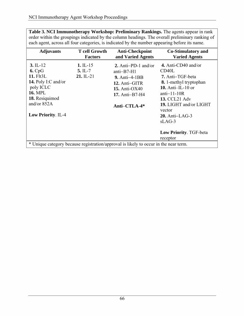
NCI Immunotherapy Agent Workshop Proceedings
Table 3. NCI Immunotherapy Workshop: Preliminary Rankings. The agents appear in rank order within the groupings indicated by the column headings. The overall preliminary ranking of each agent, across all four categories, is indicated by the number appearing before its name.
Adjuvants T cell Growth Factors
Anti-Checkpoint and Varied Agents
Co-Stimulatory and Varied Agents
3. IL-12 6. CpG 11. Flt3L 14. Poly I:C and/or poly ICLC 16. MPL 18. Resiquimod and/or 852A Low Priority. IL-4
1. IL-15 5. IL-7 21. IL-21
2. Anti−PD-1 and/or anti−B7-H1 9. Anti−4-1BB 12. Anti−GITR 15. Anti-OX40 17. Anti−B7-H4 Anti−CTLA-4*
4. Anti-CD40 and/or CD40L 7. Anti−TGF-beta 8. 1-methyl tryptophan 10. Anti–IL-10 or anti−11-10R 13. CCL21 Adv 19. LIGHT and/or LIGHT vector 20. Anti−LAG-3 sLAG-3 Low Priority. TGF-beta receptor
* Unique category because registration/approval is likely to occur in the near term.
66

NCI Immunotherapy Agent Workshop Proceedings
FINAL RANKINGS Following the workshop, the participants engaged in online discussions and balloting via e-mail, culminating in the final priority rankings for the agents of interest. The essence of the comments submitted via e-mail is reflected in the paragraphs below, and the results of the final voting are tabulated in a chart. Dr. Cheever led off the e-mail discussion by listing several concerns he had with the preliminary ranking:
• Anti–TGF-beta: First, the high preliminary ranking was based on discussion at the end of the workshop. The PowerPoint presentation did not develop justification for the agent in cancer therapy. Therefore, a better justification would be required to support a final ranking of the agent in the top 10. Second, the presentation listed multiple anti–TGF-beta agents in development. Therefore, anti–TGF-beta might not ultimately meet the criterion of “Not broadly available for testing in patients.”
• Anti–IL-10 and anti–IL-10 receptor: First, the PowerPoint presentation did not provide adequate justification for the agent’s ranking. Therefore, a better justification would be required to support a final ranking of the agent in the top 10. Second, the workshop participants seemed much more enthusiastic about anti–IL-10 receptor; however, the receptor was not mentioned in the PowerPoint presentation. Therefore, some rationale would be needed to justify inclusion of the receptor among the highest-ranked agents. Third, Dr. Cheever was not aware of any IL-10 receptor agents approaching readiness for clinical development. Several lower-ranked agents (e.g., Flt3 ligand, poly I:C, MPL, and resiquimod) have proven efficacy in the clinic.
• There are hundreds of cancer vaccines in clinical trials, but a dearth of adjuvants. It seems that adjuvants with known efficacy should have a higher priority than agents with little data available such as anti-OX40 and anti–B7-H4.
Dr. Houghton agreed with Dr. Cheever’s comments about the lack of justification for the high preliminary rankings of anti–TGF-beta and IL-10 receptor. Dr. Weiner concurred with Dr. Cheever regarding the prioritization of adjuvants. Although MPL might not be as exciting and novel as some of the higher-rated compounds, it would be generally useful to the investigator community. Dr. Weber posited that the anti–IL-10 and anti-TGF antibodies should be highly ranked on grounds of broad applicability and potential clinical utility. If cancer vaccines are not very immunogenic, then MPL would not make a difference, although an agent such as anti-OX40 or anti–TGF-beta might. Dr. Jaffee generally agreed with Dr. Cheever’s comments, but added that some good adjuvants are available and that checkpoint inhibitors are just as, if not more, difficult to obtain. She recommended highly ranking a checkpoint inhibitor as well as the best adjuvant. She agreed that not enough data are available on anti–B7-H4 to justify a very high ranking, although this would
67
NCI Immunotherapy Agent Workshop Proceedings
be a good example of an agent for which we need a mechanism to have regular follow-up so that it can be assigned a higher priority should more positive data become available. It is a unique agent in the class of checkpoint inhibitors. She also recommended moving anti-GITR higher on the list because of the need to have a Treg inhibitor. Dr. Berzofsky said he thought the ranking prepared at the end of the meeting was quite good, although many choices of exact position on the list were subjective. He opined that the rankings of a few agents should be rethought, and he emphasized the need for both adjuvants and checkpoint inhibitors. He thought that although anti–TGF-beta is very important and promising, it is already in active clinical development/trials by Genzyme. He said he has been very impressed by the mouse data on anti–IL-10 receptor from the laboratories of Anne O’Garra, Giorgio Trinchieri, and others. Additionally, a very effective anti–IL-10 receptor antibody was made a number of years ago by DNAX, but it has not been made available by Schering Plough, which purchased DNAX. He opined that some of the other adjuvants should be moved up in the rankings. Dr. Mackall agreed with several others that anti–IL-10 receptor and MPL should be moved up in the rankings and anti–TGF-beta should be demoted. She reiterated Dr. Jaffee’s desire to see some of the higher-risk agents receive some kind of real priority and suggested developing another category for considering non–GMP-grade production/acquisition to further preclinical work. This line of research would be distinct from the objective of producing clinical-grade material, but arguably would be equally important and potentially less costly. Dr. Disis wrote that adjuvants that are more likely to elicit Th1 responses should receive higher priorities. Dr. Margolin expressed her expectation that the priority ranking will reflect considerable expertise and judgment and hoped that it will be used wisely by the target audiences. Dr. Prendergast agreed about assigning higher priorities to anti-GITR and MPL. Dr. Whiteside noted that she also rearranged several agents on the priority list, pointing out that some of the antibodies (e.g., anti-GITR, anti–IL-10, and anti–IL-10 receptor) lack any record of effectiveness in human cancer, and it may be premature to put them in the top part of the list. Dr. Ho opined agreed that because of the difficulty in ranking items with limited preclinical data on this list, a regular reassessment of the rankings, as several suggested, seemed to be a reasonable approach. He also seconded Dr. Mackall’s idea of a separate list for non-GMP requests. Dr. Urba observed that it may be difficult to arrive at a consensus on some of the details of the ranking. He posited that the preliminary ranking was adequate in that the important molecules were represented in a reasonable order. He acknowledged others’ comments about the rankings of TGF-beta and anti–IL-10 receptor antibodies but did not agree with rating MPL higher, because it will likely become available to the clinical community because GSK is using it as an adjuvant. It might have to be purchased, but it would be available. He agreed with Dr. Jaffee that
68
NCI Immunotherapy Agent Workshop Proceedings
checkpoint inhibitors are very important and recommended keeping their priority rankings as they were. Dr. Calzone took a different approach. He placed the prioritized candidates into two groups on his ballot: “Deny” or “To Next Step.” The main reason for placing an agent in the “Deny” group was that clinical development was in progress or very likely. He placed eight candidates in the “To Next Step” category and prioritized them. MPL was advanced to number one because it seemed as if it would enhance a wide range of tumor vaccine studies. The rest in the “To Next Step” category were cytokines whose adjuvant potential requires further clinical study. Dr. Calzone pointed out that he is employed by Amgen, which holds the intellectual property rights to the molecules IL-15 and Flt3 ligand. He wrote that he had learned that Amgen’s discussions with NIH/NCI on Flt3 ligand for in vivo clinical trials have been long, complex, and frustrating on both sides. However, IL-15 is a different matter. He offered to arrange the proper discussion of IL-15 and Flt3 ligand with Amgen, depending on the ultimate RAID prioritization of these agents. Dr. Palucka said that she approached this priority exercise by asking what would be needed to vaccinate today and what is available or could be available for clinical testing soon. On that basis, she assigned higher priorities to agents needed to mobilize APCs, serve as adjuvants, help T cells via cytokines and/or co-stimulation, and control regulatory/suppressor mechanisms. Dr. Sondel said he would like to emphasize adjuvants and agents that might be applied broadly to a variety of diseases or combined with a variety of therapeutic strategies (e.g., Flt3 ligand, MPL, CD-40 ligand/anti-CD40).
69
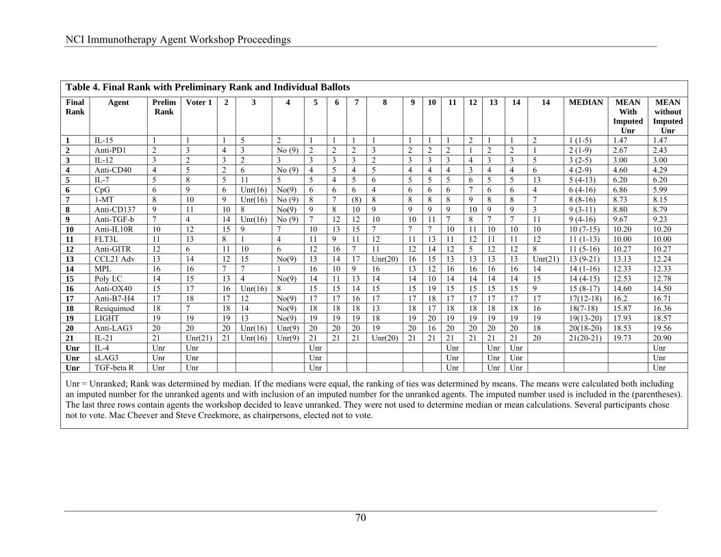
mmunotherapy Agent Workshop Proceedings
70
Table 4. Final Rank with Preliminary Rank and Individual Ballots Final Rank
Agent Prelim Rank
Voter 1 2 3 4 5 6 7 8 9 10 11 12 13 14 14 MEDIAN MEAN With
Imputed Unr
MEAN without Imputed
Unr 1 IL-15 1 1 1 5 2 1 1 1 1 1 1 1 2 1 1 2 1 (1-5) 1.47 1.47 2 Anti-PD1 2 3 4 3 No (9) 2 2 2 3 2 2 2 1 2 2 1 2 (1-9) 2.67 2.43 3 IL-12 3 2 3 2 3 3 3 3 2 3 3 3 4 3 3 5 3 (2-5) 3.00 3.00 4 Anti-CD40 4 5 2 6 No (9) 4 5 4 5 4 4 4 3 4 4 6 4 (2-9) 4.60 4.29 5 IL-7 5 8 5 11 5 5 4 5 6 5 5 5 6 5 5 13 5 (4-13) 6.20 6.20 6 CpG 6 9 6 Unr(16) No(9) 6 6 6 4 6 6 6 7 6 6 4 6 (4-16) 6.86 5.99 7 1-MT 8 10 9 Unr(16) No (9) 8 7 (8) 8 8 8 8 9 8 8 7 8 (8-16) 8.73 8.15 8 Anti-CD137 9 11 10 8 No(9) 9 8 10 9 9 9 9 10 9 9 3 9 (3-11) 8.80 8.79 9 Anti-TGF-b 7 4 14 Unr(16) No (9) 7 12 12 10 10 11 7 8 7 7 11 9 (4-16) 9.67 9.23 10 Anti-IL10R 10 12 15 9 7 10 13 15 7 7 7 10 11 10 10 10 10 (7-15) 10.20 10.20 11 FLT3L 11 13 8 1 4 11 9 11 12 11 13 11 12 11 11 12 11 (1-13) 10.00 10.00 12 Anti-GITR 12 6 11 10 6 12 16 7 11 12 14 12 5 12 12 8 11 (5-16) 10.27 10.27 13 CCL21 Adv 13 14 12 15 No(9) 13 14 17 Unr(20) 16 15 13 13 13 13 Unr(21) 13 (9-21) 13.13 12.24 14 MPL 16 16 7 7 1 16 10 9 16 13 12 16 16 16 16 14 14 (1-16) 12.33 12.33 15 Poly I:C 14 15 13 4 No(9) 14 11 13 14 14 10 14 14 14 14 15 14 (4-15) 12.53 12.78 16 Anti-OX40 15 17 16 Unr(16) 8 15 15 14 15 15 19 15 15 15 15 9 15 (8-17) 14.60 14.50 17 Anti-B7-H4 17 18 17 12 No(9) 17 17 16 17 17 18 17 17 17 17 17 17(12-18) 16.2 16.71 18 Resiquimod 18 7 18 14 No(9) 18 18 18 13 18 17 18 18 18 18 16 18(7-18) 15.87 16.36 19 LIGHT 19 19 19 13 No(9) 19 19 19 18 19 20 19 19 19 19 19 19(13-20) 17.93 18.57 20 Anti-LAG3 20 20 20 Unr(16) Unr(9) 20 20 20 19 20 16 20 20 20 20 18 20(18-20) 18.53 19.56 21 IL-21 21 Unr(21) 21 Unr(16) Unr(9) 21 21 21 Unr(20) 21 21 21 21 21 21 20 21(20-21) 19.73 20.90 Unr IL-4 Unr Unr Unr Unr Unr Unr Unr Unr sLAG3 Unr Unr Unr Unr Unr Unr Unr Unr TGF-beta R Unr Unr Unr Unr Unr Unr Unr
Unr = Unranked; Rank was determined by median. If the medians were equal, the ranking of ties was determined by means. The means were calculated both including an imputed number for the unranked agents and with inclusion of an imputed number for the unranked agents. The imputed number used is included in the (parentheses). The last three rows contain agents the workshop decided to leave unranked. They were not used to determine median or mean calculations. Several participants chose not to vote. Mac Cheever and Steve Creekmore, as chairpersons, elected not to vote.
NCI I

NCI Immunotherapy Agent Workshop Proceedings
71
PARTICIPANT LIST CHAIRPERSONS Martin A. “Mac” Cheever, M.D. Director of Solid Tumor Research Clinical Research Division Fred Hutchinson/University of Washington Cancer Research Consortium Seattle Cancer Care Alliance 825 Eastlake Ave East G4-810 Seattle, WA 98040 Phone: (206) 288-6370 Fax: (206) 288-1119 Email: [email protected] Steve Creekmore, M.D., Ph.D. Chief Biological Resources Branch Developmental Therapeutics Program, DCTD NCI-Frederick P.O. Box B, Building 1052, Room 253 Frederick, MD 21702 Phone: (301) 846-1100 Fax: (301) 846-5429 Email: [email protected] WORKSHOP PARTICIPANTS Jay Berzofsky, M.D.,Ph.D. Chief Vaccine Branch, CCR National Cancer Institute Building 10, Room 6B-04 Bethesda, MD 20982-1578 Phone: (301) 496-6874 Fax: (301) 480-0681 Email: [email protected]
Frank Calzone, Ph.D. Scientific Director, Oncology Research Department of Hematology Oncology Research Amgen, Inc One Amgen Center, 5-2-A Thousand Oaks, CA 91320 Phone: (805) 447-6696 Fax: Email: [email protected] Mary Lenora Disis, M.D. Professor, Department of Oncology University of Washington 815 Mercer Street Seattle, WA 98109 Phone: (206) 616-1823 Fax: (206) 685-3128 Email: [email protected] William Ho, M.D., Ph.D Associate Medical Director Exploratory Clinical Development Genentech, Inc. 1 DNA Way, MS-442B South San Francisco, CA 94080 Phone: (650) 467-4559 Fax: (650) 467-3165 Email: [email protected] Alan Houghton, M.D. Virginia & Daniel K. Ludwig Chair in Clinical Investigation Attending Physician & Vice-Chair Department of Medicine Professor of Medicine and Immunology Weill Medical School and Graduate School of Medical Sciences of Cornell University Memorial Sloan Kettering Cancer Center 1275 York Avenue New York, NY 10021 Phone: (646) 888-2315 Fax: (646) 422-0453 Email: [email protected]
NCI Immunotherapy Agent Workshop Proceedings
Anna Karolina Palucka, M.D., Ph.D Elizabeth Jaffee, M.D. Investigator Professor of Oncology Baylor Research Institute 1650 Orleans Street Baylor Institute for Immunology Research Johns Hopkins University School of
Medicine 3434 Live Oak Dallas, TX 75228 Building CRB 1, Rm. 4M06 Phone: 214 820-7450 Baltimore, MD 21231 Fax: 214 820-4813 Phone: (410) 955-2957
Email: [email protected] Email: [email protected] Crystal Mackall, M.D. Drew Pardoll, M.D., Ph.D. Acting Branch Chief and Professor of Oncology Head, Immunology Section Bunting-Blaustein CRB Pediatric Oncology Branch Johns Hopkins University National Cancer Institute, NIH Suite 444 Building 10-CRC, Room 1W-3940 Baltimore, MD 21231 Bethesda, MD 20892-1104 Phone: (410) 955-7866 Phone: 301 402-5940 Fax: (410) 614-0549 Fax: 301 451-7052 Email: [email protected] Email: [email protected]
George Prendergast, Ph.D. Kim Margolin, M.D. Professor & President, Chief Executive
Officer Associate Director of Clinical Research Department of Medical Oncology Lankenau Institute for Medical Research City of Hope 100 Lancaster Avenue 1500 East Duarte Road Wynnwood, PA 19096 Duarte, CA 91010 Phone: 610 645-8475 Phone: 626 359-8111 Fax: 610 645-8533 Fax: 626 301-8233 Email: [email protected]
Email: [email protected] Ellis Reinherz, M.D.
Michael Morin, Ph.D. Professor of Medicine Vice President, Discovery Chief, Laboratory of Immunobiology Antibacterials, Immunology and Cancer Harvard Medical School Pfizer Groton / New London Labs Dana Faber Cancer Institute Eastern Point Road 4 Blackfan Circle, Room 419 MS:8118W-B3 Boston, MA 02115 Groton, CT 06340 Phone: 617 632-3412 Phone: 860 441-5476 Fax: 617 632-3351 Email: [email protected] Email: [email protected]
72
NCI Immunotherapy Agent Workshop Proceedings
Steven Rosenberg, M.D., Ph.D. Chief of Surgery Surgery Branch, CCR National Cancer Institute, NIH Building 10, Room 3W3-3940 Bethesda, MD 20892 Phone: (301) 496-4164 Fax: (301) 402-1738 Email: [email protected] Jeffrey Schlom, Ph.D. Chief Laboratory of Tumor Immunology and Biology National Cancer Institute, NIH Building 10, Room 8B09 Bethesda, MD 20892 Phone: (301) 496-4343 Fax: (301) 496-2756 Email: [email protected] Paul Sondel, M.D., Ph.D. Walker Professor Departments of Pediatrics and Human Oncology University of Wisconsin Associate Director for Translational Research, Paul P. Carbone Comprehensive Cancer Center 600 Highland Avenue Room K4/448 Madison, WI 53792 Phone: 608 263-9069 Email: [email protected] Walter Urba, M.D., Ph.D Director, Robert W. Franz Cancer Research Center Providence Portland Medical Center 4805 N.E. Glisan Street, 5F40 Portland, OR 97213 Phone: 503 215-6259 Fax: 503 215-6841 Email: [email protected]
Thomas A. Waldmann, MD Chief, Metabolism Branch Center for Cancer Research National Cancer Institute Building 10, Room 4N115 10 Center Drive Bethesda, MD 20892-1374 Phone: (301) 496-6656 Fax: (301) 496-9956 E-mail: [email protected] Jeffrey Weber, M.D., Ph.D Professor, Interdisciplinary Oncology Department of Melanoma H. Lee Moffitt Cancer Center 12902 Magnolia Drive, SRB-2 Tampa, FL 33612 Phone: 813 745-1536 Fax: 503-745-6188 Email: [email protected] Louis Weiner, M.D. Vice President, Translational Research Chairman, Department of Medical Oncology Fox Chase Cancer Center 333 Cottman Avenue, Room 6315 Philadelphia, PA 19111 Phone: 215 728-2480 Fax: 215 728-5338 Email: [email protected] Theresa Whiteside, Ph.D. Professor of Pathology, Director Immunology and Otolaryngology, Immunologic Monitoring and Cellular Products Laboratory University of Pittsburgh Cancer Institute Hillman Cancer Center 5117 Center Avenue, Suite 127 Pittsburgh, PA 15213 Phone: 412 624-0096 Fax: 412 624-0264 Email: [email protected]
73
NCI Immunotherapy Agent Workshop Proceedings
Jon Wigginton, M.D. Director, Oncology Merck and Co., Inc Email: [email protected] ORGANIZING COMMITTEE MEMBERS Richard Camalier Coordinator, NCI-RAID Program Office of the Associate Director, Developmental Therapeutics Program, DCTD National Cancer Institute Executive Plaza North, Room 8024 6130 Executive Boulevard Rockville, MD 20852 Phone: (301) 496-3392 Fax: (301) 402-0831 Email: [email protected] Jerry Collins, Ph.D. Associate Director Developmental Therapeutics Program, DCRD National Cancer Institute 6130 Executive Boulevard, Room 8018 Rockville, MD 20852 Phone: (301) 496-8720 Fax: (301) 402-0831 Email: [email protected] Toby Hecht, Ph.D. Program Director Biological Resources Branch Developmental Therapeutics Program, DCTD NCI-Frederick P.O. Box B, Building 1052; Room 247 Frederick, MD 21702 Phone: (301) 846-5692 Fax: (301) 846-5249 Email: [email protected]
Kevin Howcroft, Ph.D. Program Director Division of Computational Bioscience National Cancer Institute Executive Plaza North, Room 5060 6130 Executive Boulevard Bethesda, MD 20892 Phone: (301) 496-7815 Fax: (301) 480-2844 Email: [email protected] Jill Johnson Developmental Therapeutics Program, DCTD National Cancer Institute, NIH 6130 Executive Boulevard, Room 8020 Rockville, MD 20852 Phone: (301) 435-9152 Fax: (301) 402-0831 Susan McCarthy, Ph.D. Program Director for Molecular Immunology Cancer Immunology and Hematology Branch, Division of Cancer Biology National Cancer Institute Executive Plaza North, Room 5058 6130 Executive Boulevard Rockville, MD 20852 Phone: 301-496-7815 or 301-594-8785 Fax: 301-480-2844 Email: [email protected] Robert Mufson, Ph.D. Chief Cancer Immunology and Hematology Branch, Division of Cancer Biology National Cancer Institute, NIH EPN 6130 Executive Blvd, Room 5062 Rockville, MD 20852 Phone: 301-496-7815 Fax: 301-480-2844 Email: [email protected]
74

NCI Immunotherapy Agent Workshop Proceedings
Howard Streicher, M.D. Senior Investigator Investigational Drug Branch, CTEP National Cancer Institute Executive Plaza North, Room 7126 6130 Executive Boulevard Rockville, MD 20892 Phone: 301-435-9129 Fax: 3014020428 Email: [email protected] James Zweibel, M.D. Acting Branch Chief Investigational Drug Branch, CTEP National Cancer Institute Executive Plaza North, Room 7131 6130 Executive Boulevard Bethesda, MD 20892 Phone: (301) 496-1196 Fax: (301) 402-0428 Email: [email protected] INVITED OBSERVERS Kimberly Benton, Ph.D. Division of Cellular and Gene Therapies and Gene Therapies Center for Biologics Evaluation and Research U.S. Food and Drug Administration 1401 Rockville Pike, HFM-720 Rockville, MD 20852 Phone: (301) 827-5102 Fax: (301) 827-9796 Email: [email protected] Lothar Finke, M.D. Chief Medical Officer, Vice President of Regulatory Affairs Department of Clinical/Regulatory Argos Therapeutics, Inc. 4233 Techonology Drive Durham, NC 27704 Phone: (919) 287-3421 Fax: (919) 827-4892 Email: [email protected]
Jesus Gomez-Navarro, M.D. Director Clinical Research and Development Pfizer Global 50 Pequot Avenue MS6025-A3120 New London, CT 06320 Phone: (860) 732-2079 Fax: (860) 715-8439 Email: [email protected] Steve Herrman, Ph.D. Research Fellow Biological Technologies Wyeth Research 87 Cambridge Park Drive Cambridge, MA 02140 Phone: (617) 665-8159 Fax: (617) 665-7519 Email: [email protected] Raj Puri, M.D., Ph.D. Director, Division of Cellular and Gene Therapies Center for Biologics Evaluation and Research U.S. Food and Drug Administration NIH Building 29B, Room 2NN20 Bethesda, MD 20892-4555 Phone: 301 827-0471 Fax: 301 827-0449 Email: [email protected] Amy Rosenberg, M.D. Division Director Division of Therapeutic Proteins U.S. Food and Drug Administration Building 29A, Room 2D-16 8800 Rockville Pike Bethesda, MD 20892 Phone: (301) 827-1790 Fax: (301) 480-3256 Email: [email protected]
75

NCI Immunotherapy Agent Workshop Proceedings
Daniel Takefman, Ph.D. Chief, Gene Therapy Branch Division of Cellular & Gene Therapies Center for Biologics Evaluation and Research U.S. Food and Drug Administration 1401 Rockville Pike Room 205N, HFM-720 Rockville, MD 20852 Phone: 301 827-5150 Fax: 301 827-9796 Email: [email protected] David Urdal, Ph.D. Chief Scientific Officer 3005 1st Avenue Dendreon Corporation Seattle, WA 98121 Phone: 206 829-1530 Fax: 206 219-1060 Email: [email protected] Robert Wiltrout, Ph.D. Director Center for Cancer Research National Cancer Institute Building 31, Room 3A-11 Bethesda, MD 20892 Phone: (301) 496-4345 Fax: (301) 496-0775 Email: [email protected]
76