national biology - sciencepress excretion, respiration and water 101 70 excretory organs 102 71 the...
TRANSCRIPT

• Kerri Humphreys •
NATIONAL BIOLOGYUnit 2 Cells and Multicellular
Organisms

© Science Press 2015
Science PressBag 7023 Marrickville NSW 1475 AustraliaTel: (02) 9516 1122 Fax: (02) 9550 [email protected]
All rights reserved. No part of this publication may be reproduced, stored in a retrieval system, or transmitted in any form or by any means, electronic, mechanical, photocopying, recording or otherwise, without the prior permission of Science Press. ABN 98 000 073 861

Cells and Multicellular Organisms iiiScience Press
Surfing National Biology
Contents
Introduction ivWords to Watch iv
Cells as the Basis of Life
1 Assumed Knowledge 22 Characteristics of Living Things 33 Developing the Cell Theory 44 Cell Theory and Robert Hooke 65 Cell Theory and Robert Brown 76 Technology and the Development of the 8
Cell Theory7 The Modern Light Microscope 108 The Electron Microscope 129 The Stereo Microscope 1310 Experiment – The Light Microscope 1411 Experiment – Using a Light Microscope 1512 Experiment – Making a Wet Mount 1613 Experiment – Drawing Biological Diagrams 1714 Writing a Practical Report 1815 The Light Microscope and Cell Organelles 2016 Experiment – Plant Cells 2217 Experiment – Animal Cells 2318 The Electron Microscope and Cell Organelles 2419 Mitochondria 2520 Chloroplasts 2621 Golgi Bodies 2722 Development of the Model of the Cell Membrane 2823 Cell Organelles Summary 3024 Chemicals in Cells 3125 Experiment – Substances in Tissues 3326 Cell Input and Output 3427 Passive Transport 3628 Diffusion 3729 Active Transport 3830 Movement Across the Cell Membrane 3931 Experiment – Membranes, Diffusion and Osmosis 4132 Experiment – Surface Area to Volume Ratio 42
and Diffusion33 Experiment – Plasmolysis 4434 Material Exchange and Concentration Gradient 4535 Material Exchange and the Nature of the Material 4636 Experiment – Surface Area and Rate of Reaction 4737 Prokaryotes 4838 Eukaryotes 5039 Enzymes 5140 Enzymes and Reactions 5241 Experiment – Enzyme Activity and pH 5342 Experiment – Enzyme Activity and Temperature 5443 Experiment – Enzyme Activity and Substrate 55
Concentration44 Enzymes and Inhibitors 5645 Allosteric Activation and Inhibition 5746 Enzyme Deficiencies 58
47 Photosynthesis 5948 Play – Inside Photosynthesis 6049 Light, Chlorophyll and Photosynthesis 6450 Experiment – Chlorophyll and Photosynthesis 6651 Experiment – Light and Photosynthesis 6752 Reactions in Photosynthesis 6853 Photosynthesis and Productivity 7054 Respiration 7155 Reactions in Respiration 72
Multicellular Organisms
56 Cells, Tissues, Organs and Systems 7657 Differentiation and Specialised Cells 7758 Stem Cells 7959 Human Organ Systems 8160 Organ and Tissue Transplantation 8261 Bioartificial Organs 8362 The Digestive System 8463 Teeth 8664 Carnivore, Herbivore and Nectar Feeder 88
Digestive Systems65 Play – Amino and Lipidet, a Most 90
Lamentable Tragedy66 Respiratory Systems 9567 Gas Exchange, Breathing and Ventilation 9868 Circulatory Systems in Animals 9969 Excretion, Respiration and Water 10170 Excretory Organs 10271 The Kidney 10372 Osmosis and Diffusion 10573 Renal Hormones 10674 Renal Dialysis 10775 Excretion in Other Organisms 10876 Plant Tissues and Cells 11077 Plant Organs 11278 Root Structure 11479 Leaves 11580 Stomates 11781 Experiment – Transpiration 11982 Transport Systems in Plants 12183 Experiment – Movement in Xylem 12484 Adaptations in Plants 12585 Radioisotopes Track the Movement of 127
Substances in Organisms86 Chemical Monitoring 12987 Experiment – Carbon Dioxide and pH 13088 CAT and MRI Scans 13189 Adaptations in Invertebrates 13290 Adaptations in Vertebrates 13391 Animal Ethics 135
Topic Test 136Answers 142Index 178

Cells and Multicellular OrganismsivScience Press
Surfing National Biology
Introduction
Each book in the Surfing series contains a summary, with occasional more detailed sections, of all the mandatory parts of the syllabus, along with questions and answers.
All types of questions – multiple choice, short response, structured response and free response – are provided. Questions are written in exam style so that you will become familiar with the concepts of the topic and answering questions in the required way.
Answers to all questions are included.
A topic test at the end of the book contains an extensive set of summary questions. These cover every aspect of the topic, and are useful for revision and exam practice.
Words To Watch
account, account for State reasons for, report on, give an account of, narrate a series of events or transactions.
analyse Interpret data to reach conclusions.
annotate Add brief notes to a diagram or graph.
apply Put to use in a particular situation.
assess Make a judgement about the value of something.
calculate Find a numerical answer.
clarify Make clear or plain.
classify Arrange into classes, groups or categories.
comment Give a judgement based on a given statement or result of a calculation.
compare Estimate, measure or note how things are similar or different.
construct Represent or develop in graphical form.
contrast Show how things are different or opposite.
create Originate or bring into existence.
deduce Reach a conclusion from given information.
define Give the precise meaning of a word, phrase or physical quantity.
demonstrate Show by example.
derive Manipulate a mathematical relationship(s) to give a new equation or relationship.
describe Give a detailed account.
design Produce a plan, simulation or model.
determine Find the only possible answer.
discuss Talk or write about a topic, taking into account different issues or ideas.
distinguish Give differences between two or more different items.
draw Represent by means of pencil lines.
estimate Find an approximate value for an unknown quantity.
evaluate Assess the implications and limitations.
examine Inquire into.
explain Make something clear or easy to understand.
extract Choose relevant and/or appropriate details.
extrapolate Infer from what is known.
hypothesise Suggest an explanation for a group of facts or phenomena.
identify Recognise and name.
interpret Draw meaning from.
investigate Plan, inquire into and draw conclusions about.
justify Support an argument or conclusion.
label Add labels to a diagram.
list Give a sequence of names or other brief answers.
measure Find a value for a quantity.
outline Give a brief account or summary.
plan Use strategies to develop a series of steps or processes.
predict Give an expected result.
propose Put forward a plan or suggestion for consideration or action.
recall Present remembered ideas, facts or experiences.
relate Tell or report about happenings, events or circumstances.
represent Use words, images or symbols to convey meaning.
select Choose in preference to another or others.
sequence Arrange in order.
show Give the steps in a calculation or derivation.
sketch Make a quick, rough drawing of something.
solve Work out the answer to a problem.
state Give a specific name, value or other brief answer.
suggest Put forward an idea for consideration.
summarise Give a brief statement of the main points.
synthesise Combine various elements to make a whole.

Cells as the Basis of Life
Cells and Multicellular Organisms 1Science Press
Surfing National Biology

Cells and Multicellular Organisms2Science Press
Surfing National Biology
1 Assumed Knowledge
QUESTIONS
1. Identify seven properties of living organisms.2. The cell is the basic unit of life. What structural
features of cells are possessed by all living things?3. Draw a fully labelled diagram of a plant cell as seen
under a light microscope.4. Draw a fully labelled diagram of an animal cell as
seen under a light microscope.5. Identify the following parts of a light microscope
and use by a person.A
B
C
D
E
F
Figure 1.1 Light microscope.
6. Describe one safety precaution you should follow while using a light microscope.
7. What is the function of the nucleus of a cell?8. What is the function of the cell membrane?9. What is cytoplasm?10. Defineprotoplasm.11. Describe a chloroplast.12. Define photosynthesis.13. Which group of organisms can photosynthesise?14. Identify the materials required by multicellular
organisms for photosynthesis.15. Why is photosynthesis an important process in
ecosystems?16. Name the four basic groups of organic compounds.17. What are inorganic compounds?18. What is the function of the digestive system?19. Figure 1.2 shows the human digestive tract. Identify
each part.20. For each of the following parts of the digestive
system, outline its structure and its main function. (a) Mouth. (b) Oesophagus. (c) Stomach. (d) Small intestine. (e) Large intestine. (f) Anus.
DE
C
B
A
J
I
H
G
F
Figure 1.2 Human digestive tract.
21. Define respiration.22. In humans, what structures make up the respiratory
system?23. Outline the function of the respiratory system.24. Outline the function of the circulatory system.25. In humans, what is the function of the heart in the
circulatory system?26. Identify the components of the human circulatory
system.27. In plants, what is the function of each of the
following? (a) Xylem. (b) Phloem. (c) Leaves. (d) Roots.28. Definetranspiration.29. Studytheflowchartbelow.
Mature cell‘parent’
(Division)
Small cell‘daughter’
Small cell‘daughter’
(Increase in size)
Mature cell
(Increase in size)
Mature cell
Figure 1.3 Division of a cell.
What type of division is indicated by this chart?30. Definemitosis.31. Explain why cell division is important.32. How is genetic information transferred during cell
reproduction?33. Defineagene.34. Define cytokinesis.

Cells and Multicellular Organisms 3Science Press
Surfing National Biology
2 Characteristics of Living Things
There are nine characteristics of living things. These characteristicsareusedtodefinelife.
1. Growth and development
Growth involves an increase in mass. This can occur due to an increase in the size of individual cells and/or an increase in the number of cells.
2. Reproduction
Reproduction is the ability to produce offspring. Reproduction can be asexual or sexual. Asexual reproduction involves one parent producing offspring that are genetically identical to the parent, e.g. by binary fission,buddingorvegetativepropagation.Sexual reproduction involves the union of two gametes in fertilisation to form a zygote.
3. Respiration
All living things can respire. Cellular respiration is a series of chemical reactions in which cells obtain energy from food. Each step in the series of reactions is controlled by enzymes with the energy being released at different stages in the process. Some of the energy is transferred to other molecules becoming available for other reactions.
4. Respond to stimuli
All living things respond to stimuli from both their external environment and their internal environment. The stimuli can be physical or chemical changes in the environment, e.g. a response to the intensity and direction of light or a changeinthecarbondioxidelevelsinbodyfluids.
5. Movement and locomotion
Movement can be very obvious, e.g. a running animal or very slow and involve only part of the organism, e.g. a plant leaf moving to catch the maximum amount of sunlight. Locomotion is the ability to move from one place to another.
6. Nutrition or feeding
Nutrition is a process by which organisms obtain matter to produce their physical structure and energy to continue the functions of life. Autotrophs can make their own organic nutrients from inorganic materials, e.g. plants and cyanobacteria can use the energy from sunlight in photosynthesis and bacteria living in hot springs or oceanic hydrothermal vents use the energy in hydrogen sulfide(H2S) in chemosynthesis. Heterotrophs consume other organisms to obtain organic nutrients. Their food needs to be broken down before it can be used.
7. Assimilation
Assimilation is the process of converting food into the living material of life.
8. Metabolism
Metabolism is the sum of all chemical reactions within the organism. In anabolic reactions small molecules are combined to form complex molecules, e.g. photosynthesis. In catabolic reactions chemical bonds are broken and complex molecules are broken down into smaller units, e.g. digestion. Sometimes energy is released.
9. Excretion
Excretion is the removal of unwanted waste products of metabolic reactions.
QUESTIONS
1. Construct a table to summarise the nine characteristics of living things.
2. Distinguish between asexual reproduction and sexual reproduction.
3. Definefertilisation.4. Crystals can grow in size. Explain why crystals
are not considered to be living though they show a characteristic of living things.
5. Distinguish between autotrophic and heterotrophic nutrition.
6. Distinguish between photosynthesis and chemosynthesis.
7. The diagram shows one of the features of living things.Nutrition
digestion
A BC
Food (proteins, carbohydrates, lipids)
Amino acids
LipidsGlucose
CO2 from the air
Sucrose, proteins,lipids
Wat
er
GlucoseH 2O + CO 2
Sunlight
Food to rest of plant
SaltsFrom roots
2NH3 + 3O2 → 2HNO2 + 2H2O + energy
BacteriaNitrosomonas spp
Figure 2.1 A feature of living things.
(a) For the types of nutrition labelled A, B and C, which are autotrophic and which are heterotrophic?
(b) What is the energy source for the bacteria Nitrosomas spp?
8. Distinguish between an anabolic reaction and a catabolic reaction.
9. Is respiration a catabolic or anabolic reaction? Explain your reasoning.

Cells and Multicellular Organisms4Science Press
Surfing National Biology
3 Developing the Cell Theory
The cell theory states the following.
• All living things are made of cells and of substances produced by cells.
• All cells come from pre-existing cells.• The cell is the basic unit in which the
processes of living take place.
The development of the cell theory is linked with the invention of technology that enabled scientists to see cells and to investigate the properties of cells.Humans have made observations and recorded their findingsaboutlivingthingssincethefirstmarkwasputona cave wall, clay tablet or sheet of papyrus paper. These observations record the macroscopic appearance of living things – the features observed with the naked eye.
The development of glass lenses and the construction of thefirstmicroscopeusingaglasslensenabledscientiststoobserve the microscopic appearance of living things. The firstcellswereobservedandtheideasaboutthestructureof the building blocks of life were forever changed.
Hans and Zacharias Jansen
Hans and Zacharias Jansen are believed to have created the first compound light microscope around the 1590s. They placed several lenses in a tube and realised the objectneartheendofthetubecouldbemagnifiedmorethan using a single lens in a magnifying glass.
Zacharias Jansen Francesco Redi Anton van Leeuwenhoek
Robert Brown
Figure 3.1 Jansen, Redi, van Leeuwenhoek and Brown.
Robert Hooke
In 1663 Robert Hooke observed cork under a microscope and introduced the term ‘cell’. He published his microscopical observations in 1665 in his book Micrographia. This book led to public interest in microscopy.
Francesco Redi
In 1668 Francesco Redi published the results of his experimentwithinsectswhichwasoneofthefirststeps in proving that living things do not arise from spontaneous generation.Heshowedthatflymaggotsdidnot spontaneously arise from dead meat as meat kept in jars covered with gauze did not get maggots.
Anton van LeeuwenhoekAnton van Leeuwenhoek produced higher quality lensesthatgavegreatermagnificationandaidedthedevelopment of the light microscope. He is considered to be the ‘Father of Microbiology’. In 1674 he was the firsttoobserveanddescribesingle-celled organisms which he called animalcules. In 1676 when he sent his drawings of single-celled organisms to the Royal Society of London, his credibility was questioned. In 1680 his observations were vindicated after others observed the unicells. He discovered and made drawings of protozoa, bacteria, the vacuole of the cell, the banded pattern of musclefibresandspermatozoa.
Robert Brown
Robert Brown described the nucleus in cells of the orchid and gave the structure its name. He travelled to Australia in 1801 on the HMS Investigator as the naturalist at the request of the commander of the vessel, Matthew Flinders. He collected many specimens and left Australia in 1805. In 1831 he read a paper about the cell nucleus to the Linnaean Society and published this work in 1833. Although the nucleus had been drawn by others, e.g. van Leeuwenhoek and Franz Bauer, Brown gave the structure its name. His observations of the random movement of pollen grains led to the naming of the phenomena now known as Brownian motion.
Matthias Schleiden
In 1838 Matthias Schleiden wrote Contributions to Phytogenesis and proposed that different parts of plants are made of cells. With Schwann he was the firsttoproposethecell theory. He also recognised the importance of the cell nucleus and its possible relationship with cell division.
Theodor Schwann
Theodor Schwann noted that parts of animals are made of cells and that non-cellular parts, e.g. nails, feathers and tooth enamel had a cellular origin. In 1839 he extended Schleiden’s cell theory to animals and proposed that all living things are made of cells and cell products. The cell was the basic unit of life. This is now called the Schleiden and Schwann cell theory. Schwann also observedthecellsassociatedwithnervefibreswhicharenow called Schwann cells.

Cells and Multicellular Organisms 5Science Press
Surfing National Biology
Rudolf Virchow
In 1855 Rudolf Virchow published a work that proposed that the origin of cells was the division of pre-existing cells and the cell theory was expanded to include the point that every cell originated from another living cell like it. This rejected the concept of spontaneous generation. Virchow is known as the ‘father of modern pathology’ and he developed a standard method of autopsy procedure.
Rudolf VirchowTheodor SchwannMatthias Schleiden
Figure 3.2 Matthias Schleiden, Theodor Schwann and Rudolf Virchow.
Louis Pasteur
In 1861 Louis Pasteur published his experiments demonstrating that fermentation was caused by micro-organismswhichfinallydisprovedthetheoryofspontaneous generation. The experiment also supported the germ theory.
Friedrich Miescher
In 1869 Friedrich Miescher isolated nucleic acids which he called nuclein from the nuclei of white blood cells. ThiswasthefirsttimeDNAhadbeenpurifiedandledtoinvestigations into its composition, properties and structure.
Camillo Golgi
In 1898 Camillo Golgi described the Golgi apparatus by stainingcellswithsilvernitrate.Atfirstsomebelievedthestructure was an optical illusion caused by the staining technique. The invention of the electron microscope in the 20th century proved the existence and shape of this organelle.
Camillo GolgiFriedrich MiescherLouis Pasteur
Figure 3.3 Louis Pasteur, Friedrich Miescher and Camillo Golgi.
Max Knoll and Ernst Ruska
In 1932 Max Knoll and Ernst Ruska invented the transmission electron microscope. The higher magnificationandhigherresolutionmeantgreaterdetailsofthe ultrastructure of cells could be observed and analysed and new structures were discovered, e.g. ribosomes.
QUESTIONS
1. State the cell theory.2. Construct a table to summarise the historical
development of the cell theory.3. Explain how the invention of the light microscope is
linked with the development of the cell theory.4. Explain why Anton van Leeuwenhoek is known as
the ‘Father of Microbiology’.5. Suggest why Leeuwenhoek’s discovery of
animalculeswasatfirstdisbelievedandexplainwhyitwasfinallyaccepted.
6. Outline the discovery and naming of the nucleus.7. What are the two points of the cell theory proposed
by Schleiden and Schwann?8. What is the theory of spontaneous generation?9. Discuss how the theory of spontaneous generation
wasfinallydisproved.10. How is the theory of spontaneous generation linked
with the development of the cell theory?11. (a) Why did people question the actual existence of
the Golgi body in cells? (b) What evidence proved the existence of the
Golgi body?12. Explain how the invention of the electron
microscope aided the development of knowledge about cell structure.
13. WhowasthefirstpersontoisolateDNA? (A) Robert Hooke. (B) Robert Brown. (C) Rudolf Virchow. (D) Friedrich Miescher.14. The timeline shows
events in the development of the cell theory.From this timeline which event occurred before the nucleus was named?
(A) Single-celled organisms were observed.
(B) Golgi apparatus was described.
(C) Nucleic acids were isolated.
(D) Cell theory was expanded. Figure 3.4 Cell theory timeline.
Single-celled organisms observed1660
1900
1700
1740
1780
1820
1860
Cork observed and cells named
Nucleus named
Golgi apparatus described
Nucleic acids isolatedMicro-organisms cause decayCell theory expandedCell theory proposed

Cells and Multicellular Organisms6Science Press
Surfing National Biology
4 Cell Theory and Robert Hooke
Robert Hooke (1635-1703) was an experimental scientist who was interested in physics, astronomy, chemistry, biology, geology, architecture and naval technology, although his primary interest was microscopy, mechanics and instrumentation. There are no authenticated surviving portraits of Robert Hooke.
Robert Hooke invented a compound light microscope, i.e. a microscope with more than one lens and devised an illumination system for this microscope. Using this microscope he observed many organisms and drew accurateanddetaileddrawingsofhisfindings.Healsodeveloped a micrometer; the universal, or Hooke’s joint found in all cars; the spring control of the balance wheel inwatches;thefirstreflectingtelescopeandwasinvolvedin creating different types of barometers.
Robert Boyle was Hooke’s patron when Hooke began studying at Oxford. However, Hooke clashed with Isaac Newton and this may have affected his fame in history. It is believed that Newton destroyed the only portrait known to exist of Hooke.
RobertHooke’sfirst publication was printed in 1661 and was a pamphlet on capillary action. In 1665, he published a book, Micrographia, which contained many drawings and records of his observations under the microscope, e.g. diagrams of insects, sponges, foraminifera, bird feathers and bryozoans. It also included a theory of light. The diagrams in Micrographia created a public interest in microscopy with some people believing it was a great advanceinscientificknowledgeandotherscallingitatriflingpursuit.
When Hooke observed a slice of cork under his microscope he discovered plant cells. He coined the term ‘cells’ to describe what he saw.
‘Yet it was not unlike a Honeycomb in these particulars … these pores, or cells, … consisted of a great many little Boxes.’
He also put fossils under his microscope and observed the similarities of fossil shells with living mollusc shells. He noted that dead wood could be turned to stone due to minerals being deposited throughout the wood. Hooke suggested the fossils gave clues to the past history of life on Earth.
Figure 4.1 Hooke’s microscope.
In 1678 Anton van Leeuwenhoek (1632-1723) reported the presence of ‘little animals’ in lake water. The Royal Society of London asked Robert Hooke to investigate thesefindingsandwhenheconfirmedthepresenceofsmall organisms, Leeuwenhoek’s work was accepted.
(a) Drawing of a flea. (b) Drawing of a slice of cork.
Figure 4.2 Drawings by Robert Hooke from his book Micrographia.
QUESTIONS
1. Name two instruments invented by Robert Hooke. 2. What is meant by a ‘compound’ microscope?3. Discussthesignificanceoftheinventionofthe
compound microscope.4. How did the term ‘cell’ originate as used in biology?5. Robert Hooke observed a honeycomb appearance
under the microscope which he called ‘cells’. What was he actually viewing?
6. The lenses used by Hooke were relatively low quality and caused some image distortion and separated colours giving a rainbow ‘fringe’ effect. Explain how opponents of Robert Hooke used these detailstodiscredithisfindings.
7. In Micrographia Hooke drew the microscopic structure of fossilised wood and compared its structure to a piece of rotten oak wood. Why did some people reject his conclusions about fossils showing extinction and past life forms?
8. In his book Micrographia Robert Hooke drew many diagrams.
Figure 4.3 Hooke’s drawing of Mucor from Micrographia.
Discuss why the drawings in Micrographia were important in making the book a ‘best seller’.

Cells and Multicellular Organisms 7Science Press
Surfing National Biology
5 Cell Theory and Robert Brown
Robert Brown (1773-1858) was a Scottish botanist and protégé of Joseph Banks. He sailed on the HMS Investigator in 1801 under Captain Matthew Flinders. When he reached Australia, then called New Holland, he collected more than 500 plant species and made many drawings and notes of animals and plants, naming more than 140 new genera and over 1700 new species. He returned to England in 1805 and spentfiveyearsdescribingthe2200specieshehadobserved.He published his notes in 1811 in Prodromus Florae Novae Hollandiae et insulae Van-Diemen and further notes in 1814 in ‘General Remarks, Geographical and Systemic, on the Botany of Terra Australis’ as an appendix to Matthew Flinders’ Voyage to Terra Australis.
Figure 5.1 Brown’s microscope.
From 1805 he was Secretary to the Royal Linnean Society and from 1810 to 1820 he was the personal librarian to Joseph Banks. Brown was given the care of Sir Joseph Banks’ home and collections when Banks died and when Brown organised the transfer of the specimens to the British Museum, he became the curator and Keeper of the British Museum for the rest of his life.
Using microscopes all through his adult life, in 1827Brownwasthefirstpersontonoticetheconstantmovement of suspended particles and since then this movement has been called ‘Brownian motion’.
Figure 5.2 View obtained by Brown of cell nucleus in orchid epidermal cells.
In 1833, Brown observed the epidermis of orchids under his microscope and discovered an ‘opaque spot’ which he called the nucleus.
Early microscopists had drawn nuclei in animal cells Leeuwenhoekin1700figurednucleiinredbloodcellsofsalmon in a letter to the Royal Society, but it was Hooke who coined the term ‘nucleus’.
‘In each cell of the epidermis of a great part of this family [Orchidaceae], … a singular areola, generally somewhat more opaque, … is observable … This areola, or nucleus of the cell as perhaps it might be termed, is not confined to the epidermis …’
Robert Brown continued to use his microscope and discovered nuclei in a range of plant tissues. At this stage the importance of the nucleus was still unknown.
QUESTIONS
1. Outline the epic expedition of discovery of Robert Brown.
2. Name two publications of Robert Brown.3. How did the work of Robert Brown increase
knowledge about Australian plants and animals?4. What is ‘Brownian motion’?5. How did Brown discover the plant nucleus?6. What term was coined by Brown?7. Comment on the statement:
‘RobertBrownwasthefirsttodescribethestructureand function of the nucleus’.
8. When Robert Brown described ‘… a singular areola, generally somewhat more opaque’ spot in a cell, what organism was he studying?
(A) Mouse epidermis. (B) Orchid epidermis. (C) Slice of cork. (D) Pond water.9. The diagram shows a leaf epidermis with stomates
under high power of a light microscope.
ABC
D
Figure 5.3 High power view of leaf epidermis.
Which structure is the nucleus of a leaf epidermal cell? (A) A (B) B (C) C (D) D10. Matthias Schleiden built on the work of Robert
Brown. Schleiden believed that the nucleus is the most important part of a cell and has a vital role in cell reproduction. Why would Schleiden freely talk about his indebtedness to Robert Brown?
(A) Brownwasthefirstpersontogiveadescriptionof a cell.
(B) Brown invented the staining techniques used by Schleiden.
(C) Brown had named the nucleus and noted its presence in a range of different plants.
(D) Brown showed that cells are formed from pre-existing cells.
Cells and Multicellular Organisms8Science Press
Surfing National Biology
6 Technology and the Development of the Cell Theory
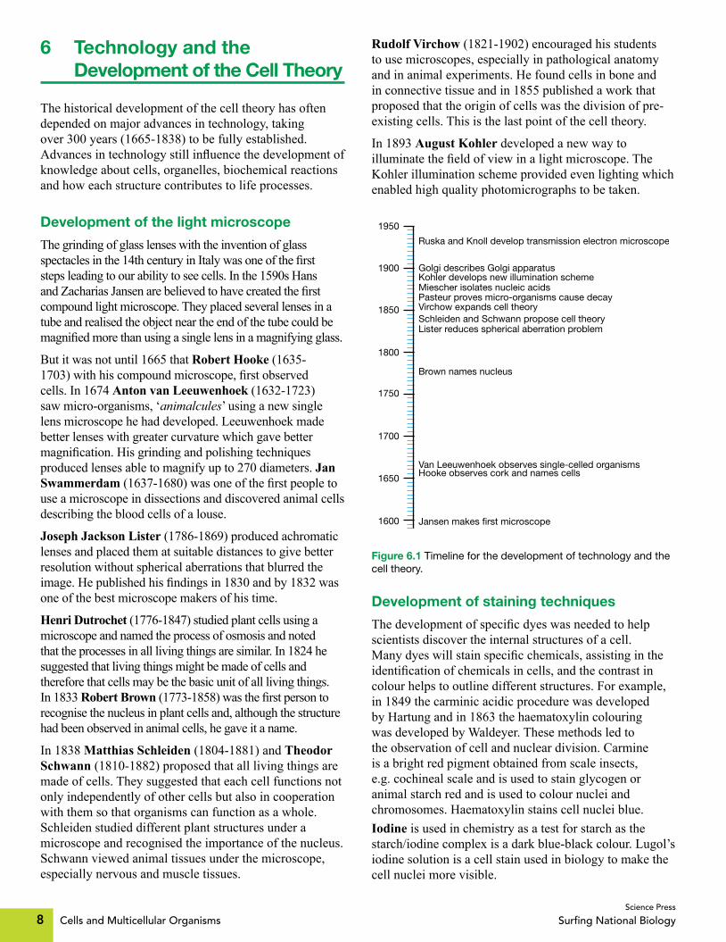
The historical development of the cell theory has often depended on major advances in technology, taking over 300 years (1665-1838) to be fully established. Advancesintechnologystillinfluencethedevelopmentofknowledge about cells, organelles, biochemical reactions and how each structure contributes to life processes.
Development of the light microscope
The grinding of glass lenses with the invention of glass spectaclesinthe14thcenturyinItalywasoneofthefirststeps leading to our ability to see cells. In the 1590s Hans andZachariasJansenarebelievedtohavecreatedthefirstcompound light microscope. They placed several lenses in a tube and realised the object near the end of the tube could be magnifiedmorethanusingasinglelensinamagnifyingglass.
But it was not until 1665 that Robert Hooke (1635-1703)withhiscompoundmicroscope,firstobservedcells. In 1674 Anton van Leeuwenhoek (1632-1723) saw micro-organisms, ‘animalcules’ using a new single lens microscope he had developed. Leeuwenhoek made better lenses with greater curvature which gave better magnification.Hisgrindingandpolishingtechniquesproduced lenses able to magnify up to 270 diameters. Jan Swammerdam (1637-1680)wasoneofthefirstpeopletouse a microscope in dissections and discovered animal cells describing the blood cells of a louse.
Joseph Jackson Lister (1786-1869) produced achromatic lenses and placed them at suitable distances to give better resolution without spherical aberrations that blurred the image.Hepublishedhisfindingsin1830andby1832wasone of the best microscope makers of his time.
Henri Dutrochet (1776-1847) studied plant cells using a microscope and named the process of osmosis and noted that the processes in all living things are similar. In 1824 he suggested that living things might be made of cells and therefore that cells may be the basic unit of all living things. In 1833 Robert Brown (1773-1858)wasthefirstpersontorecognise the nucleus in plant cells and, although the structure had been observed in animal cells, he gave it a name.
In 1838 Matthias Schleiden (1804-1881) and Theodor Schwann (1810-1882) proposed that all living things are made of cells. They suggested that each cell functions not only independently of other cells but also in cooperation with them so that organisms can function as a whole. Schleiden studied different plant structures under a microscope and recognised the importance of the nucleus. Schwann viewed animal tissues under the microscope, especially nervous and muscle tissues.
Rudolf Virchow (1821-1902) encouraged his students to use microscopes, especially in pathological anatomy and in animal experiments. He found cells in bone and in connective tissue and in 1855 published a work that proposed that the origin of cells was the division of pre-existing cells. This is the last point of the cell theory.
In 1893 August Kohler developed a new way to illuminatethefieldofviewinalightmicroscope.TheKohler illumination scheme provided even lighting which enabled high quality photomicrographs to be taken.
Jansen makes first microscope1600
1900
1950
1650
1700
1750
1800
1850
Hooke observes cork and names cells Van Leeuwenhoek observes single-celled organisms
Brown names nucleus
Schleiden and Schwann propose cell theory Virchow expands cell theory Pasteur proves micro-organisms cause decayMiescher isolates nucleic acids
Golgi describes Golgi apparatus
Lister reduces spherical aberration problem
Kohler develops new illumination scheme
Ruska and Knoll develop transmission electron microscope
Figure 6.1 Timeline for the development of technology and the cell theory.
Development of staining techniques
Thedevelopmentofspecificdyeswasneededtohelpscientists discover the internal structures of a cell. Manydyeswillstainspecificchemicals,assistingintheidentificationofchemicalsincells,andthecontrastincolour helps to outline different structures. For example, in 1849 the carminic acidic procedure was developed by Hartung and in 1863 the haematoxylin colouring was developed by Waldeyer. These methods led to the observation of cell and nuclear division. Carmine is a bright red pigment obtained from scale insects, e.g. cochineal scale and is used to stain glycogen or animal starch red and is used to colour nuclei and chromosomes. Haematoxylin stains cell nuclei blue. Iodine is used in chemistry as a test for starch as the starch/iodine complex is a dark blue-black colour. Lugol’s iodine solution is a cell stain used in biology to make the cell nuclei more visible.

Cells and Multicellular Organisms 9Science Press
Surfing National Biology
New methods in making slides have also enabled cells to be seen more clearly. Fixing specimens, e.g. in formalin will harden tissues before embedding in wax and then cutting. New instruments, e.g. microtomes have enabled extremely thin slices of specimens to be cut more easily.
Electromagneticlenses
Electromagneticlens
Source ofelectrons
Source ofelectrons
Specimen
Specimen
Eye
EyeFinal imageon photographicplate or screen
Detector
Beamdeflector
Image on TV viewing screen
(a) (b)
Figure 6.2 Comparing electron microscopes. (a) Transmission electron microscope. (b) Scanning electron microscope.
Development of the electron microscope
In 1932 Ernst Ruska (1906-1988) and Max Knoll (1897-1969)developedthefirsttransmissionelectronmicroscope (TEM). The development of the electron microscope with its greater resolution and greater magnificationcomparedtothelightmicroscopehasenabled scientists to see more details within the cell, e.g. the structure of the nuclear membrane, as well as discovering new parts, e.g. ribosomes. The transmission electron microscope uses a beam of electrons instead of a beam of light and uses electromagnets instead of glass lenses. It shows the internal appearance of cells.
Magnification refers to making things appear larger and resolution is the ability to distinguish between two points and makes detail appear more clearly.
QUESTIONS
1. Why is the development of the light microscope associated with the development of glass lenses?
2. Whenwerecellsfirstobservedandwhywasthispossible?
3. Outline the contribution of Anton van Leeuwenhoek to the development of the microscope.
4. WhatwasthesignificanceofKohler’sdevelopmentof a new illumination method?
5. Which aspect of the cell theory was not correctly understood by Schleiden and Schwann?
6. What was the contribution of Virchow?
7. Construct a table to show the contributions of at least six people to the cell theory giving the year, the person and their contribution.
8. Describe the evidence used by Hooke to support the beginning of the cell theory.
9. Discuss how the electron microscope has assisted our understanding of cell structure.
10. Ernst Abbe (1840-1905) developed many optical instruments and from experiments worked out a formula to determine the resolution limit of a microscope. He showed resolution was inversely proportional to the wavelength of light. Use this information and the fact that electron beams have much shorter wavelengths than the wavelengths of visible light to compare the resolution of a light microscope with the resolution of an electron microscope.
11. Distinguishbetweenmagnificationandresolution.12. Draw a timeline to show the stages in the
development of the cell theory including the work of Hooke, Leeuwenhoek, Dutrochet, Brown, Schleiden and Schwann and Virchow.
13. Assess the impact of the development of technology on the development of the cell theory.
14. The diagram shows a cross-section of a leaf as seen under low power light microscope.
A B C D
Figure 6.3 Cross-section of a leaf, low power light microscope.
Which arrow does not point to a nucleus of a cell? (A) A (B) B (C) C (D) D15. Which statement is not part of the cell theory? (A) All living things are made of cells. (B) All cells have a nucleus. (C) All cells come from pre-existing cells. (D) The cell is the basic unit of life in which the
processes of living occur.16. Name the scientist who proposed that all cells come
from pre-existing cells. (A) Hooke. (B) Brown. (C) Leeuwenhoek. (D) Virchow.

Cells and Multicellular Organisms10Science Press
Surfing National Biology
7 The Modern Light Microscope
The compound light microscope operates on the main principle that an objective lens with a very short focal lengthcanformahighlymagnifiedrealimageoftheobject. Visible light passes through the specimen and then a series of lenses. The resolution of the microscope is limited by the shortest wavelength of light used to view the specimen.
Images from a light microscope can be captured with a camera to produce a photomicrograph. Digital images can be shown directly on a computer screen.
Table 7.1 Features of the modern light microscope.
Feature Light microscope
Magnification Effective up to 1000´.
Resolution Up to 0.2 μm.
Stains Allows the use of many different coloured stains to identify substances, structures and provide contrast for easier viewing.
Living specimen The light microscope allows viewing of living specimen and processes occurring within a cell or within an organism.
Mounting The specimen is mounted on a glass slide in air.
Focusing By glass lenses.
Energy source for viewing
A beam of light is passed through the specimen.
How a light microscope works
The objective lens is brought close to the specimen to create an enlarged image of the object. The image is inverted. In most modern light microscopes the eyepiece is a compound lens near the back of an eyepiece tube. Light travels from the light source up the microscope to form an image at the eye.
Final image
Projector lens
Objective
Specimen
Light
Condenser lens
Eye
Eyepiece
Figure 7.1 How light travels through a light microscope.
High power (HP) objective Collects light and magnifies image x40.
x40
Eyepiece Contains the ocular lens (usually x10).
Nosepiece If the microscope is parfocal, the nosepiece can be rotated to change lenses and the image stays in focus.
Lower power (LP)objective Collectslight and magnifies image ´10.
Mirror If the microscope does not have its own light source the mirror is used to direct light up through the sample. The flat surface is used for a lamp and the curved surface is used for daylight.
Stage Holds theslide/sample.
Condenser Focuses light on to the sample.
Iris diaphragm leverMoving the lever controls the amount of light on to the stage.
Base Strong support for the structure.
Fine focus knobUsed to get a clearimage after usingthe coarse adjustment knob.
Coarse focus knobUsed to get the initialfocus on low power.
´40
´10
Figure 7.2 Features of the modern light microscope.
Advantages of a light microscope
The main advantages of light microscopes are that:
• Living cells can be observed. • Coloured stains can be used. • The specimens are easy to prepare. • The microscopes are relatively inexpensive
(compared to the cost of an electron microscope). • Their size means they are relatively easy to store.
Disadvantages of a light microscope
The main disadvantages of the light microscope are:• Itslimitedmagnification(effectivemagnification
begins to reduce after 1000´). • Its limited resolution. During the 20th century many different illumination techniques and other developments have increased the detection power of the light microscope for observing living cells.
Phase contrast microscopes
The phase contrast microscope uses interference rather than absorption of light to increase the contrast in unstained cells by amplifying variations in density within the cell. It improves our ability to study living, unpigmented cells in biological and medical research. Many dyes and stains stop chemical processes in cells which means the phase contrast microscope has improved our ability to see detail in living cells, e.g. the process of cell division. Frits Zernike was awarded with the Nobel Prize in Physics, 1953 for the development of phase contrast illumination.

Cells and Multicellular Organisms 11Science Press
Surfing National Biology
Fluorescence microscopes
Inthefluorescencemicroscopethespecimenisilluminatedthrough objective lenses with a narrow set of light wavelengths.Thespecimeneitherfluorescesinitsnaturalform,e.g.chlorophyllorhasbeentreatedwithfluorescingchemicalsorantibodies.ThefluorescentsubstancesabsorbUVlightandemitvisiblelightsothatthefluorescenceshowsthelocationofspecificmoleculesinthecell.
Other illumination techniques
Other illumination techniques include:
• Bright field – passes light through the specimen and contrast comes from the absorbance of light in the specimen. If the specimen is unstained or unpigmented there is little contrast.
• Cross-polarised – contrast occurs when polarised light is rotated through the sample.
• Confocal –isatypeoffluorescencemicroscopyusing optical sectioning by scanning lasers of fluorescently-stainedspecimens.
Stains and dyes used with the light microscope
There are many stains that are used to highlight structures being viewed under a light microscope.
• Gram staining – uses several stains, e.g. crystal violet, iodine, fuchsin or safranin to stain cell cells to differentiate bacteria into Gram positive (purple/blue colour occurs) and Gram negative (pink/red colour occurs).Thisisusuallythefirststepinidentifyingbacteria.
• DAPI–isafluorescentnuclearstainthatshowsabluefluorescencewhenboundtoDNAandviewedwith ultraviolet light.
• Eosin – is used as a counterstain with haematoxylin in H&E staining and gives a pink/red colour to cytoplasmic material, red blood cells and cell membranes.
QUESTIONS
1. Outline the basic principle behind the operation of a light microscope.
2. When using a light microscope identify the function of: (a) The condenser. (b) The iris diaphragm.3. What restricts the resolution of the light microscope?4. Construct a table to compare the advantages and
disadvantages of a light microscope.5. What is a photomicrograph?6. Outlinethebenefitofthephasecontrastmicroscope.7. Outlinethebenefitoffluorescencemicroscopy.
8. The table shows some objects and their sizes.Object Size ( µm)
Frog egg 1100
Paramecium 100
Plant epithelial cell 60
Human ovum 10
Red blood cell 7 to 8
Mitochondrion 1.1
Nanobes 0.025
Identify which objects could be seen with the naked eye and/or light microscope.
9. Use an example to show how the use of stains and the light microscope have increased our understanding of processes in cells.
10. Most modern light microscopes in schools have at least two different objectives. Name objectives you have used and state the purpose of each objective.
11. What is a parfocal microscope?12. The diagram shows red blood cells as seen and as
drawn by a biology student using a light microscope.
(a) (b)
Figure 7.3 Red blood cells seen using a light microscope. (a) Seen under a light microscope. (b) As drawn by a student.
Red blood cells are usually about 6 to 8 micrometres in diameter.Which objective would the student most likely have been using to view these cells as seen in the diagram? Explain your answer.
13. Explain why the Gram stain is used.14. What is the correct order of the movement of light
when an object is viewed under a light microscope? (A) Light → condenser → objective → eyepiece
→ eye (B) Light → objective → condenser → eyepiece
→ eye (C) Light → objective → eyepiece → condenser
→ eye (D) Eye → eyepiece → objective → condenser
→ light15. What is the limit of resolution of a light microscope? (A) 0.2 centimetres (B) 0.2 millimetres (C) 0.2 micrometres (D) 0.2 nanometres

Cells and Multicellular Organisms142Science Press
Surfing National Biology
Answers1 Assumed Knowledge1. Living organisms can: 1. Respire. 2. Assimilate food and
synthesise organic molecules. 3. Grow. 4. Reproduce. 5. Respond to stimuli from their environment. 6. Excrete. 7. Move about.
2. The cells of living things have a cell membrane, cytoplasm and DNA. 3. Plant cell.
VacuoleCell membraneNucleus Cell wall
CytoplasmChloroplast
4. Animal cell.
Nucleus Cytoplasm
Cell membrane
5. A = eye of person using the light microscope, B = ocular lens, C = objective lens, D = specimen, E = condenser lens, F = light source
6. When using a light microscope, you should always wear shoes with covered toes, as the microscope is heavy and if you drop it you could damage exposed skin on your feet.
7. The nucleus stores information needed to control all cell activities.8. The cell membrane surrounds the cell contents from the external
environment and controls the substances that can leave or enter the cell.
9. Cytoplasm is a general term for the contents of a cell outside the nucleus and within the cell membrane.
10.Protoplasmisthesemifluidtransparentsubstancethatmakesupthe living matter of plant and animal cells including the nucleus and cytoplasm.
11. A chloroplast is a green organelle found in green tissues of plants that captures sunlight in photosynthesis to manufacture sugars from carbon dioxide and water.
12. Photosynthesis is a process where the energy of sunlight is used to convert carbon dioxide and water into sugars and oxygen.
13. Groups of organisms that can photosynthesise include plants, algae and photosynthetic bacteria.
14. Carbon dioxide and water are needed for photosynthesis using light energy and in the presence of chlorophyll.
15. Photosynthesis is important in ecosystems as it converts light energy into chemical energy to begin most food chains on Earth and also provides oxygen which is needed for respiration.
16. The four basic groups of organic compounds are proteins, carbohydrates, lipids and nucleic acids.
17. Inorganic compounds are molecules that do not contain carbon (excluding some carbonates and simple oxides of carbon).
18. The function of the digestive system is to break down ingested food into smaller particles so that nutrients can be absorbed into the body.
19. A = salivary glands, B = oesophagus, C = stomach, D = liver, E = gall bladder, F = pancreas, G = large intestine, H = small intestine, I = appendix, J = anus
20.
21. Respiration is the chemical reactions in which cells obtain energy from food.
22. In humans, the respiratory system consists of the lungs and passages which bring air into and out of the lungs, e.g. trachea, bronchi and alveoli.
23. The function of the respiratory system is the intake, expulsion and exchange of oxygen and carbon dioxide.
24. The circulatory system is the transport system of the body, e.g. it carries the glucose and oxygen to the cells and distributes heat around the body.
25.Inhumanstheheartpumpsthebloodtokeepitflowingawayfromthe heart through the arteries.
26. The human circulatory system consists of the heart and blood vessels, e.g. arteries, veins and capillaries and blood.
27. (a) Xylem transports water up the plant from roots to leaves. (b) Phloem transports sugars up and down the plant. (c) Leaves are the site of photosynthesis where light energy is
changed into chemical energy to be used by the plant. (d) Roots support the plant, anchor it in the soil and are the site
of water absorption.28. Transpiration is the evaporation of water from leaves of a plant.29. Mitosis.30. Mitosis is the process during cell division in which the cell nucleus
divides into two.31. Cell division is needed for growth, repair and reproduction.32. During reproduction genetic information is transferred as DNA on
chromosomes.33. A gene is a section of DNA that relates to one characteristic.34. Cytokinesis is the division of the cell’s cytoplasm during cell
division, following the division of the nucleus.
2 Characteristics of Living Things1.
Part Structure Function
Mouth Has teeth and openings from salivary glands
Teeth break food into small pieces and salivary enzymes begins chemical digestion
Oesophagus Long tube Moves food to stomach by peristalsis
Stomach Muscles and glands in wall
Churns food and produces digestive enzyme to digest protein
Small intestine Long thin tube with villi and glands
Digestion is completed and nutrients absorbed through walls
Large intestine Long tube Water, salts and vitamins absorbed
Anus Muscular ring Eliminates faeces
Characteristic Description of characteristic
Growth and development
Involves an increase in mass due to an increase in the size of individual cells and/or and increase in the number of cells.
Reproduction Is the ability to produce offspring and can be either sexual or asexual.
Respiration Is a series of chemical reactions in which cells obtain energy from food.
Respond to stimuli Stimuli from either the internal or external environvment cause a response in or by the organism.
Movement and locomotion
Part or the whole organism can move.
Nutrition or feeding
Organisms obtain matter and energy to build their physical structure and continue the functions of life.
Assimilation Is the process of converting food into the living material of life.
Metabolism Is the sum of all chemical reactions within the organism.
Excretion Is the removal of unwanted waste products of metabolic reactions.

Cells and Multicellular Organisms 143Science Press
Surfing National Biology
2. Sexual reproduction involves the union of two gametes in fertilisation to form a zygote. Asexual reproduction only involves one parent producing offspring that are identical to the parent.
3. Fertilisation is the union of two gametes.4. Although crystals can grow in size they do not show any other
characteristic of living things, e.g. respiration, response to a stimulus.Tobeclassifiedasalivingthingacombinationofcharacteristics need to be present.
5. In autotrophic nutrition the organism can make its own organic nutrients from inorganic materials. In heterotrophic nutrition the organism needs to consume organic materials and existing foods.
6. In photosynthesis the autotroph uses energy from sunlight to create their own organic material while in chemosynthesis the autotroph uses a chemical source of energy to create their own organic material.
7. (a) A and B are autotrophic nutrition and C is heterotrophic nutrition.
(b) Nitrosomonas spp uses ammonia as its source of energy. It oxidises ammonia to nitrous acid.
8. In anabolic reactions small molecules are combined to form more complex molecules. In catabolic reactions chemical bonds are broken and complex molecules are broken down into smaller units.
9. Respiration is a catabolic reaction as complex organic molecules, e.g. glucose are broken down into smaller units, e.g. carbon dioxide and energy is released.
3 Developing the Cell Theory1. The cell theory states that: 1. All living things are made of cells and
of substances produced by cells. 2. All cells come from pre-existing cells. 3. The cell is the basic unit in which the processes of living take place.
2. Date Person Contribution
1590s Hans and Zacharias Jansen
Created the first compound light microscope.
1663 Robert Hooke Observed cork under a microscope and introduced the term ‘cell’.
1668 Francesco Redi Published the results of his experiments with insects showing living things do not arise from spontaneous generation.
1674 Anton van Leeuwenhoek
Improved the quality of lenses and aided the development of the light microscope.
Discovered unicells, e.g. protozoa and bacteria.
Discovered the vacuole and drew the banded pattern of muscle fibres and spermatozoa.
1833 Robert Brown Published a paper naming and describing the cell nucleus in orchids.
1838 Matthias Schleiden Proposed that different parts of plants are made of cells. This became part of the Schleiden and Schwann cell theory.
1839 Theodor Schwann Extended Schleiden’s cell theory to include animals.
Schleiden and Schwann cell theory: 1. All living things are made of cells and cell products. 2. The cell is the basic unit of life.
1855 Rudolf Virchow Extended the cell theory to include that every cell originated from a living pre-existing cell.
1861 Louis Pasteur Fermentation experiments finally disprove the theory of spontaneous generation.
1869 Fredrich Miescher Isolated DNA for the first time.
1898 Camillo Golgi Described the Golgi apparatus by staining cells with silver nitrate.
1932 Max Knoll and Ernst Ruska
Invented the transmission electron microscope which gave greater resolution and magnification to study the ultrastructure of cells.
3. The invention of the light microscope is tightly linked with the development of the cell theory. When Robert Hooke used his compound microscope to view cork he discovered the cellular nature of plants and called the structures ‘cells’. When van Leeuwenhoek used improved lenses to observe unicells he was not initially believed. It was the invention and development of light microscopeswithhighermagnificationandresolutionthatenabledscientists to observe and study the cellular nature of living things.
4. Anton van Leeuwenhoek produced higher quality lenses and made manymicroscopes.Theimprovedresolutionandmagnificationof his microscopes meant he was able to observe smaller objects andhewasthefirsttodrawanddescribesingle-celledorganisms,e.g. protozoa and bacteria. This work led to him being called the ‘Father of Microbiology’.
5. Leeuwenhoek’sdiscoveryofanimalculeswasatfirstdisbelievedastheideaofsingle-celledorganismsdidnotfitinwiththeearly17thcenturyconceptof‘life’.Hisworkwasfinallyacceptedsixyears later when others observed unicells under the microscope.
6. Several scientists observed and drew diagrams of cells showing the presence of a nucleus, e.g. Anton van Leeuwenhoek and Franz Bauer. Robert Brown observed the nucleus in orchid cells and named the structure in 1831.
7. Schleiden and Schwann proposed that: 1. All living things are made of cells and of substances produced by cells. 2. The cell is the basic unit and building block of life.
8. The theory of spontaneous generation proposed that living things arosefromnon-livingmatter,e.g.maggotsfromdeadflesh,ratsfrom rubbish bins.
9. FermentationexperimentscarriedoutbyLouisPasteurfinallydisproved the theory of spontaneous generation. He showed that micro-organisms were responsible for fermentation and bacteria caused the growths in boiled nutrient broths.
10. The cell theory states that all living cells come from pre-existing cells and clashes with the theory of spontaneous generation which proposes that living things can arise from non-living matter. As the cell theory developed, the theory of spontaneous generation had to be abandoned.
11. (a) Camillo Golgi discovered and used a new staining technique using silver nitrate to observe cells, e.g. in nervous tissue and to identify cell structures. Many believed that the body he found inside cells was an optical illusion caused by his staining technique.
(b) The invention of the electron microscope which had greater magnificationandresolutionthanthelightmicroscopeprovedthe existence and showed the ultrastructure of the Golgi body.
12. The invention of the electron microscope has greatly aided the development of knowledge about cell structure. The greater magnificationandresolutiongavemoredetailedinformationaboutknown cell organelles, e.g. Golgi body and also discovered new structures, e.g. ribosomes that can only be seen under an electron microscope.
13. D14. A
4 Cell Theory and Robert Hooke1. RobertHookeinvented:1.Thecompoundmicroscope.2.Thefirst
reflectingtelescope.2. A compound microscope has more than one lens arranged in such
a way that the image is larger than the object being viewed.3. The invention of a compound microscope meant that organisms
couldbeseenwithgreatermagnificationandthestructureofcellswas now visible.
4. Robert Hooke used the term ‘cell’ to describe the structures he saw when he placed a slice of cork under his compound microscope.
5. Hooke saw the cell walls of cork tissue which formed a honeycomb appearance.
6. Opponents of Robert Hooke discredited Hooke’s microscopic observations using the details about lens distortion and fringe effecttoclaimhewasdrawingartificialimagescreatedbythelenses.Thisledtosomesectionsofthescientificcommunityinitiallyrejectinghisfindings.

Cells and Multicellular Organisms144Science Press
Surfing National Biology
7. In the 17th century there were several theories to explain the origin of fossils. Some believed, similar to Aristotle that fossils formed and grew within the Earth. When Hooke suggested that the fossils were clues to the past history of life on Earth, some people found it theologically unacceptable.
8. The drawings in Micrographia are highly detailed and accurately show the structure of many common organisms. People were fascinated by this new view of life making the book a subject of interest and discussion and thus a ‘best seller’.
5 Cell Theory and Robert Brown1. Brown sailed with Mathew Flinders on the HMS Investigator
expedition 1801-1805. He travelled to Australia and Tasmania, collecting many plant specimens and describing over 2200 different plant and animal species.
2. He published his notes in 1811 in Prodromus Florae Novae Hollandiae et insulae Van-Diemen and further notes in 1814 in ‘General Remarks, Geographical and Systemic, on the Botany of Terra Australis’ as an appendix to Mathew Flinders’ Voyage to Terra Australis.
3. Robert Brown travelled to Australia and collected and made drawingsofmanyAustralianspecies.Hepublishedhisfindingsincreasing the amount of data about Australian plants and animals available to the public. He also became responsible for the work and collection of Joseph Banks which preserved specimens collected by Banks on his Australian voyage with Captain James Cook.
4. Brownian motion is the constant movement of suspended particles.5. Brown observed the epidermis of orchids under his microscope
and discovered an ‘opaque spot’ which he called the nucleus.6. Brown coined the term ‘nucleus’.7. The statement is inaccurate and untrue. It is inaccurate as Robert
Brownwasnotthefirsttodescribethenucleus–bothAntonvanLeeuwenhoek and Franz Bauer had drawn diagrams showing the presence of nuclei. It is untrue as the function of the nucleus had not yet been discovered.
8. B9. B10. C
6 Technology and the Development of the Cell Theory1. The light microscope uses glass lenses to magnify objects. Thus
the development of the light microscope is tightly linked with the ability to produce high quality glass lenses.
2. Thefirstcellswereobservedin1665,whenRobertHookeusedhiscompound microscope to observe cork.
3. Anton van Leeuwenhoek made better lenses with greater curvature whichgavebettermagnification.Healsoimprovedgrindingandpolishing techniques for making lenses.
4. August Kohler’s illumination method gave an even distribution of lightinthefieldofview.Thismeantthatbetterandhigherqualityphotomicrographs could be taken.
5. Schleiden and Schwann did not correctly understand how cells arose.6. Virchow proposed that all cells come from pre-existing cells which
provided the last point in the cell theory.7.
8. Hooke observed cells in a slice of cork using his compound microscope, providing evidence that the tissue consists of cells.
9. The electron microscope has enabled scientists to see more details within the cell, e.g. the structure of the nuclear membrane, as well as discovering new parts, e.g. ribosomes.
Year Person Contribution
1665 Hooke Observed cells in a slice of cork
1678 Leeuwenhoek Observed micro-organisms
1824 Dutrochet Living things are made of cells
1833 Brown Observes nucleus in plant cells and names structure as nucleus
1838 Schleiden and Schwann
Tissues in all living things made of cells and cells function both independently and in cooperation
1858 Virchow Cells come from pre-existing cells
10. Since the resolution of a microscope is inversely proportional to thewavelengthoftheradiation–thesmallerthewavelength,thebigger the resolution. Since the wavelength of a beam of electrons is much smaller than the wavelength of visible light the electron microscope has a much higher resolution than a light microscope.
11.Magnificationreferstomakingthingsbiggerwhileresolutionistheabilitytodistinguishbetweentwopointsandseefinedetail.
12.
13. The cell theory states that: 1. All living things are made of cells. 2. All cells come from pre-existing cells. 3. The cell is the basic unit in which the processes of living take place. The development of these ideas was only possible as instruments and stains were developed so that scientists were able to see cells and their internal structure. For example, when Robert Hooke placed a sliceofcorkunderhismicroscope,hewasthefirstpersontoseethese ‘many little boxes’, which he called cells. The discovery of staining techniques allowed the processes of cell and nuclear division to be seen. The development of the cell theory underpins our understanding of life on Earth, its structure, its physiology and why it behaves the way it does. Thus the development of technology has had a tremendous impact on the development of the cell theory as the theory could only expand as the technology allowed us to see what was present and how it behaved.
14. D15. B16. D
7 The Modern Light Microscope1. The compound light microscope passes visible light through the
specimen and then through a series of lenses. It operates on the main principle that an objective lens with a very short focal length canformahighlymagnifiedrealimageoftheobject.
2. (a) The condenser focuses light on to the sample. (b) The iris diaphragm controls the amount of light on to the stage.3. The resolution of the microscope is limited by the shortest
wavelength of light used to view the specimen (wavelengths of visible light are 400 to 700 nm).
4.
5. A photomicrograph is a photograph made through a microscope.6. Themainbenefitofthephasecontrastmicroscopeisthatit
increases the contrast in unstained cells. It improves our ability to study living, unpigmented cells in biological and medical research. Many dyes and stains stop chemical processes in cells which means the phase contrast microscope has improved our ability to see detail in living cells, e.g. the process of cell division.
7. Themainbenefitoffluorescencemicroscopyisthatthefluorescenceshowsthelocationofspecificmoleculesinthecell.Naturalfluorescingsubstances,e.g.chlorophyllcanbelocatedorthespecimencanbetreatedwithfluorescingchemicalsorantibodies to locate other particular section or substances in cells.
1665 Robert Hooke described cells in cork1678 Leeuwenhoek saw microbes
1824 Dutrochet – living things made of cells1833 Brown named nucleus in plant cells1838 Schleiden and Schwann – cell theory
1858 Virchow – cells from pre-existing cells
Advantages of light microscopeDisadvantages of light
microscope
Can view living specimens and observe processes in cells and within an organism.
Allows the use of many different coloured stains to identify substances, structures and provide contrast for easier viewing.
Specimens are easy to prepare, stain and observe in a short time frame.
Relatively inexpensive and class sets can be purchased for use in schools.
Light microscopes are not particularly large and are easy to store, e.g. in schools.
Limited magnification, e.g. effectively up to 1000´.
Resolution limited to 0.2 micrometres.

Cells and Multicellular Organisms 145Science Press
Surfing National Biology
8.
9. Stains and the light microscope have greatly increased our understanding of the processes in cells. For example, pH indicators which will show acidic or basic conditions can be used to show how the activity of micro-organisms change the pH balance of their environment such as bacterial action causing the souring of milk.
10. Objectives found on school light microscopes include 10´ and 40´. The 10´objectiveisalowpowerlensusedforfirstviewingthe specimen to locate the area to be examined. The 40´ objective is a high power lens and is used to make detailed observations of the specimen.
11. A parfocal microscope allows you to quickly focus using the lower power objective and then swivel the nosepiece to observe the specimen under a higher power objective without only minimal adjustmentofthefinefocusknob.
12.Redbloodcellsareverysmall–theyhaveadiameterof6to8micrometres. This means the student would have been using a high power objective, e.g. 40´ in the light microscope set-up to clearly observe these cells as seen in the diagram.
13.TheGramstainisusuallyusedasthefirststepinidentifyingbacteria.Itdifferentiatesbacteriaintotwogroups–Gramnegativeand Gram positive.
14. A15. C
8 The Electron Microscope1. The electron microscope has better resolution than the light
microscope as it uses a beam of electrons to view the specimen. Since the beam of electrons has a much shorter wavelength than visible light resolution is greatly improved, e.g. approximately 0.002 nm, although in practical situations it can be limited to 2 nm. Resolution is inversely related to the wavelength of radiation used by the microscope for imaging.
2. The electron microscope sends a stream of electrons through a vacuum. The electron beam is focused by electromagnets, magnifiedbyanobjectivelensandprojectedontoafluorescentscreenorphotographicfilm.
3.
4. ThemainbenefitoftheTEMisthatitshowstheinternalultrastructureofcellsathighermagnificationandresolutionthanthe light microscope. You can see objects to the order of several nanometres (10−9 m) which means new organelles have been discovered using the TEM, e.g. ribosomes and there is an increased capacity for medical, biological and materials research.
5. ThemainbenefitoftheSEMisthatitgivesathree-dimensionaldetailed view of the surface of the specimen. The image has great depthoffieldandgivesaviewoftheactualappearanceofthespecimen.
Object Size ( µm) Viewing
Frog egg 1100 Naked eye and light microscope
Paramecium 100 Light microscope
Plant epithelial cell 60 Light microscope
Human ovum 10 Light microscope
Red blood cell 7 to 8 Light microscope
Mitochondrium 1.1 Light microscope
Nanobes 0.025 Cannot be seen under a light microscope – need an electron microscope
Advantages of electron microscope
Disadvantages of electron microscope
Increased magnification, e.g. up to 300 000´.
Increased resolution, e.g. approximately 0.0005 µm
SEM shows a three-dimensional image of the specimen.
Cannot view living specimens and observe processes happening in cells.
Cannot use coloured stains.
Relatively expensive and are not available in schools.
6.
7. D
9 The Stereo Microscope1. Thestereomicroscopeiseasilyidentifiedasithastwoocular
eyepieces that each view the object from different angles. There are two separate optical paths for viewing.
2. You are likely to use a stereo microscope when dissecting small organisms or when you need to observe the external features of a specimen.
3. The parts of the stereo microscope are: A = ocular lens B = objective lens C = stage clip D = stage E = focus knob F = arm4. If a compound microscope has two eyepieces which give the same
image then the image will not be three-dimensional and give a ‘stereo’ image. To provide a three-dimensional image there must be two images each from slightly different viewing angles.
5. In a stereo microscope the lens is a distance away from the object and this gives poor resolution.
6. Atypicalschoolstereomicroscopehasamagnificationrangingfrom 10´ to 40´ while a typical school monocular microscope hasamagnificationfrom40´ to 400´.
7. Salt crystals are light coloured and usually translucent. The dark colour beneath the crystal is needed to provide contrast so that the crystal can be properly viewed.
8. When using a stereo microscope some parts of an organism may not be in focus as the specimen is three-dimensional with several levels. The microscope is focusing on one level making other levels appear out of focus.
9. A
Image A Image B
Scanning electron microscope Light microscope
Image C Image D
Light microscope Transmission electron microscope
Image E Image F
Scanning electron microscope Light microscope