nanotubular structures of zinc oxide
TRANSCRIPT

Nanotubular structures of zinc oxide
Y.J. Xinga,b, Z.H. Xia, X.D. Zhanga, J.H. Songa, R.M. Wangb, J. Xub, Z.Q. Xuea,D.P. Yub,*
aDepartment of Electronics, Peking University, Beijing 100871, People’s Republic of ChinabSchool of Physics, Electron Microscopy Laboratory, and State Key Laboratory for Mesoscopic Physics, Peking University, Beijing 100871,
People’s Republic of China
Received 10 July 2003; received in revised form 7 September 2003; accepted 10 November 2003 by Z.Z. Gan
Abstract
ZnO nanotubes with a regular polyhedral shape, hollow core, and wall thickness as small as 4 nm, have been prepared in
large-area substrate by vapor phase growth. The nanotubes can be classified into two groups consisting of either polycrystalline
or straight single crystal. The formation of the ZnO nanotubes was found closely related to the hexagonal structure of the ZnO
crystal and the peculiar growth conditions used.
q 2003 Elsevier Ltd. All rights reserved.
PACS: 61.46. þ w; 81.16. 2 c; 81.07.De; 81.05.Hd
Keywords: A. Nanostructures; A. Semiconductors; C. Scanning and transmission electron microscopy
1. Introduction
Nano-sized tubular structures have stimulated intensive
research interests [1–6], because these materials have
enabled studies on fundamental physical properties [7,8],
and served as building blocks to construct nanoscale devices
[9,10]. However, nanotubular structures have only been
grown for few materials, which have bulk lamellar
structures, such as graphite or graphite-like structures
(BN, BCN, WS2, MoS2). Little is known for direct growth
of nanotubular structures from 3-dimensional materials to
date [11], though complex lithography was used to engineer
Ge–Si thin films into nanotubes [12]. The nanophased ZnO
compound was widely studied since this material exhibits a
number of interesting fundamental properties and possible
applications, including ZnO films [13], disordered nano-
particles [14], nanobelts [15], and nanowires [16] in which
very intense stimulated UV lasing action was observed at
room temperature. Recently, the tubular structures of ZnO
with the diameters of 30/350 nm were observed in the
heterostructures of Zn core/ZnO sheath nanocables and in
Zn(NH3)42þprecursor solutions [17,18]. However, these
products have a low yield of the production and poor
morphology and crystal quality. We demonstrate, in this
communication, crystalline ZnO nanotubes with a regular
polyhedral shape, hollow core, and wall thickness of 4–
20 nm, were prepared in large-area substrate by vapor phase
growth.
2. Experimental
The ZnO nanotubes were grown using thermal evapor-
ation of Zn/ZnO powder mixture. Powders of zinc oxide
(1.0 g) and zinc (0.3 g) were well mixed and put into the
central zone of alumina tube in a tube furnace. The whole
system was evacuated to about 300 Torr, and filled with
Argon. The furnace was heated to about 1300 8C under
flowing Ar atmosphere (40 sccm) for 1 h. Si substrates
(5 £ 20 mm) were placed downstream inside the tube
(temperature 700 8C) in sequence to collect the products.
It is noted that proper amount of water was held in a glass
vessel upstream the alumina tube to keep a wet oxidation
0038-1098/$ - see front matter q 2003 Elsevier Ltd. All rights reserved.
doi:10.1016/j.ssc.2003.11.049
Solid State Communications 129 (2004) 671–675
www.elsevier.com/locate/ssc
* Corresponding author. Tel./fax: þ8610-62759474.
E-mail address: [email protected] (D.P. Yu).
ambient during the growth of the ZnO nanotubes. The
morphology of the as-grown product was analyzed using a
Dual Beam-235 focused ion beam (FIB) system. A Tecnai
F30 transmission electron microscope (TEM) equipped with
nano-beam energy dispersive spectroscopy (EDS) was used
to characterize the microstructure and chemical composition
of the nanowires.
3. Results and discussion
X-ray diffraction analysis of the nanotubes revealed a
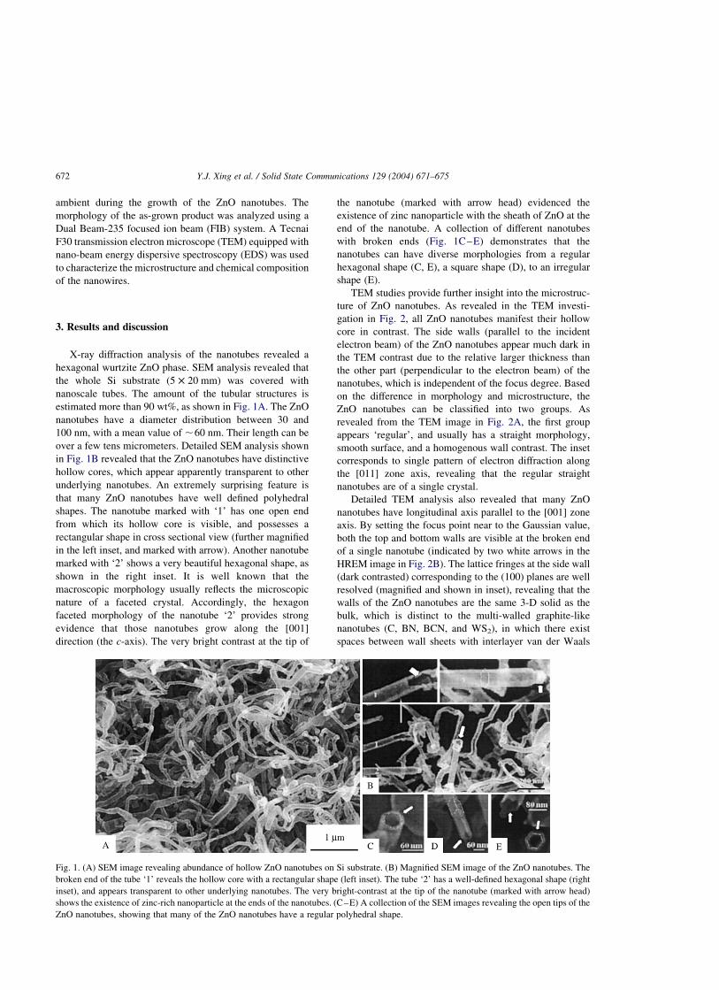
hexagonal wurtzite ZnO phase. SEM analysis revealed that
the whole Si substrate (5 £ 20 mm) was covered with
nanoscale tubes. The amount of the tubular structures is
estimated more than 90 wt%, as shown in Fig. 1A. The ZnO
nanotubes have a diameter distribution between 30 and
100 nm, with a mean value of ,60 nm. Their length can be
over a few tens micrometers. Detailed SEM analysis shown
in Fig. 1B revealed that the ZnO nanotubes have distinctive
hollow cores, which appear apparently transparent to other
underlying nanotubes. An extremely surprising feature is
that many ZnO nanotubes have well defined polyhedral
shapes. The nanotube marked with ‘1’ has one open end
from which its hollow core is visible, and possesses a
rectangular shape in cross sectional view (further magnified
in the left inset, and marked with arrow). Another nanotube
marked with ‘2’ shows a very beautiful hexagonal shape, as
shown in the right inset. It is well known that the
macroscopic morphology usually reflects the microscopic
nature of a faceted crystal. Accordingly, the hexagon
faceted morphology of the nanotube ‘2’ provides strong
evidence that those nanotubes grow along the [001]
direction (the c-axis). The very bright contrast at the tip of
the nanotube (marked with arrow head) evidenced the
existence of zinc nanoparticle with the sheath of ZnO at the
end of the nanotube. A collection of different nanotubes
with broken ends (Fig. 1C–E) demonstrates that the
nanotubes can have diverse morphologies from a regular
hexagonal shape (C, E), a square shape (D), to an irregular
shape (E).
TEM studies provide further insight into the microstruc-
ture of ZnO nanotubes. As revealed in the TEM investi-
gation in Fig. 2, all ZnO nanotubes manifest their hollow
core in contrast. The side walls (parallel to the incident
electron beam) of the ZnO nanotubes appear much dark in
the TEM contrast due to the relative larger thickness than
the other part (perpendicular to the electron beam) of the
nanotubes, which is independent of the focus degree. Based
on the difference in morphology and microstructure, the
ZnO nanotubes can be classified into two groups. As
revealed from the TEM image in Fig. 2A, the first group
appears ‘regular’, and usually has a straight morphology,
smooth surface, and a homogenous wall contrast. The inset
corresponds to single pattern of electron diffraction along
the [011] zone axis, revealing that the regular straight
nanotubes are of a single crystal.
Detailed TEM analysis also revealed that many ZnO
nanotubes have longitudinal axis parallel to the [001] zone
axis. By setting the focus point near to the Gaussian value,
both the top and bottom walls are visible at the broken end
of a single nanotube (indicated by two white arrows in the
HREM image in Fig. 2B). The lattice fringes at the side wall
(dark contrasted) corresponding to the (100) planes are well
resolved (magnified and shown in inset), revealing that the
walls of the ZnO nanotubes are the same 3-D solid as the
bulk, which is distinct to the multi-walled graphite-like
nanotubes (C, BN, BCN, and WS2), in which there exist
spaces between wall sheets with interlayer van der Waals
Fig. 1. (A) SEM image revealing abundance of hollow ZnO nanotubes on Si substrate. (B) Magnified SEM image of the ZnO nanotubes. The
broken end of the tube ‘1’ reveals the hollow core with a rectangular shape (left inset). The tube ‘2’ has a well-defined hexagonal shape (right
inset), and appears transparent to other underlying nanotubes. The very bright-contrast at the tip of the nanotube (marked with arrow head)
shows the existence of zinc-rich nanoparticle at the ends of the nanotubes. (C–E) A collection of the SEM images revealing the open tips of the
ZnO nanotubes, showing that many of the ZnO nanotubes have a regular polyhedral shape.
Y.J. Xing et al. / Solid State Communications 129 (2004) 671–675672
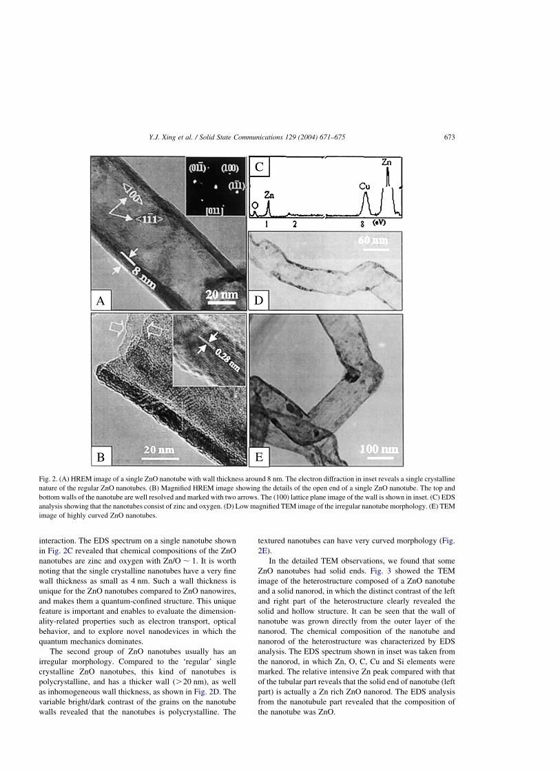
interaction. The EDS spectrum on a single nanotube shown
in Fig. 2C revealed that chemical compositions of the ZnO
nanotubes are zinc and oxygen with Zn/O , 1. It is worth
noting that the single crystalline nanotubes have a very fine
wall thickness as small as 4 nm. Such a wall thickness is
unique for the ZnO nanotubes compared to ZnO nanowires,
and makes them a quantum-confined structure. This unique
feature is important and enables to evaluate the dimension-
ality-related properties such as electron transport, optical
behavior, and to explore novel nanodevices in which the
quantum mechanics dominates.
The second group of ZnO nanotubes usually has an
irregular morphology. Compared to the ‘regular’ single
crystalline ZnO nanotubes, this kind of nanotubes is
polycrystalline, and has a thicker wall (.20 nm), as well
as inhomogeneous wall thickness, as shown in Fig. 2D. The
variable bright/dark contrast of the grains on the nanotube
walls revealed that the nanotubes is polycrystalline. The
textured nanotubes can have very curved morphology (Fig.
2E).
In the detailed TEM observations, we found that some
ZnO nanotubes had solid ends. Fig. 3 showed the TEM
image of the heterostructure composed of a ZnO nanotube
and a solid nanorod, in which the distinct contrast of the left
and right part of the heterostructure clearly revealed the
solid and hollow structure. It can be seen that the wall of
nanotube was grown directly from the outer layer of the
nanorod. The chemical composition of the nanotube and
nanorod of the heterostructure was characterized by EDS
analysis. The EDS spectrum shown in inset was taken from
the nanorod, in which Zn, O, C, Cu and Si elements were
marked. The relative intensive Zn peak compared with that
of the tubular part reveals that the solid end of nanotube (left
part) is actually a Zn rich ZnO nanorod. The EDS analysis
from the nanotubule part revealed that the composition of
the nanotube was ZnO.
Fig. 2. (A) HREM image of a single ZnO nanotube with wall thickness around 8 nm. The electron diffraction in inset reveals a single crystalline
nature of the regular ZnO nanotubes. (B) Magnified HREM image showing the details of the open end of a single ZnO nanotube. The top and
bottom walls of the nanotube are well resolved and marked with two arrows. The (100) lattice plane image of the wall is shown in inset. (C) EDS
analysis showing that the nanotubes consist of zinc and oxygen. (D) Low magnified TEM image of the irregular nanotube morphology. (E) TEM
image of highly curved ZnO nanotubes.
Y.J. Xing et al. / Solid State Communications 129 (2004) 671–675 673

The formation of the ZnO nanotubular structures is
extremely interesting, however, the growth mechanism is
yet unclear. Our growth conditions distinguished others
methods of ZnO nanowire/nanobelt growth at least in two
aspects [15,16]: the mixing of metallic zinc with zinc oxide,
and a weak wet-oxidation atmosphere. The growth of the
ZnO nanotubes can be understood on basis of the peculiar
growth conditions and the particular microstructure of the
ZnO crystal. Two important factors are responsible for the
growth of the ZnO nanotubes: the formation of the nanotube
embryos at proper nucleation sites, and unidirectional
growth of the nanotube embryos. Based on our experimental
evidences, we proposed the following model to depict the
growth of the peculiar ZnO nanotubes. The metallic zinc has
a very low melting point (419 8C) compared to zinc oxide, a
oxygen deficient vapor phase was evaporated from the
source materials at a higher temperature, forming nano-
sized metastable zinc-rich rod-like oxide (ZnOx, x , 1)
structures at the nucleation sites on the substrate. Those
metastable rods are zinc-rich, and they can serve as the
embryos (nuclei) of the nanotube growth. Once formed, the
metastable nuclei rod will absorb continuously the incoming
zinc oxide species to form a stable outer shell sheathing it.
The wet-oxidation ambient used here provides a steady
zinc-rich oxide source to support the continuous growth of
the nanotubular embryos. On the other hand, the metastable
zinc-rich embryos will decompose at 1300 8C that makes the
outer shell stable at the expense of embryos itself. The stable
outer shells will growth continuously to form ZnO
nanotubules, while the nuclei themselves will vanish.
Because k001l direction is fastest growth face of ZnO
crystal, the ZnO crystals at the nanotube walls tend to grow
along the c-axis and the (100) facet is mostly exposed [19].
The whole growth process is depicted schematically in Fig.
4. Above arguments were first confirmed by observation of
remaining zinc-rich particles wrapped inside the ZnO tubes
both in SEM and TEM analysis. Besides, a similar
metastable phase was proposed in formation of micro-
sized ZnO nanotubes [19]. It is noted that the weak wet-
oxidation conditions plays a central role in the formation of
the ZnO nanotubes embryos. In fact ZnO nanowires instead
of tubular nanostructures were observed if no water was
introduced into the growth furnace.
4. Conclusions
Nanotubular structures of zinc oxide were prepared by
physical evaporation of ZnO/Zn powder mixtures under a
weak wet oxidation atmosphere. The ZnO nanotubes had a
Fig. 3. TEM image of a short ZnO nanotube with a solid end, the white hexagon outlines the hexagonal shape of the nanotube tip. The inset is the
EDS spectrum of solid end.
Fig. 4. Schematic depiction of the formation of the ZnO nanotube:
(A) Formation of a metastable Zn-rich embryo; (B) A stable ZnO
sheathing layer was formed on the surface of the embryo; (C) The
ZnO nanotubes start to grown from the sheath layer while the
metastable embryo was decomposed and vanished.
Y.J. Xing et al. / Solid State Communications 129 (2004) 671–675674
regular polyhedral shape and a distinct hollow core. The
nanotubes can be classified into two groups consisting of
either polycrystalline or straight single crystal. The
formation of the ZnO nanotubes was found closely related
to the hexagonal structure of the ZnO crystal and the
peculiar growth conditions used.
Acknowledgements
This project was financially supported by the national
Natural Science Foundation of China (Grant No. 60071015,
60261010, 50025206, 19834080), and the Research Fund
for the Doctoral Program of Higher Education (RFDP),
China.
References
[1] S. Iijima, Nature 354 (1991) 56.
[2] N.G. Chopra, R.J. Luyken, K. Cherrey, V.H. Crespi, M.L.
Cohen, S.G. Louie, A. Zettl, Science 269 (1995) 966.
[3] O. Stephan, P.M. Ajayan, C. Colliex, P. Redlich, J.M.
Lambert, P. Bemier, P. Lefin, Science 266 (1994) 1683.
[4] R. Tenne, L. Margulis, M. Genut, G. Hodes, Nature 360
(1992) 444.
[5] L. Margulis, G. Salitra, R. Tenne, Nature 365 (1993) 113.
[6] M. Remskar, A. Mrzel, Z. Skraba, A. Jesih, M. Ceh, J. Demsar,
P. Stadelmann, F. Levy, D. Mihailovic, Science 292 (2001)
479.
[7] Z. Yao, H.W.C. Postma, L. Balents, C. Dekker, Nature 402
(1999) 273.
[8] C.L. Cheung, J. Hafner, C.M. Lieber, Proc. Natl A Sci. 97
(2000) 3809.
[9] P. Kim, C.M. Lieber, Science 286 (1999) 2148.
[10] A. Bachtold, P. Hadley, T. Nakanishi, C. Dekker, Science 294
(2001) 1317.
[11] Y.D. Li, J.W. Wang, Z.X. Deng, Y.Y. Wu, X.M. Sun, D.P. Yu,
P.D. Yang, J. Am. Chem. Soc. 123 (2001) 9904.
[12] O.G. Schmidt, K. Eberl, Nature 410 (2001) 168.
[13] D.M. Bagnall, Y.F. Chen, Z. Zhu, T. Yao, S. Koyama, M.Y.
Shen, T. Goto, Appl. Phys. Lett. 70 (1997) 2230.
[14] H. Cao, Y.G. Zhao, S.T. Ho, E.W. Seelig, Q.H. Wang, R.P.H.
Chang, Phys. Rev. Lett. 82 (2000) 2278.
[15] Z.W. Pan, Z.R. Dai, Z.L. Wang, Science 291 (2001) 1947.
[16] M.H. Huang, S. Mao, H. Feick, H.Q. Yan, Y.Y. Wu, H. Kind,
E. Weber, R. Russo, P.D. Yang, Science 292 (2001) 1897.
[17] J.J. Wu, S.C. Liu, C.T. Wu, K.H. Chen, L.C. Chen, Appl. Phys.
Lett. 81 (2002) 1312.
[18] J. Zhang, L.D. Sun, C.S. Liao, C.H. Yan, Chem. Commun. 3
(2002) 262.
[19] L. Vayssieres, K. Keis, A. Hagfeldt, S.E. Lindquist, Chem.
Mater. 13 (2001) 4395.
Y.J. Xing et al. / Solid State Communications 129 (2004) 671–675 675