nanoscale modification and functionalization of carbon ... · nanoscale modification and...
TRANSCRIPT
Nanoscale Modification and Functionalization of Carbon Electrodes for the
Detection of Harmful Organic Chemicals in Water
(Water Resources Center Annual Technical Report FY 2007)
Title: Nanoscale Modification and Functionalization of Carbon Electrodes for the Detection of Harmful Organic Chemicals in Water
Project Number: Start Date: March 1, 2007 End Date February 28, 2008 Funding Source 104B Congressional District: 1 Research Category: Engineering Focus Category: Water Quality (WQ); Water Supply (WS); Toxic Substances (TS) Descriptors: None Principal Investigators:
Dionysios (Dion) D. Dionysiou, Suzanne K. Lunsford
Dissertations and Theses (certain parts are associated with this project):
1. Yongjun Chen Ph.D., Graduated December 8, 2007
Dissertation Title: “The Role of Preparation Conditions in Sol-Gel Methods on the Synthesis of Nanostructured Photocatalytic Films for Water Treatment”
Journal Articles: 2. Yongjun Chen, Suzanne Lunsford, and Dionysios D. Dionysiou, Photocatalytic Activity and
Electrochemical Sensor Response of TiO2 Film with Macro/Mesoporous Texture, Thin Solid Films, Thin Solid Films, In Press, Available online 23 May 2008.
3. Yongjun Chen, Suzanne Lunsford, and Dionysios D. Dionysiou. Characterization and electrochemical response of sonogel carbon electrode modified with nanostructured zirconium dioxide film (in preparation).
4. Yongjun Chen, Suzanne Lunsford, and Dionysios D. Dionysiou, Mesoporous ZrO2 film modified carbon electrode for the detection of Dopamine (in preparation)
1
Conference Proceedings:
5. Suzanne K. Lunsford, Amber Yeary, Jelynn Stinson, Hyeok Choi, and Dionysios D. Dionysiou, Electrochemical Analysis of Sonogel-Carbon Electrode Modified with Titanium Oxide (TiO2) to Detect Catecholamines in the Presence of Common Interferents. Proceedings of the General Papers Session, Division of Environmental Chemistry, Vol. 47, No.2, 234th American Chemical Society (ACS) National Meeting, August 19-23, 2007, Boston, Massachusetts.
Presentations (Denotes presenter):
6. Dionysios D. Dionysiou*, Maria G. Antoniou, Hyeok Choi, Armah A. de la Cruz, Jody A. Shoemaker, and Suzanne Lunsford, Advanced Oxidation Technologies and Nanotechnologies for Water Treatment: Fundamentals, Development and Application in the Destruction of Cyanobacterial Toxins. Invited Presentation, Institute of Physical Chemistry, NCSR Demokritos Research Center, June 25, 2007, Aghia Paraskevi Attikis, Greece.
7. Dionysios D. Dionysiou*, Maria G. Antoniou, Hyeok Choi, Armah A. de la Cruz, Jody A. Shoemaker, and Suzanne Lunsford, Advanced Oxidation Technologies and Nanotechnologies for Water Treatment: Fundamentals, Development and Application in the Destruction of Cyanobacterial Toxins. Invited Presentation, Patras Technological University, June 27, 2007, Patras, Greece.
8. Dionysios D. Dionysiou*, Maria G. Antoniou, Hyeok Choi, Armah A. de la Cruz, Jody A. Shoemaker, and Suzanne Lunsford, Advanced Oxidation Technologies and Nanotechnologies for Water Treatment: Fundamentals, Development and Application in the Destruction of Cyanobacterial Toxins. Invited Presentation, June 29, 2007, University of Patras, Greece.
9. Dionysios D. Dionysiou*, Maria G. Antoniou, Hyeok Choi, Armah A. de la Cruz, Jody A. Shoemaker, and Suzanne Lunsford, Advanced Oxidation Technologies and Nanotechnologies for Water Treatment: Fundamentals, Development and Application in the Destruction of Cyanobacterial Toxins. Invited Presentation, July 3, 2007, University of Cyprus, Nicosia, Cyprus.
10. Dionysios D. Dionysiou*, Maria G. Antoniou, Hyeok Choi, Armah A. de la Cruz, Jody A. Shoemaker, and Suzanne Lunsford, Advanced Oxidation Technologies and Nanotechnologies for Water Treatment: Fundamentals, Development and Application in the Destruction of Cyanobacterial Toxins. Invited Presentation, Ecole des Mines de Nantes, July 12, 2007, Nantes, France.
2
TITLE: Nanoscale Modification and Functionalization of Carbon Electrodes for the Detection of Harmful Organic Chemicals in Water PI : Dionysios (Dion) D. Dionysiou, Ph.D. Associate Professor Department of Civil and Environmental Engineering University of Cincinnati, Cincinnati, OH Co-PI: Suzanne K. Lunsford, Ph.D. Associate Professor Department of Chemistry Wright State University, Dayton, OH Research Team: Yongjun Chen (Ph.D./Postdoctoral Fellow) START DATE: March 1, 2007 END DATE: February 28, 2008 Summary of the work The study is mainly focused on (1) fundamental understanding on the relationship of microstructure of metal oxide films to electrochemical sensors, and (2) the development of new metal oxide materials, which have great potential in the application of electrode materials. In the part A of this study, we deal with the synthesis of thick anatase TiO2 films with macro-mesoporous structure. Such a pore structure is beneficial to the improvement in mass transfer between the treated contaminants and active sites in the inner layers of such thick films. Among different types of polyethylene glycol (PEG), used as template, with different molecular weight, PEG 2000 was found as the most suitable template for improving the texture of these TiO2 films. The results show that after optimizing the PEG 2000 loading in the sol, macro-mesoporous TiO2 films are obtained which possess enhanced electrochemical response and good mechanical stability. The content of this part has been published in Thin Solid Films (in press). In part B of this study, we employed sol gel method to prepare sonogel carbon electrode modified by ZrO2 film. A systematic investigation on the relationship of the properties of electrode materials with the electrochemical response to the neurotransmitters (i.e., catechol and dopamine) was performed. It was found that sol gel derived ZrO2 film was a promising inorganic material on the application of electrochemical sensors to detect neurotransmitters. On the third part, we developed a novel sol gel route to synthesize mesoporous ZrO2 film (M-ZrO2 film), which is associated with the use of nonionic surfactant Tween 20 as template through a self-assembly pathway. It was found that sonogel carbon electrode modified by this type of M-ZrO2 film possesses high sensitivity and stability in the detection of neurotransmitters.
3
Problem and Research Objectives Recently, new advances in environmental health are revealing that anthropogenic or naturally occurring harmful organic chemicals in sources of water supply expose a great health threat to human and aquatic life. Due to their well-known carcinogenic and lethal properties, the presence of human-produced toxic chemicals such as phenol and its derivatives poses a critical threat to human health and aquatic life in such water resources of the state of Ohio. Currently, there is an urgent need to develop more innovative and effective in-situ measurement methods to detect target toxins of interest in water, and thus assess their environmental fate and distribution in water bodies and ecosystems. In order to achieve effective assessment and monitoring of the toxins, the preliminary requirement is to detect them quickly in subseconds with a more efficient, innovative, and in-situ method. Electrochemical detection of target compounds of interest using smart sensors has attracted more attention for analytical chemists due to its simplicity, rapidness and high sensitivity. However, considering the real challenges in achieving successful analysis of chemicals in the presence of common interferents in water resources, the properties of the electrode should be significantly improved before the electrode can become competitive for full-scale applications in developing such sensors to detect toxic chemicals in water. Recently, a sonogel carbon electrode modified by nanostructured TiO2 films, a new class of sensor, had been successfully developed in our group, which is associated with employing sol gel technology to coat TiO2 film on the top of sonogel graphite carbon electrode. The modified electrode had been proved to be promising sensor to detect neurotransmitters, such as catechol and dopamine. Since there are many challenging requirements to guarantee safety, reproducibility and reliability in the application of sensors, including sensitivity, selectivity and stability, it is necessary to further optimize our fabrication technology. Initially, this work was associated with the synthesis of carbon electrodes modified by metal oxides including TiO2, TiO2-ZrO2 or ZrO2 to detect toxic chemicals in water such as phenol. Preliminary data indicated that there was electrochemical response to phenol in neutral pH aqueous solution using carbon electrode modified by ZrO2 metal oxide particles. However, there were still certain challenges that needed to be overcome related to sensor sensitivity, selectivity and stability. Therefore, the project objectives were adjusted and mainly fused on the fundamental understanding on the relationship of microstructure of metal oxides to their electrochemical responses. This was a necessary and important procedure for developing accurate and smart nanostructured electrodes to detect phenol and domoic acid. In order to make a comparative study between our new methods and former studies, we perform evaluation of the as-prepared electrodes using catechol and dopamine, which were previously used in our laboratories. Research objectives: The main objectives of the study were to (1) obtain mechanistic understanding on the relationship of structure properties of metal oxides to their electrochemical response, and (2) explore new metal oxides as effective electrode materials.
4
Part A: Photocatalytic activity and electrochemical response of Titania films with macro/mesoporous texture A.1 Abstract Multifunctional anatase titania (TiO2) films with macro/mesoporous texture were prepared using a non-acidic polyethylene glycol-associated titanium alkoxide precursor sol, followed by 500 oC calcination. It was found that increasing polyethylene glycol 2000 loading in the sol not only increases the macroporous size in the films, but can also lead to an enhancement of mesopore volume (the number of mesopores). This is considered to have major contribution to increasing the final specific surface area due to inhibition of pore shrinkage or collapse at high calcination temperature (i.e., 500 oC). Good dissolution of polyethylene glycol 2000/acetonitrile solution in the isopropanol based sol can lead to the formation of the resulting TiO2 films with uniform and macroporous surface morphology. The film at optimum polyethylene glycol loading (20 g/L polyethylene glycol 2000, 500 oC) exhibits good mechanical stability, enhanced photocatalytic activity in the degradation of 2, 4-dichlorophenol in water and superior electrochemical response to catechol in the presence of ascorbic acid (a common interferent) by cyclic voltammetry. The unique textural structure, including enhanced specific surface area /pore volume and macroporous morphology with mesoporous anatase titania wall (average macrospore size in the range of 150-200 nm, mesoporous size of ~5 nm) are considered to be the main factor attributing to such a multifunction of the titania films. A.2 Introduction Development of porous TiO2 films is becoming an important topic for a variety of applications such as environmental remediation [1-13], sensors [13-15], and dye-sensitized solar cells [13, 16-18]. Currently, polyethylene glycol (i.e., PEG, in variety of molecular weights) is considered as a popular and effective polymer template for the formation of macroporous TiO2 thin films, which can be utilized for the purification of air and water because of their unique structure (i.e., high Brunauer–Emmet–Teller (BET) specific surface area and macroporous morphology) [6-12]. Recently, it has been reported that crack free TiO2 films with hierarchical macro/mesopore texture can be formed from a PEG associated acidic titanium alkoxide precursor sol by adjusting processing conditions [19]. However, such a macro/mesoporous texture of TiO2 films induced by decomposing PEG from a basic titanium alkoxide precursor sol has not yet been reported in detail. Specifically, pH is an important parameter to affect texture properties of the sol-gel derived TiO2 photocatalysts [19, 20] and a basic precursor sol (i.e. PEG-Diethanolamine (DEA)-associated sol) is beneficial to avoid acidic corrosion on metal substrate, in particular, stainless steel, during calcination. In this study, a basic PEG-associated titanium alkoxide sol is employed to investigate the effect of PEG loading on the adhesion, surface morphology, and texture in inorganic wall of TiO2 films coated on stainless steel substrate. The photocatalytic activities of such films in the degradation of 2, 4-dichlorophenol (2, 4-DCP), a toxic chlorinated organic compound in water, are evaluated. In addition, based on a former report that porous anatase TiO2 films are suitable materials for the application of
5
electrochemical sensor in the detection of catechol, a type of neurotransmitter [15], another purpose of this study is to examine the electrochemical sensor performance of these TiO2 films coated on carbon electrodes for the detection of neurotransmitters such as catechol and dopamine by utilizing cyclic voltammetry. A.3 Experimental details A.3.1 Synthesis of TiO2 films Titanium tetraisopropoxide (TTIP, 97%, Aldrich,), isopropanol (99.9%, Fisher Scientific), and diethanolamine (99%, Fisher Scientific) were used for the preparation of the conventional titania sol. Polyethylene glycol with molecular weight of 400, 1000, and 2000 (Aldrich) was used as template agent for macropore formation. Acetonitrile (99.5%, Aldrich) was used as solvent to dissolve polyethylene glycol 1000 (PEG 1000) and polyethylene glycol 2000 (PEG 2000). 304 stainless steel was used as the substrate to support the photocatalytic films. The preparation procedure for making titania sol (without PEG) is similar to that reported in former publications [21, 22]. In this study, we employed DEA with DEA/TTIP molar ratio of 1:1. Polyethylene glycol with molecular weight of 400 (PEG 400) was directly added into the titania sol to make the modified sol. PEG 1000 and PEG 2000 solutions were prepared using 0.3 g PEG /mL-acetonitrile. PEG/acetonitrile solutions were dissolved into the titania sol immediately without any precipitation. A dip-coating apparatus was used for dipping in and pulling out the coated stainless steel from the as-prepared sols. The dip coating velocity was kept constant at 12.3±0.5 cm min-1. The materials and method to prepare sonogel carbon (SGC) electrode is the same as that in a former study [15], except a 0.5 mm copper wire (Alfa Aesar) was employed. The TiO2 gel can be coated on the tip of SGC electrodes by dipping the tip of carbon electrode into the sol and taken out. The relative humidity and room temperature in the lab were ~50% and 23 oC, respectively. After the sols had been coated on the stainless steel substrate or the tip of carbon electrodes, the coated stainless steel or coated carbon electrodes were calcined in a programmable high temperature furnace. The furnace temperature was incremented at a ramp rate of 3.0 °C min-1 until 100°C; this temperature was held for 1 hour. The temperature of the oven was subsequently increased at a ramp rate of 3.0 °C min-1 to 500 oC and was held at this value for one hour. Finally, the films were cooled naturally to room temperature. A.3.2 Characterization of TiO2 films The crystal phase composition of the TiO2 films coated on stainless steel was determined by X-ray diffraction (XRD) using a Siemens Kristalloflex D500 diffractometer with Cu Kα radiation. Film morphology and film thickness on the stainless steel substrate were characterized by an environmental scanning electron microscope (ESEM, Philips XL 30 ESEM-FEG) with accelerating voltage of 10 K. To prepare and mount the film for cross-section scanning electron microscope (SEM) analysis, the transoptic mounting powder (Mark V Laboratory) together with a small coated stainless steel coupon were poured into a circular mold and heated for 15 min, then cooled down for 15 min. After the mounted coating was taken out of the mold, the top surface of
6
the resin with inserted coating was polished using sandpaper fixed to a rotating polishing disk. The bottom surface of the acrylic cylinder together with the bottom edge of the coated coupons were sequentially polished by sandpapers with Grit numbers of 240, 320, 400, 600, 800 and 1200. After finishing this procedure, the mounted coating was analyzed by cross section SEM. It should be noted that no charge fluorescent effect was observed during SEM analysis for the samples. The crystal and pore morphology of the films were determined by a JEM-2010F (JEOL) High Resolution-Transmission Electron Microscope (HR-TEM) with field emission gun at 200 kV. All powder samples were obtained by scrapping from the coating on the stainless steel and dispersed in methanol (High Performance Liquid Chromatography (HPLC) grade, Pharmco) using an ultrasonic cleaner (2510R-DH, Bransonic) for 5 min and fixed on a carbon-coated copper grid (LC200-Cu, EMS). The specific surface area and pore volume of the films were measured using a Micromeritics TriStar 3000 Gas Adsorption Analyzer. All powder samples were obtained by scraping from the coating on the stainless steel substrate and were purged with nitrogen gas for 3 h at 150 oC before N2 adsorption–desorption isotherm analysis. The strength of adhesion of the TiO2 films on stainless steel substrate was measured by the cross hatch adhesion test (ASTM D3359B-02 [23]). The cut samples were also examined by a microscope (The Buehler VersaMet®3 metallograph, U.S.A).
A.3.3 Photocatalytic activity evaluation of TiO2 films The photocatalytic activity of the TiO2 films was evaluated using 2, 4-DCP aqueous solution with an initial concentration of approximately 18 mg/L. The solution volume was 20 ml with initial pH 5.8. The photocatalytic reactor used was a round cell with inside dimensions of 9 cm. The dimensions of the coated stainless steel were 5.7 cm (length) by 5.1 cm (width). The UV source consisted of two 15 W integrally filtered low-pressure mercury UV tubes (Spectronics Corp., Westbury, New York) emitting radiation with wavelength in the range 300-400 nm and a peak at 365 nm. The average intensity of the UV radiation for each tube was approximately 72.8 µW/cm2 measured by a UV radiation meter (IL 1700 International Light, Serial No. 2547) at a distance of 16.5 cm from the center of the tube to the bottom of the reactor. The tubes were mounted in pairs in two silver-anodized housings positioned vertically on the top of the reactor at a distance of 16.5 cm. A fan (Duracraft Corporation, South Borough, Massachusetts) was used to avoid significant enhancement of temperature in reaction solution. The concentration of the 2,4-DCP in the reactor was analyzed using HPLC (Agilent 1100 series) equipped with a QuatPump, a UV–vis diode array detector and an autosampler. The column used for the analysis was an Eclipse XDB-C8 column obtained from Agilent and the mobile phase was H2SO4 (70% v/v, 0.01 N) and acetonitrile (30% v/v, HPLC grade). A.3.4 Electrochemical sensor response Electrochemical sensor response to catechol or dopamine was carried out using an Electrochemical Workstation (Epsilon, Bioanalytical Systems) based on Cyclic voltammetry (CV) with three
7
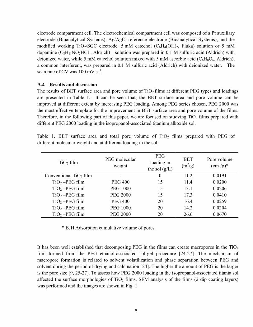
electrode compartment cell. The electrochemical compartment cell was composed of a Pt auxiliary electrode (Bioanalytical Systems), Ag/AgCl reference electrode (Bioanalytical Systems), and the modified working TiO2/SGC electrode. 5 mM catechol (C6H4(OH)2, Fluka) solution or 5 mM dopamine (C8H11NO2HCL, Aldrich) solution was prepared in 0.1 M sulfuric acid (Aldrich) with deionized water, while 5 mM catechol solution mixed with 5 mM ascorbic acid (C6H8O6, Aldrich), a common interferent, was prepared in 0.1 M sulfuric acid (Aldrich) with deionized water. The scan rate of CV was 100 mV s−1. A.4 Results and discussion The results of BET surface area and pore volume of TiO2 films at different PEG types and loadings are presented in Table 1. It can be seen that, the BET surface area and pore volume can be improved at different extent by increasing PEG loading. Among PEG series chosen, PEG 2000 was the most effective template for the improvement in BET surface area and pore volume of the films. Therefore, in the following part of this paper, we are focused on studying TiO2 films prepared with different PEG 2000 loading in the isopropanol-associated titanium alkoxide sol. Table 1. BET surface area and total pore volume of TiO2 films prepared with PEG of different molecular weight and at different loading in the sol.
TiO2 film PEG molecular weight
PEG loading in
the sol (g/L)
BET (m2/g)
Pore volume (cm3/g)*
Conventional TiO2 film - 0 11.2 0.0191 TiO2 –PEG film PEG 400 15 11.4 0.0200 TiO2 –PEG film PEG 1000 15 13.1 0.0206 TiO2 –PEG film PEG 2000 15 17.3 0.0410 TiO2 –PEG film PEG 400 20 16.4 0.0259 TiO2 –PEG film PEG 1000 20 14.2 0.0204 TiO2 –PEG film PEG 2000 20 26.6 0.0670
* BJH Adsorption cumulative volume of pores.
It has been well established that decomposing PEG in the films can create macropores in the TiO2 film formed from the PEG ethanol-associated sol-gel procedure [24-27]. The mechanism of macropore formation is related to solvent volatilization and phase separation between PEG and solvent during the period of drying and calcination [24]. The higher the amount of PEG is the larger is the pore size [9, 25-27]. To assess how PEG 2000 loading in the isopropanol-associated titania sol affected the surface morphologies of TiO2 films, SEM analysis of the films (2 dip coating layers) was performed and the images are shown in Fig. 1.
8
(a) (b)
(c) (d)
(a) (b)
(c) (d)
1 µm 1 µm
1 µm 1 µm
(a) (b)
(c) (d)
(a) (b)
(c) (d)
1 µm 1 µm
1 µm 1 µm
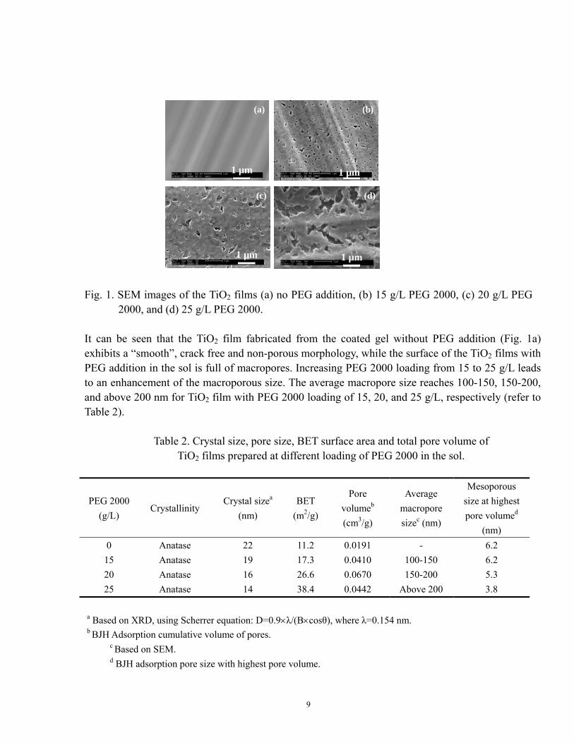
Fig. 1. SEM images of the TiO2 films (a) no PEG addition, (b) 15 g/L PEG 2000, (c) 20 g/L PEG
2000, and (d) 25 g/L PEG 2000. It can be seen that the TiO2 film fabricated from the coated gel without PEG addition (Fig. 1a) exhibits a “smooth”, crack free and non-porous morphology, while the surface of the TiO2 films with PEG addition in the sol is full of macropores. Increasing PEG 2000 loading from 15 to 25 g/L leads to an enhancement of the macroporous size. The average macropore size reaches 100-150, 150-200, and above 200 nm for TiO2 film with PEG 2000 loading of 15, 20, and 25 g/L, respectively (refer to Table 2).
Table 2. Crystal size, pore size, BET surface area and total pore volume of TiO2 films prepared at different loading of PEG 2000 in the sol.
PEG 2000 (g/L)
Crystallinity
Crystal sizea
(nm) BET
(m2/g)
Pore volumeb (cm3/g)
Average macropore sizec (nm)
Mesoporous size at highest pore volumed
(nm) 0 Anatase 22 11.2 0.0191 - 6.2
15 Anatase 19 17.3 0.0410 100-150 6.2 20 Anatase 16 26.6 0.0670 150-200 5.3 25 Anatase 14 38.4 0.0442 Above 200 3.8
a Based on XRD, using Scherrer equation: D=0.9×λ/(B×cosθ), where λ=0.154 nm. b BJH Adsorption cumulative volume of pores.
c Based on SEM. d BJH adsorption pore size with highest pore volume.
9
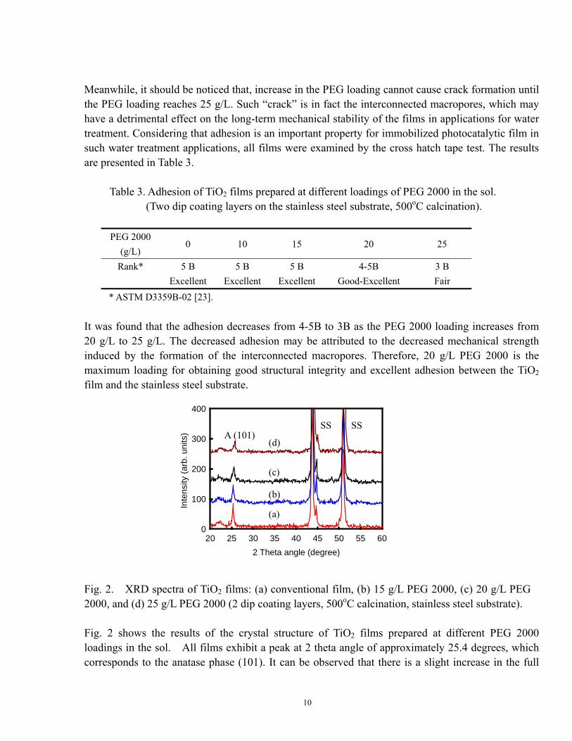
Meanwhile, it should be noticed that, increase in the PEG loading cannot cause crack formation until the PEG loading reaches 25 g/L. Such “crack” is in fact the interconnected macropores, which may have a detrimental effect on the long-term mechanical stability of the films in applications for water treatment. Considering that adhesion is an important property for immobilized photocatalytic film in such water treatment applications, all films were examined by the cross hatch tape test. The results are presented in Table 3. Table 3. Adhesion of TiO2 films prepared at different loadings of PEG 2000 in the sol. (Two dip coating layers on the stainless steel substrate, 500oC calcination).
PEG 2000 (g/L)
0 10 15 20 25
Rank* 5 B 5 B 5 B 4-5B 3 B Excellent Excellent Excellent Good-Excellent Fair
* ASTM D3359B-02 [23]. It was found that the adhesion decreases from 4-5B to 3B as the PEG 2000 loading increases from 20 g/L to 25 g/L. The decreased adhesion may be attributed to the decreased mechanical strength induced by the formation of the interconnected macropores. Therefore, 20 g/L PEG 2000 is the maximum loading for obtaining good structural integrity and excellent adhesion between the TiO2 film and the stainless steel substrate.
2 Theta angle (degree)20 25 30 35 40 45 50 55 60
Inte
nsity
(arb
. uni
ts)
0
100
200
300
400
A (101)
(a)
(b)
(c)
(d)
SS SS
2 Theta angle (degree)20 25 30 35 40 45 50 55 60
Inte
nsity
(arb
. uni
ts)
0
100
200
300
400
A (101)
(a)
(b)
(c)
(d)
SS SS
Fig. 2. XRD spectra of TiO2 films: (a) conventional film, (b) 15 g/L PEG 2000, (c) 20 g/L PEG 2000, and (d) 25 g/L PEG 2000 (2 dip coating layers, 500oC calcination, stainless steel substrate). Fig. 2 shows the results of the crystal structure of TiO2 films prepared at different PEG 2000 loadings in the sol. All films exhibit a peak at 2 theta angle of approximately 25.4 degrees, which corresponds to the anatase phase (101). It can be observed that there is a slight increase in the full
10
peak width at half maximum intensity, which suggests the increasing PEG 2000 loading could cause a decrease in the crystal size of the films. In order to check if the coated films have been better crystallized and to obtain information on the interparticle pore shape, these films were also examined by HR-TEM with Selected Area Diffraction (SAD). The results showed that all films had been well crystallized.
Slit-shaped pore
20 nm
Slit-shaped pore
20 nm
(a) (b)
Mesopore
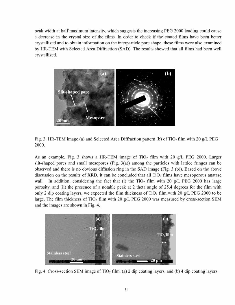
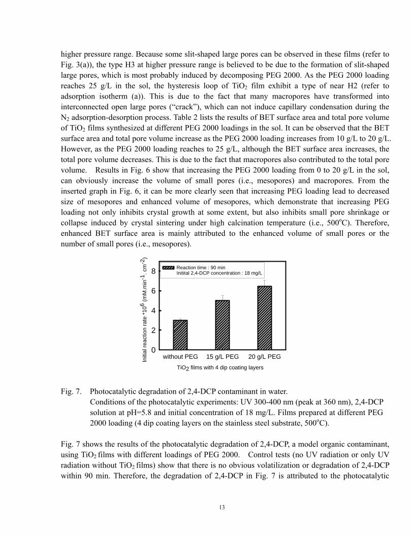
Fig. 3. HR-TEM image (a) and Selected Area Diffraction pattern (b) of TiO2 film with 20 g/L PEG 2000. As an example, Fig. 3 shows a HR-TEM image of TiO2 film with 20 g/L PEG 2000. Larger slit-shaped pores and small mesopores (Fig. 3(a)) among the particles with lattice fringes can be observed and there is no obvious diffusion ring in the SAD image (Fig. 3 (b)). Based on the above discussion on the results of XRD, it can be concluded that all TiO2 films have mesoporous anatase wall. In addition, considering the fact that (i) the TiO2 film with 20 g/L PEG 2000 has large porosity, and (ii) the presence of a notable peak at 2 theta angle of 25.4 degrees for the film with only 2 dip coating layers, we expected the film thickness of TiO2 film with 20 g/L PEG 2000 to be large. The film thickness of TiO2 film with 20 g/L PEG 2000 was measured by cross-section SEM and the images are shown in Fig. 4.
20 µm 20 µm
(a) (b)
Stainless steel Stainless steel
TiO2 filmTiO2 film
20 µm 20 µm
(a) (b)
Stainless steel Stainless steel
TiO2 filmTiO2 film
Fig. 4. Cross-section SEM image of TiO2 film. (a) 2 dip coating layers, and (b) 4 dip coating layers.
11

The results show that the film thickness is large, reaching approximately 3 μm (1 dip coating layer). It has been reported that PEG had some positive effects on the film thickness [28]. Therefore, formation of such thick films may be correlated to several factors, including isopropanol solvent, PEG 2000 and the stainless steel substrate.
P/Po0.0 .2 .4 .6 .8 1.0
Ads
orpt
ed v
olum
e (c
m3 /g
, STP
)
0
10
20
30
40
(a)
(b)
(c)
(d)
P/Po0.0 .2 .4 .6 .8 1.0
Ads
orpt
ed v
olum
e (c
m3 /g
, STP
)
0
10
20
30
40
(a)
(b)
(c)
(d)
Fig. 5. Nitrogen adsorption and desorption isotherms of TiO2 films prepared at: (a) 25 g/L PEG 2000, (b) 20 g/L PEG 2000, (c) 15 g/L PEG 2000, and (d) conventional TiO2 film (no PEG added).
Pore width (nm)0 50 100 150
Por
e vo
lum
e (c
m3 /
g)
0.00
.05
.10
.15
.20
Pore width (nm)4 8 12
Pore
vol
ume
(cm
3 /g)
0.00
.04
.08
.12 (a)(b)
(c)(d)
(a)
(b)(c)
(d)
Pore width (nm)0 50 100 150
Por
e vo
lum
e (c
m3 /
g)
0.00
.05
.10
.15
.20
Pore width (nm)4 8 12
Pore
vol
ume
(cm
3 /g)
0.00
.04
.08
.12 (a)(b)
(c)(d)
(a)
(b)(c)
(d)
Fig. 6. Pore size distribution curves of TiO2 films prepared at different PEG loadings. (a) 25 g/L PEG 2000, (b) 20 g/L PEG 2000, (c) 15 g/L PEG 2000, and (d) no PEG addition.
Figs. 5 and 6 show respectively (i) nitrogen adsorption and desorption isotherms, and (ii) pore size distribution of TiO2 films prepared at different PEG 2000 loading. From Fig. 5, it can be seen that all samples are of type IV (IUPAC classification). The hysteresis loop of TiO2 film without PEG 2000 addition only shows type H2, while the hysteresis loop of TiO2 films with PEG 2000 loading of 15 g/L and 20 g/L in the sol shows type H2 at relatively lower pressure range and type H3 at relatively
12
higher pressure range. Because some slit-shaped large pores can be observed in these films (refer to Fig. 3(a)), the type H3 at higher pressure range is believed to be due to the formation of slit-shaped large pores, which is most probably induced by decomposing PEG 2000. As the PEG 2000 loading reaches 25 g/L in the sol, the hysteresis loop of TiO2 film exhibit a type of near H2 (refer to adsorption isotherm (a)). This is due to the fact that many macropores have transformed into interconnected open large pores (“crack”), which can not induce capillary condensation during the N2 adsorption-desorption process. Table 2 lists the results of BET surface area and total pore volume of TiO2 films synthesized at different PEG 2000 loadings in the sol. It can be observed that the BET surface area and total pore volume increase as the PEG 2000 loading increases from 10 g/L to 20 g/L. However, as the PEG 2000 loading reaches to 25 g/L, although the BET surface area increases, the total pore volume decreases. This is due to the fact that macropores also contributed to the total pore volume. Results in Fig. 6 show that increasing the PEG 2000 loading from 0 to 20 g/L in the sol, can obviously increase the volume of small pores (i.e., mesopores) and macropores. From the inserted graph in Fig. 6, it can be more clearly seen that increasing PEG loading lead to decreased size of mesopores and enhanced volume of mesopores, which demonstrate that increasing PEG loading not only inhibits crystal growth at some extent, but also inhibits small pore shrinkage or collapse induced by crystal sintering under high calcination temperature (i.e., 500oC). Therefore, enhanced BET surface area is mainly attributed to the enhanced volume of small pores or the number of small pores (i.e., mesopores).
TiO2 films with 4 dip coating layers
without PEG 15 g/L PEG 20 g/L PEGIniti
al re
actio
n ra
te *1
06 (m
M.m
in-1
. cm
-2)
0
2
4
6
8 Reaction time : 90 minInitital 2,4-DCP concentration : 18 mg/L
Fig. 7. Photocatalytic degradation of 2,4-DCP contaminant in water.
Conditions of the photocatalytic experiments: UV 300-400 nm (peak at 360 nm), 2,4-DCP solution at pH=5.8 and initial concentration of 18 mg/L. Films prepared at different PEG 2000 loading (4 dip coating layers on the stainless steel substrate, 500oC).
Fig. 7 shows the results of the photocatalytic degradation of 2,4-DCP, a model organic contaminant, using TiO2 films with different loadings of PEG 2000. Control tests (no UV radiation or only UV radiation without TiO2 films) show that there is no obvious volatilization or degradation of 2,4-DCP within 90 min. Therefore, the degradation of 2,4-DCP in Fig. 7 is attributed to the photocatalytic
13

reaction of TiO2 films. The results clearly show that increasing PEG loading leads to an enhancement in photocatalytic activity of TiO2 films. This can be explained by the increase in surface area and pore volume. When PEG 2000 loading reaches 20 g/L, the initial reaction rate of photocatalytic degradation 2,4-DCP in macroporous TiO2 film can reach 6.43 ×10⁻⁶ mM min-1 cm-2, which is above two times that of TiO2 film without PEG. Considering that the macroporous TiO2 film with 20 g/L PEG 2000 has the highest photocatalytic activity and good mechanical properties, this type of thick film is considered to be a promising photocatalyst suitable for the destruction of organic contaminants in water, especially under conditions of low corrosivity. The concept of electrode modification originated over three decades ago with work of several research groups. Considering this fact, our research group has developed a titanium dioxide modified carbon electrode to be a promising electrochemical sensor for the detection of catecholamines, a class of neurotransmitters in the neuroscience field [15]. In this study, the modified TiO2 films are coated on the tip of sonogel carbon electrode, in order to evaluate their electrochemical response for the detection of catechol in the presence of ascorbic acid, a common interferent.
Potential, E (V)0.0.2.4.6.8
Cur
rent
, i (
Fig. 8. Cyclic voltammograms of sonogel carbon electrode (a) 5 mM catechol , 2 scans; (b) 5 mM catechol, 20 scans; (c) 5 mM catechol in the presence of 5mM ascorbic acid, 2 scans; (d) 5 mM catechol in the presence of 5mM ascorbic acid, 20 scans; scan rate 100mV/s, electrolyte 0.1 M sulfuric acid.
μA)
-100-80-60-40-20
6080
02040
Col 1 vs Col 3
Potential, E (V)0.0.2.4.6.8
Cur
rent
, i (μ
A)
-100-80-60-40-20
020406080
Potential, E (V)0.0.2.4.6.8
Cur
rent
, i ( μ
Α)
-100-80-60-40-20
020406080
Potential, E (V)0.0.2.4.6.8
Cur
rent
, i (μ
A)
-100-80-60-40-20
020406080
Epc Catechol E Catechol
Epc Catechol Epc Catechol
pc
Epa Catechol Epa Catechol
Epa Catechol Epa Catechol
(a) (b)
(c) (d)
Epa Ascorbic acid Epa Ascorbic acid
Potential, E (V)0.0.2.4.6.8
Cur
rent
, i ( μ
A)
-100-80-60-40-20
6080
02040
Col 1 vs Col 3
Potential, E (V)0.0.2.4.6.8
Cur
rent
, i (μ
A)
-100-80-60-40-20
020406080
Potential, E (V)0.0.2.4.6.8
Cur
rent
, i ( μ
Α)
-100-80-60-40-20
020406080
Potential, E (V)0.0.2.4.6.8
Cur
rent
, i (μ
A)
-100-80-60-40-20
020406080
Epc Catechol Epc Catechol
Epc Catechol Epc Catechol
Epa Catechol Epa Catechol
Epa Catechol Epa Catechol
(a) (b)
(c) (d)
Epa Ascorbic acid Epa Ascorbic acid
14
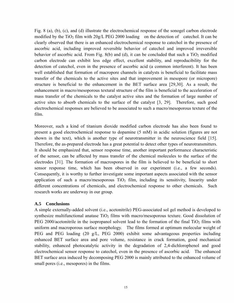
Fig. 8 (a), (b), (c), and (d) illustrate the electrochemical response of the sonogel carbon electrode modified by the TiO2 film with 20g/L PEG 2000 loading on the detection of catechol. It can be clearly observed that there is an enhanced electrochemical response to catechol in the presence of ascorbic acid, including improved reversible behavior of catechol and improved irreversible behavior of ascorbic acid. From Fig. 8(b) and (d), it can be concluded that such a TiO2 modified carbon electrode can exhibit less edge effect, excellent stability, and reproducibility for the detection of catechol, even in the presence of ascorbic acid (a common interferent). It has been well established that formation of macropore channels in catalysts is beneficial to facilitate mass transfer of the chemicals to the active sites and that improvement in mesopore (or micropore) structure is beneficial to the enhancement in the BET surface area [29,30]. As a result, the enhancement in macro/mesoporous textural structure of the film is beneficial to the acceleration of mass transfer of the chemicals to the catalyst active sites and the formation of large number of active sites to absorb chemicals to the surface of the catalyst [3, 29]. Therefore, such good electrochemical responses are believed to be associated to such a macro/mesoporous texture of the film. Moreover, such a kind of titanium dioxide modified carbon electrode has also been found to present a good electrochemical response to dopamine (5 mM) in acidic solution (figures are not shown in the text), which is another type of neurotransmitter in the neuroscience field [15]. Therefore, the as-prepared electrode has a great potential to detect other types of neurotransmitters. It should be emphasized that, sensor response time, another important performance characteristic of the sensor, can be affected by mass transfer of the chemical molecules to the surface of the electrodes [31]. The formation of macrospores in the film is believed to be beneficial to short sensor response time, which has been observed in our experiment (i.e., a few seconds). Consequently, it is worthy to further investigate some important aspects associated with the sensor application of such a macro/mesoporous TiO2 film, including its sensitivity, linearity under different concentrations of chemicals, and electrochemical response to other chemicals. Such research works are underway in our group.
A.5 Conclusions A simple externally-added solvent (i.e., acetonitrile) PEG-associated sol gel method is developed to synthesize multifunctional anatase TiO2 films with macro/mesoporous texture. Good dissolution of PEG 2000/acetonitrile in the isopropanol solvent lead to the formation of the final TiO2 films with uniform and macroporous surface morphology. The films formed at optimum molecular weight of PEG and PEG loading (20 g/L, PEG 2000) exhibit some advantageous properties including enhanced BET surface area and pore volume, resistance in crack formation, good mechanical stability, enhanced photocatalytic activity in the degradation of 2,4-dichlorophenol and good electrochemical sensor response to catechol, even in the presence of ascorbic acid. The enhanced BET surface area induced by decomposing PEG 2000 is mainly attributed to the enhanced volume of small pores (i.e., mesopores) in the films.
15
A.6 References
[1] J. C. Yu, L. Zhang, J. Yu, Chem. Mater. 14 (2002), p. 4647.
[2] J. C. Yu, J. G. Yu, J. C. Zhao, Appl. Catal. B: Environ. 36 (2002), p. 31
[3] Y. J. Chen, D. D. Dionysiou, Appl. Catal. A: General. 317 (2007), p. 129.
[4] Y. J. Chen, E. Stathatos, D. D. Dionysiou, Sur. Coat. Technol. 202 (2008), p.1944.
[5] H. Choi, E. Stathatos and D. D. Dionysiou, Appl. Catal. B: Environ. 63 (2006), p. 60.
[6] G. Lei, M. H. Xu, J. Sol-Gel Sci. Technol. 43 (2007), p. 1.
[7] T. Kitamura, H. Kumazawa. Chem. Eng. Comm. 192 (2005), p. 795.
[8] B. Guo, Z. L. Liu, L. Hong, H. X. Jiang, J. Y. Lee, Thin Solid Films 479 (2005), p. 310.
[9] B. Guo, Z. l. Liu, L. Hong, H. X. Jiang. Surf. Coat. Technol. 198 (2005), p. 24.
[10] Y. Kotani, T. Matoda, A. Matsuda, T. Kogure, M. Tatsumisago, T. Minami, J. Mater.
Chem. 11 (2001), p. 2045.
[11] J. G. Yu, X. J. Zhao, Q. N. Zhao, Thin Solid Films 379 (2000), p. 7.
[12] N. Nobuaki, T. Koji, I. Takashi, J. Sol-Gel Sci. Technol. 13 (1998), p. 691.
[13] X. B. Chen, S. S. Mao, Chem. Rev. 107 (2007), p. 2891.
[14] K. D. Benkstein, S. Semancik. Sens. Actuators, B, 113 (2006), p. 445.
[15] S. K. Lunsford, H. Choi, J. Stinson, A. Yeary, D. D. Dionysiou, Talanta, 73 (2007), p.
172.
[16] M. Okuya, K. Nakade, S. Kaneko, Sol. Energy Mater. Sol. Cells 70, (2002), p. 425.
[17] E. Stathatos, P. Lianos, J. Nanosci. Nanotechnol. 7 (2007), p. 555.
[18] E. Stathatos , P. Lianos, C. Tsakiroglou, Microporous Mesoporous Mater. 75 (2004), p.
255.
[19] M. C. Fuertes and G. J. A. A. Soler-Illia. Chem. Mater. 18 (2006) p. 2109.
[20] G. J. A. A. Soler-Illia, C. Sanchez, B. Lebeau, J. Patarin, Chem. Rev. 102 (2002), p. 4093.
[21] Y. Takahashi, Y. Matsuoka, J. Mater. Sci. 23 (1988), p. 2259.
[22] Y. J. Chen, D. D. Dionysiou, Appl. Catal. B: Environ. 69 (2006), p.24.
[23] American Society for Testing and Materials, ASTM, West Conshohocken, PA, D-3359-02
cross-cut tape test for adhesion.
[24] S. J. Bu, Z. G. Jin, X. X. Liu, L. R. Yang, Z. J. Cheng, J. Sol–Gel Sci. Technol. 30 (2004), p.
239.
[25] K. Kato, A. Tsuzuki, Y. Torii, H. Taoda, T. Kato, Y. Butsugan, J. Mater. Sci. 30 (1995), p.
16
837.
[26] J. G. Yu, X. I. Zhao, J. C. Du, W. M. Chen. J. Sol–Gel Sci. Technol. 17 (2000), p. 163.
[27] S. J. Bu, Z. G. Jin, X. X. Liu, L. R. Yang, Z. J. Cheng, J. Sol–Gel Sci. Technol. 30 (2004), p. 239.
[28] T. Miki, K. Nishizawa, K. Suzuki, K. Kato. J. Mater. Sci. 39 (2004), p. 699.
[29] X. C. Wang, J. C. Yu, C. M. Ho, Y.D. Hou, X. Z. Fu. Langmuir 21 (2005), p. 2552.
[30] Y. Zhang, M. Koike, N. Tsubaki. Catal. Letters. 91 (2005), p.193.
[31] P. R. Warburton, M. P. Pagano, R. Hoover, M. Logman, K. Crytzer, Y. J. Warburton. Anal. Chem., 70 (1998), p. 998.
17
Part B: Characterization and electrochemical response of sonogel carbon
electrode modified with nanostructured zirconium dioxide
B.1 Abstract This part reports on a sonogel carbon electrode modified by zirconium dioxide (ZrO2) film crystallized from a zirconium acetylacetonate precursor. The electrode materials including sol gel derived ZrO2 and carbon matrix are characterized using XRD, EDS, SEM, TEM, N2 adsorption and desorption. It was found that as-prepared ZrO2 film has a relatively high BET surface area (69 m2/g), tetragonal crystalline phase with crystalline size of ~15 nm and mesoporous structure with average pore size of ~2.6 nm, while graphite carbon matrix has mainly microporous structure with relative high total porosity (~0.04 cm3/g) which is beneficial to good adhesion between ZrO2 film and carbon matrix. The results of electrochemical characterization showed that the presence of ZrO2 film leads to a good electrochemical response to catechol and dopamine, including less edge effect, excellent stability, and reproducibility, even in the presence of ascorbic acid (a common interferent). This study suggested that zirconium dioxide film prepared by such a wet chemistry route is a kind of promising inorganic metal oxide for the sonogel carbon based electrode in the detection of neurotransmitters. B.2 Introduction Recently, a sonogel carbon electrode modified by nanostructured TiO2 films, a new class of sensor, has been successfully developed in our group, which is associated with employing sol gel technology to coat TiO2 film on the top of sonogel graphite carbon electrode. The modified electrode has been proved to be a promising sensor to detect neurotransmitters, such as catechol and dopamine. Since there are many challenging requirements to guarantee safety, reproducibility and reliability in the application of sensors, including sensitivity, selectivity and stability, it is necessary to further optimize our previous sensor fabrication technology. In this part of the study, we report a new sonogel carbon electrode modified by ZrO2 film. The advantage for employing ZrO2 is associated to its super hardness, which can enhance electrode endurance, especially at extreme conditions. B.3 Experimental part B.3.1 Synthesis of ZiO2 films Zirconium (IV) propoxide solution 70 wt. % in 1-propanol (Aldrich,), ethanol (200 proof, Aldrich), acetylacetone (99%, Fisher Scientific) and deionized water were used for the preparation of the zirconium sol. The molar ratio of zirconium(IV) propoxide : water : acetylacetone : ethanol was 2:4:1:62. The final zirconium propoxide concentration was 0.43 M. The materials and method to prepare sonogel carbon (SGC) electrode was the same as that of part A. The ZrO2 gel was coated
18
on the tip of SGC electrodes by dipping carbon electrode into the sol and taken out. The relative humidity and room temperature in the lab were ~50% and 23 oC, respectively. After the sols had been coated on the top of carbon electrodes, the coated carbon electrodes were calcined in a programmable high temperature furnace. The furnace temperature was incremented at a ramp rate of 15°C min-1 to 100 oC and hold for 15 min, then continued to increase at a ramp rate of 3.0 °C min-1 until 500 oC and was held at this value for 30 min. Finally, the films were cooled naturally to room temperature. B.3.2 Characterization of ZiO2 films and sonogel carbon material The crystal phase composition of the ZiO2 coated on sonogel carbon material was determined by X-ray diffraction (XRD) using a Siemens Kristalloflex D500 diffractometer with Cu Kα radiation. Powder samples used for XRD and TEM analysis were obtained from calcined sonogel graphite carbon powder mixed with zirconium sol (about 3 ml zirconium sol to impregnate 1 g sonogel graphite carbon powders). The heat treatment (calcination) procedure was the same as the above. Morphology on sonogel carbon material was characterized by an environmental scanning electron microscope (ESEM, Philips XL 30 ESEM-FEG) with accelerating voltage of 10 K. The crystal and pore morphology of the films were determined by a JEM-2010F (JEOL) High Resolution-Transmission Electron Microscope (HR-TEM) with field emission gun at 200 kV. All powder samples were dispersed in methanol (High Performance Liquid Chromatography (HPLC) grade, Pharmco) using an ultrasonic cleaner (2510R-DH, Bransonic) for 5 min and fixed on a carbon-coated copper grid (LC200-Cu, EMS). The specific surface area and pore volume of the films were measured using a Micromeritics TriStar 3000 Gas Adsorption Analyzer.
B.3.3 Electrochemical sensor response Electrochemical sensor response to catechol or dopamine was carried out using an Electrochemical Workstation (Epsilon, Bioanalytical Systems) based on Cyclic voltammetry (CV) with three electrode compartment cell. The electrochemical compartment cell was composed of a Pt auxiliary electrode (Bioanalytical Systems), Ag/AgCl reference electrode (Bioanalytical Systems), and the modified working TiO2/SGC electrode. 5 mM catechol (C6H4(OH)2, Fluka) solution or 5 mM dopamine (C8H11NO2HCL, Aldrich) solution was prepared in 0.1 M sulfuric acid (Aldrich) with deionized water, while 5 mM catechol solution mixed with 5 mM ascorbic acid (C6H8O6, Aldrich), a common interferent, was prepared in 0.1 M sulfuric acid (Aldrich) with deionized water. The scan rate of CV was 100 mV s−1.
19
B.4 Results and discussion
2 Theta angle (degree)
10 15 20 25 30 35 40 45 50 55 60 65 70
Inte
nsity
(arb
.uni
ts)
0
200
400
600
800
1000
1200
1400
(b)
(a)
� t-ZrO2 (011)
*Carbon(002)
*(004)*(100) *(101)
� (112)� (121)
2 Theta angle (degree)
10 15 20 25 30 35 40 45 50 55 60 65 70
Inte
nsity
(arb
.uni
ts)
0
200
400
600
800
1000
1200
1400
(b)
(a)
� t-ZrO2 (011)
*Carbon(002)
*(004)*(100) *(101)
� (112)� (121)
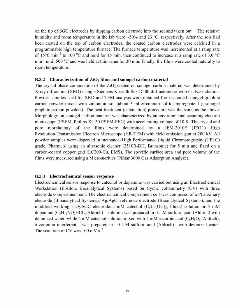
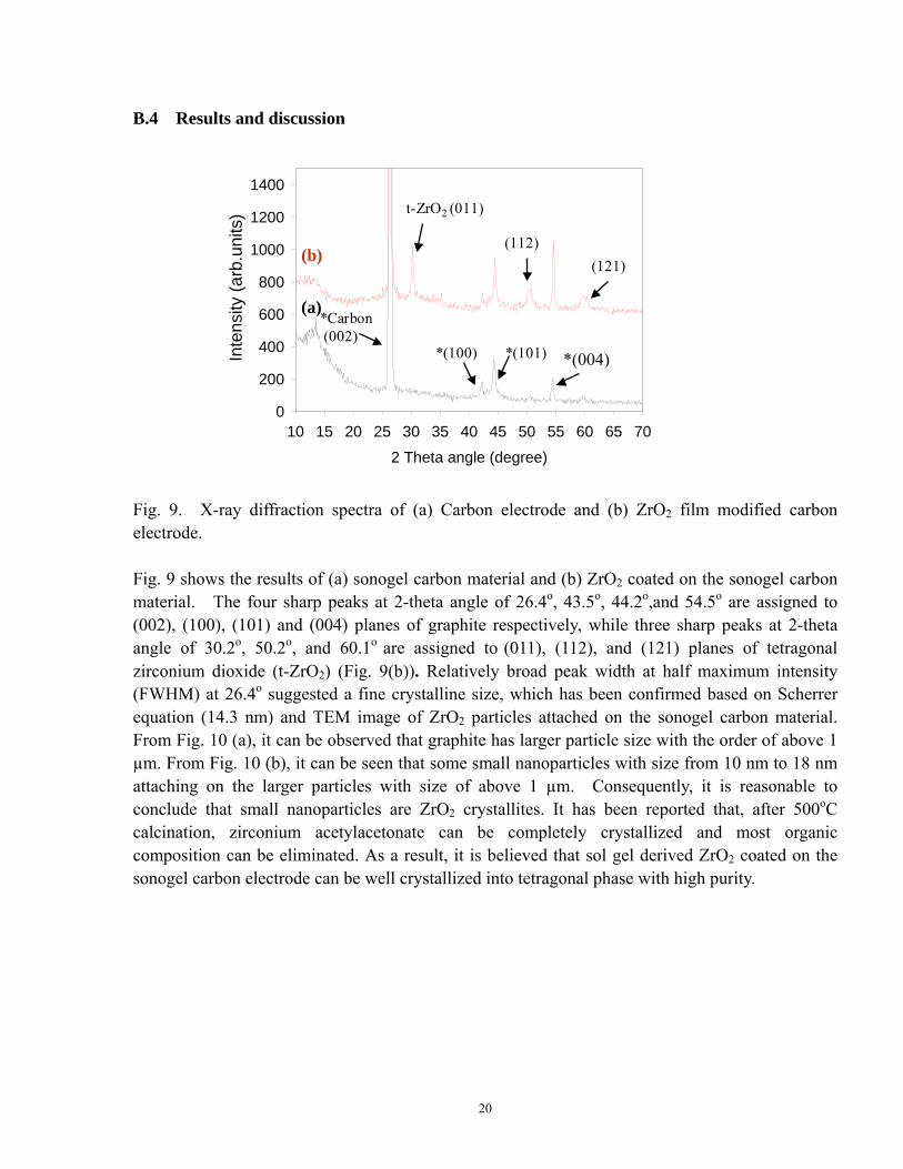
Fig. 9. X-ray diffraction spectra of (a) Carbon electrode and (b) ZrO2 film modified carbon electrode. Fig. 9 shows the results of (a) sonogel carbon material and (b) ZrO2 coated on the sonogel carbon material. The four sharp peaks at 2-theta angle of 26.4o, 43.5o, 44.2o,and 54.5o are assigned to (002), (100), (101) and (004) planes of graphite respectively, while three sharp peaks at 2-theta angle of 30.2o, 50.2o, and 60.1o are assigned to (011), (112), and (121) planes of tetragonal zirconium dioxide (t-ZrO2) (Fig. 9(b)). Relatively broad peak width at half maximum intensity (FWHM) at 26.4o suggested a fine crystalline size, which has been confirmed based on Scherrer equation (14.3 nm) and TEM image of ZrO2 particles attached on the sonogel carbon material. From Fig. 10 (a), it can be observed that graphite has larger particle size with the order of above 1 µm. From Fig. 10 (b), it can be seen that some small nanoparticles with size from 10 nm to 18 nm attaching on the larger particles with size of above 1 µm. Consequently, it is reasonable to conclude that small nanoparticles are ZrO2 crystallites. It has been reported that, after 500oC calcination, zirconium acetylacetonate can be completely crystallized and most organic composition can be eliminated. As a result, it is believed that sol gel derived ZrO2 coated on the sonogel carbon electrode can be well crystallized into tetragonal phase with high purity.
20
(a) (b)
(a) (b)
Fig. 10. TEM images of (a) carbon material and (b) ZrO2 material
(a) 1 dip layer (b) 2 dip layers(a) 1 dip layer (b) 2 dip layers
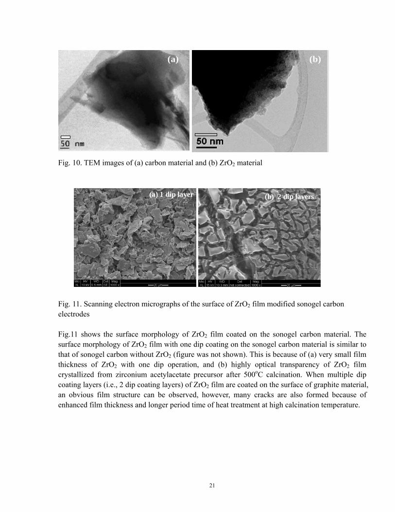
Fig. 11. Scanning electron micrographs of the surface of ZrO2 film modified sonogel carbon electrodes Fig.11 shows the surface morphology of ZrO2 film coated on the sonogel carbon material. The surface morphology of ZrO2 film with one dip coating on the sonogel carbon material is similar to that of sonogel carbon without ZrO2 (figure was not shown). This is because of (a) very small film thickness of ZrO2 with one dip operation, and (b) highly optical transparency of ZrO2 film crystallized from zirconium acetylacetate precursor after 500oC calcination. When multiple dip coating layers (i.e., 2 dip coating layers) of ZrO2 film are coated on the surface of graphite material, an obvious film structure can be observed, however, many cracks are also formed because of enhanced film thickness and longer period time of heat treatment at high calcination temperature.
21
Element Wt % At %
C K 57.53 78.96
O K 14.31 14.74
SiK 02.98 01.75
ZrL 25.18 04.55
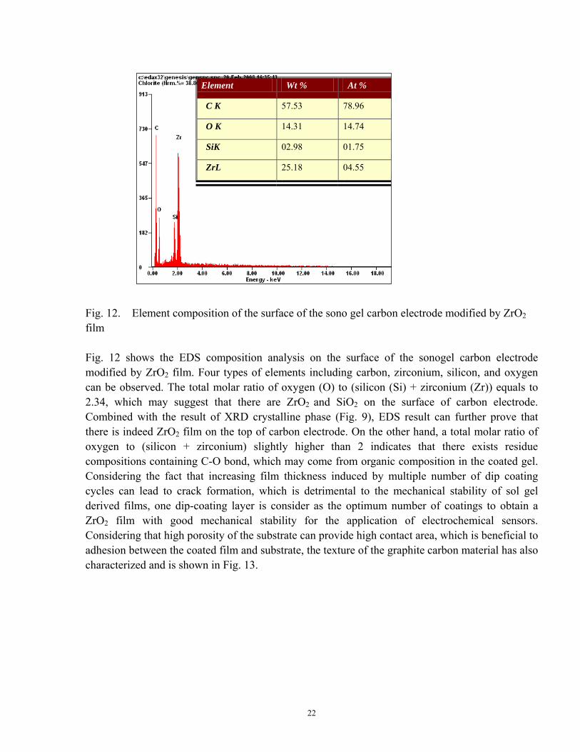
Fig. 12. Element composition of the surface of the sono gel carbon electrode modified by ZrO2 film Fig. 12 shows the EDS composition analysis on the surface of the sonogel carbon electrode modified by ZrO2 film. Four types of elements including carbon, zirconium, silicon, and oxygen can be observed. The total molar ratio of oxygen (O) to (silicon (Si) + zirconium (Zr)) equals to 2.34, which may suggest that there are ZrO2 and SiO2 on the surface of carbon electrode. Combined with the result of XRD crystalline phase (Fig. 9), EDS result can further prove that there is indeed ZrO2 film on the top of carbon electrode. On the other hand, a total molar ratio of oxygen to (silicon + zirconium) slightly higher than 2 indicates that there exists residue compositions containing C-O bond, which may come from organic composition in the coated gel. Considering the fact that increasing film thickness induced by multiple number of dip coating cycles can lead to crack formation, which is detrimental to the mechanical stability of sol gel derived films, one dip-coating layer is consider as the optimum number of coatings to obtain a ZrO2 film with good mechanical stability for the application of electrochemical sensors. Considering that high porosity of the substrate can provide high contact area, which is beneficial to adhesion between the coated film and substrate, the texture of the graphite carbon material has also characterized and is shown in Fig. 13.
22
Pore diameter (nm)0.0 2.5 5.0 7.5 10.0 12.5 15.0 17.5 20.0
Ads
orbe
d po
re v
olum
e (c
m3 /
g)
0.00.02.04.06.08.10.12.14
(a) sonogel carbon,500oC(b) ZrO2,500oC
(a)
(b)
Pore diameter (nm)0.0 2.5 5.0 7.5 10.0 12.5 15.0 17.5 20.0
Ads
orbe
d po
re v
olum
e (c
m3 /
g)
0.00.02.04.06.08.10.12.14
(a) sonogel carbon,500oC(b) ZrO2,500oC
(a)
(b)
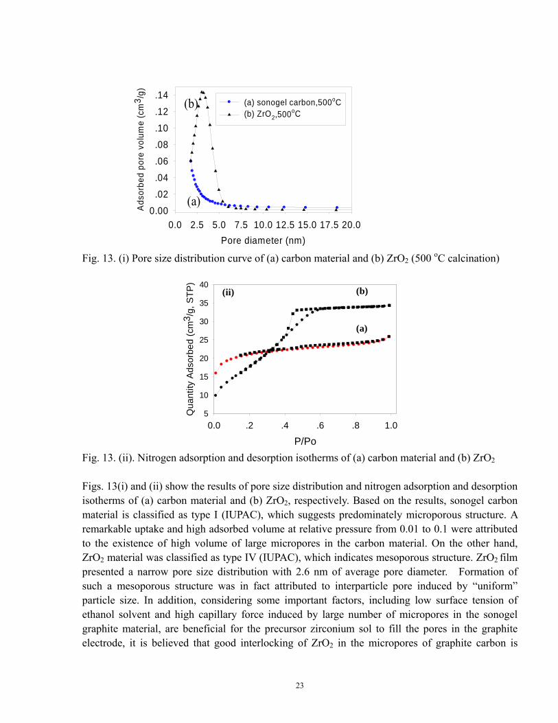
Fig. 13. (i) Pore size distribution curve of (a) carbon material and (b) ZrO2 (500 oC calcination)
P/Po0.0 .2 .4 .6 .8 1.0
Qua
ntity
Ads
orbe
d (c
m3 /
g, S
TP)
5
10
15
20
25
30
35
40
(a)
(b)
P/Po0.0 .2 .4 .6 .8 1.0
Qua
ntity
Ads
orbe
d (c
m3 /
g, S
TP)
5
10
15
20
25
30
35
40
(a)
(b)(ii)
Fig. 13. (ii). Nitrogen adsorption and desorption isotherms of (a) carbon material and (b) ZrO2
Figs. 13(i) and (ii) show the results of pore size distribution and nitrogen adsorption and desorption isotherms of (a) carbon material and (b) ZrO2, respectively. Based on the results, sonogel carbon material is classified as type I (IUPAC), which suggests predominately microporous structure. A remarkable uptake and high adsorbed volume at relative pressure from 0.01 to 0.1 were attributed to the existence of high volume of large micropores in the carbon material. On the other hand, ZrO2 material was classified as type IV (IUPAC), which indicates mesoporous structure. ZrO2 film presented a narrow pore size distribution with 2.6 nm of average pore diameter. Formation of such a mesoporous structure was in fact attributed to interparticle pore induced by “uniform” particle size. In addition, considering some important factors, including low surface tension of ethanol solvent and high capillary force induced by large number of micropores in the sonogel graphite material, are beneficial for the precursor zirconium sol to fill the pores in the graphite electrode, it is believed that good interlocking of ZrO2 in the micropores of graphite carbon is
23
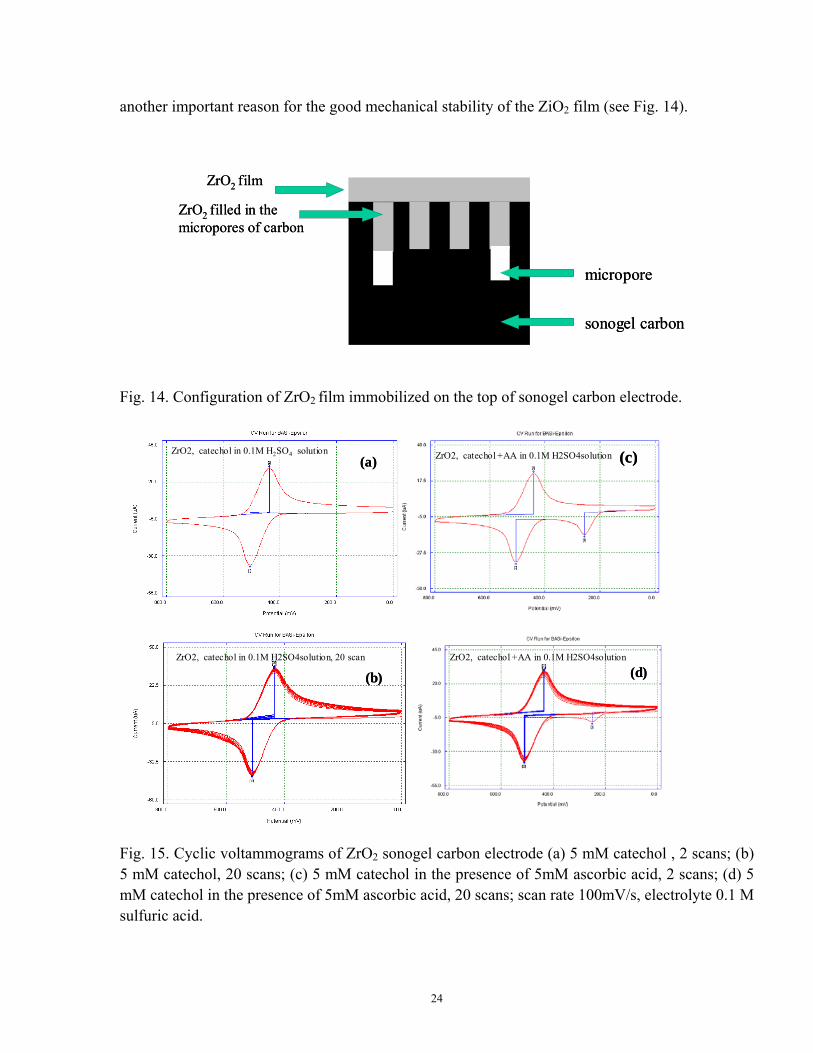
another important reason for the good mechanical stability of the ZiO2 film (see Fig. 14).
ZrO2 film
ZrO2 filled in the micropores of carbon
micropore
sonogel carbon
ZrO2 film
ZrO2 filled in the micropores of carbon
micropore
sonogel carbon
Fig. 14. Configuration of ZrO2 film immobilized on the top of sonogel carbon electrode.
ZrO2, catechol in 0.1M H2SO4 solution
ZrO2, catechol in 0.1M H2SO4solution, 20 scan
ZrO2, catechol +AA in 0.1M H2SO4solution
ZrO2, catechol +AA in 0.1M H2SO4solution
(a)
(b)
(c)
(d)
ZrO2, catechol in 0.1M H2SO4 solution
ZrO2, catechol in 0.1M H2SO4solution, 20 scan
ZrO2, catechol +AA in 0.1M H2SO4solution
ZrO2, catechol +AA in 0.1M H2SO4solution
(a)
(b)
(c)
(d)
(a)
(b)
(c)
(d)
Fig. 15. Cyclic voltammograms of ZrO2 sonogel carbon electrode (a) 5 mM catechol , 2 scans; (b) 5 mM catechol, 20 scans; (c) 5 mM catechol in the presence of 5mM ascorbic acid, 2 scans; (d) 5 mM catechol in the presence of 5mM ascorbic acid, 20 scans; scan rate 100mV/s, electrolyte 0.1 M sulfuric acid.
24

ZrO2 , Dopamine in 0.1 M H2SO4 solution, 2 scans ZrO2 , Dopamine in 0.1 M H2SO4 solution, 20 scansZrO2 , Dopamine in 0.1 M H2SO4 solution, 2 scans ZrO2 , Dopamine in 0.1 M H2SO4 solution, 20 scans
(a) (b)(a) (b)
Fig. 16. Cyclic voltammograms of ZrO2 sonogel carbon electrode (a) 5 mM dopamine, 2 scans; and (b) 5 mM catechol, 20 scans. Scan rate 100mV/s, electrolyte 0.1 M sulfuric acid. Fig. 15 shows the results of the electrochemical characterization of sonogel carbon electrode modified by zirconium dioxide for the detection of catechol. A good electrochemical response to catechol in the presence of ascorbic acid, such as less edge effect, excellent stability, and reproducibility, can be observed. In addition, from Fig. 16, it can be deduced that such an electrode has also a good electrochemical response to dopamine. Based on the above discussion on the properties of electrode materials, it is reasonable to conclude that good electrochemical behavior is mainly attributed to good adhesion between ZrO2 film and the graphite carbon matrix as well as to the formation of ZrO2 film with special microstructure, including high BET surface area (69 m2/g), and tetragonal crystal phase for high conductivity.
B.5 Conclusions The following conclusions were made from the study in part B: (1) Sol gel derived ZirO2 film formed from zirconium acetylacetone precursor proved to be a
promising inorganic material for good electrochemical response to catechol and dopamine. (2) After 500oC calcination, the as-prepared ZrO2 film has a tetragonal crystalline structure with
small crystal size of 15 nm and mesoporous film texture with narrow pore size of ~2.6 nm. (3) Graphite carbon material, as matrix, has mainly microporous structure with high total porosity
(~0.04 cm3/g), which is beneficial to good adhesion between sol gel derived inorganic films and carbon matrix.
25

Part C: Surfactant self-assembling sol gel synthesis of mesoporous ZrO2 and its
application as effective electrochemical sensor material C.1 Abstract Mesoporous ZrO2 film (M-ZrO2) was successfully synthesized using a surfactant self-assembling sol gel method, which involves the use of nonionic surfactant Tween 20 as template through a self assembly pathway. It was found that sono gel carbon electrode modified by this type of M-ZrO2 film could present high sensitivity and stability in the detection of neurotransmitters. C.2 Experimental details C.2.1 Synthesis of ZiO2 films Zirconium(IV) propoxide solution 70 wt. % in 1-propanol (Aldrich), ethanol (200 proof, Aldrich), Acetylacetone (99%, Fisher Scientific) and de-ionized water were used for the preparation of the zirconium sol. The molar ratio of zirconium(IV) propoxide : water : acetylacetone : ethanol was 2:4:1:62. Surfactant Tween 20 was mixed with zirconium sol with volume ratio of 1: 1. After vigorously stirring, a transparent and homogenous modified sol was formed. The materials and methods to prepare sonogel carbon (SGC) electrode were the same as those reported in Parts A and B. The ZrO2 gel was coated on the tip of SGC electrodes by dipping carbon electrode into the sol and taken out. For the synthesis of mesoporous ZrO2 film, Tween 20 was mixed with the zirconium sol with 1:1 (v/v). The relative humidity and room temperature in the lab were ~50% and 23 oC, respectively. After the sols had been coated on the top of carbon electrodes, the coated carbon electrodes were calcined in a programmable high temperature furnace. The furnace temperature was incremented at a ramp rate of 15°C min-1 to 100 oC and was hold for 15 min, then continued to increase at a ramp rate of 3.0 °C min-1 until 500 oC and was held at this value for 30 min. Finally, the films were cooled naturally to room temperature. C.2.2 Characterization of metal oxide films and sonogel carbon material The crystal phase composition of mesoporous ZrO2 coated on sonogel carbon material was determined by X-ray diffraction (XRD) using a Siemens Kristalloflex D500 diffractometer with Cu Kα radiation. Powder samples used for XRD and TEM analysis were obtained from calcined sono-gel graphite carbon powder mixed with zirconium sol (about 3 ml zirconium sol to impregnate 1 g sono-gel graphite carbon powders). The heat treatment (calcination) procedure was the same as that reported in Parts A and B. Film morphology on sonogel carbon material was characterized by an environmental scanning electron microscope (ESEM, Philips XL 30 ESEM-FEG) with accelerating voltage of 10 K. The crystal and pore morphology of the films were determined by a JEM-2010F (JEOL) High Resolution-Transmission Electron Microscope (HR-TEM) with field emission gun at 200 kV. All powder samples were dispersed in methanol (High Performance Liquid Chromatography (HPLC) grade, Pharmco) using an ultrasonic cleaner
26
(2510R-DH, Bransonic) for 5 min and fixed on a carbon-coated copper grid (LC200-Cu, EMS). The specific surface area and pore volume of the films were measured using a Micromeritics TriStar 3000 Gas Adsorption Analyzer.
C.2.3 Electrochemical sensor response Electrochemical sensor response to catechol or dopamine was carried out using an Electrochemical Workstation (Epsilon, Bioanalytical Systems) based on Cyclic voltammetry (CV) with three electrode compartment cell. The electrochemical compartment cell was composed of a Pt auxiliary electrode (Bioanalytical Systems), Ag/AgCl reference electrode (Bioanalytical Systems), and the modified working TiO2/SGC electrode. 5 mM catechol (C6H4(OH)2, Fluka) solution or 5 mM dopamine (C8H11NO2HCL, Aldrich) solution was prepared in 0.1 M sulfuric acid (Aldrich) with deionized water, while 5 mM catechol solution mixed with 5 mM ascorbic acid (C6H8O6, Aldrich), a common interferent, was prepared in 0.1 M sulfuric acid (Aldrich) with deionized water. The scan rate of CV was 100 mV s−1. The detection limit of catechol or dopamine was also determined. C.3 Results and Discussion
2 Theta Angle (Degree)10 15 20 25 30 35 40 45 50 55 60 65
Inte
nsity
( ar
b.un
its)
0
500
1000
1500
2000
*Carbon (002)
�t-ZrO2 (011)�(112)
�(121)
*(100)
*(101)
*(004)
2 Theta Angle (Degree)10 15 20 25 30 35 40 45 50 55 60 65
Inte
nsity
( ar
b.un
its)
0
500
1000
1500
2000
*Carbon (002)
�t-ZrO2 (011)�(112)
�(121)
*(100)
*(101)
*(004)
(b)
(a)
2 Theta Angle (Degree)10 15 20 25 30 35 40 45 50 55 60 65
Inte
nsity
( ar
b.un
its)
0
500
1000
1500
2000
*Carbon (002)
�t-ZrO2 (011)�(112)
�(121)
*(100)
*(101)
*(004)
2 Theta Angle (Degree)10 15 20 25 30 35 40 45 50 55 60 65
Inte
nsity
( ar
b.un
its)
0
500
1000
1500
2000
*Carbon (002)
�t-ZrO2 (011)�(112)
�(121)
*(100)
*(101)
*(004)
(b)
(a)
(b)
(a)
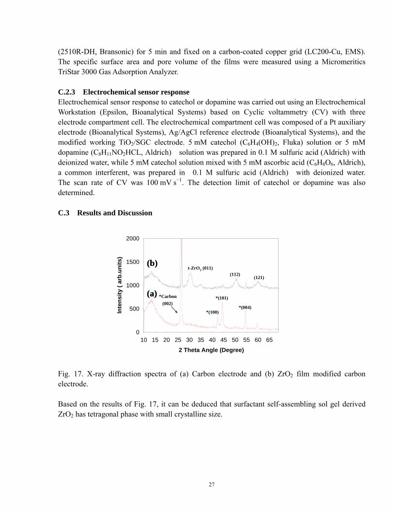
Fig. 17. X-ray diffraction spectra of (a) Carbon electrode and (b) ZrO2 film modified carbon electrode. Based on the results of Fig. 17, it can be deduced that surfactant self-assembling sol gel derived ZrO2 has tetragonal phase with small crystalline size.
27
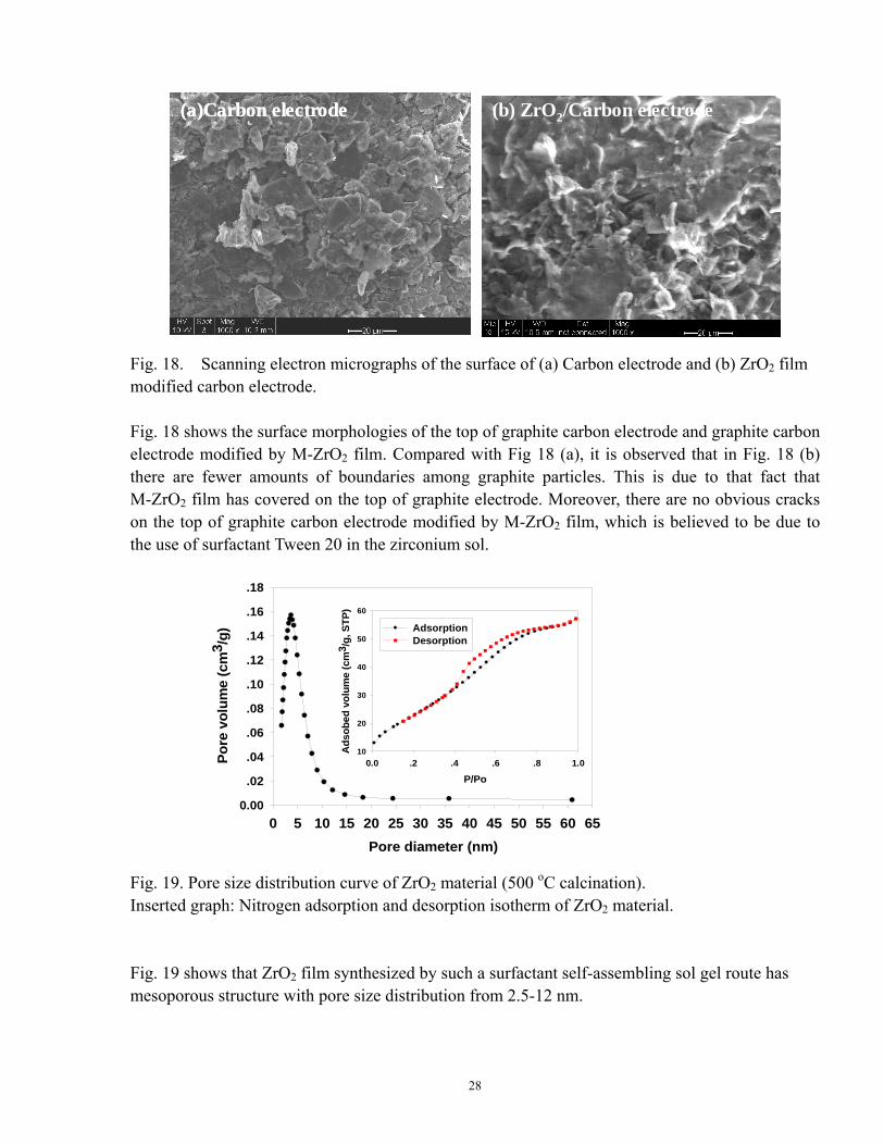
(a)Carbon electrode (b) ZrO2/Carbon electrode(a)Carbon electrode (b) ZrO2/Carbon electrode(a)Carbon electrode (b) ZrO2/Carbon electrode Fig. 18. Scanning electron micrographs of the surface of (a) Carbon electrode and (b) ZrO2 film modified carbon electrode. Fig. 18 shows the surface morphologies of the top of graphite carbon electrode and graphite carbon electrode modified by M-ZrO2 film. Compared with Fig 18 (a), it is observed that in Fig. 18 (b) there are fewer amounts of boundaries among graphite particles. This is due to that fact that M-ZrO2 film has covered on the top of graphite electrode. Moreover, there are no obvious cracks on the top of graphite carbon electrode modified by M-ZrO2 film, which is believed to be due to the use of surfactant Tween 20 in the zirconium sol.
Pore diameter (nm)0 5 10 15 20 25 30 35 40 45 50 55 60 65
Pore
vol
ume
(cm
3 /g)
0.00
.02
.04
.06
.08
.10
.12
.14
.16
.18
P/Po0.0 .2 .4 .6 .8 1.0
Ads
obed
vol
ume
(cm
3 /g,
STP
)
10
20
30
40
50
60
AdsorptionDesorption
Pore diameter (nm)0 5 10 15 20 25 30 35 40 45 50 55 60 65
Pore
vol
ume
(cm
3 /g)
0.00
.02
.04
.06
.08
.10
.12
.14
.16
.18
P/Po0.0 .2 .4 .6 .8 1.0
Ads
obed
vol
ume
(cm
3 /g,
STP
)
10
20
30
40
50
60
AdsorptionDesorption
Fig. 19. Pore size distribution curve of ZrO2 material (500 oC calcination). Inserted graph: Nitrogen adsorption and desorption isotherm of ZrO2 material. Fig. 19 shows that ZrO2 film synthesized by such a surfactant self-assembling sol gel route has mesoporous structure with pore size distribution from 2.5-12 nm.
28
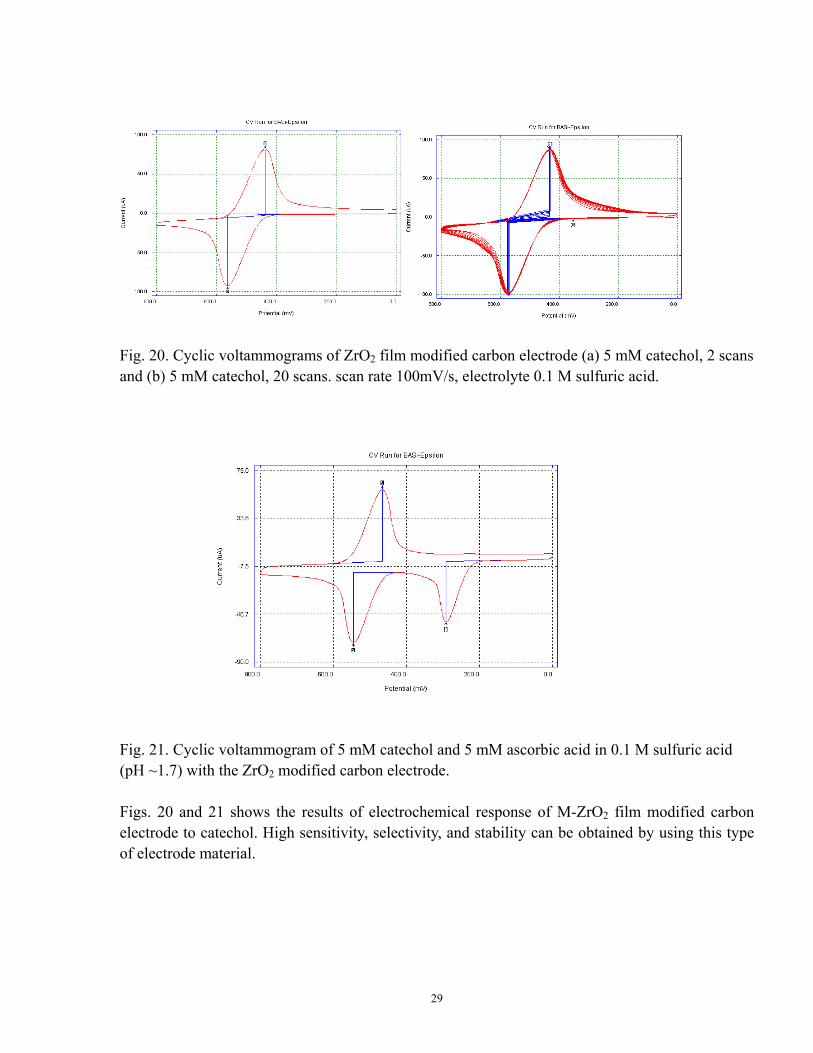
Fig. 20. Cyclic voltammograms of ZrO2 film modified carbon electrode (a) 5 mM catechol, 2 scans and (b) 5 mM catechol, 20 scans. scan rate 100mV/s, electrolyte 0.1 M sulfuric acid.
Fig. 21. Cyclic voltammogram of 5 mM catechol and 5 mM ascorbic acid in 0.1 M sulfuric acid (pH ~1.7) with the ZrO2 modified carbon electrode. Figs. 20 and 21 shows the results of electrochemical response of M-ZrO2 film modified carbon electrode to catechol. High sensitivity, selectivity, and stability can be obtained by using this type of electrode material.
29
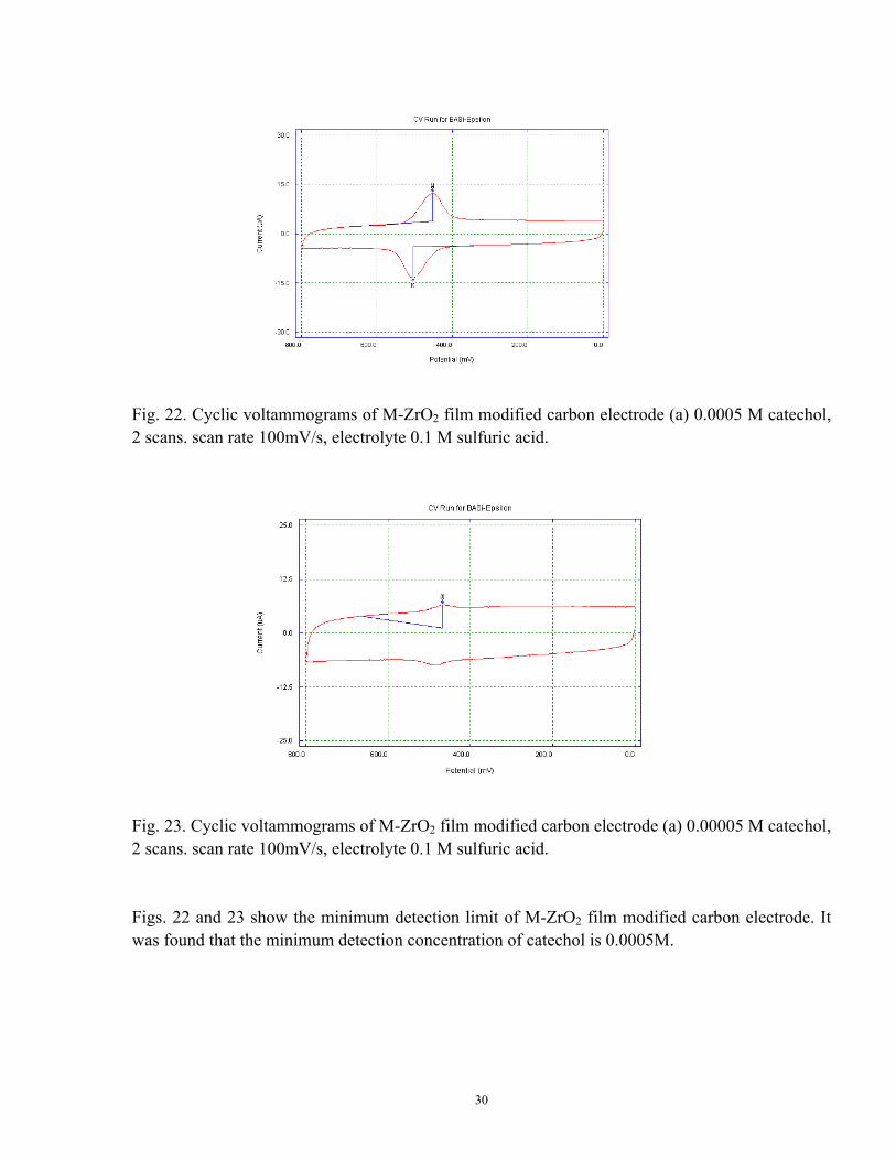
Fig. 22. Cyclic voltammograms of M-ZrO2 film modified carbon electrode (a) 0.0005 M catechol, 2 scans. scan rate 100mV/s, electrolyte 0.1 M sulfuric acid.
Fig. 23. Cyclic voltammograms of M-ZrO2 film modified carbon electrode (a) 0.00005 M catechol, 2 scans. scan rate 100mV/s, electrolyte 0.1 M sulfuric acid. Figs. 22 and 23 show the minimum detection limit of M-ZrO2 film modified carbon electrode. It was found that the minimum detection concentration of catechol is 0.0005M.
30

(a) (b)(a) (b)
Fig. 24. Cyclic voltammograms of ZrO2 film modified carbon electrode (a) 5 mM dopamine, 2 scans and (b) 5 mM dopamine, 20 scans. scan rate 100mV/s, electrolyte 0.1 M sulfuric acid.
Fig. 25. Cyclic voltammogram of 5 mM dopamine and 5 mM ascorbic acid in 0.1 M sulfuric acid (pH ~1.7) with the M- ZrO2 modified carbon electrode. Figs. 24 and 25 shows the results of electrochemical response of M-ZrO2 film modified carbon electrode to dopamine in 0.1 M sulfuric acid. It was found that such an electrode can also have good electrochemical response to dopamine.
31
Principal Findings and Significance A simple externally-added solvent (i.e., acetonitrile) PEG-associated sol gel method was developed to synthesize multifunctional anatase TiO2 films with macro/mesoporous texture. Good dissolution of PEG 2000/acetonitrile in isopropanol solvent lead to the formation of the final TiO2 films with uniform and macroporous surface morphology. The films formed at optimum molecular weight of PEG and PEG loading (20 g/L, PEG 2000) exhibit some advantageous properties including enhanced BET surface area and pore volume, resistance in crack formation, good mechanical stability, enhanced photocatalytic activity in the degradation of 2,4-dichlorophenol (2,4-DCP) and good electrochemical sensor response to catechol even in the presence of ascorbic acid. Sol gel derived ZrO2 film based on the zirconium acetylacetone precursor has been proved to be a promising inorganic material for good electrochemical response to catechol, even in the presence of ascorbic acid. After 500oC calcination, the as-prepared ZrO2 film has a tetragonal crystalline structure with small crystal size of 15 nm and mesoporous film texture with narrow pore size of ~2.6 nm. Surfactant self-assembling sol gel method has been proved to be a promising technique to synthesize mesoporous ZrO2 film with excellent electrochemical response to neurotransmitters (i.e., catechol), even in the presence of ascorbic acid Based on our studies, it can be concluded that film microstructure, such as crystalline structure and pore structure, is an important film property to affect the electrochemical behavior of the electrodes. Sol gel derived ZrO2 has been developed in this study, which has been proved to be a promising electrode material in the detection of organic molecules of concern. Further work is under way on (1) fabrication and optimization of nanostructured metal oxide modified carbon electrode with tailor-designed structural properties for facilitating adsorption of phenol and domoic acid onto the metal oxides and charge transfer between carbon electrode and the toxins, and (2) functionalization of metal oxide-modified carbon electrode with tyrosinase-chitosan composite film for enhancing the selectivity and sensitivity of the electrode towards phenol and domoic acid.
32