nanaji deshmukh veterinary science, university, jabalpur
TRANSCRIPT

Unit-8 (EMBRYOLOGY)
Topic - General embryology
Course Instructors –Dr. S.K.Gupta
Dr. S.K.Karmore
Dr. Alka Suman
Nanaji Deshmukh Veterinary Science, University, Jabalpur
College Of Veterinary Science And A.H. , Mhow
Department of Veterinary Anatomy and Histology

Embryology
The study of developmental events that occur during the prenatal
stage.
The branch of biology concerned with the study ofembryos
and their development.
Ontogeny: all the developmental events that occur during the
existence of a living organism
Phylogeny: it pertains to the evolutionary history or development
of a group of organisms, such as a tribe or a racial group.
Phylogeny vs. ontogeny
Both phylogeny and ontogeny deals with the origin and the
development of organisms. They are both concerned with the
developmental histories. However, ontogeny is different from
phylogeny in a way that it looks through the historical
development of an organism within its own timeline (e.g. from its
simplest to the most complex form) and not on its evolutionary
history. Thus, ontogeny is to the development of an individual
organism as phylogeny is to the evolution of a species.
BRANCHES OF EMBRYOLOGY-
1. DESCRIPTIVE EMBRYOLOGY: This field of embryology is associated with the morphological description of different embryonic stages in the ontogenetic development of individuals of different species. This involves the initial work of embryologists till 18th century.
2. COMPARATIVE EMBRYOLOGY: It embraces the comparative study of embryology of different animal groups.
3. EXPERIMENTAL EMBRYOLOGY: It involves all those studies that attempt to understand the various fundamental mechanism in the development of different animals, like fertilization, Cleavage, Gastrulation, Embryonic induction, determination and differentiation.
4. CHEMICAL EMBRYOLOGY: This branch of embryology includes all those studies which employ various biochemical, biophysical and physiological techniques for understanding embryological events at molecular level.
5. TERATOLOGY: It is the branch of embryology concerned with the study of malformations or birth defects. The substances that cause birth defects are called tetratogens. Eg.Phocomalia (poorly developed arms child), Ectomalia (arm less child)

History of embryology Embryonic development has been a source of
wonder…
Aristotle’s (384-322 B.C.) studies – a shift from superstitions to observation.
Galen (130-200 A.D) – learned about advanced fetuses but the minute dimensions resisted analysis
De Graaf in 1672 – described ovarian follicle
Hamm and Leeuwenhoek in 1677 – have seen the sperm cells
Theory of Preformation
Spermists - sperm contained new individual in miniature and only nourished in the ovum
Ovists- thought the same and that the seminal fluid only stimulates it.
Bonnet (1745) – discovered eggs of some insects undergoing parthenogenesis
Spallanzani (1729-1799) – demonstrated that both male and female sex products are necessary for the initiation of development
Wolff (1733–1794) – thesis on epigenesis(embryological development occurs through progressivegrowth and differentiation)-Von Baer (1828) – discovered mammalian egg, firstemphasized that the more general basic features of anyanimal group appear earlier in the development thando special features of different members of the group–Von Baer’s law (Demonstrated existence of germ layers)
-The formulation of cell theory by Matthias
-Schleiden and Theodore Schwann laid down the foundation ofmodern embryology as a science.
-Ernst Haeckel (1834 -1919) – drafted the Biogenetic Law ofMuller and Haeckel – Haeckel’s Law of Recapitulation
Ontogeny recapitulates phylogeny Eg. Tail in vertebrates
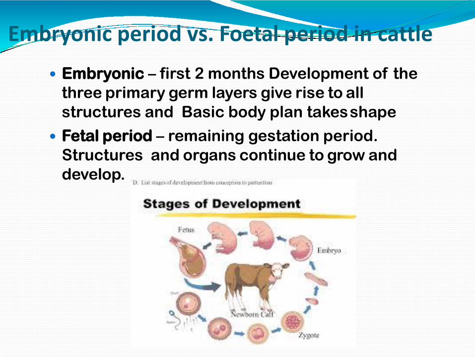
Embryonic period vs. Foetal period in cattle
Embryonic – first 2 months Development of the
three primary germ layers give rise to all
structures and Basic body plan takesshape
Fetal period – remaining gestation period.
Structures and organs continue to grow and
develop.

Stages of Development
Embryogenesis
1. Fertilization
2. Cleavage
3.Gastrulation
4.Organogenesis
5.Maturation

Fertilization: The process of fusion or union of the spermatozoon with the
mature ovum is known as conception /fertilizaiton/impregnantation.
Which produced the fertilized single mono-nucleatedcell called thezygote.
Embryogenesis: Theformation and development of anembryo.1. Cleavage: is a series of rapidmitotic divisions (without
cellgrowth)2. Gastrulation : is a phase early in the embryonic development
of most animals/human being, during which the single-layered blastula is reorganized into a trilaminar ("three-layered") structure known as the gastrula. These three germ layers are known as the ectoderm, mesoderm,and endoderm.
3. Organogenesis: The production and development of theorgans of an animal.

Events of Fertilization: 1. Attraction 2. Penitration 3. ConjugationHow fertilization occurs..? Following ovulation, the ovum is picked up by the tubal fimbriae
and is moved along by the cilia and by peristaltic movement of thetube.
At the time the cervix under the influence of estrogen, secretes a f low of alkaline mucus that deposited in the vagina, only thousands capacitated spermatozoa enter the uterine tube while 300-500 reach the ovum, and remainderare destroyed by the acid medium of thevagina.
It takes about 1 hour for sperm to reach thesite.

The sperm release the enzyme, Hylluronidase whichallows penetration of the zona pellucida and the cell membrane surrounding theovum.
Many sperm are needed for this to take place but onlyone will enter the ovum.
After this the membrane is sealed to prevent entry of any further sperm and the nucluei of the two cell fuse.
The sperm and ovum contribute half (n) thecomplement of chromosomes to make a diploidnumber (2n).
The sperm (n) and ovum (n) is known as the male and female gametes and the fertilizedovum as thezygote (2n)

. Fertilization

Normal site for Conception..? The most common site of conception is the ampullary part
(Ampulla ) of the fallopian tube which is the widest partlocated closed to theovary
The sex of the new individual at the time of conception is determined by sexchromosomes.
Every cattle cell contains 60 chromosomes, which are made up of 58 autosomechromosome and 2 sex chromosomes.
The sex chromosome are X and Y . Woman have no Y chromosome and male has Y chromosome
(male 58+X+Y) (female 58+X+X).
There for e, in mammals sex of young one isalways determined by Sire (While reverse is found in case of Birds)

Development of fertilized ovum/ZygoteMorula:1. After fertilization, the Zygote divides into 2 cells
(blastomere) (mitosis division)in about 30 hours after fertilization.
2. The blastomeres continue to divide by binary division through 4, 8, and 16 cell stage until a cluster of cells is formed– Morula, resemblibg a mulberry
3. The morula after spending about 3 days(72hours) in the uterine tube enters the uterine cavity through the narrow uterine ostium(1mm) on the 4th day
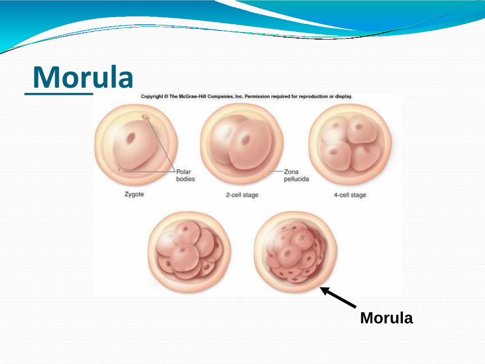
Morula
Morula

Blastocyst: Morula, once entering the uterine cavity, floats
freely(next 2 days) and is covered byendometrial f luid and mucus.
This fluid is absorbed through the canaliculi of the zona pellucida and Morula begins toaccumulatefluid and forms a cavity between its cells.
Once cavity appears, it is now called a blastocyst.

Blastocyst The zona pellucida
becomes stretched, thinned and gradually disappear soon prior to implantation.
The cell of the outer cell mass forms the wall of the blastocyst and is knownas trophoblast.
The inner cell massis concerned with the development of the embryo.

Two Distinct Cell Types
1. Trophoblasts – will form the invading placenta
2. Innercell mass – will form the embryo
Trophoblasts

Blastocyst
Trophoblast Inner cell mass
Placenta Chorion Fetus Amnion umbilical cord

DEVELOPMENT OF PLACENTAFrom zygote to the placenta formation

DEVELOPMENT OF PLACENTA➢The placenta is a foetomaternal composite structure
formed by the association of embryo and extra embryonicmembrane with uterine tissue for exchange of foodmaterials , oxygen and waste materials
➢Placenta develops from two sources:Foetal part– From chorio-allantoic membraneMaternal part– From Endometrium(decidua basils)
➢Placenta begins to develop upon implantation of theblastocyst into the maternal endometrium (That meansdevelopment of placenta starts when blastocyst attached tothe endometrium)
➢Once blastocyst is embeded in the endometrial wall,endometrium changed into Decidua and secretory activityof endometrium started, glycogen and lipids are stored andvacuole appear into the stroma
➢Placenta grows throughout the pregnancy
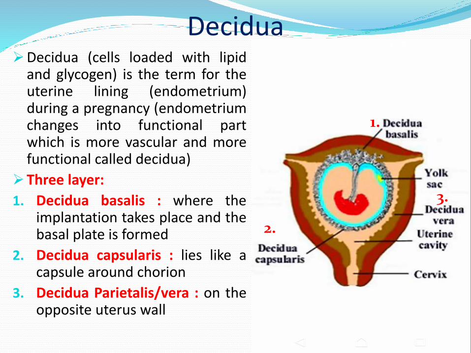
Decidua➢Decidua (cells loaded with lipid
and glycogen) is the term for theuterine lining (endometrium)during a pregnancy (endometriumchanges into functional partwhich is more vascular and morefunctional called decidua)
➢Three layer:
1. Decidua basalis : where theimplantation takes place and thebasal plate is formed
2. Decidua capsularis : lies like acapsule around chorion
3. Decidua Parietalis/vera : on theopposite uterus wall
1.
2.
3.
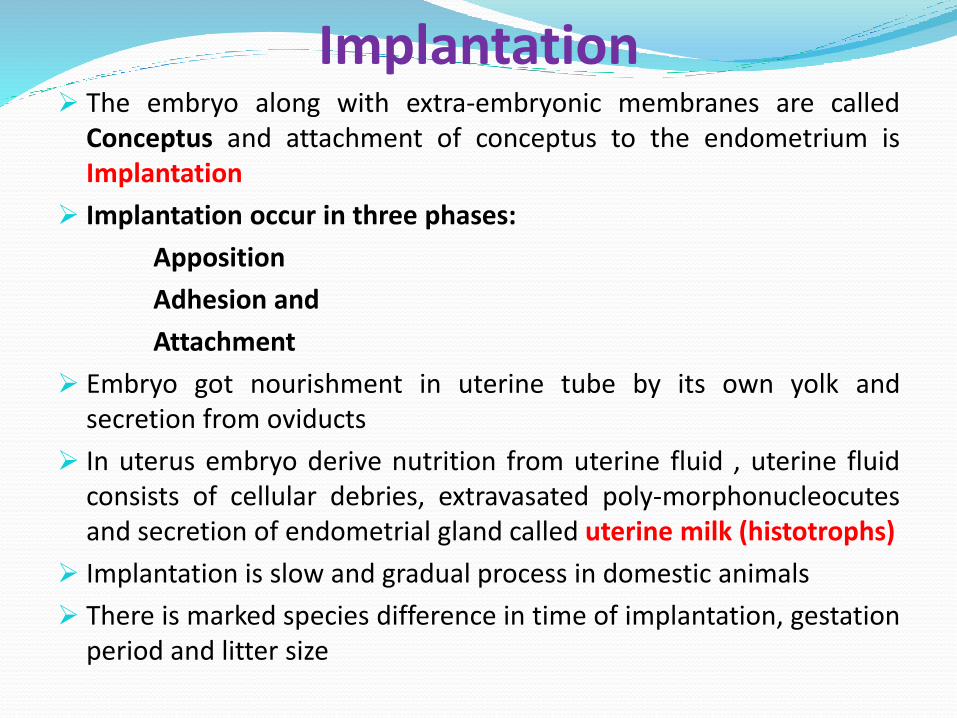
Implantation➢ The embryo along with extra-embryonic membranes are called
Conceptus and attachment of conceptus to the endometrium isImplantation
➢ Implantation occur in three phases:
Apposition
Adhesion and
Attachment
➢ Embryo got nourishment in uterine tube by its own yolk andsecretion from oviducts
➢ In uterus embryo derive nutrition from uterine fluid , uterine fluidconsists of cellular debries, extravasated poly-morphonucleocutesand secretion of endometrial gland called uterine milk (histotrophs)
➢ Implantation is slow and gradual process in domestic animals
➢ There is marked species difference in time of implantation, gestationperiod and litter size

SpeciesTime of
implantation (in days)
Gestation period
(in days)Litter size(numbers)
Cow 28-35 282 (277-290)
01
Ewe 17-20 148 (144-152)
1-2
Sow 17-24 114 (110-116)
08-12
Mare 49-70 338 (330-345)
01
Bitch 14-21 61 (58-64)
06-10
Cat 14-21 64 (60-68)
04

Types of Implantation
➢ Three types of Implantation:1. Superficial/Centric: The
chorionic vesicle remainwithin uterine cavity andexpands to fill its lumenEx. Domestic Animals
2. Eccentric : The chorionicvesicle become partiallyembedded in pockets of theuterine wallEx. Rat , Squirrel
3. Interstitial: The blastocystpenetrate into the wall ofuterus and develops thereuntil parturitionEx. Primates
1. 2. 3.

Implantation

➢At the time of implantation Zona pellucida becomes disappear
➢The trophoblastic layer differentiates into two parts:
Inner layer - Cytotrophoblast
Outer layer - Syncytotrophoblast
➢ Syncytotrophoblast proliferates into multilayered, multinucleatedprotoplasmic mass
➢Cytotrophoblast differentiates into layer of primary mesoderm
Primarymesoderm
Cytotophoblast Syncytotrophoblast
CHORION
➢ Inside syncytotrophoblast a number of lacunar spaces appearand syncytial cells form cords between the lacunar space, calledTrabeculae
➢Cords of cytotrophoblast invade the trabeculae and convert intoPrimary chorionic villi , lacunar space are now calledintervillous space
➢Primary chorionic villi are transformed into Secondary chorionicvilli when primary mesodermic layer invade into the primary villi
➢ Secondary villi are transformed into Tertiary villi when the foetalblood vessels appear within primary mesoderm and theirbranches project into secondary villi
➢ Later on within primary mesoderm vacuoles are appearedsubsequently they coalase to form extra-embryonic coelomebetween amniotic cavity and primary mesoderm


➢ In Birds and in some Farm animals like Cattle, Sheep, Goatand Pig the allantoic vesicle expands into the extraembryoniccoelom and surrounds the whole amniotic cavity
➢ It occupies the space between the amnion and the chorion(serosa), the outer wall of amnion fuses with chorion andtherefore forms Chorioallantioic Type of Placenta
➢The amniotic cavity contains Amniotic fluids within whichembryo becomes float. Amniotic fluid contains salt, water,protein and sugar . It gives protection to the foetus byneutralizing shock and pressure. It also acts as lubricants at thetime of birth
➢At the time of birth the placenta is discarded along with theamnion and referred as Afterbirth

➢ In Bovines, attachment between maternal and foetalmembranes occur throughout the endometrium of the horn inthe sporadic manner as per distribution of cotyledones
➢ In Sow and Mare the union of chorion and uterine wall issuperficial and their separation at the time of birth withoutinjury to maternal tissue this type of placenta is calledDeciduate placenta
➢ In Carnivores The villi occupy on the girdle like band aroundthe middle of chorionic sac
➢ In Humans, The chorionic villi develop rapidly at theembryonic pole of blastocyst called chorionic frondosum
➢ In Primates The union between foetal and maternal tissue isso intimate and damage of uterine tissue at the time of birth,that's why there is extensive bleeding at the time of birth inprimates

➢Exchange of metabolites occurs directly through foetal andmaternal blood circulation
➢There is no direct mixing of foetal and maternal blood inplacenta
➢The chorio-allantoic placenta directly absorb nutrition frommaternal blood is called Haemotrope
➢Therefore placenta formation (contact between foetalmembrane and endometrium) occurs in various zones whichdiffers characteristically depending upon the species
THANKS