name, address, telephone and fax numbers, and e-mail ... · licausi, giacomo novi, ottavio beretta,...
TRANSCRIPT

1
Running head: Transcript profiling of the anoxic rice coleoptile
Name, address, telephone and fax numbers, and e-mail address of author to whom all
correspondence:
Pierdomenico Perata, Phone: +39-050-883355; fax: +39-0502216532; e-mail:
Journal research area most appropriate for the paper: Environmental Stress and Adaptation
Plant Physiology Preview. Published on March 16, 2007, as DOI:10.1104/pp.106.093997
Copyright 2007 by the American Society of Plant Biologists
www.plantphysiol.orgon February 18, 2019 - Published by Downloaded from Copyright © 2007 American Society of Plant Biologists. All rights reserved.

2
Transcript Profiling of the Anoxic Rice Coleoptile
Rasika Lasanthi-Kudahettige1, Leonardo Magneschi1, Elena Loreti, Silvia Gonzali, Francesco
Licausi, Giacomo Novi, Ottavio Beretta, Federico Vitulli, Amedeo Alpi, Pierdomenico Perata*
Plant & Crop Physiology Laboratory, Scuola Superiore Sant'Anna, Via Mariscoglio 34, 56124 Pisa,
Italy ( R. L-K.; L.M.; S.G.; F.L; G.N.; P.P.); IBBA-CNR, Via del Borghetto 80, 56100 Pisa, Italy
(E. L.); Department of Crop Plant Biology, University of Pisa, Via Mariscoglio 34, 56124 Pisa,
Italy (A.A.); Genopolis, University of Milano-Bicocca, Dept. of Biotechnology and Bioscience,
Piazza della Scienza 4, 20126 Milano, Italy (O.B; F.V).
www.plantphysiol.orgon February 18, 2019 - Published by Downloaded from Copyright © 2007 American Society of Plant Biologists. All rights reserved.

3
Footnotes:
1 These authors contributed equally to the paper.
*Corresponding author; e-mail: [email protected]; fax: +39-0502216532
www.plantphysiol.orgon February 18, 2019 - Published by Downloaded from Copyright © 2007 American Society of Plant Biologists. All rights reserved.

4
ABSTRACT
Rice (Oryza sativa L.) seeds can germinate in complete absence of oxygen. Under anoxia, the rice
coleoptile elongates, reaching a length greater than that of the aerobic one. In this paper, we
compared and investigated the transcriptome of rice coleoptiles grown under aerobic and anaerobic
conditions. The results allow drawing a detailed picture of the modulation of the transcripts
involved in the anaerobic carbohydrate metabolism, suggesting the up-regulation of the steps
required to produce and metabolize pyruvate and its derivatives. Sugars appear to play a signalling
role under anoxia, with several genes indirectly up-regulated by anoxia-driven sugar starvation. The
analysis of the effects of anoxia on the expansin gene families revealed that EXPA7 and EXPB12
are likely to be involved in the rice coleoptile elongation under anoxia. Genes coding for Ethylene
Response Factors (ERFs) and Heat Shock Proteins are among the genes that are modulated by
anoxia in both rice and Arabidopsis. The identification of anoxia-induced ERFs is suggestive, since
genes belonging to this gene family play a crucial role in rice tolerance to submergence, a process
closely related to, but independent from, the ability to germinate under anoxia. Genes coding for
some enzymes requiring oxygen for their activity are dramatically down-regulated under anoxia,
suggesting the existence of an energy-saving strategy in the regulation of gene expression.
www.plantphysiol.orgon February 18, 2019 - Published by Downloaded from Copyright © 2007 American Society of Plant Biologists. All rights reserved.

5
INTRODUCTION
Higher plants are aerobic organisms that rapidly die when oxygen availability is limited due
to soil flooding (Voesenek et al., 2006). Species originating from semi-aquatic environments are,
however, able to cope with flooding stress. They can survive complete submergence for weeks and
some even have the capacity to grow vigorously and produce flowers and seeds in permanently
water-saturated soils. In this context, a well-known crop is rice that produces high yields even when
it is grown in waterlogged rice paddies. A broad range of metabolic and morphological adaptations
characterizes these tolerant species. Flood-tolerant plants have developed the capacity to generate
ATP without the presence of oxygen (fermentative metabolism) and/or to develop specific
morphologies (e.g. air channels, enhanced shoot elongation) that improve the entrance of oxygen
(Jackson, 1985; Crawford, 1992; Perata and Alpi, 1993; Armstrong et al., 1994; Drew et al., 2000;
Sauter, 2000; Gibbs and Greenway, 2003; Colmer, 2003; Voesenek et al., 2006). Gene expression
studies on plants exposed to low oxygen revealed the up-regulation of genes coding for
transcription factors (Hoeren et al., 1998; Liu et al., 2005), signal transduction components (Baxter-
Burrell et al., 2002), non-symbiotic haemoglobin (Dordas et al., 2004), ethylene biosynthesis
(Vriezen et al., 1999), nitrogen metabolism (Mattana et al., 1994), and cell wall loosening (Saab
and Sachs, 1996). At the protein level, low oxygen selectively induces the synthesis of the proteins
known as anaerobic proteins, most of which are enzymes involved in sugar metabolism, glycolysis
and fermentation pathways (Sachs et al., 1980; Huang et al., 2005).
Despite the knowledge on adaptive mechanisms and on the regulation at the gene and protein level,
our understanding of the mechanism(s) behind plant responses to anaerobiosis is very limited. Even
flood-intolerant species, such as Arabidopsis, switch on many genes that are generally associated
with responses to flooding (Klok et al., 2002; Branco-Price et al., 2005; Loreti et al., 2005; Gonzali
et al. 2006). This strongly suggests that the regulation of flooding tolerance in plants is far more
complex than anticipated for many years.
In rice, the flooding tolerance - defined as the survival after several days of complete submergence
of the entire plant - is strongly affected by a locus (Sub1) on chromosome 9 (Siangliw et al., 2003).
This locus explains a large proportion of the variation in flooding tolerance between indica
(tolerant) and japonica (intolerant) rice cultivars (Toojinda et al., 2003). Two Ethylene Response
Factors (ERFs) on this locus inhibit ethylene production and underwater elongation and stimulate
glycolysis and fermentation (Fukao et al., 2006). The importance of low elongation rates for the
flooding tolerance of rice was already established by Setter and Laureles (1996). The introduction
of the Sub1 locus into the background of a japonica rice cultivar significantly increased its flooding
tolerance, thus demonstrating the importance of the Sub1 locus for flooding tolerance (Fukao et al.,
2006). The identification of the Sub1 locus and the elucidation of its role in the adaptation of rice to
www.plantphysiol.orgon February 18, 2019 - Published by Downloaded from Copyright © 2007 American Society of Plant Biologists. All rights reserved.

6
submergence is a breakthrough in plant adaptation to anaerobiosis. It has to be highlighted however
that the ability of rice seeds to germinate under complete anoxia (Alpi and Beevers, 1983) is not
likely to be explained in terms of Sub1 genes. Ethylene, the trigger for Sub1A expression, is not
produced under anoxia and indeed the M202 and Nipponbare cultivars, both lacking the Sub1A
gene (Xu et al., 2006), display a better germination under anoxia when compared with the FR13A
genotype (P. Perata, unpublished), that possesses the Sub1A gene (Xu et al., 2006). The molecular
mechanisms allowing rice to elongate the coleoptile in complete absence of oxygen - a behaviour
not observed in any other cereal - are thus largely unknown. In this paper, we present the transcript
profiling of the anoxic rice coleoptile compared to its aerobic counterpart.
RESULTS AND DISCUSSION
The Anaerobic Responses in Rice
The ability of rice to germinate under anoxia is comparable to that in air, but only the
coleoptile elongates (Figure 1A), while both the root and the primary leaf fail to grow (Alpi and
Beevers, 1983). Rice coleoptiles show an enhanced expression of alcohol dehydrogenase genes, and
distinct expression patterns are observed for ADH1 and ADH2, the latter being induced at a higher
level (Figure 1B). When air-germinated seedlings are transferred to anoxia, the coleoptile fails to
elongate further and only the aerobic root continues to grow (Figure 1C). Both ADH1 and ADH2
are rapidly induced in the coleoptile and root from aerobic seedlings transferred to anoxia (Figure
1D). Twenty-day-old rice plants transferred to anoxia for 3h show an induction of ADH1 in leaves,
internodes and roots, while after 3-6h of anoxia a strong expression of ADH2 is observed, mostly in
the roots (Figure 1E). We chose to use four-day-old coleoptiles for exploring the anoxic coleoptile
transcriptome. Four days after germination, both the anoxic and the aerobic coleoptiles are close to
their maximal length (Figure 1A), and the aerobic coleoptile is large enough to allow a fast and
clean removal of the primary leaf, avoiding the extraction of leaf mRNA that would otherwise
contaminate coleoptile mRNA preparations. The analysis of the microarray results (Table S2)
indicate that anoxia exerted a dramatic effect on the coleoptiles, with 1,364 probe sets showing
increased expression and 1,770 probe sets indicating a decreased expression after data filtering and
selection of differentially expressed genes using a low (1%) “false discovery rate” threshold (DEG,
Table S2).
Anoxia induces genes involved in glycolysis, pyruvate metabolism, fermentation and a futile
cycle of starch synthesis and degradation.
www.plantphysiol.orgon February 18, 2019 - Published by Downloaded from Copyright © 2007 American Society of Plant Biologists. All rights reserved.

7
Several genes encoding glycolytic enzymes show an enhanced mRNA accumulation, thus
confirming the previous results based on enzymatic assays (Guglielminetti et al., 1995a). The
hexokinase activity, which is crucial for channelling glucose into glycolysis, is enhanced under
anoxia in rice (Guglielminetti et al., 1995a) and our microarray data reveal that OsHXK7
(Os05g09500) encodes for the previously measured anoxic hexokinase activity (Guglielminetti et
al., 1995a). The transcripts coding for the other 9 rice hexokinase genes (Cho et al., 2006) are
expressed at low levels in the anoxic coleoptiles (see Table S1, S2). Remarkably, OsHXK7 is a
starvation-induced gene (Cho et al., 2006), and its induction under anoxia is likely a consequence of
the lower sugar content of the anoxic coleoptiles (Alpi and Beevers, 1993). Interestingly, the genes
involved in pyruvate metabolism are strongly up-regulated (Figure 2). A transcript encoding
phosphoenolpyruvate carboxykinase (PCK) is among the genes showing a high induction by
anoxia, and pyruvate orthophosphate dikinase (PPDK) and pyruvate decarboxylase (PDC) are also
strongly induced (Figure 2). The PPDK induction at the protein level in rice under hypoxia has been
reported by Moons et al. (1998) and by Huang et al. (2005). It is suggested that PPDK may provide
pyrophosphate (PPi) for the PPi-dependent activity of phosphofructokinase (PPi-PFK), whose
mRNA accumulation is increased under anoxia (Figure 2). PPi is also needed for the activity of the
PPi-dependent vacuolar H+ translocase (H+-PPase, Figure 2; Carystinos et al., 1995). The activity
of PEP-carboxylase (PEPC) is repressed under anoxia (Figure 2). PEPC is highly expressed in the
aerobic coleoptile and would drain PEP from glycolysis in a reaction that, if not down-regulated
under anoxia, would compete with PCK, which is absent under aerobic conditions, but is strongly
expressed under anoxia (Figure 2; Table S2). The role of PCK in the anaerobic metabolism is
unexplored, but it is tempting to speculate that its activity may be useful to channel amino acids,
through OAA, into glycolysis through a cataplerotic reaction (Owen et al., 2002). The expression of
aspartate aminotransferase (Asp-AT), an enzyme involved in cataplerosis (Owen et al., 2002), is
high and enhanced under anoxia (Figure 2; Table S2).
Genes coding for pyruvate decarboxylase (PDC) and alcohol dehydrogenase (ADH2) are highly
expressed in the anoxic coleoptiles, and a mitochondrial aldehyde dehydrogenase (ALDH2a), which
is involved in the metabolism of the potentially toxic acetaldehyde (Perata and Alpi, 1991), is also
induced under anoxia (Nakazono et al., 2000; Figure 2). The expression of ALDH2a is usually
associated to the ability to metabolize acetaldehyde during re-aeration, since, although the mRNA
accumulates during low-oxygen treatment and not upon re-aeration, the enzyme activity increases
only when the plants are transferred from submergence to aerobic conditions (Tsuji et al., 2003).
ALDH activity correlates with anoxia tolerance in varieties of Echinocloa crus-galli, and it has
been proposed that ALDH may detoxify acetaldehyde formed through alcoholic fermentation
during anaerobic germination (Fukao et al., 2003). Alternatively, PDC and ALDH2a may represent
www.plantphysiol.orgon February 18, 2019 - Published by Downloaded from Copyright © 2007 American Society of Plant Biologists. All rights reserved.

8
part of a pyruvate dehydrogenase bypass (PDH, down regulated under anoxia; Figure 2) as
proposed by Mellema et al. (2002).
The strong expression of alpha-amylase (RAmy3D) indicates that starch plays a role also in
the coleoptiles, besides the known role of starch degradation in the seed endosperm under anoxia
(Perata et al., 1992; 1993; Guglielminetti et al., 1995b). The anoxic rice coleoptiles contain
amyloplasts with low starch content (Perata and Alpi, 1993), a likely consequence of active starch
degradation. Interestingly, also some genes involved in starch synthesis are up-regulated by anoxia,
which suggests that an apparently futile cycle of starch synthesis degradation is active in the anoxic
coleoptiles. The need to avoid starch-depleted amyloplasts, which would negatively affect the
gravitropic response of the coleoptiles (Kutschera et al., 1991; Kutschera and Hoss, 1995), may
explain the operation of a possible moderate flux of sugars to starch even under anoxia. Only a few
genes involved in carbohydrate metabolism are repressed under anoxia. Sucrose transport appears
to be severely impaired, with most translocators showing reduced expression under anoxia. The
inability to transport sugars from the starchy endosperm to the growing coleoptile may explain why
rice coleoptiles suffer from sugar starvation (Alpi and Beevers, 1983) despite the availability of
starch degradation products in the endosperm (Perata et al., 1992). Sucrose degradation in the
vacuole appears to be repressed, as well as the translocation of glucose-6-phosphate to the
amyloplasts, a pathway that would drain Glc-6P from glycolysis.
Effects of Anoxia on α-amylases
Seven α-amylase genes, out of a total of 10 genes in the rice genome (Huang et al., 1990),
are represented on the rice GeneChip. Only RAMY3D, is significantly modulated by anoxia (879
fold, see Table S2). A detailed analysis of the expression pattern of RAMY3D in the anoxic rice
coleoptile reveals that the induction under anoxia is much stronger when anoxia is prolonged up to
6-8 days, with an anoxic mRNA level 15,000-fold higher than in air (Figure 3A). Anoxia induces
RAMY3D also in the coleoptiles and roots of the seedlings transferred from air to anoxia (Figure
3B). Since RAMY3D is known to be a starvation-induced gene (Yu et al., 1996; Loreti et al., 2003),
we analysed and compared the carbohydrate content of the aerobic and anoxic coleoptiles. While
starch is present at a relatively low and constant level in the aerobic coleoptiles, this polysaccharide
is present in the anoxic coleoptiles from 2-days old seedlings, a likely consequence of a slower use
of starch in this tissue during the initial phases of germination under anoxia. Starch is however
rapidly degraded during the subsequent days of anoxic germination (Figure 3C), indicating that the
expression of RAMY3D has relevant consequences on starch metabolism in the anoxic coleoptiles.
While the aerobic coleoptile display a relatively high sucrose and fructose content during the first 4
www.plantphysiol.orgon February 18, 2019 - Published by Downloaded from Copyright © 2007 American Society of Plant Biologists. All rights reserved.
9
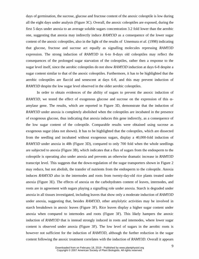
days of germination, the sucrose, glucose and fructose content of the anoxic coleoptile is low during
all the eight days under analysis (Figure 3C). Overall, the anoxic coleoptiles are exposed, during the
first 5 days under anoxia to an average soluble sugars concentration 3.2 fold lower than the aerobic
one, suggesting that anoxia may indirectly induce RAMY3D as a consequence of the lower sugar
content of the anoxic coleoptiles, also in the light of the results of Umemura et al. (1998) indicating
that glucose, fructose and sucrose act equally as signalling molecules repressing RAMY3D
expression. The strong induction of RAMY3D in 6-to 8-days old coleoptiles may reflect the
consequences of the prolonged sugar starvation of the coleoptiles, rather then a response to the
sugar level itself, since the aerobic coleoptiles do not show RAMY3D induction at days 6-8 despite a
sugar content similar to that of the anoxic coleoptiles. Furthermore, it has to be highlighted that the
aerobic coleoptiles are flaccid and senescent at days 6-8, and this may prevent induction of
RAMY3D despite the low sugar level observed in the older aerobic coleoptiles.
In order to obtain evidences of the ability of sugars to prevent the anoxic induction of
RAMY3D, we tested the effect of exogenous glucose and sucrose on the expression of this α-
amylase gene. The results, which are reported in Figure 3D, demonstrate that the induction of
RAMY3D under anoxia is completely abolished when the coleoptiles are incubated in the presence
of exogenous glucose, thus indicating that anoxia induces this gene indirectly, as a consequence of
the low sugar content of the coleoptile. Comparable results were obtained using sucrose as
exogenous sugar (data not shown). It has to be highlighted that the coleoptiles, which are dissected
from the seedling and incubated without exogenous sugars, display a 40,000-fold induction of
RAMY3D under anoxia in 48h (Figure 3D), compared to only 700 fold when the whole seedlings
are subjected to anoxia (Figure 3B), which indicates that a flux of sugars from the endosperm to the
coleoptile is operating also under anoxia and prevents an otherwise dramatic increase in RAMY3D
transcript level. This suggests that the down-regulation of the sugar transporters shown in Figure 2
may reduce, but not abolish, the transfer of nutrients from the endosperm to the coleoptile. Anoxia
induces RAMY3D also in the internodes and roots from twenty-day-old rice plants treated under
anoxia (Figure 3E). The effects of anoxia on the carbohydrates content of leaves, internodes, and
roots are in agreement with sugars playing a signalling role under anoxia. Starch is degraded under
anoxia in all tissues investigated, including leaves that show only a moderate induction of RAMY3D
under anoxia, suggesting that, besides RAMY3D, other amylolytic activities may be involved in
starch breakdown in anoxic leaves (Figure 3F). Rice leaves display a higher sugar content under
anoxia when compared to internodes and roots (Figure 3F). This likely hampers the anoxic
induction of RAMY3D that is instead strongly induced in roots and internodes, where lower sugar
content is observed under anoxia (Figure 3F). The low level of sugars in the aerobic roots is
however not sufficient for the induction of RAMY3D, although the further reduction in the sugar
content following the anoxic treatment correlates with the induction of RAMY3D. Overall it appears
www.plantphysiol.orgon February 18, 2019 - Published by Downloaded from Copyright © 2007 American Society of Plant Biologists. All rights reserved.

10
that sugars act as signalling molecules under anoxia, but the level of sugars itself appears to be
required but not sufficient to activate the signalling pathway triggering induction of genes such as
RAMY3D. We tested the effect of exogenous glucose on the expression of ADH1 and ADH2 genes,
and the results indicate that glucose is unable to counteract the anoxic induction of ADH genes,
although the induction is reduced by about 40% when glucose is added to the incubation medium
(data not shown). Therefore, glucose availability appears to play an important role in the
modulation of some anoxia-induced genes, although the regulation of genes, such as ADH, is
relatively independent of sugar availability.
Rice expansins EXPA7 and EXPAB12 are up-regulated in the anoxic rice coleoptile
The coleoptile elongation under anoxia is due to cell expansion, and expansins likely play a
major role in this process (Cosgrove, 1999; Huang et al., 2000). Twenty-two alpha-expansins
(EXPA) and twelve beta-expansins (EXPB) are represented on the GeneChip, but only 6 are
differentially expressed in the anoxic rice coleoptile (Figure 4A). Only EXPA7 and EXPB12 appear
to be more expressed under anoxia than in air (Figure 4A). A previous report indicated EXPA2 and
EXPA4 as possibly involved in the anoxic coleoptile elongation (Huang et al., 2000). We therefore
compared the patterns of expression of EXPA2, EXPA4, EXPA7 and EXPB12 in coleoptiles from
aerobic and anaerobic seedlings. The results, which are shown in Figure 4B, indicate that, while the
expression of both EXPA2 and EXPA4 in air is compatible with the growth of the aerobic
coleoptile, these mRNAs are comparatively less abundant under anoxia during the first 4 days of
germination, when most of the anoxic coleoptile growth is observed (Figure 1A). The mRNA level
of EXPA7 and EXPB12 are instead higher under anoxia at day 2-3 after germination that coincides
with the anoxic coleoptile elongation phase, which makes these expansins good candidates for
explaining the anoxic growth of the rice coleoptile. EXPA2, EXPA4 and EXPA7 are not affected by
the transfer of seedlings or twenty-day-old rice plants from air to anoxia (Figures 4C and 4D), thus
suggesting that EXPA7 is developmentally regulated in the anoxic rice coleoptile. EXPB12 is
instead transiently up-regulated in coleoptiles and roots transferred from air to anoxia (Figure 4C)
and moderately induced in the internodes kept for 6h under anoxia (Figure 4D).
Genes coding for HSPs are induced by Anoxia-Driven Sugar Starvation
Heat shock proteins (HSPs) are not only expressed in plants experiencing high temperature
stress, but they are also expressed in response to a wide range of other environmental stresses, such
as water, salinity, osmotic, cold and oxidative stress (reviewed by Wang et al., 2004). Indeed, HSPs
www.plantphysiol.orgon February 18, 2019 - Published by Downloaded from Copyright © 2007 American Society of Plant Biologists. All rights reserved.

11
play a crucial role in protecting plant cells against stressful conditions and in re-establishing cellular
homeostasis (Wang et al., 2004). It has recently been demonstrated that a heat pre-treatment
strongly enhances anoxia-tolerance in Arabidopsis (Loreti et al., 2005), suggesting that HSPs may
play a role in anoxia tolerance. A large number of genes coding for HSPs are represented on the rice
GeneChip and thirteen are differentially expressed under anoxia, nine are upregulated and four
down regulated (Figure 5A). Rice HSP20 (Os02g52150) shows a higher expression under anoxia,
with a peak in 6-to-8 days old anoxic rice coleoptiles (Figure 5B). A nearly identical pattern of
expression was observed analysing the pattern of expression of rice HSP17.4 (Os03g16030, data
not shown). We compared the expression of rice HSP20 with the expression of an Arabidopsis
sHSP known to be anoxia-enhanced (Loreti et al., 2005). In Arabidopsis, the induction of HSP25.3-
P (At5g59720) by anoxia is rapid, with a 700-fold induction in the leaves of seedlings after only 6h
under anoxia (Figure 5C). All other anoxia-induced Arabidopsis sHSP genes (Loreti et al., 2005)
show a very similar fast induction under anoxia (data not shown). In rice, instead, the induction of
HSP20 (Os02g52150) is a slow process, with an anoxic induction comparable to that observed in
Arabidopsis only after 1 week of anoxic growth of the rice coleoptile (Figure 5B). We checked
whether the faster induction of HSP25.3-P observed in Arabidopsis (Figure 5C) is a consequence of
the air-to-anoxia transfer of the seedling. Aerobically germinated rice seedlings transferred to
anoxia show an induction of HSP20 in both the coleoptile and roots (Figure 5D) at a level well
below that of Arabidopsis leaves and roots (Figure 5C).
In Arabidopsis, the anoxic induction of HSPs is enhanced by exogenous sucrose (Loreti et
al., 2005). When we tested the effect of exogenous sugars on the expression of HSP20 in rice, we
realized that the induction by anoxia is totally abolished in the presence both of glucose (Figure 5E)
and of sucrose (not shown). It appears, therefore, that HSP20 is induced by anoxia as a consequence
of the sugar starvation occurring in the coleoptiles, which had been grown under anoxia for several
days, rather than being a direct consequence of anoxia. This unveils a different effect of sugars on
the anoxic induction of HSPs in Arabidopsis and rice. In Arabidopsis, a lipid-storing seed,
exogenous sucrose is needed to fuel the fermentative metabolism (P. Perata, unpublished), but,
although sucrose feeding prolongs Arabidopsis survival to anoxia, it cannot prevent the rapid death
of the seedlings. We speculate that, in Arabidopsis, sucrose prevents an otherwise dramatically
rapid metabolic collapse, allowing Arabidopsis to detect the protein dysfunction requiring the
expression of HSPs (Wang et al., 2004). In rice, starch degradation delays sugar starvation (Perata
et al., 1993), and the well-coordinated up-regulation of the fermentative metabolism (Figure 2)
allows rice to avoid the stressful condition triggering HSP induction. After only 1 week of anoxic
germination, the coleoptile starts suffering from the prolonged sugar starvation, the fermentative
metabolism slows down, and the coleoptile experiences the impaired metabolism triggering the
induction of HSPs, a condition occurring only after a few hours of anoxia in Arabidopsis.
www.plantphysiol.orgon February 18, 2019 - Published by Downloaded from Copyright © 2007 American Society of Plant Biologists. All rights reserved.

12
Interestingly, HSP20 is also induced in tissues of twenty-day-old plants treated under anoxia
(Figure 5F).
ERFs are induced by Anoxia in both Rice and Arabidopsis
The mRNA level of several ERF-like transcription factors is enhanced by anoxia both in rice
(Table S2) and Arabidopsis (Loreti et al., 2005). Remarkably, the submergence tolerance of the rice
variety FR13A is linked to a major QTL, known as Submergence1 (Sub1), on chromosome 9 (Xu
and Mackill, 1996). Xu et al. (2006) revealed that the Sub1 region encodes three transcription
factors (Sub1A, Sub1B and Sub1C) belonging to the B-2 subgroup of the Ethylene Response Factors
(ERFs)/ethylene-responsive element binding proteins (EREBPs)/apetala 2-like proteins (AP2). The
transcription of both Sub1A and Sub1C is strongly up-regulated by submergence and down-
regulated by de-submergence. The third ERF, Sub1B is only slightly regulated upon submergence.
It has to be pointed out that the ability of rice to germinate under complete anoxia cannot be
explained in terms of Sub1A expression. Ethylene, the trigger for Sub1A expression, is not produced
under anoxia. Furthermore, the Nipponbare cultivar lacks the Sub1A gene (Xu et al., 2006; Fukao et
al., 2006), but shows a vigorous germination under anoxia (Figure 1A). This does not rule out the
possibility that other ERFs may play a role in anoxia tolerance at the germination stage. A large
number (111) of ERF family genes (Nakano et al., 2006) is represented on the rice GeneChip, 6 and
16 of them showing induction or repression respectively by anoxia (Figure 6A). Sub1B
(OsERF#063) is not expressed in the rice coleoptiles (see Table S1), while Sub1C (OsERF#073) is
down-regulated under anoxia (FDR p-value 0.032, Table S2), an expected result since this gene is
up-regulated by ethylene (Fukao et al., 2006), which is a hormone requiring oxygen for its
synthesis. Anoxia-induced ERFs are thus unlikely to be ethylene-modulated, and may play a role in
the adaptation of plants to a complete lack of oxygen. In order to further confirm the induction of
ERFs under anoxia, we selected three ERF genes (OsERF#060, OsERF#067, OsERF#068), all
belonging to the subgroup B-2 in the ERF gene family (Sakuma et al., 2002; corresponding to the
group VIIa, according to the classification of Nakano et al., 2006), similarly to Sub1B and Sub1C
genes (Xu et al., 2006). These three ERF genes were chosen because of their strong induction by
anoxia and because of their homology with two Arabidopsis ERFs displaying induction by anoxia
(Loreti et al., 2005; At1g72360 showing homology to OsERF#060; At2g47520 showing homology
to OsERF#067/OsERF#068; Loreti et al., 2005). The three rice ERFs display distinct patterns of
expression in the anoxic coleoptile (Figure 6B), with OsERF#060 showing a pattern closely similar
to that of ADH2 (Figure 1B) and OsERF#067 mirroring the expression of ADH1 (Figure 1B). The
patterns of expression are instead very similar in the coleoptiles and roots from aerobically
germinated seedlings transferred to anoxia (Figure 6C). In Arabidopsis seedlings treated under
www.plantphysiol.orgon February 18, 2019 - Published by Downloaded from Copyright © 2007 American Society of Plant Biologists. All rights reserved.

13
anoxia up to 6h, At1g72360 is transiently induced by anoxia, while At2g47520 induction is
prolonged during the 6h anoxic treatment (Figure 6D). In twenty-day-old rice plants the three ERFs
are anoxia-induced in all tissues, although the response in leaves is smaller (Figure 6E).
OsERF#060, OsERF#067 and OsERF#068 induction by anoxia is reduced in the presence of
exogenous glucose (Figure 6F), thus confirming that glucose availability improves the overall status
of the seedlings, with a consequent lower response in terms of anaerobic gene induction.
Xylanase Inhibitor Protein Genes are strongly induced by Anoxia-Driven Sugar Starvation
The data mining of microarray data revealed that, surprisingly, xylanase inhibitor protein
(XIP) genes were strongly up-regulated under anoxia. XIPs are known to be induced by pathogen-
related pathways, as well as by wounding and methyl jasmonate (Igawa et al., 2005). Ten genes out
of the 31 represented on the GeneChip are induced under anoxia, and only one is repressed (Figure
7A). Some of these XIPs, such as Os11g47560, are among the genes showing the strongest
induction under anoxia (Table S2). Os11g47560 is indeed induced 150,000 fold in the coleoptile
that elongated under anoxia for 8 days (Figure 7B), and induction is also observed in the roots
transferred from air to anoxia (Figure 7C), as well as in twenty-day-old rice plants (Figure 7D). The
treatment of coleoptiles with exogenous glucose revealed that Os11g47560 is, similarly to
RAMY3D, a starvation-induced gene (Figure 7E). Even in aerobic conditions, the surgically
dissected coleoptiles (and thus unable to take glucose from the seed endosperm) display an
induction of Os11g47560 when incubated in mannitol, but such an induction is dramatic under
anoxia, with a 450,000-fold induction value. The fold induction value for isolated anoxic coleoptiles
(Figure 7E) is much higher than that of coleoptiles sampled from intact seedlings (Figure 7C), thus
confirming the existence of a glucose flow from the endosperm to the anoxic coleoptile. Anoxia
may induce XIPs to protect the seedlings from pathogen infection in the moist environment of
flooded soil. Other pathogen or disease-related genes are not up regulated under anoxia (Table S2),
and this hypothesis deserves further investigation.
Genes coding for P450 are down-regulated under Anoxia
A survey of the genes that are down-regulated by anoxia revealed that 39 genes coding for
P450 enzymes are significantly down-regulated under anoxia, while only 5 genes are up-regulated
(Figure 8A). Cytochromes P450 are heme-containing oxygenases, which transfer a single O-atom
from O2 to substrates, and therefore, have an oxygen-binding capability. The down-regulation of
P450s by anoxia prompted us to analyze the detailed pattern of expression of the P450 gene
(Os11g18570), which shows the highest expression in the aerobic coleoptile and the strongest
www.plantphysiol.orgon February 18, 2019 - Published by Downloaded from Copyright © 2007 American Society of Plant Biologists. All rights reserved.

14
down-regulation under anoxia. Os11g18570 is transiently expressed in the aerobic coleoptile, with a
peak of expression coinciding with the end of the coleoptile elongation (Figure 8B). Under anoxia,
Os11g18570 is not expressed. We observed a rapid down-regulation of Os11g18570 when the
aerobically germinated seedlings were transferred to anoxia (Figure 8C). This P450 gene appears to
be expressed in the aerobic coleoptile, but not in the root (Figure 8C) of rice seedlings. In the
twenty-day-old rice plants, Os11g18570 is predominantly expressed in the internodes, where a
down-regulation by anoxia can also be observed (Figure 8D). Glucose cannot prevent the rapid
decline in the expression of Os11g18570 (not shown). Due to the diversity of pathways requiring
mono-oxygenase activity, a wide variety of environmental and developmental signals modulate the
expression of CYP450 (Dixon and Paiva, 1995; Mizutani et al., 1998). It is tempting to speculate
that the anoxia-driven down-regulation of CYP450 genes is linked to the need of avoiding the
energy-waste related to the transcription of genes whose products require oxygen.
Anoxia down-regulates Catalase
A catalase-coding transcript is strongly down-regulated under anoxia (see Table S2). A
dramatic reduction in the activity of catalase was previously observed under anoxia (Alpi and
Beevers, 1983), while hypoxia increased catalase activity (Ushimaru et al., 1999). The authors
(Ushimaru et al., 1999) speculate that catalase may convert H2O2 (generated by glyoxysome
metabolism) to oxygen under hypoxia, thus alleviating the oxygen-shortage conditions. We studied
more in detail the effects of anoxia on catalase. Three genes coding for catalase are represented on
the GeneChip. Os03g03910 is not expressed in the rice coleoptiles, Os06g51150 is moderately
expressed and unaffected by anoxia, while Os02g02400 is strongly expressed in the aerobic
coleoptile but absent under anoxia (Figure 9A). In the light of the data of Alpi and Beevers (1983)
indicating that over 90% of catalase activity is lost under anoxia, Os02g02400 is the gene, which
contributes to most of the catalase activity in the rice coleoptile. A time-course analysis shows that
Os02g02400 is transiently expressed during aerobic germination, but never under anoxia (Figure
9A). When aerobically-germinated seedlings were transferred to anoxia, the Os02g02400 transcript
disappeared within 3h in the coleoptiles, the only tissues showing expression of this gene (Figure
9B). Exogenous glucose is unable to counteract the decline in Os02g02400 transcript under anoxia
(data not shown). The expression of Os02g02400, which is strong in the internodes of twenty-day-
old rice plants, is rapidly down-regulated under anoxia (Figure 9C). It is unclear why anoxia drives
a rapid down-regulation of catalase. When submerged seedlings were exposed to air, the activities
of catalase exceeded the level of catalase activity detected in aerobically grown controls (Ushimaru
et al., 1994), which indicates that the H2O2–degradation system is rapidly reconstituted under
aerobic conditions. The absence of catalase under anoxia does not appear to affect the development
www.plantphysiol.orgon February 18, 2019 - Published by Downloaded from Copyright © 2007 American Society of Plant Biologists. All rights reserved.
15
of post-anoxic injuries, since anoxia-germinated seedlings rapidly adapt to air with root and leaves
development (not shown). It is thus likely that, in the absence of oxygen, the production of H2O2 is
negligible, making catalase superfluous in the anoxic coleoptile. The down-regulation of catalase
can therefore be part of a strategy of metabolism reorganization aimed at producing only the
enzymes that are requested for housekeeping, as well as for the anaerobic metabolism, with a
consequent energy saving, which is likely of great importance under anoxia (Perata and Alpi, 1993;
Fukao and Bailey-Serres, 2004). An alternative hypothesis can be proposed, assuming that oxygen
peroxide can act as a second messenger in low-oxygen signalling (Fukao and Bailey-Serres, 2004;
Bailey-Serres and Chang, 2005). Although it is predicted that under anoxia the H2O2 should be
negligible, H2O2 was detected under anoxia (Blokhina et al., 2001), possibly as a consequence of
traces of oxygen in the experimental system, thus suggesting that the decline in catalase activity
may contribute to the build-up of an H2O2 level acting as a signal.
CONCLUSION
The ability of the rice coleoptile to elongate under anoxia represents an unveiled enigma,
while the mechanism(s) and genes involved in the adaptation of rice plants to submergence have
recently been discovered (Fukao et al., 2006; Xu et al., 2006). When rice plants are flooded, the
ethylene produced as a consequence of submergence acts as a trigger of Sub1 genes, down-
regulating their elongation and thus preserving energy for their survival. This mechanism cannot
explain the germination of rice under anoxia, a process that is observed also in rice varieties, such
as Nipponbare, which was used in our experiments and which lacks the Sub1A gene (Xu et al.,
2006), although other anoxia-ERFs (Figure 6) may play a role in anoxia tolerance. The experiments
described in this paper contribute to a better understanding of the anoxic responses of rice to anoxia
at the molecular level. Although the transcript steady-state level detected by microarray analysis
does not take into account the possible regulation of transcript translation efficiency (Fennoy and
Bailey-Serres, 1995), our results indicate that glycolysis is up-regulated under anoxia. This is
supported by a previous work, based on enzyme activity measurements, and indicating that
anaerobic transcripts are actually translated (Guglielminetti et al., 1995a). The metabolism of
pyruvate appears to play an important role, beyond the expected induction of pyruvate
decarboxylase, with PCK playing a cataplerotic role, and the production of PPi by PPDK
supporting the ATP-independent activity of PPi-PFK and of a vacuolar H+-PPase (Figure 2). Sugar
transporters are down-regulated under anoxia and the anoxic rice coleoptiles experience sugar
starvation (Alpi and Beevers, 1983). It is not unlikely that a moderate sugar starvation may
represent a signal for the activation of the key genes required for the anaerobic metabolism,
www.plantphysiol.orgon February 18, 2019 - Published by Downloaded from Copyright © 2007 American Society of Plant Biologists. All rights reserved.

16
including hexokinase, regulating carbohydrate entry into glycolysis (Fox et al., 1998). We indeed
observed an important role for sugar signalling in the modulation of several genes induced by
anoxia, which indicates that sugars may represent important second messengers in low-oxygen
signalling. The genes coding for some enzymes that require oxygen for their activity are
dramatically down-regulated under anoxia, indicating a possible strategy aimed to prevent the
production of enzymes that would be not operative under anoxia, thus saving energy for the
expression of genes required for the adaptative response of the rice seedling. The elongation of the
coleoptile is the most remarkable phenotype of the anoxic coleoptile, and the identification of
EXPA7 and EXPB12, rather than EXPA2 and EXPA4 (Huang et al., 2000), as candidates for the
expansin involved in the anoxic coleoptile elongation provides the basis for future investigations in
this field, including transgenic approaches, as well as the study of the germplasm showing a distinct
ability to elongate the coleoptile under anoxia.
MATERIALS AND METHODS
Plant Material
Rice (Oryza sativa L. cv Nipponbare) and Arabidopsis (gl1) seeds were sterilized with
diluted bleach (15-min incubation in 1.7% sodium hypochlorite, rinsing and washing in sterile water
10 times) and germinated on sterile filter paper at 28°C (23°C for Arabidopsis) in the dark. Three
different treatments were performed: (1) seed germination under anoxia since imbibition, (2)
transfer of the aerobically (4-day-old) germinated seedlings to anoxia, and (3) anoxic treatment of
the surgically dissected coleoptiles from aerobically (4-day-old) germinated seedlings. The RNA
samples used for the microarray experiments were produced from 2 independent biological
replicates (Anoxic treatment No.1, see above), each resulting from the pooling of at least 5 petri
dishes containing 25 seedlings each. The primary leaf was removed from the coleoptiles of air-
germinated seedlings (the leaf is absent in anoxia-germinated seedlings). All the air/anoxia
treatments were performed in the dark at 28°C for rice and at 23°C for Arabidopsis. Twenty-days
old plants were used as a source of roots, internodes and leaves as indicated in the figures legends.
Rice plants were grown using a hydroponics system: seeds were sown in rockwool hydroponic
growing media in Hoagland solution. Twenty-days old plants were treated under anoxia in the light,
with the anoxic treatment starting at the beginning of the light treatment. An enclosed anaerobic
work station (Anaerobic System model 1025; Forma Scientific, Marietta, OH) was used to provide
an oxygen-free environment for seed germination or seedling incubation. This chamber uses
palladium catalyst wafers and desiccant wafers to maintain strict anaerobiosis to less than 10 µg
www.plantphysiol.orgon February 18, 2019 - Published by Downloaded from Copyright © 2007 American Society of Plant Biologists. All rights reserved.

17
mL–1 O2 (according to the specifications provided by the manufacturer). High-purity N2 was used
for purging the chamber initially and the working anaerobic gas mixture was N2:H2 proportioned at
90:10.
RNA Isolation, cRNA Synthesis, and Hybridization to Affymetrix GeneChips
Total RNA was extracted from the seedling samples using the Ambion RNAqueous kit
(Ambion). RNA quality was assessed by agarose gel electrophoresis and spectrophotometry. RNA
was processed for use on Affymetrix Rice Genome GeneChip arrays, as previously described
(Loreti et al., 2005). Hybridization, washing, staining, and scanning procedures were performed by
Genopolis (University of Milano Bicocca, Italy), as described in the Affymetrix technical manual.
Microarray analysis was performed using R/Bioconductor (Gentlemen et al., 2004). Expression
measures were obtained using GCRMA (Wu and Irizarry, 2005), a multiarray analysis method
estimating probesets signals taking into account the physical affinities between probes and targets.
Normalization was done by a quantiles method (Bolstad et al., 2003). To reduce the number of non
informative genes, two different filters were applied: the first removes the probesets presenting an
Affymetrix Absent Call (A) for both the conditions; the second eliminates the probesets showing, as
maximum signal in the two conditions, a value equal or less than the 95th percentile of the overall
Absent calls signals distribution. This filtering yielded 21484 probesets. To identify a statistically
reliable number of differentially expressed genes among the two conditions, a linear model was
performed (Wettenhall et al., 2004). For assessing differential expression, an empirical Bayesian
method (Smyth, 2004) was used to moderate the standard error of the estimated log-fold changes. In
order to control p-values in a context of multiple testing problem, a Benjamini-Hochberg's
correction of the false discovery rate (Reiner et al., 2003) was applied (adjusted P-value <= 0.01)
leading to 3134 differentially expressed probesets. Microarray datasets were deposited in a public
repository with open access (accession no. GSE6908, http://www.ncbi.nlm.nih.gov/projects/geo/).
Real-Time RT-PCR
RNA was extracted from the seedlings grown as indicated in figure legends. Total RNA,
extracted using the RNAqueous kit (Ambion) according to the manufacturer's instruction, was
subjected to a DNase treatment using the TURBO DNA free kit (Ambion). Two micrograms of
each sample were reverse transcribed into cDNA using the high-capacity cDNA archive kit
(Applied Biosystems). Real-time PCR amplification was carried out with the ABI Prism 7000
sequence detection system (Applied Biosystems), using the primers described in Table S4 and the
default ABI Prism 7000 PCR program for PCR conditions. GPDH was used as endogenous control
for rice and Ubiquitin10 (UBQ10) was used as endogenous control for Arabidopsis. Taqman probes
www.plantphysiol.orgon February 18, 2019 - Published by Downloaded from Copyright © 2007 American Society of Plant Biologists. All rights reserved.

18
specific for each gene were used. The probe sequences are reported in Table S4. PCR reactions
were carried out using 50 ng of cDNA and TaqMan Universal PCR master mix (Applied
Biosystems), following the manufacturer's protocol. Relative quantitation of each single gene
expression was performed using the comparative CT method, as described in the ABI PRISM 7700
Sequence Detection System User Bulletin #2 (Applied Biosystems).
Carbohydrate analyses
Analysis of sucrose, fructose and glucose were carried out as previously described (Guglielminetti
et al., 1995a). Samples (0.05-0.3 g fresh weight) were rapidly frozen in liquid nitrogen and ground
to a powder. Samples were then extracted as described by Tobias et al. (1992). After centrifugation,
the supernatant was used for the analysis of sucrose, fructose and glucose, while the starch-
containing pellet was extracted using 10% KOH, centrifuged, and the neutralized supernatant
treated with 2.5 units amyloglucosidase (from Rhizopus niger) for 3 hours to release glucose.
Samples were assayed by coupled enzymatic assay methods measuring the increase in A340. The
efficiency of the methods was tested by using known amounts of carbohydrates. Incubations of the
samples and standards were carried out at 37°C for 30 min. The reaction mixtures (1 mL) were as
follows. Glc: 300 mM Tris-HC1, pH 7.6, 10 mM MgCl2, 2 mM ATP, 0.6 mM NADP, 1 unit HK, 1
unit Glc6P dehydrogenase; Fru was assayed as described for Glc plus the addition of 2 units of PGI;
the increase in A340 was recorded. SUC was first broken down using 85 units of invertase (in 150
mM sodium acetate, pH 4.6) and the resulting Glc was assayed as described above. Starch was
quantified on the basis of the glucose units released after the amyloglucosidase treatment.
SUPPLEMENTAL MATERIAL
The following materials are available in the online version of this article.
Table S1: Entire data set of the microarray experiments. Signal values and changes in the
expression in rice coleoptiles from seedlings germinated in air and anoxia are reported. Column
identifiers: Call: Call Affymetrix; SigNorm: Normalized signal value; Mean: averaged signal value;
Fold(nX): fold increase (+) and decrease; p-value_RAW: p-value computed using the LIMMA
method without multiple testing correction; p-value_FDR: p-value corrected using the Benjamini-
Hochberg method;- Up-Down: flag defining the fold change direction: red = Up; yellow =
Unchanged; blue = Down; Oryza sativa TIGR gene code; BEST Arabidopsis HIT: Arabidopsis
genes showing homology to rice genes were identified using
http://www.ricearray.org/matrix.search.shtml using the Affymetrix Probe Set ID as identifiers; This
paper gene code: gene code for genes discussed in the text or figures; This paper annotation: gene
www.plantphysiol.orgon February 18, 2019 - Published by Downloaded from Copyright © 2007 American Society of Plant Biologists. All rights reserved.

19
category as discussed in the paper; Affymetrix Transcript Assignments: latest annotation (as of
16/07/2006).
Table S2: Differentially expressed genes list. The dataset reported in Table S1 was filtered as
described in Materials and Methods. Column identifiers: Call: Call Affymetrix; SigNorm:
Normalized signal value; Mean: averaged signal value; Fold(nX): fold increase (+) and decrease;
p-value_RAW: p-value computed using the LIMMA method without multiple testing correction; p-
value_FDR: p-value corrected using the Benjamini-Hochberg method;- Up-Down: flag defining the
fold change direction: red = Up; yellow = Unchanged; blue = Down; DEG: flag identifying probeset
showing a p-value_FDR lower than 1%; Oryza sativa TIGR gene code; BEST Arabidopsis HIT:
Arabidopsis genes showing homology to rice genes were identified using
http://www.ricearray.org/matrix.search.shtml using the Affymetrix Probe Set ID as identifiers; This
paper gene code: gene code for genes discussed in the text or figures; This paper annotation: gene
category as discussed in the paper; Affymetrix Transcript Assignments: latest annotation (as of
16/07/2006).
Table S3: Real-time PCR data (heat-maps in Figures)
Table S4: List of primers and Taqman probes.
Miame checklist
www.plantphysiol.orgon February 18, 2019 - Published by Downloaded from Copyright © 2007 American Society of Plant Biologists. All rights reserved.

20
References
Alpi A, Beevers H (1983) Effects of O2 concentration on rice seedlings
Plant Physiol 71: 30-34
Armstrong W, Brändle R, Jackson MB (1994) Mechanisms of flood tolerance in plants. Acta Bot
Neerl 43: 307-358
Bailey-Serres J, Chang R (2005) Sensing and signalling in response to oxygen deprivation in
plants and other organisms. Ann Bot 96: 507-518
Baxter-Burrell A, Yang ZB, Springer PS, Bailey-Serres J (2002) RopGAP4-dependent Rop
GTPase rheostat control of Arabidopsis oxygen deprivation tolerance. Science 296: 2026-2028
Bernt E, Gutman I (1974) Ethanol. Determination with alcohol dehydrogenase and NAD. In:
Bergmeyer HU, ed. Methods of enzymatic analysis. New York, USA: Academic Press, 1499–1505
Blokhina OB, Chirkova TV, Fagerstedt KV (2001). Anoxic stress leads to hydrogen peroxide
formation in plant cells. J Exp Bot 52: 1-12
Bolstad BM, Irizarry RA, Astrand M, Speed TP (2003) A comparison of normalization methods
for high density oligonucleotide array data based on variance and bias. Bioinformatics 19: 185-193.
Branco-Price C, Kawaguchi R, Ferreira RB, Bailey-Serres J (2005) Genome-wide analysis of
transcript abundance and translation in arabidopsis seedlings subjected to oxygen deprivation. Ann
Bot 96: 647-660
Carystinos Gd, Macdonald Hr, Monroy Af, Dhindsa Rs, Poole Rj (1995) Vacuolar H+-
translocating pyrophosphatase is induced by anoxia or chilling in seedlings of rice. Plant Physiol
108: 641-649
Cho JI, Ryoo N, Ko S, Lee SK, Lee J, Jung KH, Lee YH, Bhoo SH, Winderickx J, An G,
Hahn TR, Jeon JS (2006) Structure, expression, and functional analysis of the hexokinase gene
family in rice (Oryza sativa L.). Planta 224: 598-611
Colmer TD (2003) Long-distance transport of gases in plants: a perspective on internal aeration
and radial oxygen loss from roots. Plant Cell Environ 26: 17-36
Cosgrove DJ (1999) Enzymes and other agents that enhance cell wall extensibility. Annu Rev Plant
Physiol Plant Mol Biol 50: 391-417
Crawford RMM (1992) Oxygen availability as an ecological limit to plant distribution. Adv Ecol
Res 23: 93-185
Dixon RA, Paiva NL (1995). Stress-Induced Phenylpropanoid Metabolism. Plant Cell. 7: 1085-
1097
Dordas C, Hasinoff BB, Rivoal J, Hill RD (2004) Class-1 hemoglobins, nitrate and NO levels in
anoxic maize cell-suspension cultures. Planta 219: 66-72
www.plantphysiol.orgon February 18, 2019 - Published by Downloaded from Copyright © 2007 American Society of Plant Biologists. All rights reserved.

21
Drew MC, He CJ, Morgan PW. (2000) Programmed cell death and aerenchyma formation in
roots. Trends Plant Sci 5: 123-127
Fennoy SL, Bailey-Serres J (1995). Post-transcriptional regulation of gene expression in oxygen-
deprived roots of maize. Plant J 7: 287?295
Fox TC, Green BJ, Kennedy RA, Rumpho ME (1998) Changes in hexokinase activity in
Echinochloa phyllopogon and Echinochloa crus-pavonis in response to abiotic stress. Plant Physiol
118:1403-1409
Fukao T, Bailey-Serres J (2004). Plant responses to hypoxia—Is survival a balancing act? Trends
Plant Sci. 9, 449–456
Fukao T, Kennedy RA, Yamasue Y, Rumpho ME (2003) Genetic and biochemical analysis of
anaerobically-induced enzymes during seed germination of Echinochloa crus-galli varieties tolerant
and intolerant of anoxia. J Exp Bot 54: 1421-1429
Fukao T, Xu KN, Ronald PC, Bailey-Serres J (2006) A variable cluster of ethylene response
factor-like genes regulates metabolic and developmental acclimation responses to submergence in
rice. Plant Cell 18: 2021-2034
Gentlemen RC, Carey VJ, Bates DM, Bolstad B, Dettling M, Dudoit S, Ellis B, Gautier L, Ge
Y, Gentry J, Hornik K, Hothorn T, Huber W, Iacus S, Irizarry R, Leisch F, Li C, Maechler
M, Rossini AJ, Sawitzki G, Smith C, Smyth G, Tierney L, Yang JY, Zhang J (2004)
Bioconductor: open software development for computational biology and bioinformatics. Genome
Biol 5: R80
Gibbs J, Greenway H (2003) Mechanisms of anoxia tolerance in plants. I. Growth, survival and
anaerobic catabolism. Funct Plant Biol 30: 1-47
Gonzali S, Loreti E, Novi G, Poggi A, Alpi A, Perata P (2005) The use of microarrays to study
the anaerobic response in Arabidopsis. Ann Bot 96: 661-668
Guglielminetti L, Perata P, Alpi A (1995a) effect of anoxia on carbohydrate-metabolism in rice
seedlings. Plant Physiol 108: 735-741
Guglielminetti L, Yamaguchi J, Perata P, Alpi A (1995b) Amylolytic activities in cereal seeds
under aerobic and anaerobic conditions. Plant Physiol 109: 1069-1076
Hoeren FU, Dolferus R, Wu YR, Peacock WJ, Dennis ES (1998) Evidence for a role for
AtMYB2 in the induction of the Arabidopsis alcohol dehydrogenase gene (ADH1) by low oxygen.
Genetics 149: 479-490
Huang J, Takano T, Akita S (2000) Expression of alpha-expansin genes in young seedlings of rice
(Oryza sativa L.). Planta 211: 467-473
Huang N, Sutliff Td, Litts Jc, Rodriguez Rl (1990) Classification and characterization of the rice
alpha-amylase multigene family. Plant Mol Biol 14: 655-668
www.plantphysiol.orgon February 18, 2019 - Published by Downloaded from Copyright © 2007 American Society of Plant Biologists. All rights reserved.

22
Huang SB, Greenway H, Colmer TD, Millar AH (2005) Protein synthesis by rice coleoptiles
during prolonged anoxia: Implications for glycolysis, growth and energy utilization. Ann Bot 96:
703-715
Igawa T., Tokai T., Kudo T., Yamaguchi I., Kimura M. (2005) Taxi-I, is significantly induced
by biotic and abiotic signals that trigger. plant defense. Biosci Biotechnol Biochem 69, 1058–1063
Jackson MB (1985) Ethylene and the responses of plants to soil waterlogging and submergence.
Annu Rev Plant Physiol 36: 145-174
Klok EJ, Wilson IW, Wilson D, Chapman SC, Ewing RM, Somerville SC, Peacock WJ,
Dolferus R, Dennis ES (2002) Expression profile analysis of low-oxygen response in Arabidopsis
root cultures. Plant Cell 14: 2481-2494
Kutschera U, Hoss R (1995) Mobilization of starch after submergence of air-grown rice
coleoptiles - implications for growth and gravitropism. Botanica Acta 108: 266-269
Kutschera U, Siebert C, Masuda Y, Sievers A (1991) Effects of submergence on development
and gravitropism in the coleoptile of Oryza-sativa L. Planta 183: 112-119
Liu FL, VanToai T, Moy LP, Bock G, Linford LD, Quackenbush J (2005) Global transcription
profiling reveals comprehensive insights into hypoxic response in Arabidopsis. Plant Physiol 137:
1115-1129
Loreti E, Poggi A, Novi G, Alpi A, Perata P (2005) A genome-wide analysis of the effects of
sucrose on gene expression in Arabidopsis seedlings under anoxia. Plant Physiol 137: 1130-1138
Loreti E, Yamaguchi J, Alpi A, Perata P (2003) Sugar modulation of alpha-amylase genes under
anoxia. Ann Bot 91: 143-148
Mattana M, Coraggio I, Bertani A, Reggiani R (1994) Expression of the enzymes of nitrate
reduction during the anaerobic germination of rice. Plant Physiol 106: 1605-1608
Mellema S, Eichenberger W, Rawyler A, Suter M, Tadege M, Kuhlemeier C (2002) The
ethanolic fermentation pathway supports respiration and lipid biosynthesis in tobacco pollen. Plant J
30: 329-336
Mizutani M, Ward E, Ohta D (1998). Cytochrome p450 superfamily in Arabidopsis thaliana:
isolation of cDNAs, differential expression, and RFLP mapping of multiple cytochromes p450
Plant Mol Biol 37: 39-52
Moons A, Valcke R, Van Montagu M (1998) Low-oxygen stress and water deficit induce
cytosolic pyruvate orthophosphate dikinase (PPDK) expression in roots of rice, a C-3 plant. Plant J
15: 89-98
Nakano T, Suzuki K, Fujimura T, Shinshi H (2006) Genome-wide analysis of the ERF gene
family in Arabidopsis and rice. Plant Physiol 140: 411-432
www.plantphysiol.orgon February 18, 2019 - Published by Downloaded from Copyright © 2007 American Society of Plant Biologists. All rights reserved.

23
Nakazono M, Tsuji H, Li YH, Saisho D, Arimura S, Tsutsumi N, Hirai A (2000) Expression of
a gene encoding mitochondrial aldehyde dehydrogenase in rice increases under submerged
conditions. Plant Physiol 124: 587-598
Owen OE, Kalhan SC, Hanson RW (2002) The key role of anaplerosis and cataplerosis for citric
acid cycle function. J Biol Chem 277: 30409-30412
Perata P, Alpi A (1991) Ethanol-Induced Injuries To Carrot Cells - The Role Of Acetaldehyde.
Plant Physiol 95: 748-752
Perata P, Alpi A (1993) Plant responses to anaerobiosis. Plant Sci 93: 1-17
Perata P, Geshi N, Yamaguchi J, Akazawa T (1993) effect of anoxia on the induction of alpha-
amylase in cereal seeds. Planta 191: 402-408
Perata P, Pozueta-Romero J, Akazawa T, Yamaguchi J (1992) Effect of anoxia on starch
breakdown in rice and wheat seeds. Planta 188: 611-618
Reiner A, Yekutieli D, Benjamini Y (2003) Identifying differentially expressed genes using false
discovery rate controlling procedures. Bioinformatics 19: 368-375
Saab IN, Sachs MM (1996) A flooding-induced xyloglucan endo-transglycosylase homolog in
maize is responsive to ethylene and associated with aerenchyma. Plant Physiol 112: 385-391
Sachs MM, Freeling M, Okimoto R. (1980) The anaerobic proteins of maize. Cell 20: 761-767
Sakuma, Y., Liu, Q., Dubouzet, J.G., Abe, H., Shinozaki, K., and Yamaguchi-Shinozaki, K.
(2002). DNA-binding specificity of the ERF/AP2 domain of Arabidopsis DREBs, transcription
factors involved in dehydration- and cold-inducible gene expression. Biochem Biophys Res.
Commun 290: 998–1009
Sauter M (2000) Rice in deep water: “How to take heed against a sea of troubles”.
Naturwissenschaften 87: 289-303
Schuler MA (1996) Plant cytochrome P450 monooxygenases. Crit Rev Plant Sci 15: 235-284
Setter TL, Laureles EV (1996) The beneficial effect of reduced elongation growth on
submergence tolerance of rice. J Exp Bot 47: 1551-1559
Siangliw M, Toojinda T, Tragoonrung S, Vanavichit A (2003) Thai jasmine rice carrying
QTLch9 (SubQTL) is submergence tolerant. Ann Bot 91: 255-261
Smyth GK (2004) Linear models and empirical Bayes methods for assessing differential expression
in microarray experiments. Stat Appl Genet Mol Biol 3: Article 3
Tobias RB, Boyer CD, Shannon JC (1992) Alterations in carbohydrate intermediates in the
endosperm of starch-deficient maize (Zea mays L.) genotypes. Plant Physiol 99: 146-152
Toojinda T, Siangliw M, Tragoonrung S, Vanavichit A (2003) Molecular genetics of
submergence tolerance in rice: QTL analysis of key traits. Ann Bot 91: 243-253
www.plantphysiol.orgon February 18, 2019 - Published by Downloaded from Copyright © 2007 American Society of Plant Biologists. All rights reserved.

24
Tsuji H, Meguro N, Suzuki Y, Tsutsumi N, Hirai A, Nakazono M (2003) Induction of
mitochondrial aldehyde dehydrogenase by submergence facilitates oxidation of acetaldehyde during
re-aeration in rice. FEBS Lett 546: 369-373
Umemura T, Perata P, Futsuhara Y, Yamaguchi J (1998) Sugar sensing and �-amylase gene
repression in rice embryos. Planta 204: 420-428
Ushimaru T, Kanematsu S, Shibasaka M, Tsuji H (1999) Effect of hypoxia on the antioxidative
enzymes in aerobically grown rice (Oryza sativa) seedlings. Physiol Plant. 107: 181-187.
Ushimaru T, Shibasaka M, Tsuji H (1994) Resistance to oxidative injury in submerged rice
seedlings after exposure to air. Plant Cell Physiol. 35: 211-218
Voesenek LACJ, Colmer TD, Pierik R, Millenaar FF, Peeters AJM (2006) How plants cope
with complete submergence. New Phytol 170: 213-226
Vriezen WH, Hulzink R, Mariani C, Voesenek LACJ (1999) 1-aminocyclopropane-1-
carboxylate oxidase activity limits ethylene biosynthesis in Rumex palustris during submergence.
Plant Physiol 121: 189-195
Wang W, Vinocur B, Shoseyov O, Altman A. (2004) Role of plant heat-shock proteins and
molecular chaperones in the abiotic stress response. Trends Plant Sci. 9:244-252
Wettenhall JM, Smyth GK (2004) LimmaGUI: a graphical user interface for linear modeling of
microarray data. Bioinformatics 20: 3705-3706
Wu Z, Irizarry RA (2005) Stochastic models inspired by hybridization theory for short
oligonucleotide arrays. J Comput Biol 12: 882-893
Xu K, Xu X, Fukao T, Canlas P, Maghirang-Rodriguez R, Heuer S, Ismail AM, Bailey-Serres
J, Ronald PC, Mackill DJ (2006) Sub1A is an ethylene-response-factor-like gene that confers
submergence tolerance to rice. Nature 442: 705-708
Xu KN, Mackill DJ (1996) A major locus for submergence tolerance mapped on rice chromosome
9. Mol Breed 2: 219-224
Yu SM, Lee YC, Fang SC, Chan MT, Hwa SF, Liu LF (1996) Sugars act as signal molecules and
osmotica to regulate the expression of alpha-amylase genes and metabolic activities in germinating
cereal grains. Plant Mol Biol 30: 1277-1289
www.plantphysiol.orgon February 18, 2019 - Published by Downloaded from Copyright © 2007 American Society of Plant Biologists. All rights reserved.

25
FIGURE LEGENDS
Figure 1. Effects of anoxia on rice coleoptile growth and ADH gene expression. A. Growth pattern
of the rice coleoptiles in air and anoxia; rice seeds were germinated in air and anoxia and the
coleoptile length has daily been recorded. Data are mean of twenty replicates ±SD. B. Pattern of
expression of ADH1 (Os11g10480) and ADH2 (Os11g10510) genes in the coleoptiles from rice
seeds germinated in air and anoxia. The relative expression level (REL), measured by real-time RT-
PCR, is shown as a heat map (Relative expression level: 1 = expression data from the aerobic
coleoptile at day 2). Data are mean of three replicates ±SD. C. Growth pattern of the rice coleoptiles
and roots from seedlings germinated under aerobic conditions for 4 days, and then transferred to
anoxia for an additional 48h period; the coleoptile and root length was recorded during the 48-hour
anoxic treatment, as well as in air. Data are mean of twenty replicates ±SD. D. Pattern of expression
of ADH1 and ADH2 genes in the coleoptiles and roots from rice seeds germinated in air for 4 days,
and then transferred to anoxia for an additional 48h period; the relative expression level (REL),
measured by real-time RT-PCR, is shown as a heat map (Relative expression level: 1 = expression
data from the aerobic coleoptile at the beginning of the experiment T=0); data are mean of three
replicates (see Table S3 for ±SD values). E. Pattern of expression of ADH1 and ADH2 genes in the
leaves, internodes and roots from rice plants grown in air for 20 days, and then transferred to anoxia
for an additional 3-6h period; the relative expression level (REL), measured by real-time RT-PCR,
is shown as a heat map; (Relative expression level: 1 = expression data from the aerobic leaf at the
beginning of the experiment T = 0); data are mean of three replicates (see Table S3 for ±SD values).
Figure 2. Effects of anoxia on carbohydrate metabolism in rice coleoptiles. Data mining of the
transcriptome of aerobic and anoxic coleoptiles allowed the identification of the genes involved in
starch and sucrose metabolism, sugar transport, and glycolysis. The genes showing a statistically
significant change in expression when the aerobic dataset was compared with the anoxic dataset
(see Material and Methods) are reported on the metabolic pathway shown in this figure. Dotted
lines summarize metabolic steps catalysed by several enzymes. The red arrows highlight the
metabolic steps that, based on the transcripts level changes, are strongly up-regulated under anoxia,
while the blue arrows indicate the down-regulation. It should be remarked that increased mRNA
level relative to an enzymatic step does not necessarily imply increased activity of the enzyme in
vivo. The fold change (nX, air vs. anoxia) is shown together with the gene abbreviation (see text).
Fold changes are mean of two biological replicates.
Figure 3. Effects of anoxia on α-amylase gene expression. A. Pattern of expression of the RAMY3D
(Os08g36910) gene in the coleoptiles from rice seeds germinated in air and anoxia; the expression
www.plantphysiol.orgon February 18, 2019 - Published by Downloaded from Copyright © 2007 American Society of Plant Biologists. All rights reserved.

26
level, measured by real-time RT-PCR, is shown (Relative expression level: 1 = expression data
from the aerobic coleoptile at day 2); data are mean of three replicates ±SD. B. Pattern of
expression of the RAMY3D gene in the coleoptiles and roots from rice seeds germinated in air for 4
days, and then transferred to anoxia for an additional 48h period; the relative expression level
(REL), measured by real-time RT-PCR, is shown as a heat map (Relative expression level: 1 =
expression data from the aerobic coleoptile at the beginning of the experiment T = 0); data are mean
of three replicates. (see Table S3 for ±SD values). C. Starch, sucrose, fructose, and glucose content
in the coleoptiles from rice seeds germinated in air and anoxia. Data are mean ± SD of three
replicates. D. Effects of glucose (100 mM) and mannitol (100 mM, used as an osmotic control) on
the expression of the RAMY3D gene in the coleoptiles dissected from four-day-old aerobically
germinated seedlings; the expression level, measured by real-time RT-PCR, is shown in the graph
(Relative expression level: 1 = expression data from the aerobic coleoptile at the beginning of the
experiment T = 0); data are mean of three replicates ±SD. E. Pattern of expression of the RAMY3D
gene in the leaves, internodes and roots from rice plants grown in air for 20 days, and then
transferred to anoxia for an additional 3-6h period; the relative expression level (REL), measured by
real-time RT-PCR, is shown as a heat map; (Relative expression level: 1 = expression data from the
aerobic leaf at the beginning of the experiment T = 0); data are mean of three replicates (see Table
S3 for ±SD values). F. Starch, sucrose, fructose, and glucose content in leaves, internodes and roots
from twenty-day-old rice plants treated under anoxia for 3-6h. Data are mean of three replicates (±
SD did not exceed 20% of the reported data).
Figure 4. Effects of anoxia on expansin gene expression. A. Pattern of expression of the expansin
genes differentially regulated by anoxia; data are expressed as a heat map showing the signal
intensity from the microarray experiment (data are mean of two GeneChip experiments) performed
on coleoptiles from rice seeds germinated in air and anoxia for 4 days. Fold change is shown on the
right side of the heat map (FDR p-value <0.01). TIGR gene codes are as follows:
EXPA2=Os01g60770; EXPA7=Os03g60720; EXPB2=Os10g40710; EXPB6=Os10g40700;
EXPB11=Os02g44108; EXPB12=Os03g44290. B. Pattern of expression of the expansin genes
EXPA2, EXPA4 (Os05g39990), EXPA7 and EXPB12 in the coleoptiles from rice seeds germinated
in air and anoxia; the expression level, measured by real-time RT-PCR, is shown (Relative
expression level: 1 = expression data from the aerobic coleoptile at day 2); data are mean of three
replicates ±SD. C. Pattern of expression of the expansin genes EXPA2, EXPA4, EXPA7 and
EXPB12 in the coleoptiles and roots from rice seeds germinated in air for 4 days, and then
transferred to anoxia for an additional 48h period; the relative expression level (REL), measured by
real-time RT-PCR, is shown as a heat map (Relative expression level: 1 = expression data from the
aerobic coleoptile at the beginning of the experiment T = 0); data are mean of three replicates (see
www.plantphysiol.orgon February 18, 2019 - Published by Downloaded from Copyright © 2007 American Society of Plant Biologists. All rights reserved.

27
Table S3 for ±SD values). D. Pattern of expression of the expansin genes EXPA2, EXPA4, EXPA7
and EXPB12 in the leaves, internodes and roots from rice plants grown in air for 20 days, and then
transferred to anoxia for an additional 3-6h period; the relative expression level (REL), measured by
real-time RT-PCR, is shown as a heat map; (Relative expression level: 1 = expression data from the
aerobic leaf at the beginning of the experiment T = 0); data are mean of three replicates (see Table
S3 for ±SD values).
Figure 5. Effects of anoxia on heat shock protein (HSP) gene expression. A. Pattern of expression
of HSP genes differentially regulated by anoxia; data are expressed as a heat map showing the
signal intensity from the microarray experiment (data are mean of two GeneChip experiments)
performed on coleoptiles from rice seeds germinated in air and anoxia for 4 days. HSP categories
(left side of the heat map) are as described by Wang et al. (2004). Fold change is shown on the right
side of the heat map (FDR p-value <0.01). B. Pattern of expression of Os02g52150 in the
coleoptiles from rice seeds germinated in air and anoxia; the expression level, measured by real-
time RT-PCR, is shown (Relative expression level: 1 = expression data from the aerobic coleoptile
at day 2); data are mean of three replicates ±SD. C. Pattern of expression of At5g59720 in
Arabidopsis shoots and roots (10-day-old seedlings germinated in air, and then transferred to anoxia
for up to 6h. The expression level, measured by real-time RT-PCR, is shown in the graph (Relative
expression level: 1 = expression data from the Arabidopsis aerobic leaves at T0); data are mean of
three replicates ±SD. D. Pattern of expression of Os02g52150 in the coleoptiles and roots from rice
seeds germinated in air for 4 days, and then transferred to anoxia for an additional 48h period; the
expression level, measured by real-time RT-PCR, is shown in the heat map (Relative expression
level: 1 = expression data from the aerobic coleoptile at the beginning of the experiment T = 0);
data are mean of three replicates (see Table S3 for ±SD values). E. Effects of glucose (100 mM)
and mannitol (100 mM, used as an osmotic control) on the expression of the HSP gene Os02g52150
in the coleoptiles dissected from four-day-old aerobically germinated seedlings; the expression
level, measured by real-time RT-PCR, is shown in the graph (Relative expression level: 1 =
expression data from the aerobic coleoptile at the beginning of the experiment T = 0); data are mean
of three replicates ±SD. F. Pattern of expression of the HSP gene Os02g52150 in the leaves,
internodes and roots from rice plants grown in air for 20 days, and then transferred to anoxia for an
additional 3-6h period; the relative expression level (REL), measured by real-time RT-PCR, is
shown as a heat map; (Relative expression level: 1 = expression data from the aerobic leaf at the
beginning of the experiment T = 0); data are mean of three replicates (see Table S3 for ±SD values).
Figure 6. Effects of anoxia on ERF gene expression. A. Pattern of expression of ERF genes
differentially regulated by anoxia; Data are expressed as a heat map showing the signal intensity
www.plantphysiol.orgon February 18, 2019 - Published by Downloaded from Copyright © 2007 American Society of Plant Biologists. All rights reserved.

28
and fold change from the microarray experiment (data are mean of two GeneChip experiments)
performed on the coleoptiles from rice seeds germinated in air and anoxia for 4 days. ERF
categories (left side of the heat map) are as described by Nakano et al. (2006). Fold change is
shown on the right side of the heat map (FDR p-value <0.01). See Nakano et al. (2006) or Table S2
for the corresponding TIGR gene codes. B. Pattern of expression of the ERF genes in coleoptiles
from rice seeds germinated in air and anoxia; the expression level, measured by real-time RT-PCR,
is shown (Relative expression level: 1 = expression data from the aerobic coleoptile at day 2); data
are mean of three replicates ±SD. C. Pattern of expression of rice ERF genes in the coleoptiles and
roots from rice seeds germinated in air for 4 days, and then transferred to anoxia for an additional
48h period; the relative expression level (REL), measured by real-time RT-PCR, is shown as a heat
map (Relative expression level: 1 = expression data from the aerobic coleoptile at the beginning of
the experiment T = 0); data are mean of three replicates (see Table S3 for ±SD values). D. Pattern
of expression of the ERF genes in Arabidopsis seedling (four-day-old seedlings germinated in air,
and then transferred to anoxia for up to 6h). The relative expression level, measured by real-time
RT-PCR, is shown in the graphs (Relative expression level: 1 = expression data from the
Arabidopsis aerobic seedlings at T0); data are mean of three replicates ±SD. E. Pattern of
expression of rice ERF genes in the leaves, internodes and roots from rice plants grown in air for 20
days, and then transferred to anoxia for an additional 3-6h period; the relative expression level
(REL), measured by real-time RT-PCR, is shown as a heat map (Relative expression level: 1 =
expression data from the aerobic leaf at the beginning of the experiment T = 0); data are mean of
three replicates (see Table S3 for ±SD values). F. Effects of glucose (100 mM) and mannitol (100
mM, used as an osmotic control) on the expression of rice ERF genes in the coleoptiles dissected
from four-day-old aerobically germinated seedlings; the expression level, measured by real-time
RT-PCR, is shown in the graph (Relative expression level: 1 = expression data from the aerobic
coleoptile at the beginning of the experiment T = 0); data are mean of three replicates ±SD.
Figure 7. Effects of anoxia on xylanase inhibitor protein (XIP) gene expression. A. Pattern of
expression of XIP genes differentially regulated by anoxia; data are expressed as a heat map
showing the signal intensity from the microarray experiment (data are mean of two GeneChip
experiments) performed on the coleoptiles from rice seeds germinated in air and anoxia for 4 days.
Fold change is shown on the right side of the heat map (FDR p-value <0.01). B. Pattern of
expression of the Os11g47560 gene in the coleoptiles from rice seeds germinated in air and anoxia;
the expression level, measured by real-time RT-PCR, is shown in the graph (Relative expression
level: 1 = expression data from the aerobic coleoptile at day 2); data are mean of three replicates
±SD. C. Pattern of expression of the Os11g47560 gene in the coleoptiles and roots from rice seeds
germinated in air for 4 days, and then transferred to anoxia for an additional 48h period; the relative
www.plantphysiol.orgon February 18, 2019 - Published by Downloaded from Copyright © 2007 American Society of Plant Biologists. All rights reserved.

29
expression level (REL), measured by real-time RT-PCR, is shown as a heat map (Relative
expression level: 1 = expression data from the aerobic coleoptile at the beginning of the experiment
T = 0); data are mean of three replicates (see Table S3 for ±SD values). D. Pattern of expression of
the Os11g47560 gene in the leaves, internodes and roots from rice plants grown in air for 20 days,
and then transferred to anoxia for an additional 3-6h period; the relative expression level (REL),
measured by real-time RT-PCR, is shown as a heat map; (Relative expression level: 1 = expression
data from the aerobic leaf at the beginning of the experiment T = 0); data are mean of three
replicates (see Table S3 for ±SD values). E. Effects of glucose (100 mM) and mannitol (100 mM,
used as an osmotic control) on the expression of the Os11g47560 gene in the coleoptiles dissected
from four-day-old aerobically germinated seedlings; the expression level, measured by real-time
RT-PCR, is shown in the graph (Relative expression level: 1 = expression data from the aerobic
coleoptile at the beginning of the experiment T = 0); data are mean of three replicates ±SD.
Figure 8. Effects of anoxia on P450 gene expression. A. Pattern of expression of P450 genes
differentially regulated by anoxia; data are expressed as a heat map showing the signal intensity
from the microarray experiment (data are mean of two GeneChip experiments) performed on the
coleoptiles from rice seeds germinated in air and anoxia for 4 days. Fold change is shown on the
right side of the heat map (FDR p-value <0.01). B. Pattern of expression of the Os11g18570 gene in
the coleoptiles from rice seeds germinated in air and anoxia; the expression level, measured by real-
time RT-PCR, is shown in the graph (Relative expression level: 1 = expression data from the
aerobic coleoptile at day 2); data are mean of three replicates ±SD. C. Pattern of expression of the
Os11g18570 gene in the coleoptiles and roots from rice seeds germinated in air for 4 days, and then
transferred to anoxia for an additional 48h period; the relative expression level (REL), measured by
real-time RT-PCR, is shown as a heat map (Relative expression level: 1 = expression data from the
aerobic coleoptile at the beginning of the experiment T = 0); data are mean of three replicates (see
Table S3 for ±SD values). D. Pattern of expression of the Os11g18570 gene in the leaves,
internodes and roots from the rice plants grown in air for 20 days, and then transferred to anoxia for
an additional 6h period; the relative expression level (REL), measured by real-time RT-PCR, is
shown as a heat map; (Relative expression level: 1 = expression data from the aerobic leaf at the
beginning of the experiment T = 0); data are mean of three replicates (see Table S3 for ±SD values).
Figure 9. Effects of anoxia on catalase gene expression. A. Pattern of expression of the
Os02g02400 gene in the coleoptiles from rice seeds germinated in air and anoxia; the expression
level, measured by real-time RT-PCR, is shown in the graph (Relative expression level: 1 =
expression data from the aerobic coleoptile at day 2); data are mean of three replicates ±SD. B.
Pattern of expression of the Os02g02400 gene in the coleoptiles and roots from rice seeds
www.plantphysiol.orgon February 18, 2019 - Published by Downloaded from Copyright © 2007 American Society of Plant Biologists. All rights reserved.

30
germinated in air for 4 days, and then transferred to anoxia for an additional 48h period; the relative
expression level (REL), measured by real-time RT-PCR, is shown as a heat map (Relative
expression level: 1 = expression data from the aerobic coleoptile at the beginning of the experiment
T = 0); data are mean of three replicates (see Table S3 for ±SD values). C. Pattern of expression of
the Os02g02400 gene in the leaves, internodes and roots from rice plants grown in air for 20 days,
and then transferred to anoxia for an additional 6h period; the relative expression level (REL),
measured by real-time RT-PCR, is shown as a heat map (Relative expression level: 1 = expression
data from the aerobic leaf at the beginning of the experiment T = 0); data are mean of three
replicates (see Table S3 for ±SD values).
www.plantphysiol.orgon February 18, 2019 - Published by Downloaded from Copyright © 2007 American Society of Plant Biologists. All rights reserved.

www.plantphysiol.orgon February 18, 2019 - Published by Downloaded from Copyright © 2007 American Society of Plant Biologists. All rights reserved.

www.plantphysiol.orgon February 18, 2019 - Published by Downloaded from Copyright © 2007 American Society of Plant Biologists. All rights reserved.

www.plantphysiol.orgon February 18, 2019 - Published by Downloaded from Copyright © 2007 American Society of Plant Biologists. All rights reserved.

www.plantphysiol.orgon February 18, 2019 - Published by Downloaded from Copyright © 2007 American Society of Plant Biologists. All rights reserved.

www.plantphysiol.orgon February 18, 2019 - Published by Downloaded from Copyright © 2007 American Society of Plant Biologists. All rights reserved.

www.plantphysiol.orgon February 18, 2019 - Published by Downloaded from Copyright © 2007 American Society of Plant Biologists. All rights reserved.

www.plantphysiol.orgon February 18, 2019 - Published by Downloaded from Copyright © 2007 American Society of Plant Biologists. All rights reserved.

www.plantphysiol.orgon February 18, 2019 - Published by Downloaded from Copyright © 2007 American Society of Plant Biologists. All rights reserved.

www.plantphysiol.orgon February 18, 2019 - Published by Downloaded from Copyright © 2007 American Society of Plant Biologists. All rights reserved.