myelinated number in unaffectedby alzheimer's disease · visual pathway in ad. some authors...
TRANSCRIPT

British Journal of Ophthalmology 1995; 79: 596-600
Myelinated axon number in the optic nerve isunaffected by Alzheimer's disease
D C Davies, P McCoubrie, B McDonald, K A Jobst
AbstractAims/Background-Visual symptoms area common but not invariable feature ofAlzheimer's disease (AD) and suchsymptoms appear to become more pro-nounced as the severity of the dementiaincreases. Pathology in both the pre-geniculate and cortical parts of the visualsystem has been suggested to underlie thevisual deficits in AD. In order to investi-gate the former possibility, the effect ofAD on the optic nerve was investigated.Methods-Intraorbital segments of opticnerve were taken at autopsy from ninepatients with AD and seven patients withno history of psychiatric or neurologicaldisease and no abnormal neuropathology.All patients had functional vision beforedeath and appeared free of retinal, opticnerve, or microvascular disease. Theoptic nerves were processed into resin,semi-thin sections cut perpendicular tothe long axis of each optic nerve, andstained with paraphenylenediamine. Thesections were then investigated using animage analysis system and standardmorphometric techniques.Results-There was no significant differ-ence in the mean cross sectional neuralarea of AD compared with control opticnerves. Neither were there any significantdifferences between myelinated axon sur-face density, total axon number, or meancross sectional axon area in AD comparedwith control optic nerves.Conclusion-These results indicate thatoptic nerve degeneration is not a featureofAD and suggest that the visual deficitsin the disease result from cortical dys-function. This view is supported by thefact that visuospatial dysfunction appearsto be the most common visual problem inAD.(Bry Ophthalmol 1995; 79: 596-600)
Almost half of patients with senile dementia ofthe Alzheimer type (SDAT - that is, patientswith a clinical diagnosis of Alzheimer-typedementia but without subsequent histopatho-logical confirmation) have been reported to bevisually impaired' and some present with aspecific set of visual symptoms.2 3 In contrast,other patients with SDAT do not suffer visualimpairment.4 5 Visual disturbances are mostpronounced in patients with severe dementia6 7and visual symptoms diagnosed early in SDATappear to increase with the severity of thedementia.8 However, it must be borne inmind that testing of visual function becomes
increasingly difficult as the dementia pro-gresses. 1 6 9
There appear to be specific patterns of visualdysfunction in SDAT, with disturbances ofoculomotor, basic visual, and complex visualfunction. Abnormal oculomotor activity includ-ing saccadic pursuit'0 hypometric saccades,increased saccadic latencies, fixational insta-bility, and acquired oculomotor apraxia' 511have all been reported in SDAT. Visual acuityis rarely impaired, but abnormal electro-retinograms,12 13 delayed visual evoked poten-tials,8 14 depressed contrast sensitivities, visualfield deficits, and dyschromatopsia6 all occur.Complex visual disturbances are also a majorfeature of SDAT, since patients are impaired inthe visual evaluation of common objects,famous faces, spatial locations, and complexfigures.'15The precise nature of the pathological
changes that underlie visual dysfunction inAlzheimer's disease (AD), is a matter of somedebate. However, there is general agreementthat AD severely affects visual associationcortices, with relative sparing of the primaryvisual cortex.'6-2' Moreover, brains frompatients with AD and Balint's syndrome (aspecific deficit in visuospatial skills), havesignificantly more AD pathology in occipitalvisual areas than those from patients with ADalone.322 Furthermore, patients with SDATthat exhibit visual symptoms show reducedglucose metabolism in their secondary andvisual association cortices compared with thosewithout visual symptoms.5
Little is known about the involvement of thevisual thalamus in AD. The data of Scholtzet al 23 suggest that total neuronal number andmean neuronal diameter in the lateral geni-culate nucleus (LGN) are similar, whereasmean nucleolar volume, cytoplasmic RNA,lipofuscin content, and tetraploid glia are allsignificantly reduced in AD compared withcontrol brains. These results, together with thefact that senile plaques occur in all layers of theLGN in AD,24 suggest that although neuronaldeath in the LGN is not a feature of AD,degenerative change may occur.There is far from unanimous agreement
about the involvement of the pregeniculatevisual pathway in AD. Some authors havereported abnormal electroretinograms'2 13 inSDAT and degeneration of retinal ganglioncells2629 in AD, while others have reportednormal electroretinograms9 25 and retinalganglion cell number to be unaffected.30-32Sadun and colleagues26 29 have reporteddegenerating axons in, and axonal loss from,the optic nerves of some patients with AD.However, in neither of these reports was
Department ofAnatomy, St George'sHospital MedicalSchool, LondonD C DaviesP McCoubrie
Department ofNeuropathology,Radcliffe InfirmaryTrust, OxfordB McDonald
Oxford Project toInvestigate Memoryand Ageing(OPTIMA), RadcliffeInfirmary Trust,OxfordK A Jobst
Correspondence to:Dr D C Davies, Departmentof Anatomy, St George'sHospital Medical School,Cranmer Terrace, Tooting,London SW17 ORE.
Accepted for publication2 February 1995
596
on 1 April 2019 by guest. P
rotected by copyright.http://bjo.bm
j.com/
Br J O
phthalmol: first published as 10.1136/bjo.79.6.596 on 1 June 1995. D
ownloaded from

Myelinated axon number in the optic nerve is unaffected by Alzheimer's disease
Table 1 Subject details
Age PostmortemSubject Condition (years) Sex delay (hours) Cause of death
1 AD 84 M 36-0 Pulmonary embolism2 AD 76 M 29-5 Bronchopneumonia3 AD 89 F 3 0 Pneumonia4 AD 76 F 15-5 Acute bronchitis5 AD 73 F 28-0 Carcinomatosis6 AD 90 F 47 0 Bronchopneumonia7 AD 75 M 22-5 Bronchopneumonia8 AD 72 F 19-0 Bronchopneumonia9 AD 92 F 61-0 Bronchopneumonia10 Control 74 F 12-5 Metastatic lung cancer11 Control 66 F 34 0 Multiple lung abscesses12 Control 62 F 104-0 Myocardial infarction13 Control 78 M 36-0 Bronchopneumonia14 Control 93 F 45-5 Bronchopneumonia15 Control 80 M 13-0 Acute cardiac failure16 Control 68 F 41-0 Bronchopneumonia
AD=Alzheimer's disease.
adequate information given about the subjectgroups or methodology and insufficient datawere presented to allow critical evaluation ofthe results. In view of these shortcomings andthe fact that there is controversy over theinvolvement of retinal ganglion cells in AD, theeffect of AD on the optic nerve was investi-gated using morphometric techniques.
Materials and methodsThe right optic nerve was taken at autopsyfrom nine patients (mean age 80-7 (SEM 2-7)years, range 72-92 years) with a clinical diag-nosis of SDAT according to both the DSM-III-R34 and NINCDS-ADRDA35 criteria andwas fixed by immersion in neutral bufferedformalin solution. The clinical diagnosis wasconfirmed subsequently by histopathologicalexamination of the brains.36 The right opticnerve was also taken from seven patients withno history of neurological or psychiatricdisease (mean age 74.7 (4 0) years, range62-93 years). Histopathological assessment ofthe brains from these patients revealed them tobe free of any neuropathological abnormality.All subjects used in this study had functionalvision (although subject 6 had a densecataract) and appeared free of retinal, opticnerve and microvascular disease. Subjectdetails are given in Table 1.A segment (approximately 5 mm long) was
dissected from the intraorbital part of eachoptic nerve, immersed in a solution of 1%osmium tetroxide in 0X1 M phosphate buffer(pH 7 2) for 4 h and processed into Aralditeresin. The tissue blocks were then coded sothat subsequent investigation could be con-ducted 'blind'. Semi-thin sections (1-2 ,umthick) were cut from each tissue block, per-pendicular to the long axis of the optic nerve,so that the entire cross section of the opticnerve was present in each section. The sectionswere then mounted on to glass slides andstained with a 1% solution ofparaphenylenedi-amine (Sigma) in absolute methanol37 for 10minutes and placed under a cover slip.Low power images of three sections from
each optic nerve were transmitted from a lightmicroscope via a video camera to an imageanalysis system (Sight Systems Ltd) and thetotal surface area of each optic nerve sectionoccupied by nerve fascicles determined. This
'neural area' excluded connective tissue andblood vessels. A photomicrograph was taken ofone entire cross section of each optic nerve andprinted at a final magnification of x62. Thephotomicrograph of each optic nerve was thendivided radially into eight segments, whichwere subdivided into inner and outer regionsof approximately equal area. The resulting 16divisions of each optic nerve were then used asa sampling guide, for counting myelinatedaxons systematically throughout the nervedirectly from the semi-thin section, using alight microscope, video camera, and an imageanalysis system. One image of a sample areawithin a nerve fascicle was captured from thecentre of each of the 16 regions and displayedon the system monitor at a final magnificationof X4600, representing an actual area of900 pum2. The total number of myelinatedaxons and the surface area of axoplasm withinthe myelin sheath of each axon were thenrecorded from each image.The mean neural area, myelinated axon
surface density, mean myelinated axon crosssectional area, and total axon number werethen calculated for each optic nerve. The tissuecodes were broken and the mean neural area,myelinated axon density, myelinated axoncross sectional area, and total axon number ofAD and control optic nerves were compared bymeans of the Mann-Whitney U test.
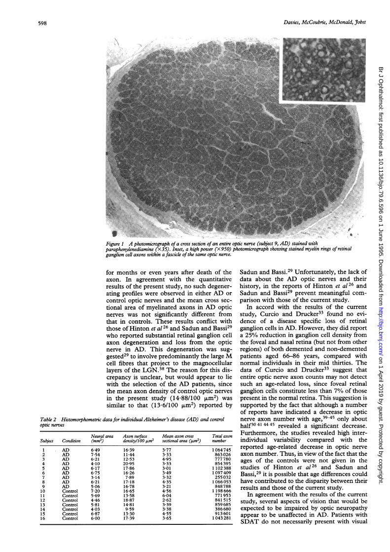
ResultsMyelinated axons in the semi-thin sections ofoptic nerve stained with paraphenylenedi-amine, appeared as dark brown rings of myelinsurrounding unstained axoplasm (Fig 1).There was no significant difference (p=0.71)in the mean cross sectional neural area of con-trol (5.73 (SEM 0 44) mm2) compared withAD (5 74 (0 46) mm2) optic nerves. There wasno significant difference (p=0 87) in the meanmyelinated axon surface density withinfascicles of control (0 1488 (0 0117)/4im2)compared in AD (0 1525 (0 0131)/4tm2) opticnerves. Neither was there any significant differ-ence (p=0 63) in the mean total number ofmyelinated axons in control (859 000 (96 000)compared with AD (881 000 (89 000)) opticnerves. The mean (SEM) myelinated axoncross sectional area in control optic nerves(4 03 (0.42) pum2) was not significantly differ-ent from that in optic nerves from AD patients(3-64 (0-21) [Lm2). The data for individualoptic nerves are given in Table 2.
DiscussionThe results of the current study demonstratethat in subjects with functional vision, AD hasno significant effect on the cross sectionalneural area of the optic nerve, myelinatedaxon surface density within optic nerve fasci-cles, or the total number of myelinated axonsin the optic nerve. Degenerating axons,characterised by large homogeneous darkbrown circular profiles in paraphenylene-diamine stained material, have been previ-ously reported37 to remain in the optic nerve
597
on 1 April 2019 by guest. P
rotected by copyright.http://bjo.bm
j.com/
Br J O
phthalmol: first published as 10.1136/bjo.79.6.596 on 1 June 1995. D
ownloaded from
Davies, McCoubnie, McDonald, Jobst
Figure 1 A photomicrograph of a cross section of an entire optic nerve (subject 9, AD) stained withparaphenylenediamine (X3S) Inset, a high power (X 950) photomicrograph showing stained myelin rings of retinalganglion cell axons within a fascick of the same optic nerve.
for months or even years after death of theaxon. In agreement with the quantitativeresults of the present study, no such degener-ating profiles were observed in either AD orcontrol optic nerves and the mean cross sec-tional area of myelinated axons in AD opticnerves was not significantly different fromthat in controls. These results conflict withthose of Hinton et al 26 and Sadun and Bassi29who reported substantial retinal ganglion cellaxon degeneration and loss from the opticnerve in AD. This degeneration was sug-gested29 to involve predominantly the large Mcell fibres that project to the magnocellularlayers of the LGN.38 The reason for this dis-crepancy is unclear, but would appear to liewith the selection of the AD patients, sincethe mean axon density of control optic nervesin the present study (14 88/100 jim2) wassimilar to that (13 6/100 jim2) reported by
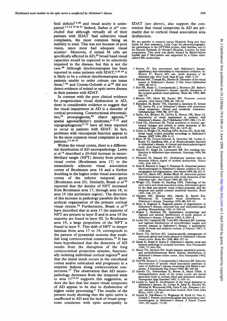
Table 2 Histomorphometric data for individualAlzheimer's disease (AD) and controloptic nerves
Neural area Axon surface Mean axon cross Total axonSubject Condition (mm2) density/100 pum2 sectional area (,trm2) number
1 AD 6 49 16-39 3-77 10647452 AD 7-54 11-44 3-33 8630263 AD 6-21 12-53 4*95 7777804 AD 4-10 20-95 3-33 8543955 AD 6-17 17-86 3-01 1 1023886 AD 6-75 16-26 3-49 10974097 AD 3-14 7-89 3-32 2550328 AD 6-21 17-18 4-35 10660539 AD 5-06 16-78 3-21 84878810 Control 7-20 16-65 4-56 1 19866611 Control 5-69 13-58 6-04 771 95312 Control 4-46 18-87 2-62 841 51513 Control 5-81 14-81 3-39 85968514 Control 4 03 9-59 3-38 38668015 Control 6-87 13-30 4-55 913 60116 Control 6-00 17-39 3-65 1 043281
Sadun and Bassi.29 Unfortunately, the lack ofdata about the AD optic nerves and theirhistory, in the reports of Hinton et al 26 andSadun and Bassi29 prevent meaningful com-parison with those of the current study.
In accord with the results of the currentstudy, Curcio and Drucker33 found no evi-dence of a disease specific loss of retinalganglion cells in AD. However, they did reporta 25% reduction in ganglion cell density fromthe foveal and nasal retina (but not from otherregions) of both demented and non-dementedpatients aged 66-86 years, compared withnormal individuals in their mid thirties. Thedata of Curcio and Drucker33 suggest thatentire optic nerve axon counts may not detectsuch an age-related loss, since foveal retinalganglion cells constitute less than 7% of thosepresent in the normal retina. This suggestion issupported by the fact that although a numberof reports have indicated a decrease in opticnerve axon number with age,39-45 only abouthalf3041 4445 revealed a significant decrease.Furthermore, the studies revealed high inter-individual variability compared with thereported age-related decrease in optic nerveaxon number. Thus, in view of the fact that theages of the controls were not given in thestudies of Hinton et al 26 and Sadun andBassi,29 it is possible that age differences couldhave contributed to the disparity between theirresults and those of the current study.
In agreement with the results of the currentstudy, several aspects of vision that would beexpected to be impaired by optic neuropathyappear to be unaffected in AD. Patients withSDAT do not necessarily present with visual
598
on 1 April 2019 by guest. P
rotected by copyright.http://bjo.bm
j.com/
Br J O
phthalmol: first published as 10.1136/bjo.79.6.596 on 1 June 1995. D
ownloaded from
Myelinated axon number in the optic nerve is unaffected by Alzheimer's disease
field deficits5 646 and visual acuity is unim-paired.5 6 14 15 46 47 Indeed, Sadun et al6 con-cluded that although virtually all of theirpatients with SDAT 'had subjective visualcomplaints, the most common being aninability to read. This was not because of poorvision, since most had adequate visualacuities'. Moreover, if retinal M cells arespecifically affected in AD,29 broad band visualcapacities would be expected to be selectivelyimpaired in the disease, but this is not thecase.48 Although dyschromatopsia has beenreported in some patients with SDAT,6 46 49 itis likely to be a cortical dyschromatopsia sincepatients unable to order colours can namethem,5 46 and Cronin-Golomb et a149 did notdetect evidence of retinal or optic nerve diseasein their patients with SDAT.
In contrast with the poor clinical evidencefor pregeniculate visual dysfunction in AD,there is considerable evidence to suggest thatthe visual impairment in AD is a disorder ofcortical processing. Constructional abnormali-ties,50 prosopagnosia,46 object agnosia,15spatial agnosia/Balint's syndrome,6 15 22 andtopographagnosia5l 52 have all been reportedto occur in patients with SDAT. In fact,problems with visuospatial function appear tobe the most common visual complaints in suchpatients.2 6 46 53
Within the visual cortex, there is a differen-tial distribution ofAD neuropathology. Lewiset al 18 described a 20-fold increase in neuro-fibrillary tangle (NFT) density from primaryvisual cortex (Brodmann area 17) to theimmediately adjacent visual associationcortex of Brodmann area 18 and a furtherdoubling in the higher order visual associationcortex of the inferior temporal gyrus(Brodmann area 20). Similarly, Braak et a120reported that the density of NFT increasedfrom Brodmann area 17, through area 18, toarea 19 (the peristriate region). The directionof this increase in pathology parallels the hier-archical organisation of the primate corticalvisual system.54 Furthermore, Braak et a120have described that in area 17 the majority ofNFT are present in layer II and in area 18 themajority are found in layer III. In Brodmannarea 19, a large proportion of the NFT isfound in layer V. This shift ofNFT to deeperlaminae from area 17 to 19, corresponds tothe pattern of pyramidal neurons that estab-lish long corticocortical connections.55 It hasbeen hypothesised that the dementia of ADresults from the disruption of the longcorticocortical projection systems, function-ally isolating individual cortical regions56 andthat the initial insult occurs in the entorhinalcortex and/or subiculum and progresses in astepwise fashion along corticocortical con-nections.57 The observation that AD neuro-pathology decreases from the temporal stemto area 171820 supports this suggestion, asdoes the fact that the major visual symptomsof AD appear to be due to dysfunction ofhigher order processing.2 The results of thepresent study showing that the optic nerve isunaffected in AD and the lack of visual symp-toms consistent with optic neuropathy in
SDAT (see above), also support the con-tention that visual symptoms in AD are pri-marily due to cortical visual association areadysfunction.We are grateful to research nurses Elizabeth King and AmySmith for their assistance, Celia Cope for photomicrography,the participants in the OPTIMA project, their families, and tothe Keratec Eyebank, St George's Hospital, London, for theircooperation. This work was partly funded by Bristol-MyersSquibb and P McCoubrie was supported by a Glaxo inter-calated BSc studentship.
1 Hutton JT. Eye movements and Alzheimer's disease:significance and relationship to visuospatial confusion. In:Hutton JT, Kenny AD, eds. Senile dementia of theAlzheimer type. New York: Alan R Liss, 1985: 3-33.
2 Mendez MF, Tomsak RL, Remler B. Disorders of the visualsystem in Alzheimer's disease. J Clin Neuro-Ophthalmol1990; 10: 62-9.
3 Hof PR, Boras C, Constantinidis J, Morrison JH. Balint'ssyndrome in Alzheimer's disease: specific disruption ofthe occipito-parietal visual pathway. Brain Res 1989; 494:368-75.
4 Benson DF, Davis RJ, Snyder BD. Posterior corticalatrophy. Arch Neurol 1988; 45: 789-93.
5 Kiyosawa M, Bosley TM, Chawluk J, Jamieson D, SchatzNJ, Savino PJ, et al. Alzheimer's disease with prominentvisual symptoms: clinical and metabolic evaluation.Ophthalmology 1989; 96: 1077-86.
6 Sadun AA, Bochert M, DeVita E, Hinton DR, Bassi CJ.Assessment of visual impairment in patients withAlzheimer's disease. AmJY Ophthalmol 1987; 104: 113-20.
7 Trick GL, Silverman SE. Visual sensitivity to motion: age-related changes and deficits in senile dementia of theAlzheimer type. Neurology 1991; 41: 1437-40.
8 Orwin A, Wright CE, Harding GFA, Rowan DC, Rolfe EB.Serial visual evoked potential recordings in Alzheimer'sdisease. BMJ 1986; 293: 9-10.
9 Rizzo JF III, Cronin-Golomb A, Growdon JH, Corkin S,Rosen TJ, Sandberg MA, et al. Retinocalcarine functionin Alzheimer's disease. A clinical and electrophysiologicalstudy. Arch Neurol 1992; 49: 93-101.
10 Hutton JT, Nagel JA, Loewensen RB. Eye tracking dys-function in Alzheimer-type dementia. Neurology 1984; 34:99-102.
11 Pirozzolo PJ, Hansch EC. Oculomotor reaction time indementia reflects degree of cerebral dysfunction. Science1981; 214: 349-51.
12 Katz B, Rimmer S, Iragui V, Katzman R. Abnormal patternelectroretinogram in Alzheimer's disease: evidence for reti-nal ganglion cell degeneration. Ann Neurol 1989; 26: 221-5.
13 Trick GL, Barris MC, Bickler-Bluth M. Abnormal patternelectroretinograms in patients with senile dementia of theAlzheimer type. Ann Neurol 1989; 26: 226-31.
14 Wright CE, Drasdo N, Harding GFA. Pathology of theoptic nerve and visual association areas. Information givenby the flash and pattern visual evoked potential, and thetemporal and spatial contrast sensitivity function. Brain1987; 110: 107-20.
15 Mendez MF, Mendez MA, Martin R, Smyth KA,Whitehouse PJ. Complex visual disturbances inAlzheimer's disease. Neurology 1990; 40: 439-43.
16 Brun A, Englund E. Regional pattern of degeneration inAlzheimer's disease; neuronal loss and histopathologicalgrading. Histopathology 1981; 5: 549-64.
17 Rogers J, Morrison JH. Quantitative morphology andregional and laminar distributions of senile plaques inAlzheimer's disease. J Neurosci 1985; 5: 2801-8.
18 Lewis DA, Campbell MJ, Terry RD, Morrison JH. Laminarand regional distributions of neurofibrillary tangles andneuritic plaques: in Alzheimer's disease: a quantitativestudy of visual and auditory cortices. J Neurosci 1987; 7:1799-808.
19 Beach TG, McGeer EG. Lamina-specific arrangement ofastrocytic gliosis and senile plaques in Alzheimer's diseasevisual cortex. Brain Res 1988; 463: 357-61.
20 Braak H, Braak E, Kalus P. Alzheimer's disease: areal andlaminar pathology in occipital isocortex. Acta Neuropathol1989; 77: 494-506.
21 Beach TG, McGeer EG. Senile plaques, amyloid P protein,and acetylcholinesterase fibres: laminar distributions inAlzheimer's disease striate cortex. Acta Neuropathol 1992;83: 292-9.
22 Hof PR, Bouras C, Constantinidis J, Morrison JH. Selectivedisconnection of specific visual association pathways incases of Alzheimer's disease presenting with Balint's syn-drome. J Neuropathol Exp Neurol 1990; 49: 168-84.
23 Scholtz CL, Swettenham K, Brown A, Mann DMA. Ahistoquantitative study of the striate cortex and lateralgeniculate body in normal, blind and demented subjects.NeuropatholApplNeurobiol 1981; 7: 103-14.
24 Leuba G, Saini K. Involvement of the visual thalamus inAlzheimer's disease. In: Corian B, Iqbal K, Nicolini M,Winblad B, Wisniewski HM, Zatta P, eds. Alzheimer's dis-ease: advances in clinical and basic research. New York: JohnWiley, 1993: 139-49.
25 Strenn K, Dal-Bianco 0, Weghaupt H, Koch G, Vass C,Gottlob I. Pattern electroretinogram and luminance elec-troretinogram in Alzheimer's disease. J Neural Transm1991; 33 (suppl): 73-80.
599
on 1 April 2019 by guest. P
rotected by copyright.http://bjo.bm
j.com/
Br J O
phthalmol: first published as 10.1136/bjo.79.6.596 on 1 June 1995. D
ownloaded from
Davies, McCoubrie, McDonald, Jobst
26 Hinton DR, Sadun AA, Blanks JC, Miller CA. Optic nervedegeneration in Alzheimer's disease. New Engl J Med1986; 315: 485-7.
27 Blanks JC, Hinton DR, Sadun AA, Miller CA. Retinalganglion cell degeneration in Alzheimer's disease. BrainRes 1989; 501: 364-72.
28 Blanks JC, Torigoe Y, Spee C, Gauderman WJ, BlanksRHI. Ganglion cell loss in the macula of patients ofAlzheimer's disease. Invest Ophthalmol Vis SCi 1990; 31:356.
29 Sadun AA, Bassi CJ. Optic nerve damage in Alzheimer'sdisease. Ophthalmology 1990; 97: 9-17.
30 Martin LI, Pardo CA, Price PL, Troncoso JC. Neuriticpathology in the retina in Alzheimer's disease and aging.J Neuropathol Exp Neurol 1991; 50: 302.
31 Price PL, Pardo CA, Silva JC, Martin 14, Troncoso JC. Theretina and optic nerve in Alzheimer's disease (AD) andaging: a histological and immunocytochemical study.NeurobiolAging 1990; 11: 275.
32 Drucker DN, Curcio CA. Retinal ganglion cells are lostwith aging but not in Alzheimer's disease. InvestOphthalmol Vis SCi 1990; 31: 356.
33 Curcio CA, Drucker DN. Retinal ganglion cells inAlzheimer's disease and aging. Ann Neurol 1993; 33:248-57.
34 American Psychiatric Association. Diagnostic and statisticalmanual of mental disorders. 3rd ed, revised. Washington,DC.
35 McKhann G, Drachman D, Folstein M, Katzman R, PriceD, Stadlan EM. Clinical diagnosis of Alzheimer's disease:report of the NINCDS-ADRDA work group under theauspices ofDepartment ofHealth and Human Services taskforce on Alzheimer's disease. Neurology 1984; 34: 939-44.
36 Mirra SS, Heyman A, McKeel D, Sumi SM, Crain BJ,Brownlee LM, et al, and participating CERAD neuro-pathologists. The consortium to establish a registry forAlzheimer's disease (CERAD). Part II. Standardization ofthe neuropathologic assessment of Alzheimer's disease.Neurology 1991; 41: 479-86.
37 Sadun AA, Smith LEH, Kenyon KR. Paraphenylene-diamine: a new method for tracing human visual pathways.JNeuropathol Exp Neurol 1983; 42: 200-6.
38 Shapley R, Perry VH. Cat and monkey retinal ganglion cellsand their visual functional roles. Trends Neurosci 1986; 9:229-35.
39 Dolman CL, McCormick AQ, Drance SM. Aging of theoptic nerve. Arch Ophthalmol 1980; 98: 2053-8.
40 Balazsi AG, Rootman J, Drance SM, Schulzer M, DouglasGR. The effect of age on the nerve fiber population of thehuman optic nerve. Am J Ophthalmol 1984; 971 760-6.
41 Johnson BM, Miao M, Sadun AA. Age-related decline ofhuman optic nerve axon populations. Age 1987; 10: 5.
42 Mikelberg FS, Drance SM, Schulzer M, Yidegiligne HM,Weis MM. The normal human optic nerve. Ophthalmology1989; 86: 1325.
43 Repka MX, Quigley HA. The effect of age on normalhuman optic nerve fiber number and diameter.Ophthalmology 1989; 96: 26-32.
44 Jonas JB, Muller-Bergh JA, Schl6tzer-Schrehardt UM,Naumann GOH. Histomorphometry of the human opticnerve. Invest Ophthalmol Vis Sci 1990; 31: 736-44.
45 Jonas JB, Schmidt AM, Muiller-Bergh JA, Schlotzer-Schrehardt UM, Naumann GOH. Human optic nervefiber count and optic disc size. Invest Ophthalmol Vis Sci1992; 33: 2012-8.
46 Cogan DG. Visual disturbances with focal progressivedementing disease. Am J Ophthalmol 1985; 100: 68-72.
47 Schlotterer G, Moscovitch M, Crapper-McLachlan D.Visual processing deficits as assessed by spatial frequencycontrast sensitivity and backward masking in normal age-ing and Alzheimer's disease. Brain 1983; 107: 309-35.
48 Kurylo DD, Corkin S, Dolan RP, Rizzo JF III, Parker SW,Growdon JH. Broad-band visual capacities are not selec-tively impaired in Alzheimer's disease. Neurobiol Aging1994; 15: 305-11.
49 Cronin-Golomb A, Rizzo JF, Corkin S, Growdon JH.Visual function in Alzheimer's disease and normal aging.Ann NYAcad Sci 1991; 640: 28-35.
50 Moore V, Wyke MA. Drawing disability in patients withsenile dementia. Psychol Med 1984; 14: 97-105.
51 Landis T, Cummings JL, Benson DF, Prather Palmer E.Loss oftopographic familiarity: an environmental agnosia.Arch Neurol 1986; 43: 132-6.
52 Henderson VW, Mack W, Williams BW. Spatial disorien-tation in Alzheimer's disease. Arch Neurol 1989; 49: 391-4.
53 Neary D, Snowden JS. Perceptuospatial disorder inAlzheimer's disease. Semin Ophthalmol 1987; 2: 141-58.
54 Van Essen DC. Functional organisation of primate visualcortex. In: Peters A, Jones EG, eds. Cerebral cortex. Vol 3.New York: Plenum Press, 1985: 259-329.
55 Jones EG. Laminar distribution of cortical efferent cells. In:Peters A, Jones EG, eds. Cerebral cortex. Vol 1. New York:Plenum Press, 1984: 521-53.
56 Pearson RCA, Esiri MM, Hiorns RW, Wilcock GK, PowellTPS. Anatomical correlates of the distribution of thepathological changes in the neocortex in Alzheimer'sdisease. Proc NadtAcad Sci USA 1985; 82: 4531-4.
57 DeLacoste M-C, White CL III. The role of cortical connec-tivity in Alzheimer's disease pathogenesis: a review andmodel system. Neurobiol Aging 1993; 14: 1-16.
600
on 1 April 2019 by guest. P
rotected by copyright.http://bjo.bm
j.com/
Br J O
phthalmol: first published as 10.1136/bjo.79.6.596 on 1 June 1995. D
ownloaded from