mycotoxigenic fungi and mycotoxin contamination of
TRANSCRIPT

Mycotoxigenic Fungi and Mycotoxin Contamination of Traditionally
Fermented Milk (Mursik) in Soliat Location Kericho County, Kenya.
Keith Kiplangat Talaam
A thesis submitted in partial fulfillment for the Degree of Master of
Science in Molecular Medicine in the Jomo Kenyatta University of
Agriculture and Technology.
2015

ii
DECLARATION
This thesis is my original work and has not been presented for a degree in any other
University.
Signature ….……………….. Date…………………
Talaam Kiplangat Keith
This thesis has been submitted for examination with our approval as University
supervisors.
Signature: ……………… Date……………………
Dr. Bii Christine.
KEMRI, Kenya.
Signature: ……………… Date……………………
Prof. Zipporah Ng’ang’a
JKUAT, Kenya.

iii
DEDICATION
I dedicate this work to my mother; Mrs. Sarah Tanui, my brothers and sisters for their
moral and financial support they gave me through the period of my study.

iv
ACKNOWLEDGEMENTS
I acknowledge Prof. Z. Ng’ang’a of Jomo Kenyatta University of Agriculture and
Technology and Dr. Christine Bii of Centre for Microbiology Research (CMR)-KEMRI for
their guidance and full support during proposal development of this study through to
making the write up of this thesis.
I also acknowledge the KEMRI-CMR staff for their humble time and technical support
which saw me through my bench work. I thank all the farmers who accepted to be part of
this study by allowing me have samples of their fermented milk.
I acknowledge Mr. Kipkurui Mugun for his generosity during my study and Mr Langat
Bernard for his guidance in the thesis write up.

v
TABLE OF CONTENTS
DECLARATION.................................................................................................................. ii
DEDICATION..................................................................................................................... iii
ACKNOWLEDGEMENTS ............................................................................................... iv
TABLE OF CONTENTS .................................................................................................... v
LIST OF FIGURES ............................................................................................................ ix
LIST OF TABLES ............................................................................................................... x
LIST OF PLATES .............................................................................................................. xi
LIST OF APPENDICES ................................................................................................... xii
LIST OF ABBREVIATIONS AND ACRONYMS ........................................................ xiii
DEFINITION OF TERMS................................................................................................ xv
ABSTRACT ....................................................................................................................... xvi
CHAPTER ONE .................................................................................................................. 1
1 INTRODUCTION .......................................................................................................... 1
1.1 Background Information ............................................................................................................... 1
1.2 Statement of the Problem .............................................................................................................. 3
1.3 Justification ................................................................................................................................... 4
1.4 Research questions ........................................................................................................................ 4
1.5 Hypothesis..................................................................................................................................... 5
1.6 Objectives ..................................................................................................................................... 5
1.6.1 General Objective ................................................................................................................. 5

vi
1.6.2 Specific Objectives ............................................................................................................... 5
CHAPTER TWO ................................................................................................................. 6
2 LITERATURE REVIEW .............................................................................................. 6
2.1 Mursik ........................................................................................................................................... 6
2.2 Mursik and its contaminants ....................................................................................................... 10
2.3 Mycotoxigenic fungi ................................................................................................................... 12
2.4 Mycotoxin genes and toxin synthesis ......................................................................................... 15
2.5 Mycotoxins ................................................................................................................................. 19
2.5.1 Mycotoxin Contamination in milk ...................................................................................... 19
2.5.2 Effects of Mycotoxin Contamination .................................................................................. 20
2.5.3 Mechanism of Action of Mycotoxins ................................................................................. 21
2.6 Factors Influencing Fungal Growth and Mycotoxin Production in Food ................................... 22
2.7 Aflatoxins .................................................................................................................................... 22
2.8 Fumonisins .................................................................................................................................. 23
2.9 Deoxynivalenol (DON) or Vomitoxin ........................................................................................ 24
CHAPTER THREE ........................................................................................................... 27
3 MATERIALS AND METHODS ................................................................................ 27
3.1 Study area .................................................................................................................................... 27
3.2 Sample size determination .......................................................................................................... 28
3.3 Study design ................................................................................................................................ 28
3.4 Investigation variables ................................................................................................................ 29
3.5 Mursik Sample collection............................................................................................................ 29
3.6 Storage of samples ...................................................................................................................... 29
3.7 Safety Measures in the Laboratory ............................................................................................. 29

vii
3.8 Mycological Investigations of Mursik ........................................................................................ 29
3.8.1 Identification- Chromogenic Agar Candida (ChroMagar) .................................................. 30
3.8.2 Analytical Profile Index (API 20C AUX- Biomeriux) ....................................................... 30
3.9 Molecular analysis of Mycotoxigenic fungi ............................................................................... 31
3.9.1 Genomic DNA Extraction ................................................................................................... 31
3.9.2 Cloning of Fungal beta-tubulin (Bt) and Internal Transcribed Spacer (ITS) using
Polymerase Chain Reaction (PCR) procedure .................................................................................... 31
3.10 Mycotoxin analysis ..................................................................................................................... 32
3.10.1 Mycotoxin Extraction ......................................................................................................... 32
3.10.2 Mycotoxin Quantification ................................................................................................... 33
3.11 Data Analysis .............................................................................................................................. 34
3.12 Ethical Considerations ................................................................................................................ 34
CHAPTER FOUR .............................................................................................................. 35
4 RESULTS ...................................................................................................................... 35
4.1 Samples Collected in Four Sub-locations in Soliat Location ...................................................... 35
4.2 Fungal Isolates from Mursik ....................................................................................................... 35
4.3 Agarose Gel Electrophoresis of Quantified Genomic DNA ....................................................... 40
4.4 Mycotoxins ................................................................................................................................. 41
4.5 Other variables crucial in mursik processing .............................................................................. 44
CHAPTER FIVE: .............................................................................................................. 46
5 DISCUSSION, CONCLUSIONS AND RECOMMENDATIONS .......................... 46
5.1 Discussion ................................................................................................................................... 46
5.1.1 Fungal contaminants ........................................................................................................... 46
5.1.2 Fungal mycotoxin genes ..................................................................................................... 55
5.1.3 Aflatoxins, Fumonisins and Deoxynivalenol (DON) or Vomitoxin ................................... 56

viii
5.2 Conclusions ................................................................................................................................. 61
5.3 Recommendations ....................................................................................................................... 62
REFERENCES ................................................................................................................... 63
APPENDICES .................................................................................................................... 93

ix
LIST OF FIGURES
Figure 3.1 Map showing the area of study (Soliat Location)…………………………. 27
Figure 4.1 Sample distribution in Soliat Location…………………………………… 35
Figure 4.2 Aflatoxin levels in mursik collected in Soliat Location in 2013…………. 44

x
LIST OF TABLES
Table 4.1 Sample population contaminated with different yeast species………. 36
Table 4.2 Yeast isolates and Colony colors of Candida isolates on
CHROMagar Candida after 72 h incubation at 30oC…………………
37
Table 4.3 Yeast fermentation of sugars; analytical profile index………………. 39
Table 4.4 Quantified levels of fumonisin toxin in mursik collected from Soliat
Location in 2013……………………………………………...............
42
Table 4.5 Quantified levels of deoxynivalenol (DON) toxin in mursik collected
from Soliat Location in 2013…………………………………………
42
Table 4.6 The mean and range of the mycotoxins levels in mursik collected in
Soliat Location in 2013…………………………..…………………..
43

xi
LIST OF PLATES
Plate 4.1 Primary culture plates; showing different colonies of yeast
species……………………………………………………………..
36
Plate 4.2 Macroscopic appearance of Rhodotorula species and Geotrichum
candidum plus the microscopic appearance of Geotrichum
candidum…………………………………………………………..
38
Plate 4.3 Different Candida species on CHROMagar Candida after
incubation at 30 °C for 48 hours…………………………………..
38
Plate 4.4 PCR amplification profiles of beta tubulin gene region of
mycotoxigenic fungi........................................................................
40
Plate 4.5 PCR amplification profiles of rDNA ITS gene region of
mycotoxigenic fungi………………………………………………
41

xii
LIST OF APPENDICES
Appendix I Questionaire……………………………………………… 93
Appendix II: Informed consent document……………………………... 97
Appendix III Analytical Profile Index (API) yeast fermentation of
sugars…..….......................................................................
105
Appendix IV Aflatoxin Quantified levels……………………………… 106
Appendix V Investigative variable primary results………………….. 108
Appendix VI Ethical clearance…………………………………………. 110

xiii
LIST OF ABBREVIATIONS AND ACRONYMS
A. Aspergillus
ACH Acetaldehyde
AFB1 Aflatoxin B1
AFM1 Aflatoxin M1
API Analytical Profile Index
Bt beta tubulin
C. Candida
CFU colony forming units
CMA Corn meal agar
DNA Deoxyribonucleic acid
DON Deoxyvanelol
E. Escherichia
EU European Union
F Fusarium
FAO Food and Agriculture Organisation
FDA Food and Drug Administration
IARC International Agency for Research on Cancer
ITS internal transcribed spacer
KEMRI Kenya Medical research Institute
OSCC Esophageal squamous cell cancer
PCR Polymerase Chain reaction
Ppb parts per billion
PPE Personal Protective Equipment
ppt parts per trillion
rpm revolution per minute
SD Standard Deviation
spp. Species

xiv
USA United States of America
USAID United States Agency for International Development
WHO World Health Organisation
YPDA yeast potato dextrose agar

xv
DEFINITION OF TERMS
Amasi Traditionally fermented milk of South Africa and Zimbabwe.
Kule naoto Traditionally fermented milk among the Maasai community.
Kwerionik Ugandan traditionally fermented milk
Mala Kiswahili reference of fermented milk produced commercially.
Mursik Traditionally fermented milk among the Kalenjin community.
Nono Traditionally fermented milk prepared from unboiled cow milk in
Nigeria.
Sameel Traditionally fermented milk of Saudi Arabia.
Senna
didymobotrya
Plant used by Kalenjin community of Kenya to treat calabashes and milk
before the spontaneous fermentation.
Suusac Traditionally fermented milk among Somalis of Kenya.
Wara White soft non-ripened cheese processed by the addition of a plant extract
(Calotropis procera).
Wo’sek Ashes obtained from Senna didymobotrya used in treating calabashes
before use in milk fermentation.

xvi
ABSTRACT
Mursik is traditional fermented milk. Fungi are among the major contaminants of milk
including mycotoxigenic fungi. Mycotoxigenic fungi may grow in mursik and produce
mycotoxins that can cause poisoning to consumers. This study aimed to enumerate fungal
species contaminants including fungi responsible for mycotoxin production and
quantification of the mycotoxins. Microbiological analysis was done on 194 samples from
Soliat Location and 4 samples from commercial outlets where fungal enumeration was
carried out on Potato Dextrose Agar. Polymerase Chain Reaction (PCR) was used to detect
moulds and mycotoxins extracted using Envirologix procedure and quantified using
quicktox kit. Data was analyzed using SPSS version 17 for descriptive statistics. Yeast
species isolated from mursik samples were: Geotricum candidum 124 (32.56%) as the
predominant strain, Rhodotorula species 19 (4.99%), Sacharomyces cerevisiae 13
(3.41%), Candida parapsilosis 41 (10.76%), Candida albicans 22 (5.77%), Candida
tropicalis 67 (17.59%), Candida glabrata 53 (13.91%) and Candida krusei 21 (5.51%) and
moulds were of Aspergillus, Penicillium and Fusarium genus. Aflatoxin was detected in
196/198 (99.0%) of the samples where levels ranged between 2-12 parts per billion
exceeding the required levels of 0.05ppb. Fumonisin toxin was detected in only 3 (1.5%) of
the samples mean of 0.008ppb and Deoxynivalenol toxin was detected in 1 (0.5%) sample
with the level of 0.001. Both fumonisin and Deoxynivalenol levels were below the standard
levels. Eighty percent of the mursik producers milk their cows in the open and store their
milk and their milk fermenting calabashes at a maize store and/or in the living rooms which
are risk factors for contamination of mursik with mycotoxigenic fungi and other
microorganisms. Microorganisms isolated are responsiple for mycoses and mycotoxins
cause mycotoxicoses. These results will help the authorities to develop measures to tame
the contamination of milk and milk products.
1
CHAPTER ONE
1 INTRODUCTION
1.1 Background Information
In Kenya, dairy industry is increasingly becoming a smallholder farmers’ domain presently,
owning over 80% of the 3 million heads of dairy cattle. It contributes to approximately
56% of the total milk production and 80% of the marketed milk. Similarly, in many third
world countries, livestock rearing and their products is an important part of the national
economy. Subsistence and semi-commercial smallholder farming systems are dominated
by resource constrained farm households (Lanyasunya et al., 2005).
Milk fermentation is a very common practice in Africa. In South Africa, fermented milk
known as Sethemi is made from naturally fermenting milk. Among the Maasais’, fermented
milk is known as Kule naoto and Suusac by the Somalis of Kenya while it’s known as
Amasi in both South Africa and Zimbabwe (Chelule et al., 2010). Among the Kalenjin
community in Kenya, fermented milk is known as Mursik. It is fermented from raw and
boiled milk and prepared in calabashes (gourds) where the calabashes are first treated with
charcoal like material obtained from burning plant called Senna didymobotrya. The milk is
treated with the same and left to ferment naturally for three to four days (Mureithi et al.,
2000). The production of fermented milk in Kenya and other African countries does not
involve the use of starter cultures, suggesting that the fermentation arises spontaneously
from microbes originating from the environment, processing equipment, or processors
(Eyassu et al., 2012).
Milk forms favorable medium for growth and multiplication of microorganisms because of
its high nutrient content. It contains proteins, carbohydrates, minerals and vitamins which
support the growth of many forms of fungi and yeasts. These include species from the
genus, Aspergillus, Penicillium, and Candida (Abd El- Aziz et al., 2011). Thus, besides its
beneficial effects on nutrition, milk though can also act as a vehicle for the transmission of
diseases of bacterial (like brucellosis, tuberculosis, salmonellosis, listeriosis), viral (like

2
hepatitis, foot-and-mouth-disease), rickettsia (Q-fever) or parasitological (toxoplasmosis,
giardiasis) origin. Milk is an excellent culture and protective medium for certain
microorganisms, particularly bacterial pathogens, whose multiplication depends mainly on
temperature and competing microorganisms and their metabolic products. Where milk is
produced under poor hygienic conditions and is not cooled, the main contaminants are
usually lactic acid producers which cause rapid souring. Lactic acid has an inhibitory effect
on pathogenic bacteria but this cannot be depended upon to provide a safe milk product
(Heeschen, 1994).
According to Saadia (2010), Aspergillus spp., Alternaria spp., Fusarium spp., Neurospora
spp. are the most common fungi species found in milk products. Other documented milk
contaminants includes: Aspergillus glaucus, A. niger, Alternaria spp., Cheotomium
candidum, Cladosporium herbarum, Fusarium spp., Monilia spp., Mucor rouxii,
Neurospora spp., Penicillium expansum, Penicillium spp., Rhizopus nigricans,
Sporotrichum carinis and Thamnidium elegans. Fungi of the genus, Fusarium, Aspergillus,
Penicillium, Cladosporium, Geotrichum and Cladosporium species have been isolated in
milk during different seasons in different geographical locations (Pešić-Mikulec et al.,
2005). Most of these fungi have been known to be mycotoxin producers (Uzeh et al.,
2006).
With the numerous species of fungi present in milk with mycotoxic potential, there is a
possibility that there will be contamination with multiple toxins (Whitlow et al., 2005). Use
of available feed resources is further complicated by farmers’ inability to use them before
they spoil especially during wet season. Colonization of molds in wet season results in
mycotoxin risk mainly associated with favorable climatic conditions, poor feed handling
and storage practices. Mycotoxins are passed to milk when the cows are fed with
contaminated feeds and also fungi responsible for these toxin productions can colonize
milk thus producing significant amounts of toxins under poor handling practices
(Lanyasunya et al., 2005).
According to Kang’ethe and Lang’a (2009), in a study carried out to determine the level of
aflatoxin in milk in urban towns, thirty five percent (in Nairobi, Nakuru, Nyeri, Eldoret and

3
Machakos) of the positive samples had aflatoxin levels exceeding 0.05ppb, the FAO/WHO
and EU acceptable level of aflatoxin M1 and aflatoxin M2 for milk. A larger number of
people consume fresh and fermented milk in the rural areas. No report has been
documented on the mycotoxin contamination of the milk in these areas which poses a
health risk. Additionally, fermented milk undergoes a process which is traditionally
practiced, providing favorable conditions for milk contamination with mycotoxin
producing fungi. Thus, the objective of this study was to enumerate fungal contaminants
including fungi responsible for mycotoxin production and quantification of the mycotoxins
in mursik collected in Soliat Location.
1.2 Statement of the Problem
Mursik is a common diet among the Kalenjin community. Fungi and other microbes can
colonize fermented milk since they can survive a wide range of temeperatures and
fermentation. Warm humid conditions of 18oC-37
oC, poor livestock feeds and conditions of
storing milk and milk products handling favour the growth of mycotoxigenic fungi. The
formation of mycotoxins in nature is considered a global problem though in certain
geographical areas of the world, some mycotoxins are produced more readily than others
(Lawlor & Lynch, 2005). In most European countries aflatoxins are not considered to be a
major problem. However, vomitoxin, ochratoxin, Zearalenone are found more frequently.
Aflatoxins are common in Asian and African countries and certain parts of Australia. In a
study done in Iran, where detection of Aflatoxin M1 (aflatoxin metabolite) in Iranian milk,
showed that 80% of the samples had AFM1 greater than the maximum tolerance limits
(0.05ppb) accepted by the European Union and Iranian national standards (Tabari, 2010).
In United States, 10% excess of primary liver-cell cancer was observed in the Southeast,
where the estimated average daily intake was high (IARC, 1993). In Kenya, Kang’ethe and
Lang’a (2009), indicated that 99% of pasteurized market milk was contaminated with
aflatoxin M1 where 31% of the contaminated samples exceeded the WHO/FAO levels of
0.05ppb. Contamination of mycotoxins will lead to mycotoxicoses in consumers.

4
1.3 Justification
Owing to its high nutrient content milk is a good culture media for microorganisms.
Fermentation of milk allows the growth of some microbes while inhibiting the growth of
others. Research has shown that fungi can colonize fermented milk since they can survive a
wide range of pH and temperatures. There was therefore need to research on fungal
contaminants in Mursik as these can survive processes like boiling done to freshly retrieved
milk from the cows before fermenting. Also, to determine the presence of mycotoxins as
these may be produced by fungal contaminants or passed along the food chain through
consumption of contaminated feeds. Fungal contamination is accelerated by poor milk
handling methods and storage of animal feeds. Mycotoxins pose a health hazard to public
health and no research has been done in Kenya to determine the presence of fungal and
mycotoxin contaminants in Mursik. The rise in chronic diseases including cancer may be
associated with mycotoxins exposure. The study site has all the favorable conditions for
fungal growth and people ferment milk traditionally and practice mixed farming. Isolation
of fungi responsible for mycotoxins has been done majorly in fresh milk. Aflatoxin is one
of the most widely occurring and dangerous mycotoxins and is the most studied toxin
(C.A.S.T. 1989).Thus this study included the study of other mycotoxin like, fumonisin,
vomitoxin by quantifying them because they have been known to cause illnesses in both
humans and animals. The study focused on quantifying mycotoxins in traditionally
fermented milk (Mursik) and detects mycotoxigenic fungi responsible for mycotoxin
production which has not been conducted by the fore researchers. This is essential given
the public health implication of mycotoxin exposure and the risk of carcinogenic,
hepatotoxic and mutagenic potential of mycotoxins (Riley et al., 2001). This study aimed
to enumerate fungal species contaminants including fungi responsible for mycotoxin
production and quantification of the mycotoxins.
1.4 Research questions
i. What are the types of fungi contaminating traditionally fermented milk (mursik)
from Soliat Location, Kericho County?
ii. What are the types of mycotoxins contaminating the milk and their levels?

5
iii. Which genes coding for mycotoxigenic fungi are present in fermented milk from
Soliat location; Kericho County?
1.5 Hypothesis
Null hypothesis: Traditionally fermented milk (mursik) is contaminated with
mycotoxigenic fungi and mycotoxins.
1.6 Objectives
1.6.1 General Objective
To investigate mycotoxigenic fungi and mycotoxin contamination of fermented milk
produced in Soliat location, Kericho County.
1.6.2 Specific Objectives
1. To identify fungi from fermented milk (mursik) from Kericho county.
2. To detect genes of fungi responsible for mycotoxin production in mursik.
3. To establish the level of aflatoxin, fumonisins and deoxynivalenol contamination in
the fermented milk (mursik).
6
CHAPTER TWO
2 LITERATURE REVIEW
2.1 Mursik
Spontaneous fermentation and preferential selection of those foodstuffs whose
sweetness was improved by this process, remains the origin of consumable fermented
products (Stiles, 1996) and allowed our ancestors to keep foodstuffs at ambient
temperature. Fermented milk was first produced around 10’000 years ago (Tamime, 2002)
when the Neolithic Revolution transformed hunters and gatherers into settled land-
based communities. The first evidence that cattle, sheep and goats were domesticated for
milk production dates back to 5000 BC (Teuber, 1995). The actual origin of fermented
milks is unknown but there is no doubt that their consumption dates back to prehistoric
times (Helferich & Westhoff, 1980). Milk fermentation is dating to 1300 BC cited in the
Old Testament (Roginski, 1988). Cheese and butter consumption by the general population
in the ancient Kingdom of Meroe (690 BC – AD 323) is recorded in detail (Abdelgadir et
al., 1998).
In many third world countries, Kenya included, livestock rearing and their products is an
important part of the national economy (Lanyasunya et al., 2005). In regard to its high
nutrient content, milk form a favourable habitat for growth and multiplication of
microorganisms. Milk supports the growth of many forms of fungi and yeasts. These
include species from the genus Aspergillus, penicillium, and Candida (Abd El- Aziz et al.,
2011). Fermentation is a method of food preservation, used from old age. The traditional
practice of fermenting milk products is common in Asia, Africa, the Middle East, and
Northern and Eastern Europe (Savadogo et al., 2004). For more than 330 years ago, it
became part of the cultural and traditional norm among the indigenous communities mostly
in the third world nations, majorly in Africa (Chelule et al., 2010; Abeer et al., 2009).
Traditional fermentation is a way of food processing, where microbes, lactic acid bacteria
for example, Lactobacillus, Lactococcus, Streptococcus and Leuconostoc species are used.

7
Fermentation of milk mainly involves lactic acid bacteria, but micrococci coryneforms,
yeasts and moulds can be involved (Zamfir et al., 2006; Ogier et al., 2004; Hassan et al.,
2006). In lactic acid (non-alcoholic) fermentation, lactic acid is the main by-product of the
fermentation process, the pH of the ferment is always lower than 5. Foods processed using
fermentation process include: beverages, dairy products, cereals and even meat products
(Chelule et al., 2010).
In general, fermented food products constitute a major portion of the daily diet in Africa
(Jespersen, 2003; Sanni, 1993). The use and production of fermented milk is an old
tradition among Kenyans. Every tribe has their own unique methods of producing
fermented milk, and the differences between fermented milks are based on the location of
tribal communities and the different production processes. Fermentation of milk is an
ancient practice of man which has been passed down from generation to generation. The
aim of fermenting was to obtain products with characteristic flavor, aroma and consistency
and at the same time, could be stored unspoiled for a longer time than untreated raw milk
(Chelule et al., 2010).
In Kenya, traditional fermented milk products are mainly produced by pastoral
communities such as the Maasai, Borana, Kalenjins, Gusii and Somali. They are mainly
produced by spontaneous fermentation of the milk in traditional containers such as gourds
and skin bags. Some of these fermented milks have been reported to have health beneficial
properties such as Kule naoto produced by the Maasai, mursik produced by the Kalenjin
and ambere amaruranu produced by the Gusii (Mathara et al., 2008; Mathara et al., 1995;
Mokua, 2004). Mursik is mainly produced by the Kalenjin community in Kenya, through
spontaneous fermentation of cow milk in a traditionally prepared gourd (Mathara et al.,
1995; FAO, 1990). It forms a major part of the Kalenjin diet due to its delicious taste and
belief that it improves health (Mathara et al., 1995). The Kalenjin also value it as a special
drink that is shared in special occasions to symbolize success of certain activities such as
successful marriage negotiations and weddings, victory in athletics among other events.
In the Kalenjin tribe the fermented milk, mursik, is produced in specific calabash gourds,
also known as sotet. Sotet, a traditionally treated gourd is made from the hollowed out dried

8
fruit of the plant Lagenaria siceraria. Some days before the milk is treated, a small branch
of an Ite or Senetwet tree (Senna didymobotrya) is cut and allowed to dry. One end of this
tree is first burned in a fire and a dried, cleaned gourd is rubbed on the inner surface gently
with it. A chopped end stick from the tree Olea africana locally known as Enkidogoe in
Maasai and Sosiot in Kalenjin is used to break charcoal inside (Mathara, 1999).This is
repeated several times until the gourd is fully coated inside with charcoal dust. This process
reduces the porosity of the gourd and improves the flavor. The gourd is filled with milk
which is also treated with charcoal from Senna dydimobotrya tree and then closed by a
special cap obtained from a cow hide and left to ferment naturally by microbes from the
environment or those pre-exist in milk. Repeated use of naturallyoccurring microbiota to
inoculate fresh milk, commonly referred to back-slopping, has been the
developmental keystone of traditional fermented milk products worldwide (Wouters et
al., 2002). After fermentation, the product is gently shaken before consumption. Some
Kalenjin groups also add small quantities of blood obtained from prickling a vein in the
neck region of a healthy bull, and from which fibrin has been removed by gentle stirring.
Addition of blood can impact the microbial metabolism, as iron is an important cofactor for
a number of essential cellular processes. A thick bluish layer forms on the surface when
mursikis ready. It is shaken well before drinking, to ensure that a uniformly thick emulsion
is formed. In some Kalenjin households, fermented milk is consumed several times daily
(Mikko et al., 2012).
Fermentation and all of these additions to the process are used to improve the odor, the
taste, and the flavor of fresh milk. In an interview with farmers, Mureithi and colleagues
noted that farmers were of the opinion that fresh milk smells and tastes like cow urine and
had to be improved before it can be consumed. Also, lack of refrigeration, and the need to
store milk for the dry season (when milk production decreases due to a lack of pasture)
required that excess milk be stored for a longer time. For example, the Pokot developed
chekha mwaka, specially treated milk that could be stored for more than a year without
getting spoiled (Mureithi et al., 2000).
Some of the herbs have been known suitable for the purposeof imparting the preservative
and aromatic effect to milk. Some of the plant herbs used by Kalenjins in Kericho County
9
include sertwet (Acacia meansii), simotwet (Ficus thoningii), suriat (Rhus natalensis),
muokiot (Lippia kituiensis), Olea Afrikana (emitiot) with a majority preferring to use
Senna didymobotrya (senetwet), mostly citing its ease of availability (Wangila et al.,
2014). Products of S. didymobotrya used in the treatment of calabashes and fresh milk
have been known to be medicinal among the Kalenjin community. In Kenya, traditionally
Kipsigis community has been using these plants to control malaria as well as diarrhea. In
addition, they use to treat skin conditions of humans and livestock infections as well. S.
didymobotrya is useful in the treatment of fungal and bacterial infections (Raja, 2012).
Mathara et al., 1995 and Nakamura et al., 1999, established that Mursik culture consist of
lactic acid bacteria (LAB) species which include Lactococcus lactis subsp. lactis,
Leuconostoc mesenteroides subsp. dextranicum, Lb. curvatus, Leuc. paramesenteroides
and Lb. planturum. The local dairy products produced and widely consumed by Fulanis in
Nigeria are Nono and wara; Nono is prepared from unboiled cow milk collected in a
calabash and allowed to ferment naturally while Wara is a white soft non-ripened cheese
processed by the addition of a plant extract (Calotropis procera) to the non-pasteurized
whole milk from cattle. The nature of fermented products varies from one region to
another. It depends on the local indigenous microflora, which in turn reflects the climatic
conditions of the area (Savadogo et al., 2004). Studies have shown that; Aspergillus niger,
Aspergillus fumigatus, Penicillium chrysogenum, Rhizopus spp., Fusarium moniliforme,
and Trichoderma reesii are the fungi mostly isolated from the two products (Uzeh et al.,
2006; Abeer et al., 2009).
Fermented dairy products for example, yoghurt, cheese and sour milk are protein-rich milk
products. They have been acclaimed both by popular believe and some research findings as
being more nutritious and health promoting than fresh milk. Reports by Platt (1964) stated
that fermented milk is a good source of the B vitamins, including vitamin B12. There are
also claims that the digestibility of the milk proteins is improved by fermentation (Marshal,
1986). Yoghurt, a fermented milk product, has the ability to kill pathogens and build the
immune system against invading organisms. People of Finland consume large amounts of
yoghurt and it has been suggested that they harbor numerous intestinal lactobacilli that
have anticarcinogenic properties (O’ Sullivan et al., 1992). According to Mathara et al.,

10
1995 some of the LAB has antimicrobial properties against pathogenic bacteria species
such as Staphylococcus aureus and Salmonella typhimurium.
Lactobacillus bacteria have been known to produce exopolysaccharides (EPS), components
which contribute to the beneficial health effects. These exopolysaccharides are excreted by
microorganisms onto the surface of their cell walls as cohesive layers or into the growth
medium as slime (ropy) exopolysaccharides. A wide variety of microorganisms
including moulds, yeasts, bacteria and algae have been reported to produce these EPS
(Kumar et al., 2007). EPS have been reported to contain properties such as oligofructans,
glucooligosaccharides and β-glucan are prebiotic or bifidogenic thus boosting health to the
consumers (Badel et al., 2011; Korakli et al., 2003). EPS has other health beneficial
effects which include reduction of blood cholesterol levels, ant-carcinogenic, anti-
temporal and immunomodulation activity (Ruas and Reyes, 2005). LAB EPS enhance
the attachment and colonization of the gut by probiotics and this antagonizes the effects
of pathogenic microorganisms. Lactobacillus rhamnosus RW-9595M produces EPS
which increase production of the cytokine IL-10 by the macrophages, and these
prevent development of inflammatory conditions (immunesuppression) in the gut
(Lebeer et al., 2010; O’connor et al., 2005).
2.2 Mursik and its contaminants
Fermentation does not take place in controlled systems or sterilized conditions; as a result
contamination with yeasts and some pathogenic bacteria may arise, thus making fermented
milks to become major vehicles of transmission for many food borne pathogens. Food
borne diseases caused by these pathogens were problematic and must have continually
preoccupied early man (Uzeh et al., 2006). Unhygienic milking procedures and equipment
used for milking, filtering, cooling, storing or distributing milk is also an important source
of microorganisms. This situation is aggravated if the equipment is not properly cleaned
and sanitized after use. Milk residues left on equipment and utensil surfaces provide
nutrients to support the growth of many microorganisms, including pathogens (Bryan,
1983). Poor hygiene, practiced by handlers of food products, may lead to introduction of
pathogenic microorganisms into the products and since they do not undergo further

11
processing before consumption, these foods may pose risk to consumers (Kang’ethe &
Lang’a, 2009).
Fermented products have been reported to exhibit the highest incidence and level of yeast
contamination due to the low pH which forms a selective environment for their growth
(Vogel et al., 2002). Hence, different yeast species have been isolated from fermented
dairy products (Mathara et al., 2004). Their presence is due to various properties such as
fermentation or assimilation of lactose, production of extracellular proteolytic and lipolytic
enzymes, assimilation of lactic acid and citric acid, growth at low temperatures and
tolerance to elevated salt concentrations (Akalin et al., 2006).
Yeasts are widely distributed in nature and are therefore often found as contaminants in
both commercial and traditional fermented milk and have been enumerated and identified
in a wide variety of African fermented food products including milk (Fleet and Mian, 1987;
Fleet, 2007; Jespersen, 2003). The presence of yeasts indicates that they are able to
proliferate during milk fermentation and positively react with LAB (Pereira-Dias et al.,
2000; Gadaga et al., 2001). In yoghurt, their occurrence is mainly a consequence of the
contamination and hence they are a major cause of yoghurt spoilage (Fleet, 1990).
However, in traditionally fermented milk, yeasts are part of the indigenous microflora,
coming into the product with the raw milk or from the environment and containers (Gadaga
et al., 2000).
The yeast species frequently found in dairy products include Debaryomyces hansenii,
Candida famata, Kluyveromyces marxianus, Candida kefyr, Candida stellata,
Saccharomycopsis lipolytica, Saccharomyces cerevesiae, Candida krusei, Rhodotorula
glutinis and Rhodotorula rubra. However, D. hansenii and Kluy. marxianus, as well as
their asporagenous equivalents C. famata and C. kefyr respectively, emerge as the most
prevalent ones (Fleet, 1990). Al-Otaibi, (2012) isolated Candida lusitania, Cryptococcus
laurentii, Saccharomyces cerevisiae and Candida kefyr Sameel, traditionally fermented
milk in Saudi Arabia. Saccharomyces cerevisiae, Candida Lusitania, Candida colliculosa
and Saccharomyces dairenensis were the predominant strains in isolated Zimbwabwean
traditional fermented milk (Gadaga et al., 2000). Abdelgadir et al. (2001), in Sudan and

12
Shuangquan et al., (2006) in Mongolia, found that the predorminant yeast strains in the
fermented milk were Sacharomyces cerevisiae and Candida kefyr.
The yeast isolated in susaac, Kenyan traditionally fermented camel were, Candida krusei,
Geotricum penicillatum and Rhodotorula muciloginosa (Lore et al., 2005). This shows that
different yeast species were predorminant in different fermented milk products. Moulds
which have been known mainly in milk and cheese fermentation and include, Penicillium,
Mucor, Geotrichium, and Rhizopus species. Uzeh et al., (2006), isolated moulds of the
species, Aspergillus niger, Aspergillus fumigatus, Penicillium chrysogenum, Rhizopus spp,
Fusarium moniliforme, and Trichoderma reesii in nono and wara, traditionally fermented
milk products of Nageria. The fungi Aspergillus, Penicillium, Rhizopus, Fusarium, and
Trichoderma species which were isolated are identified spore formers, which therefore
mean that they can easily contaminate the dairy products which are usually exposed during
processing, storage, and hawking. The fungi of the genus Aspergillus, Peniccillium,
Fusarium have been among the contaminants of raw milk and are major spoilage
organisms of carbohydrate foods (Alborzi et al., 2005; Rhodes & Fletcher, 1966).
However, their growth can result in the production and accumulation of mycotoxins which
are of public health and economic importance.
2.3 Mycotoxigenic fungi
Mycotoxigenic fungi are mould which produce mycotoxins as their secondary metabolites
that exert toxic effects on animals and humans. The toxic effect of mycotoxins on animal
and human health is referred to as mycotoxicosis (CAST, 2003). Mycotoxigenic fungi are
readily isolated from diverse environmental samples, soil and plant tissues or residues, are
considered the natural habitat of these fungi (Cotty, 2004). Soil serves as a reservoir for
primary innocullum for the contamination (Abbas et al., 2006). Mycotoxigenic fungi has
been enabled by their phenotypic and metabolic flexibility to colonize a broad range of
agriculturally important products and to adapt to a range of environmental conditions.They
are structurally diverse, deriving from a number of biosynthetic pathways and their effect
upon consumers is equally diverse ranging from acutely toxic to immunosuppressive or
carcinogenic (Moretti et al., 2013).

13
Colonization of mycotoigenic fungi on foods, affects the quality of food, their organoleptic
attributes, and nutritional quality. Fungi same as other microorganisms will assimilate and
utilize the most readily available nutrients in the materials they grow upon thus resulting in
food spoilage (Cegielska-Radziejewska et al., 2013). Fungi of the specis Aspergillus niger,
Aspergillus fumigatus, Penicillium chrysogenum, Rhizopus spp, Fusarium moniliforme, and
Trichoderma reesii has been known to contaminate fermented milk of all kinds at different
geographic locations (Uzeh et al., 2006; Pesic-Mikulec et al., 2005). Moulds of genera,
Aspergillus, Penicillium, Rhizopus, Fusarium, and Trichoderma species which were
isolated in traditionally fermented milk nono of Nigeria were identified as spore formers,
which therefore mean that they can easily contaminate the dairy products which are usually
exposed during processing (Uzeh et al., 2006). Species that belong to the
genera Aspergillus, Penicillium, Fusarium, and Alternaria have been considered the most
toxic which produce mycotoxins such as aflatoxins, zearalenone, T2-toxin, deoxynivalenol,
ochratoxin A, fumonisins, and patulin the most common mycotoxins found in animal feeds
and food (Pitt & Hocking, 2009; Hussein & Brasel, 2001). Mycotoxin production is a
complex secondary metabolite process (Geiser et al., 1998).
In the recent past, molecular techniques have increased the possibilities to characterize milk
microbial ecology. Amplification of specific DNA fragments using polymerase chain
reaction (PCR) and specific probes is extensively sensitive and has the potential to detect
the presence of fungi in agricultural commodities (Manonmani et al., 2005). The use of
PCR to identify mycotoxin fungi is attracting considerable attention (Haughland et al.,
2004).
Genotyping techniques have shed new light on mycotoxin producing fungi and provided
the foundation for advances in detection methodology. Historically, fungi have been
identified on the basis of traditional taxonomic characteristics (for example, morphological
features); more recently, the tools of molecular biology have enabled genetic analysis and
classification on the basis of nucleic acid sequence. Since analytical methods for detecting
mycotoxins have become more prevalent, sensitive, and specific, surveillance of foods for
mycotoxin contamination has become more common (Michealsen et al., 2006).

14
Internal Transcribed spacer (ITS) regions contain the most conserved sequence at the
terminal region and also contain the hyper variable sequences distinguishing between
species. The use of ITS region as compared with other molecular probes is advantageous
due to many reasons including increased sensitivity because of existence of more than 100
copies per genome (Mirhedi et al., 2007).
Until recently, the molecular identification of fungi to species level has been based mostly
on the use of variable internal transcribed spacer (ITS) regions. The non-coding ITS
regions have a high copy number in the fungal genome as part of tandemly repeated
nuclear rDNA. These regions benefit from a fast rate of evolution, which results in higher
variation in sequence between closely related species, in comparison with the more
conserved coding regions of the rRNA genes (Michealsen et al., 2006).
As a consequence, the DNA sequences in the ITS region generally provide greater
taxonomic resolution than those from coding regions (Lord et al., 2002; Anderson et al.,
2003). In addition, the DNA sequences in the ITS region are highly variable, divergent, and
distinctive, and might serve as markers for taxonomically more distant groups (Michealsen
et al., 2006).
The coding portions of many fungal 18S, 5.8S and 28S rDNA genes are greatly preserved
and primers to these regions have been generated (Brown et al., 2001). These allow the
isolation of the internal transcribed spacer sequences, which lie between the coding
regions, from a wide range of fungi. The ITS region is amplified from the target fungus and
polymorphism within the ITS region is generally at the level of species, rather than
between isolates of the same species, making it an ideal target for the development of
species-specific PCR assays (Nule, 2001).
The tubulin gene family consists of three major highly conserved sub-families, alpha-,
beta-, and gamma-tubulin, which arose from a series of gene duplications in early
eukaryotic evolution (Keeling & Doolittle, 1996). The tubulin genes are made up of highly
conserved proteins which are the principle structural and functional components of
eukaryotic microtubules that are major components of the cytoskeleton, mitotic spindles,
and flagella of eukaryotic cells (Thon & Royse, 1999).

15
Several studies have made use of beta-tubulin genes to examine relationships among fungi
at all levels, and has been found to be a useful tool in both deep level phylogenetic studies
and studies of complex species groups (O'Donnell et al., 1998a). Studies that have used the
beta-tubulin gene for characterization of Fusarium species include Geiser et al., (2001),
Mach et al., (2004), Reischer et al., (2004), and Yli-Mattila et al., (2004). Geiser et al.,
(2001) used the beta-tubulin gene, as well as the EF-la gene, to identify and characterize F.
hostae. Mach et al., (2004) employed the beta-tubulin gene for the early and specific
detection of Fusarium langsethiae, and distinguishing it from related species of section
Sporotrichiella. Yli Mattila et al., (2004) demonstrated that the beta-tubulin gene was able
to distinguish between Fusarium poae, Fusarium sporotrichioides, Fusarium langsethiae
and Fusarium kyushuense.
2.4 Mycotoxin genes and toxin synthesis
Mycotoxin production is a complex secondary metabolite process. Most of the specific
enzymatic activities required for mycotoxin production are encoded in a gene cluster but
additional unlinked loci also are required (Geiser et al., 1998). Aflatoxins are toxic
secondary metabolites produced by a 70-kb cluster of genes in Aspergillus flavus. The
cluster genes are coordinately regulated and reside as a single copy within the genome
(Carrie et al., 2007). Jiujiang et al., (1995) showed that at least nine genes involved in the
aflatoxin biosynthetic pathway are located within a 60-kb DNA fragment.
The genes, nor-1, aflR, ver-1, and omtA, have been cloned in A. flavus and A. parasiticus.
In addition, five other genes, pksA, uvm8, aad, ord-1, and ord-2 have been cloned in A.
parasiticus. The pksA, aad, and uvm8 genes exhibit sequence homologies to polyketide
synthase, aryl-alcohol dehydrogenase, and fatty acid synthase genes, respectively. The ord-
1 and ord-2 genes involved in later steps of aflatoxin biosynthesis, have been determined;
the ord-1 gene product exhibits homology to cytochrome P-450-type enzymes. Order of
aflatoxin pathway genes within 60-kb DNA region has been found to be pksA, nor-1, uvm8,
aflR, aad, ver-1, ord-1, ord-2, and omtA in A. parasiticus and nor-1, aflR, ver-1, ord-1, ord-
2, and omtA in A. flavus. The order is related to the order in enzymatic steps required for
aflatoxin biosynthesis (Jiujiang et al., 1995).

16
Significant progress in understanding of fungal secondary metabolism include the
discovery of sterigmatocystin (ST) biosynthetic gene clusters and the discovery of a G-
protein-mediated growth pathway in A. nidulans regulating secondary metabolism
production (Tag et al., 2000; Brown et al., 1996). According to Barnes et al. (1994)
sterigmatocystin is a highly toxic intermediate in the AFBl biosynthetic pathway. AFBl
biosynthetic pathway in A. flavus and A. parasiticus and the ST biosynthetic pathway in A.
nidulans are believed to be similar. It is now apparent that structural genes required for
secondary metabolite production are usually clustered and that the regulation of the
clustered genes is largely dependent on pathway-specific transcription factors (Tsuji et al.,
2000; Keller & Hohn, 1997).
Linkage of aflatoxin pathway genes was first evidenced in an A. parasiticus cosmid clone,
NorA that contains both nor-1and ver-1 genes (Skory et al., 1992). A physical and
transcriptional map of the 35-kb genomic DNA insert in cosmid NorA suggested that
several genes involved particularly in the early stages of aflatoxin B1biosynthesis are
clustered on one chromosome in A. parasiticus. These include the pksA gene, which codes
for a polyketide synthase, the nor-1gene, which codes for a reductase that converts
norsolorinic acid to averantin, the ver-1gene, which is involved in the conversion of
versicolorin A to sterigmatocystin and the omtA gene, coding for an S-adenosyl
methionine-dependent O-methyltransferase that converts sterigmatocystin to O-methyl
sterigmatocystin and dihydrosterigmatocystin to dihydro-O-methylsterigmatocystin (Chang
et al., 1992; Skory et al., 1992; Cleveland et al., 19987). The omtA gene has been cloned
and characterized for both A. parasiticus and A. flavus (Yu et al., 1993). In addition to
these structural genes, a regulatory gene, aflR (previously named afl-2 for A. flavus and
apa-2 for A. parasiticus), that codes for a regulatory factor (Aflr protein) has been cloned
and was shown to be involved in the activation of pathway gene transcription (Chang et al.,
1993).
The step for the conversion of O-methyl sterigmatocystin and dihydro-O-
methylsterigmatocystin to AFB1and AFB2 appears to be catalyzed by an oxidoreductase
enzyme complex, which may consist of two or more subunits. Genes that code for the
polypeptides of this enzyme complex have not yet been characterized. In addition, a

17
putative fatty acid synthase gene, uvm8, potentially involved in polyketide backbone
synthesis, first identified by complementation of a UV mutation and a gene, aad,
homologous to aryl-alcohol dehydrogenase potentially involved in an intermediate step of
aflatoxin biosynthesis have been cloned (Jiujiang et al., 1995).
Aspergillus nidulans, produce the mycotoxin sterigmatocystin (ST), which also serves as
the penultimate precursor in the aflatoxin (AF) biosynthetic pathway (Cole & Cox, 1981).
Both AF and ST are among the most toxic, carcinogenic and mutagenic compounds. Study
of the A. nidulans ST pathway has led to identification of a 60 kb gene cluster (the ST
Cluster; stc) that includes 25 co-regulated genes, many of which have been shown to
function in ST biosynthesis (Brown et al., 1996). Transcription of all of these genes is
dependent upon the activity of aflR, a pathway-specific regulatory gene found within the
ST cluster that encodes a zinc binuclear cluster type DNA binding protein (Woloshuk et
al., 1994). aflR expression is regulated during the life cycle such that aflR mRNA begins to
accumulate early in the stationary phase and activation of other genes required for ST
biosynthesis quickly follows (Yu et al., 1996a).
All fumonisin biosynthetic (FUM) genes characterized to date are localized within a 42.5
kb region of the F. Verticillioides genome (Proctor et al., 2003). The clustering of genes
involved in the biosynthesis of secondary metabolites in filamentous ascomycetes is
common. A fumonisin biosynthetic gene (FUM) cluster has been described in species
Fusarium proliferatum, Fusarium verticillioides and one of the rare fumonisin-producing
strains (FRC O-1890) of F. oxysporum and in the more distantly related fungus Aspergillus
niger (Proctor et al., 2008; Khaldi & Wolfe, 2011). The Fusarium cluster includes 16 genes
that encode biosynthetic enzymes as well as regulatory and transport proteins. Each
selected FUM gene (FUM1, FUM3, FUM6, FUM7, FUM8, FUM10, FUM13, FUM14 and
FUM19) is required for wild-type fumonisin production in F. verticillioides, and together
they span almost the entire length of the FUM cluster (Alexander et al., 2009). The PM
genes included eight genes (CAL1, CPR1, HIS3, RED1, RPB1, RPB2, TEF1and TUB2) that
have been used previously to infer phylogenetic relationships within Fusarium; two global
regulatory genes, LAE1 and FLB2, that also affect fungal secondary metabolism and a
18
dehygrogenase gene, ZBD1, that flanks the FUM cluster in F. verticillioides (Proctor et al.,
2003; Proctor et al., 2009; Wiemann et al., 2010).
Functions of most of the genes in fumonisin biosynthesis in F. verticillioides have been
determined by gene inactivation and heterologous expression analyses. Within the cluster,
the FUM1gene encodes a polyketide synthase that catalyses synthesis of a linear polyketide
that forms the backbone structure of fumonisins. In addition, the FUM8 gene encodes anα-
oxoamine synthase that mediates whether fusaria produce FBs or FCs by catalysing the
condensation of the linear polyketide with alanine to produce FBs or with glycine to
produce FCs. The number, order and orientation of genes within FUM clusters in F.
verticillioides, F. oxysporum and F. proliferatum are the same (Proctor et al., 2008).
Many of the Deoxynivalenol (DON) biosynthesis genes are localized in a gene cluster of at
least 10 genes. The genes in this cluster include those for trichodiene synthetase (tri5),
P450 oxygenase (tri4 and tri11), acetyltransferase (tri3 and tri7), transcription factors (tri6
and tri10), a toxin efflux pump (tri12), and two unidentified hypothetical proteins (tri8 and
tri9). Another acetyltransferase gene (tri101) is unlinked to the cluster (Kimura et al.,
1998). Recently, two F. sporotrichioides genes, tri13 and tri14, were found to be under the
control of tri10, but the functions of these genes are unknown. Homologs of tri genes have
been reported for G. zeae (Tag et al., 2001). Lee et al., (2001), analyzed the sequences of
tri genes from G. zeae DON chemotypes and reported that, of the 10 tri gene homologs in
the tri gene cluster, all except tri7 were conserved.
tri5 encodes a trichodiene synthase that catalyses the first committed reaction in the
trichothecene biosynthetic pathway. The nucleotide sequence of the tri5 gene has been
characterized in several Fusarium species (Hohn & Desjardins, 1992). The tri6 gene
encodes a protein that regulates the trichothecene biosynthesis genes and has been
sequenced in F. sporotrichioides (Proctor et al., 1995).

19
2.5 Mycotoxins
2.5.1 Mycotoxin Contamination in milk
Mycotoxins are secondary metabolites of fungi that are toxic. The word Mycotoxin plainly
means poison from fungi. Amid the thousands of species of fungi, only about 100 are
known to produce mycotoxins. There are three major genera of fungi that produce
mycotoxins: Aspergilius, Fusarium and Penicilium. Although between 300 and 400
mycotoxins are known, those mycotoxins of most concern, based on their toxicity and
occurrence, are aflatoxin, deoxynivalenol (DON) or vomitoxin and fumonisin (Lanyasunya
et al., 2005).
Mycotoxins are responsible for mycotoxicoses in animals and humans. The expression of
toxicity in animals is diverse since different fungal species produce different compounds
(Tarık et al., 2005). Cattle feed is at a risk of contamination from activities of microbes
like yeast and fungi invading the feeds and producing mycotoxins, toxic compounds which
pose threat to the health of dairy cattle. Cattle feed basically comprises of cereals mixed
with green fodder. Cereals form very good substrates for fungal growth such as:
Aspergillus, Penicillum and Fusarium species and susceptible to mycotoxin contamination
(Sultana & Hanif, 2009). Maize can be contaminated by mycotoxins such as aflatoxins,
ochratoxin A, trichothecenes, and fumonisins (Wang & Liu, 2006; Sangare-Tigori et al.,
2006; Pietri et al., 2004; Nikiema et al., 2004; Pietri et al., 2004; Arino et al., 2007).
Mycotoxins are not only accumulated in muscles of all animal species, but through
metabolism it is excreted in urine and feaces. It is also found in eggs of poultry and animal
milk. Attention must be paid to lactating animals with regard to the possible excretion of
metabolites in milk. The mean rate of presence in milk varies according to the mycotoxins’
minimum levels which range from 0.3-2.2% for AFB1 to 0.05% for FB1 and T2-toxin.
Ochratoxin A and Vomitoxin residues can only be found in cow’s milk when high
quantities of toxins have been administered to animals. Occurrence of AFM1 in milk is a
matter of concern in relation to the transfer of mycotoxins in the dairy food chain (Whitlow
et al., 2005).

20
Several mycotoxin contamination interventions measures are recommended including:
good storage practices, addressing food shortages, public mycotoxicological food safety
and educational campaigns on dangers of mycotoxin exposure. This multiple approach is
not only a cost effective way of reducing mycotoxin exposure but it also attracts long term
sustainability (Kang’the & Lang’a, 2009).
Effective mycotoxin detection and control strategies have eliminated mycotoxicosis in
developed countries (Guo et al., 2000). These strategies include; inclusion of sorbent
materials in feed or addition of enzymes or microorganisms capable of detoxifying
mycotoxins has been reported to be reliable methods for mycotoxin prevention in feeds,
also by incorporating pro-biotics or lactic acid bacteria into the diet (Jard et al., 2011; Hell
& Mutegi, 2011). However, while bentonite and aluminosilicate clays have been used as
binding agents for reducing aflatoxin intoxication in cattle without causing digestive
problems when mixed with aflatoxin-contaminated feed (Diaz et al., 1997). Devegowda
and Castaldo (2000) found that using glucomannan supplementation at 0.05% of diet of
dairy cows that consumed aflatoxin-contaminated feed; there was a reduction of 58% in
aflatoxin in the cow’s milk. While in developing countries prevention of mycotoxins from
entering the food chain may not currently be receiving sustainable attention or focus as in
developed countries because of different food systems, financial constraints, availability of
food policies, levels of food safety education and technological development posing as
some of the challenges (Adegoke & Letuma, 2013).
2.5.2 Effects of Mycotoxin Contamination
Fungal metabolites are toxic to humans and farm animals when concentrations are higher
than 5ppb (parts per billion) FAO/WHO limits (Kang’ethe & Lang’a, 2009; Bennett &
Klich, 2003). Mycotoxicoses are diseases caused by mycotoxins and are examples of
“poisoning by natural means” and thus are analogous to the pathologies caused by exposure
to pesticides or heavy metal residues. The symptoms of a mycotoxicosis depend on the type
of mycotoxin; the amount and duration of the exposure; age, health, and sex of the exposed
individual; and many poorly understood synergistic effects involving genetics, dietary
status, and interactions with other toxic compounds. Thus, the severity of mycotoxin

21
poisoning can be compounded by factors such as vitamin deficiency, caloric deprivation,
alcohol abuse, and infectious disease status. In turn, mycotoxicoses can heighten
vulnerability to microbial diseases, worsen the effects of malnutrition, and interact
synergistically with other toxins (Calderone et al., 2002).
Mycotoxins cause a variety of adverse effects in humans ranging from allergic responses to
immunosuppression and cancer (Pitt, 2000). Mycotoxins are responsible for a decrease in
breeding, production and subsequent economic losses for farmers (Hussein & Brasel,
2001). The occurrence of these fungal metabolites may also be of public health concern.
Some mycotoxins are now linked with the incidence of certain types of diseases and have
brought global concern over feed and food safety, more so for milk and milk products.
Aflatoxin B1 (AFB1) which is also known as Aflatoxin M1 after metabolic breakdown of
aflatoxin B1 and can be detected in the milk of lactating cows which have consumed
significant quantities of aflatoxin B1 (Lanyasunya et al., 2005). Aflatoxin B1 has been
known to be carcinogenic in humans leading to hepatocarcinoma (IARC, 1993). Aflatoxin
M1 cannot be denatured through pasteurization or in yoghurt and cheese processing
(Chelule et al., 2010). Ochratoxin A is probable carcinogen causing urinary tract cancer
and kidney damage in humans while fumonisins appear to be the cause of oesophageal
cancer in man (Marasas et al., 2001). Acute mycotoxin poisoning results in the weakening
of liver or kidney functions, in severe cases results in deaths (Pitt, 2000).
The majority of mycotoxicoses, result from eating contaminated foods. Skin contact with
mold infested substrates and inhalation of spore-borne toxins are also important sources of
exposure. Except for supportive therapy (diet, hydration), there are no treatments for
mycotoxin exposure, although Fink-Gremmels, (1999) described a few methods for
veterinary management of mycotoxicoses, and there is some evidence that some strains of
Lactobacillus effectively bind dietary mycotoxins ( El-Nezami et al., 1998)
2.5.3 Mechanism of Action of Mycotoxins
According to Pitt (2000), mycotoxins have diverse modes of action. Some mycotoxins act
by interfering with protein synthesis thus resulting in adverse effects ranging from skin

22
sensitivity or necrosis to extreme immunodeficiency. Aflatoxin B1 is genotoxic in that, it
induces DNA changes hence known as a cancer initiator (Mace et al., 1997). In contrast,
AFB1 does not interact with DNA, but probably modifies the cell death and proliferation
mechanisms, acting as a cancer promoter (Riley et al., 2001). The co-contamination of
foods by many mycotoxins, with different modes of action raises the problem of a possible
synergy amongst these toxic metabolites. That is the reason why the presence of
mycotoxins in foods and feeds has evoked regulation in many countries (FAO, 2004).
2.6 Factors Influencing Fungal Growth and Mycotoxin Production in Food
Aspergillus flavus known to produce aflatoxins in corn is favored by heat and drought
stress related with warmer climates. In wheat, high moisture at flowering is associated with
increased incidence of mycotoxin formation. In corn, Fusarium diseases are mostly
associated with warm conditions at silking, insect damage and wet situation late in the
growing season. Penicillium molds grow in wet and cool conditions and some require
oxygen. Molds flourish on the surfaces of cheeses where oxygen is present and low pH
being selective for them. Molds commonly found growing in vacuum-packaged cheese
include Penicillium spp. and Cladosporium spp. (Loralyn et al., 2009). These are potential
mycotoxin producing fungi (Uzeh et al., 2006).
2.7 Aflatoxins
Aflatoxins, are secondary metabolites produced by species of Aspergilus, specifically
Aspergilus flavus and parasiticus fungi, which are naturally occurring contaminants of food
and elaborate the toxins under favorable conditions of temperature, relative humidity and
poor storage conditions. There is high risk of farmers feeding AFB1contaminated animal
feeds to their animals. Aflatoxin M1 (AFM1) is the principal hydroxylated AFB1
metabolite present in milk of cows fed with a diet contaminated with AFB1 and excreted
within 12 hours of administration of contaminated feeds (Kang’ethe & Lang’a, 2009).
Aflatoxins are a family of extremely toxic, mutagenic, and carcinogenic compounds. Two
structural types of aflatoxins are known, B and G types, of which aflatoxin B1 is considered
the most potent and mostly found in Kenya during outbreaks (Azziz-Baumgartner et al.,

23
2005; Sherphard, 2003). Toxigenic A. flavus species produce aflatoxins B 1 and B2, and
toxigenic A. parasiticus species produce aflatoxins B 1, B2, GI, and G2. Aflatoxin B1 is a
carcinogen and is excreted in milk in the form of aflatoxin M1 (Kang’ethe & Lang’a,
2009).
Exposure to aflatoxins is a result of ingestion of contaminated foods. Ingestion of aflatoxin
poisoned food causes hepatic and gastrointestinal injury, immunosuppression, teratogenic,
and oncogenic effects (Henry et al., 1999; 2002). Constant exposure to low-level of
aflatoxin enhances the danger of hepatocellular carcinoma (Chao et al., 1991). Acute liver
injury, morbidity and mortality have been associated with high exposure to aflatoxins.
Intake of 2-6 ppb of aflatoxin for a month can result in acute hepatitis leading to death
(Patten, 1981). Aflatoxin in swine leads to reduced weight gain, immunosuppresion,
hepatitis and death (Peraica et al., 1999). The Food and Drug Administration (FDA) has
established action levels of 20 parts per billion (ppb) for grain and feed products, and 0.5
ppb for milk. Grain, feed, or milk having aflatoxin at or above these levels cannot be
allowed for consumer consumption (FAO, 1997). Recommended limits in feed are: 20 ppb
for dairy animals, 100 ppb for breeding cattle, breeding swine, and mature poultry (USAID,
2004).
2.8 Fumonisins
Fumonisin is a secondary metabolite produced by the fungus of the genus Fusarium.
Fumonisin is responsible for production of trichothecene mycotoxins. Trichothecenes are
found mainly on small grain cereals such as wheat, barley, oats and rye, whereas
fumonisins are mainly linked with maize. The most common forms of fumonisins are B1,
B2 and B3 (Rheeder et al., 2002; Marasas, 2001). Ingestion of these secondary metabolites
may cause a range of adverse effects in different animal species. Variety of malignancies in
many consumers has been associated with the consumption of fumonisin contaminated
maize (Gelderblom et al., 1988).
Ingestion of these secondary metabolites may cause a range of adverse effects in different
animal species. Variety of malignancies in many consumers has been associated with the
consumption of fumonisin contaminated maize (Gelderblom et al., 1988). Fumonisin has

24
been known as the causative agent of equine leukoencephalomalacia, a neurotoxicological
aberration in horses and pulmonary oedema in pigs (Marasas et al., 1988; Ross et al.,
1990). It has been established that fumonisin causes hepatic cancer in experimental mice
and rat models (Marasas, 2001). Fumonisin in humans has been related with neural tube
defects in people that rely on maize as a staple food, but most prominently it has been
indicated as a potential cause of oesophageal cancer in humans (Missmer et al., 2006;
Marasas, 2001). In certain regions around the world including Bomet County in Kenya,
where high daily intake of maize and maize-derived commodities occurs, an associations
between either mouldy maize, F. verticillioides or fumonisin and the incidence of
oesophageal cancer have been reported (Marasas, 2001; Doko & Visconti, 1994).
The toxic concentrations of fumonisin differ much depending on the animal species
(Rheeder et al., 2002). Absorption of about 5 - 10 mg/kg fumonisin in feed induces
neurotoxic effects in horses (Marasas et al., 1988). In pigs the ingestion of 4 - 16 mg/kg
body weight may result in liver cirrhosis and more than 16 mg/kg body weight result in
pulmonary edema (Marasas, 1995). Chickens can withstand higher concentrations of
fumonisin in feed, up to 75 mg/kg and cattle seem not to be affected by high fumonisin
concentrations (Gelderblom et al., 1991).
The fumonisins are highly water-soluble and unlike all other food mycotoxins because they
do not have an aromatic structure or a unique chromophore for easy analytical detection.
They are primary amines with 2 tricarballylic groups, which contribute to their water-
solubility. Study of metabolic pathways is in progress. Fumonisin synthesis is known to
involve acetate precursors and alanine and several of the genes involved in fumonisin
synthesis have been identified as a cluster on chromosome 1 in F. verticillioides
(Desjardins et al., 2000).
2.9 Deoxynivalenol (DON) or Vomitoxin
Deoxynivalenol (DON) is a secondary metabolite produced by a Fusarium species. It is
one of the most mycotoxin commonly detected in feed. DON is also called vomitoxin
because it was first associated with vomiting in swine. DON has can be detected in milk
due to contamination of milk by Fusarium species (Uzeh et al., 2006) or through

25
contamination of cattle feeds (Gajecki et al., 2010). Dairy cattle consuming feeds
contaminated primarily with DON have led to reduction in milk production and can be
detected in milk. Lactating cows are more susceptible to infections caused by feed-borne
mycotoxins than beef cattle due to their very high performance levels (Seeling et al., 2006).
In healthy animals, DON is very quickly converted into de-epoxide in the rumen. De-
epoxide is far less toxic, which explains the animals tolerance to the presence of this
substance in feed. In animals with a previous history of ruminal acidosis, DON is not fully
broken down, and its presence is determined in the blood (Seeling et al., 2006). Grass
haylage contaminated with deoxynivalenol causes the toxic syndrome in cattle, which is
characterized by more pronounced inflammations, enteritis, mastitis and laminitis (Weaver
et al., 1980; Whittlow et al., 2005). Research has shown that DON causes polyribosomal
breakdown in mammalian cell lines. Cell signaling pathways are activated by 1mg DON,
through gene induction and activation of several nitrogen-activated protein kinases. DON
activates hematopoetic cell kinase and double-stranded RNA-activated protein kinase,
which leads to apoptosis. Numerous studies have shown that DON is immunotoxic in
animal models (Briani et al., 2008).
The existence of DON in the gastrointestinal tract encourages the production of mucosal
antibodies and autoantibodies (Baumgart & Carding, 2007). DON raises IgA secretion
from Peyer’s patch lymphocytes through the activation of MAP kinases and
proinflammatory cytokines, and COX-2 gene expression (Pestka & Smoliński, 2005). DON
has nephropathic properties, and it causes dysfunctions in IgA secretion (Baumgart &
Carding, 2007; Briani et al., 2008). It therefore indicates that when DON is administered in
low doses to monogastric organisms; DON increases the production of IgA which interacts
with many self-antigens and intestinal bacteria (Maresca & Fantini, 2010).
Human exposure to DON may be within the range of doses shown to be immunotoxic in
rodents, human exposures and responses to this toxin are ill defined (Whittlow et al.,
2005). Gastrointestinal symptoms including vomiting were apparent in humans after high
levels of DON intake in China (Luo et al., 1988). A similar outbreak was observed in India
when local villages consumed rain damaged wheat that contained DON and other
26
trichotheces (Bhat et al., 1989). Also, along with Fumonisin, DON outbreak was also
observed in Transkei, South Africa (Isaacson, 2005)

27
CHAPTER THREE
3 MATERIALS AND METHODS
3.1 Study area
Fermented milk samples were collected from Soliat location, Kericho County Kenya. This
area of study consists of many households, where each household has livestock and
prepares fermented milk. This area is located at the lower part of Kericho District where its
temperatures are of hot and humid; temperature range from 18oC-37
oC (Kang’ethe &
Langa, 2009), conditions known to favor the growth of mycotoxigenic fungi although there
is no documentation in the country showing fungal and mycotoxin contamination in the
area (Figure 3.1).
Figure 3. 1 Map showing the area of study (Soliat Location).
KERICHO COUNTY
SOLIAT LOCATION (STUDY SITE)

28
3.2 Sample size determination
194 samples were collected from the area of study, assuming rate of 17% of milk is
contaminated by fungi and mycotoxins as reported by Kang’ethe and Lang’a, (2009).
Prevalence at 5% accuracy.
The following formula by Fisher et al., (1998) was used:
N = Z2 (P (1-P))
D2
Where;
N= sample size
Z= standard error from the mean at 1.96
P= prevalence (17% rate of contamination of milk with mycotoxins)
D= absolute precision (at 5%)
a= level of significance (at 5%)
Hence, N= 1.962 0.17(1-0.17) = 216.8 or 217 samples.
0.052
This means at 5% accuracy, a minimum of 217 samples was to be collected in the area;
however, due to unavailability of mursik in the rest of selected (23) households in the area,
194 samples were collected.
3.3 Study design
The study was laboratory based carried out on mursik samples collected from households
from Soliat Location, Kericho County. It was conducted between February and August,
2013. All mursik samples were processed at mycology laboratory, Center for Microbiology
Research (CMR), KEMRI.
Sampling was done by using simple random sampling method. The number of households
in the area was determined by obtaining the area chief’s records; the households were
numbered and randomly selected by random numbers.

29
3.4 Investigation variables
Preparation of fermented milk by use of boiled and raw milk, use of calabashes, use or no
use of charcoal like material (Wo’sek) to treat their calabashes before use, the interval of
milk addition to their continuous cultures during the period of fermentation, storage of
fermenting milk and calabash conditions and washing or no washing of calabashes before
the next batch. This information was obtained by administering a questionnaire to the
persons from whom Mursik was collected (Appendix I).
3.5 Mursik Sample collection
According to simple random sampling technique; each household in each sublocation was
given a number, mixed and picked randomly. Mursik samples were collected in the 4 sub
locations. The owner of each household was provided with a sterile, glass a 50ml bottle
where they pour the fermented milk in to it, tightly closed with the lid and transported to
the laboratory in a cool box, to arrest further fermentation.
3.6 Storage of samples
Samples were stored at 4oC for 72 hours before processing or at -40
oC when longer storage
was anticipated.
3.7 Safety Measures in the Laboratory
All procedures were carried out in accordance to the general laboratory bio-safety and
waste disposal guidelines. These included working under level two biosafety cabinets,
wearing of personal protective equipments (PPE) and careful handling of glassware and
other tools while working with isolates.
3.8 Mycological Investigations of Mursik
Fermented milk samples were collected from Soliat location, Kericho County and cultured
for fungal isolation. Briefly, 1ml of Mursik sample was suspended in 10ml of sterile water
and vortexed for 1 minute. This was repeated in all the samples under investigation. One
milliliter of the sample was then inoculated onto Yeast Potato Dextrose Agar (Oxoid)

30
(YPDA) supplemented with chloramphenical to inhibit bacterial contamination and
incubated at 25o
C for 72 hours. After incubation, soft, moist white/cream colonies grew on
the plates in 24 to 48 hours and were considered positive. Fungal colonies were sub-
cultured onto new YPDA plates for purity. This was done by touching a specific colony
using an inoculating applicator sticks and streaking on the new YPDA and incubated for 72
hours at at 25oC (Nakavuma et al., 2011). The identification of fungal colonies was done as
described by Larone (2002).
Microscopy was done to determine the micromorphological features of each fungal isolate.
Briefly, a small part of each colony was picked and smears of the colonies were made on
heat sterilized glass slides. The slides were then flooded with lactophenol blue solution for
up to one minute, excess lactophenol from slides was removed and blotted and covered
with a cover slip. The slides were examined under the oil immersion lens at x100 (David
and Joseph, 2000).
3.8.1 Identification- Chromogenic Agar Candida (ChroMagar)
It is a primary medium for differentiating between various Candida species based on their
colony colour and morphology. Preliminary identification of yeasts was done by sub-
culturing onto ChroMagar candida where each colony was transferred by touching using an
inoculating applicator sticks and streaking on the ChroMagar and incubated at 30oC for 72
hours (Fujita et al., 2001).
3.8.2 Analytical Profile Index (API 20C AUX- Biomeriux)
This was used as a confirmatory test for all the Candida spp.; it is based on fermentation
and utilization of substrates such as glucose, maltose, sucrose and lactose and to observe
fermentation of glucose and maltose and absence in sucrose. The basal medium in the
ampoules were thawed by placing them in a boiling water bath for two minutes
and allowed to cool; an incubation tray was prepared and 20 ml of water dispensed and
strip placed into the incubation tray; the ampoules were opened by breaking the
glass tip inside its tip, the molten medium was inoculated with two colonies (>2 mm
diameter) of the test yeast fungus; the inoculated media was introduced onto the strip

31
(20 cupules; approximately 0.2 ml each) following the manufacturer's directions; the
lid was replaced on the tray and incubated at 28 to 30oC for 72 hours (Karin & Joel,
1993).
Results were read and recorded after 24, 48, and 72 hours of incubation. Growth in each
cupule was compared to cupule 0 which was used as negative control. A cupule more
turbid than the control was recorded as a positive reaction. Identification was obtained
with numerical profile. On the result sheet, the test was separated into groups of three and
a number 1, 2 and 4 was indicated for each group sequentially. The numbers
corresponding to positive reactions within each group were added to obtain a
number which constituted the numerical profile (Appendix III).
3.9 Molecular analysis of Mycotoxigenic fungi
3.9.1 Genomic DNA Extraction
DNA extraction was done by pipetting 500µl of mursik into a vial, centrifuged at 3000 rpm
for 20 minutes, the supernatant discarded and the concentrate resuspended in 200 µl of
sterile distilled water, heated at 100o C for 10 minutes a water bath and again centrifuged to
obtain a supernatant. The supernatant was transferred to another vial for use a template in
the PCR (Dragan et al., 2010).
3.9.2 Cloning of Fungal beta-tubulin (Bt) and Internal Transcribed Spacer (ITS)
using Polymerase Chain Reaction (PCR) procedure
Fungal genes of Beta-tubulin (βt) and Internal Transcribed Spacers (ITS) region of the
ribosomal RNA gene were amplified by PCR. Amplification was carried out in 50 µl
reaction mixture containing: The β-tubulin gene was amplified and the primers were Bt
forward primer Bt2a 5’-GGT AAC CAA ATC GGT GCT GCT TTC-3’ and Bt reverse
primer Bt2b 5’-ACC CTC AGT GTA GTG ACC CTT GGC-3’ which amplified a fragment
between 400-500 bp. The 18S – 28S ITS region of ribosomal DNA was amplified using the
ITS5 forward primer and ITS4 reverse primer with sequences, 5’-GGA AGT AAA AGT
AAC AAG G-3’ and 5’-TCC TCC GCT TAT TGA TAT GC-3’ respectively,
amplifying the region of 600bp. Amplification was performed in a thermocycler in 50 µL

32
reaction mixtures containing 5µL of 10× PCR buffer, 1µl of dNTP 10mM of each, 2.5 µl
of MgCl2, 1µM of each primer, 1.5 µl of Taq polymerase, and 1µL of DNA template and
PCR water to add up to 50 µL. The PCR conditions were: initial denaturation at 95°C for
5min, then 35 cycles of denaturation at 95°C for 30 sec, annealing at 55°C for 30 sec
and extension at 72°C for 30 sec. Final extension was carried out at 72°C for 7 min,
then the amplified product was kept at 4°C (Dragan et al., 2010). Monoplex PCR was used
for each primer set as described by Dragan et al., (2010) technique.
Gel electrophoresis was used to detect the presence of the amplified PCR product. A
volume of 2µl of PCR products was subjected to electrophoresis on a 5% agarose gel
stained with ethidium and viewed under ultraviolet light to detect the presence and size of
the amplified DNA product (Dragan et al., 2010).
3.10 Mycotoxin analysis
3.10.1 Mycotoxin Extraction
Aflatoxin
Briefly, 25ml of Mursik was measured into a disposable sample cup, 50mL of 50% ethanol
was added; the sample cup was capped tightly and shaken using a hand for 2 minutes. The
extract immediately separated into two layers. The top yellowish layer containing aflatoxin
residues was used in testing. Using a calibrated pipette with a new tip, 100µl of DB4 Buffer
provided in the kit was put into a reaction vial. Exactly, 100µl of the top yellowish layer of
the sample was then added into the reaction vial containing the buffer. The buffer and
sample extract were then mixed thoroughly by drawing the liquids up and down in the
pipette tip (http://www.envirologix.com).
Fumonisin
Briefly, 25ml of Mursik was measured into a disposable sample cup, 50mL of 50% ethanol
was added; the sample cup was capped tightly and shaken using a hand for 2 minutes. The
extract immediately separated into two layers. The top yellowish layer containing
fumonisin residues was used in testing. 2 sets of vials were used for each sample being
tested: the first set for dilution, the second set for testing (http://www.envirologix.com).

33
Dilution vial set number 1. Using a calibrated pipette with a new tip, 100 µL 50% ethanol
was pipetted into the dilution vial. Using the same tip 100 µL from the top yellowish layer
of extract was pipetted and added to the dilution vial containing the ethanol and mixed well
with pipette by drawing liquids up and down in the pipette tip
(http://www.envirologix.com).
Dilution vial set number 2. Using a new pipette tip, 100 µL of DB2 Buffer was added to
the (second) reaction vial, used for testing. Using the same tip, 100 µL of the well mixed
diluted sample extract from the dilution (first vial) was transferred into the reaction
(second) vial containing the DB2 Buffer, and mixed well by drawing the liquids up and
down in the pipette tip until the mixture was uniformly light yellow
(http://www.envirologix.com).
Deoxynivalenol
Briefly, 10ml of Mursik was measured into a disposable sample cup, 50mL of tap water
was added; the sample cup was capped tightly and shaken using a hand for 30 seconds. The
sample was allowed to settle until two distinct separate layers were visible. The top layer
containing DON residues was used in testing. Using a calibrated pipette with a new tip,
0.25µl of dilution buffer provided in the kit was put into a reaction vial. Exactly, 150µl of
the top yellowish layer of the sample was then added into the reaction vial containing the
buffer. The buffer and sample extract were then mixed thoroughly by stiring with pipette
tip (http://www.envirologix.com).
3.10.2 Mycotoxin Quantification
Mycotoxins were quantified using a QuickTox Quickscan kit. QuickTox Strips canisters
was removed the refrigerator and allowed to come to room temperature before opening.
Quick to srips to be used were removed and the canister resealed immediately. The strips
were placed into each reaction vial containing the buffer and the extract with the arrow at
the end of the strip pointing into the reaction vial. The sample extract travelled up the strip
and allowed to develop for 5 minutes. Immediately the arrow tape of the strip was cut off
and discarded and strip inserted into the Quickscan reader for quantification where a strip

34
was inserted face down in the carrier. The carrier was inserted into the reader and the strips
were read by touching or clicking on the “Read Test” area of the screen. Results were
recorded in an electronic worksheet (http://www.envirologix.com). This procedure was
used in the quantification of aflatoin, fumonisin and Deoxynivalenol.
Results were reported in parts per billion (ppb); the quantification units and the samples
which contained less amounts of mycotoxins were reported as <LOD (less than Limit of
Detection).
3.11 Data Analysis
The data collected was entered using Ms. Excel, a computer package, and uploaded to
statistical computer software, SPSS version 17 and analyzed for descriptive statistics.
3.12 Ethical Considerations
All protocols and procedures used in the study were reviewed and approved by KEMRI’s
Scientific Steering Committee (SSC) and Ethical Review Commitee (ERC); SSC Protocol
Number 2347 (Appendix IV) prior to the study. The participants were recruited randomly
where each household in each sublocation was given a number, mixed and picked without
discrimination by using the documentation of the local authority; area chief. Consent from
individuals was done before sampling their milk by having them read and sign the consent
form and for the people who are not able to read, another person who understood the
language well read for them to understand what was going on and let them decide if an
individual was part of the study without influence. A copy of the consent form remained
with the participant. All signed documents were filed and kept away from access by other
people apart from the principal investigator (Appendix II). Samples were coded and
individuals were not exposed by name or home and a link to the participants was created
and kept in different logbooks where the Principal Investigator could access. The collected
data were recorded in logbooks which were kept in drawers under lock and key to avoid
access by other people. Also the data was stored in a computer where the computer have
password known only by the principal investigator.

35
CHAPTER FOUR
4 RESULTS
4.1 Samples Collected in Four Sub-locations in Soliat Location
A total of 198 mursik samples were used in the study. Study sites comprised of 4 sub
locations, all within Soliat Location. Of these samples, 30% (59), 27% (53), 22% (44) and
19% (38) were collected from Soliat, Kongeren, Motero and Kamasega sub locations,
respectively. In addition, 2% (4), used as controls, these were the fermented milk (Mala)
were obtained from the commercial shops found in the stated areas (Figure 4.1).
Figure 4. 1: Sample distribution in Soliat Location
4.2 Fungal Isolates from Mursik
Fungal contaminants grew on Yeast Potato Dextrose agar (YPDA) yielding mixed colonies
of different fungal species (Plate 4.1). Out of 198 mursik samples 62.6% (124/198) were
contaminated with Geotrichum candidum giving a higher prevalence. 33.8% (67/198) of
the samples were contaminated with Candida tropicalis, 26.8% of the samples were
contaminated with Candida glabrata, and 20.7% (41/198) colonized with C. parapsilosis.
C. albicans contaminated only 11.1% of the mursik samples while 9.6% (19/198) of
samples had Rhodotorula species. Both C. kefyr and C. krusei contaminated 10.6% of
samples and Sacharomyces cerevisiae contaminated only 6.6% (13/198) of samples (Table
4.1).
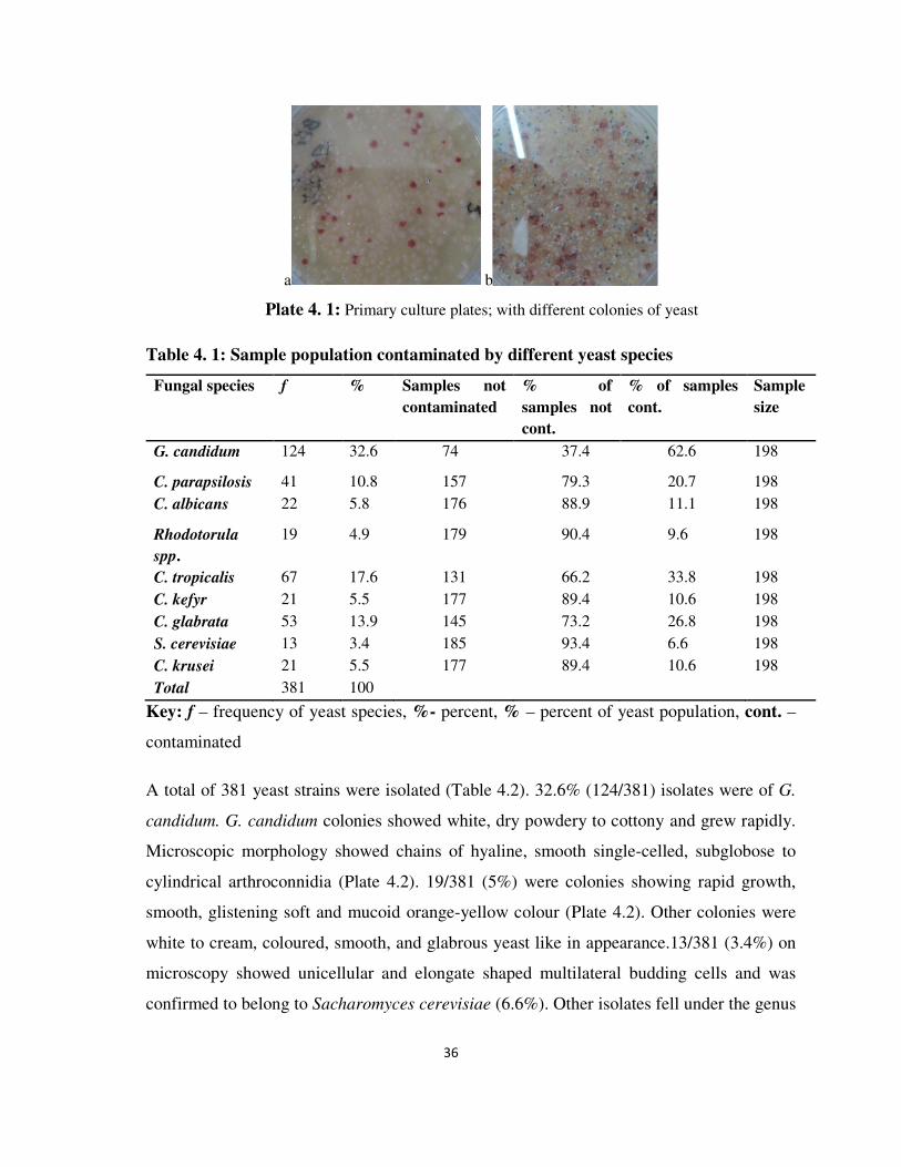
36
a b
Plate 4. 1: Primary culture plates; with different colonies of yeast
Table 4. 1: Sample population contaminated by different yeast species
Fungal species f % Samples not contaminated
% of samples not cont.
% of samples cont.
Sample size
G. candidum 124 32.6 74 37.4 62.6 198
C. parapsilosis 41 10.8 157 79.3 20.7 198
C. albicans 22 5.8 176 88.9 11.1 198
Rhodotorula
spp. 19 4.9 179 90.4 9.6 198
C. tropicalis 67 17.6 131 66.2 33.8 198
C. kefyr 21 5.5 177 89.4 10.6 198
C. glabrata 53 13.9 145 73.2 26.8 198
S. cerevisiae 13 3.4 185 93.4 6.6 198
C. krusei 21 5.5 177 89.4 10.6 198
Total 381 100
Key: f – frequency of yeast species, %- percent, % – percent of yeast population, cont. –
contaminated
A total of 381 yeast strains were isolated (Table 4.2). 32.6% (124/381) isolates were of G.
candidum. G. candidum colonies showed white, dry powdery to cottony and grew rapidly.
Microscopic morphology showed chains of hyaline, smooth single-celled, subglobose to
cylindrical arthroconnidia (Plate 4.2). 19/381 (5%) were colonies showing rapid growth,
smooth, glistening soft and mucoid orange-yellow colour (Plate 4.2). Other colonies were
white to cream, coloured, smooth, and glabrous yeast like in appearance.13/381 (3.4%) on
microscopy showed unicellular and elongate shaped multilateral budding cells and was
confirmed to belong to Sacharomyces cerevisiae (6.6%). Other isolates fell under the genus

37
Candida where 22/381 (5.8%) had branched pseudohyphae and true hyphae and yielded
green colonies on CHROMagar candida indicative of Candida albicans (Plate 4.3). 17%
(67/381) gave blastoconidia and developed a blue colour on CHROMagar characteristic of
C. tropicalis. 13.9% were C. glabrata, exhibiting cells, small in size with a light pinkish
colonies on CHROMagar. C. parapsilosis, 10.8%, C. kefyr and C. krusei both at 5.5%
showed pink, purple and whitish pink colours on CHROMagar respectively (Plate 4.3).
Table 4. 2: Yeast isolates and Colony colors of Candida species on CHROMagar Candida
after 72 h incubation at 30oC
Fungal species Number of Isolates
(%)
Colors observed
Geotricum candidum 124 (32.6) Blue with hyphae
Candida parapsilosis 41(10.7) Pink
Candida albicans 22 (5.8) Green
Rhodotorula species 19 (5.0) Red
Candida tropicalis 67 (17.6) Blue
Candida kefyr 21 (5.5) purple
Candida glabrata 53 (13.9) Light pink
S. cerevisiae 13 (3.4)
Candida krusei 21 (5.5) Whitish pink
Total 381 (100)
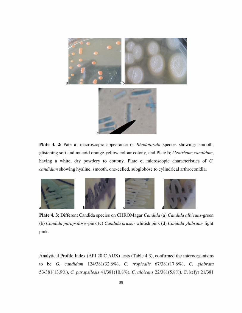
38
a b
c
Plate 4. 2: Pate a; macroscopic appearance of Rhodotorula species showing: smooth,
glistening soft and mucoid orange-yellow colour colony, and Plate b; Geotricum candidum,
having a white, dry powdery to cottony. Plate c; microscopic characteristics of G.
candidum showing hyaline, smooth, one-celled, subglobose to cylindrical arthroconidia.
a b c d
Plate 4. 3: Different Candida species on CHROMagar Candida (a) Candida albicans-green
(b) Candida parapsilosis-pink (c) Candida krusei- whitish pink (d) Candida glabrata- light
pink.
Analytical Profile Index (API 20 C AUX) tests (Table 4.3), confirmed the microorganisms
to be G. candidum 124/381(32.6%), C. tropicalis 67/381(17.6%), C. glabrata
53/381(13.9%), C. parapsilosis 41/381(10.8%), C. albicans 22/381(5.8%), C. kefyr 21/381

39
(5.5%),C. krusei 21/381(5.5%), Rhodotorula spp. 19/381(5.0%) and Sacharomyces
cerevisiae 13/381(3.4%).
Table 4. 3: Yeast fermentation of sugars; analytical profile index
Analytical Profile Index (API 20 C AUX) TESTS
Fungal isolates Glucose Maltose Galactose Sucrose Lactose
G. candidum _ _ _ _ _
S. cerevisiae + + + + _
C. albicans + + + _ _
C. parapsilosis + _ _ _ _
C. kefyr + _ + + +
C. glabrata + _ _ _ _
C. tropicalis + + +/_ + _
C. krusei + ___ _ _ _
Key; + fermentation, +/_ no clear fermentation and – no fermentation.
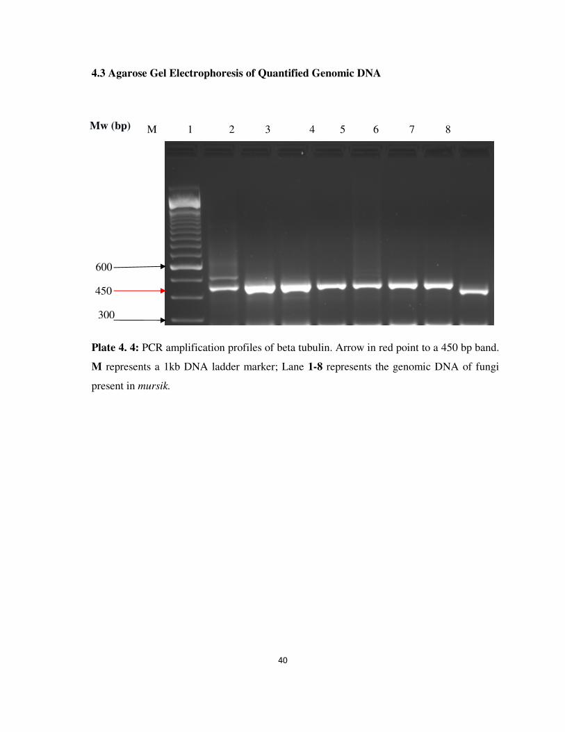
40
4.3 Agarose Gel Electrophoresis of Quantified Genomic DNA
Plate 4. 4: PCR amplification profiles of beta tubulin. Arrow in red point to a 450 bp band.
M represents a 1kb DNA ladder marker; Lane 1-8 represents the genomic DNA of fungi
present in mursik.
M 1 2 3 4 5 6 7 8 Mw (bp)
600
450
300

41
Plate 4. 5: PCR amplification profiles of rDNA ITS region. Arrow in Red points to a 600
bp band. M represents a 1kb DNA ladder marker; Lane 1-5, 7 and 8 represents the genomic
rDNA of mycotoxxigenic fungi present in mursik. Lane 6, represents a different genomic
DNA present in mursik.
4.4 Mycotoxins
Aflatoxins B1, detected in 99.5% (196/198) of the samples with the range levels of 0-
12ppb (Figure 4.2.). Fumonisin B1 was detected in 3 (1.5%) of the samples had detectable
quantities with the range levels of 0-0.77ppb (Table 4.4) and Deoxynivalenol toxins was
detected on 1 (0.5%) sample only showing a quantifiable level of 0.25ppb (Table 4.5). The
mean and range of the mycotoxins levels were calculated (Table 4.6).
M 1 2 3 4 5 6 7 8 Mw (bp)
600
300
100

42
Table 4. 4: Quantified levels of Fumonisin toxin in mursik collected in Soliat location in
2013.
Toxin level
(ppb)
Frequency % of total
0 195 98.5
0.33 1 0.5
0.39 1 0.5
0.77 1 0.5
198 100
Table 4. 5: Quantified levels of DON toxin in mursik collected in Soliat location in 2013
Toxin level
(ppb)
Frequency % of total
0 197 99.5
0.25 1 0.5
Total 198 100

43
Table 4. 6: The mean and range of the mycotoxins levels in mursik collected in Soliat
location in 2013.
Aflatoxin(ppb) Fumonisin(ppb) Deoxynivalenol(ppb)
N 198 198 198
Mean 4.671 0.008 0.001
Range 12 0.77 0.25
Minimum 0 0 0
Maximum 12 0.77 0.25

44
Figure 4. 2: Aflatoxin levels in mursik collected in Soliat location in 2013.
4.5 Other variables crucial in mursik processing
From the questionnaire results, all participants were milk fermenters. 85.3% of the farmers
obtained milk for fermentation from their own cows, 14.7% bought from other farmers.
Participants reportedly milked their cows in the open field. After milking, 90% of the
participants did not cover the milk containers. Among the respondents, 88% did not boil
milk before fermentation. All people used calabashes to ferment their milk, and left it for

45
duration of 3-4 days, with 82.2% and 17.8% keeping it for 3 and 4 days respectively.
Furthermore, none of the respondents have ever used starter cultures. Charcoal like
material (wo’sek), obtained from Senna didymobotrya plant, and was used to treat
calabashes, before pouring fresh milk, then left to ferment. Calabashes containing milk
underwent fermentation process in living rooms (among 74% of respondents) as well as
maize stores (74% of the participant). Use of hot water to clean calabashes was reported by
80% of participants, with 20% utilizing cold water. The clean calabashes were
subsequently used in further fermentation. As the capacity of calabashes varied, coupled
with the fact that cattle kept produced small quantities of milk, addition of more fresh milk
into the calabash containing already fermenting milk was reportedly done after every 1 day
by all farmers until it filled up, before leaving to ferment. Commercial feeds were not used
by participants. In addition to grazing their cattle freely in the field, they supplemented it
with nappier grass.

46
CHAPTER FIVE
5 DISCUSSION, CONCLUSIONS AND RECOMMENDATIONS
5.1 Discussion
5.1.1 Fungal contaminants
The results obtained from the microbial analysis of Mursik in this study show that product
was contaminated with microorganisms of public health concern. The fungal isolates
constituted mainly the yeasts. The present study isolated 381 strains of the following fungi,
Geotricum candidum, Candida tropicalis, C. glabrata, C. parapsilosis, C. albicans, C.
kefyr, C. krusei, Rhodotorula spp and Sacharomyces cerevisiae. Microbiological analyses
provide useful information which shows the conditions in which the milk was obtained,
processed and stored (Pietrowski et al., 2008).
G. candidum has been known to be the most important species in food and identified as the
dairy mold which contributes to flavour and aroma development in cheese and is also
responsible for the characteristic taste and aroma of gari, a fermented cassava product (Jay,
1992). It is thus expected that G. candidum plays a functional role in the development of
the taste and flavour of mursik. However, geotrichosis is an infreguent opportunistic
mycosis whose main etiologic agent is Geotrichum cancdidum though several studies have
proven that it is a commensal in humans and part of the normal flora of the skin, mouth and
gastrointestinal tract (Pottier et al., 2008). Many reported clinical cases have occurred in
immunosuppressed patients or immunosuppressive disorders, and has been known to affect
the respiratory tract (bronchi and lungs); superficial skin infections and lately invasive
infections have also been reported (Girmenia et al., 2005) and oral geotrichosis has an
association with diabetes mellitus (Hattori et al., 2007). G. candidum with its positive
contribution in fermentation, it poses treat to consumers especially persons whose immune
systems are compromised.

47
Candida tropicalis, has been isolated from sobia, a fermented beverage in the western
province of Saudi Arabia (Gassem, 2002), and some traditional fermented milk products in
Zimbabwe (Gadaga et al., 2000). C. tropicalis has been identified as an important yeast
pathogen of humans (Ahearn, 1998) and its presence in the traditionally fermented
milk indicates that these fermented foods could be a source of pathogenic
microorganisms.
C. glabrata, C. albicans and C. parapsilosis were isolated by Gogoi et al., (2014) in mark
raw milk. C. albicans causes candidiasis in humans and has been associated with
infections, as well as colonization, in both immunocompromised and immunocomptent
patients. Due to rambant cases of HIV/AIDS, some other Candida species other than C.
albicans have also emerged as significant opportunistic fungal pathogens (Richardson &
Warnock, 2003). Examples are C. glabrata, C. parapsilosis, C. kefyr and C. krusei which
have been have been responsible for increasing the proportion of cases of fungemia and
other complex cases of candidiasis (Colombo et al. 2006; Lewis et al. 2009; Miceli et
al.2011). In terms of virulence and pathogenicity, these species behave with equal or
greater virulence compared with C. albicans (Abbas et al. 2000).
Milk fermentation has been known to result in the production of ethanol resulting in
acetaldehyde (ACH) which is a carcinogen especially responsible mostly for esophageal
cancer (Mikko et al., 2012). According to Salaspuro, (2009), acetaldehyde (ACH),
resulting from ethanol or tobacco, is responsible for the pathogenesis of cancer. It has been
concluded that ACH present in alcoholic beverages, is a congenitor or formed
endogenously from ethanol and is a Group1 carcinogen in humans (Secretan et al., 2009).
Esophageal squamous cell cancer (OSCC) is unusually frequent in southern and eastern
Africa, including Western Kenya, where it is the most common malignancy in both men
and women. Of special interest is that occurrence of OSCC is in young people of less than
30 years of age in the area, especially among members of the Kalenjin tribe (Lodenyo et
al., 2005). A study done by Mikko et al., (2012), provided evidence that high levels of
ACH and alcohol are present in mursik having upto 3.8% w/v ethanol and mutagenic
concentrations of ACH. This has been postulated to be responsible for the high prevalence
of esophageal cancer cases occurring in patients 30 years of age or younger amongst the

48
Kalenjins community. Candida albicans is known to produce significant amounts of ACH
during fermentation. The presence of Candida albicans in Mursik is therefore a significant
risk factor for ACH exposure. Other yeasts that have been known to produce ACH are
Candida kefyr in conjuction with Lactobacilli bacteria.
Candida kefyr, are some of the most predominant and important yeast species in milk
(Fleet, 1990). C. kefyr is able to assimilate lactose, and therefore has a potential for growth
in milk (Gadaga et al., 2000). C. kefyr has been known to produce negligible amounts of
lactic acid and inclusion of the yeast into mixed cultures did not seem to affect production
of lactic acid by the LAB (Gadaga et al., 2001).
C. krusei as has been known to be used together with lactic acid bacteria as starter cultures
to uphold the activity and enhance the long life of the lactic acid bacteria, even
though this kind of association are not confirmed but C. krusei cannot metabolise lactose,
although is able to utilise the breakdown products made available by lactose metabolism by
Lactobacilli, which is a strong proof for the association (Frazier & Westhoff, 2001; Mikko
et al., 2012). This showed that symbiotic association between C. krusei and the lactic
acid bacteria is involved in mursik production. C. krusei is also drawn in the
fermentation of cacao beans, playing a vital role in development of the desirable
chocolate flavour of roasted beans. Flavor improvement is attributed to the proteolytic
ability of the yeasts (Jay, 1992). Thus C. krusei may too play a role in flavour
improvement in mursik.
Rhodotorula species are the mainly frequent species in foods especially R. mucilaginosa
and R. glutinis. They produce pink or red pigments and cause discolouration of foods
such as fresh poultry, fish, beef and sauerkraut (Jay, 1992; Frazier & Westhoff,
2001) giving bitter taste to the products (Muhamad et al., 2006). Pitt and Hocking (1999),
reported them to cause spoilage in dairy products such as yoghurt, butter, cream and
cheese. Consequently the presence of Rhodotorula in mursik is unwanted as its existence in
milk products results to spoilage.
Saccharomyces cerevisiae has been known to be of beneficial role in cheeses and
fermented milk for example kefir and koumiss. S. cerevisiae has been associated with the

49
production of aroma compounds and stimulation of lactic acid bacteria, improvement of
nutritional value and inhibition of undesired microorganisms (Jespersen, 2003).
Zimbabwean traditionally fermented milk S. cerevisiae enhanced growth of L. lactis subsp.
Lactis and additionally stimulated growth of Lb. paracasei in UHT milk (Gadaga et al.,
2001). These therefore indicate that presence of S. cerevisiae in mursik contributes in the
improvement of flovour; aroma and enhancement of milk lactobacilli as it inhibit the
growth of harmfull organisms improving on the value of the milk product. S. cerevisiae
has been considered as a well-established cause of nosocomially acquired yeast infection.
Its fungemia is unknown but considered a risk factor for nosocomial bloodstream infection
in patients with predisposing underlying conditions (Lherm et al., 2002). The most
consistent risk factor for S. cerevisiae fungemia is the use of probiotics. S. cerevisiae can
cause a wide variety of clinical syndromes, like liver abscess and esophagitis (Aucott et al.,
1990; Konecny et al., 1999). This implies that presesnce of S. cerevisiae mursik and its use
in other fermented foods raises clinical concerns to the end users of the products.
Yeast species can produce several extracellular enzymes, mainly proteinases,
phospholipases and lysophospholipases. These hydrolytic enzymes can modify the
membrane components, causing a misfunctioning. Pathogenic Candida albicans strains
have been known to produce toxins though toxin production appears to be a strain
characteristic and not a property of all pathogenic strains. These toxins could be in two
groups: high-molecular-weight-toxins (canditoxin) and low molecular-weight-toxins. Both
toxins affect mechanisms of humoral immunological response affecting the interaction
between macrophages and fungi. Acid phosphates with pH optima at 3-6 and 5.6, a
peptidase with optimum pH 6.6 and β-glucosidase have been isolated from both
blastospore and mycelial forms of C. albicans and have been indicted as the potential
agents of pathogenicity because of their proteolytic activities (Chattaway et al., 1971) .
During fungal investigations, moulds were not isolated. The use of charcoal (wo’sek) from
Senna didymobotrya could have inhibitory activity on moulds. Reports by Korir et al.,
(2012) indicated that the extracts from Senna didymobotrya have inhibitory properties
against filamentous fungi. The use of the charcoal from the plant could also be to act as a
detoxifying agent to binding toxins and carcinogens in the fermenting milk (Mikko et al.,

50
2012). Therefore the absence of moulds or mycotoxin producing fungi from the mursik
could be due to the antifungal properties of the plant.
However, DNA isolated from 198 samples of mursik was used as template and PCR was
carried out with two combinations of primer pairs: ITS5/ITS4 that amplifies the 18S-28S
rDNA region, and Bt2a/Bt2b that amplify the 400- 500bp region. When primer pair
Bt2a/Bt2b was used, a unique amplicon of approximately 450 bp in size was obtained for
all tested samples. No significant differences in amplicon size were noted among the
various samples tested (Plate 4.4). With the primer pair ITS5/ITS4, an amplicon of
approximately 600 bp was obtained for all samples apart from 1 sample which gave an
amplicon of approximately 580 bp a subject of further analysis through seguencing (Plate
4.5). The results suggest contamination of mursik with mycotoxigenic fungi which is in
accordance with the data from literature (Mirhendi et al., 2007). Similar amplicons, as size,
were obtained for A. ochraceus strains (Aguirre et al., 2004).
Moulds of the genus Aspergillus, Penicillium and Fumonisin were among the contaminants
of mursik. Aspergillus niger, Penicillium chrysogenum, Rhizopus spp, Fusarium
moniliforme, Trichoderma reesii, and Aspergillus fumigatus are among the contaminants of
traditionally fermented milk (Uzeh et al., 2006). Though, they have been associated to
fermentations of milk products. Several fungal starter cultures commonly used in Asia have
been used in fermentations in other places as fungi can add fiber, vitamins and proteins to
fermented foods (Nout, 1994).
Aspergillus species for instance Aspergillus oryzae and A. sojae are used in the production
of miso and soya sauce fermentations. Aspergillus oryzae and A. Niger are also used for
production of sake and awamori liquors, respectively (Nout, 1994). Aspergillus acidus is
used for fermenting Puerh tea. The presence of the Aspergillus species in the traditional
fermented milk; mursik could be responsible for its fermentation. On the other hand, some
members of the genus Aspergillus are contaminants of milk product and contribute to its
spoilage. Aspergillus and Rhizopus species were the moulds isolated as the primary
contaminants in yogurt produced in Nigeria (Ifeanyi et al., 2013). Aspergilli species have
been reported as spoilage organisms during storage of a wide range of foods where they

51
may produce mycotoxins. Mycotoxin produced by Aspergillus is known as aflatoxin which
has been among the toxins contaminating milk (Kang’ethe & Lang’a, 2009).
Moulds of the genus Penicillium is most frequently occurring on cheeses and has been used
in Europe to ripened foods, primarily cheeses and meats, usually using a Penicillium
species. This indicated that presence of penicilium could contribute to the fermentation.
However, Penicillium species are reported to produce the widest range of mycotoxins.
Among them are ochratoxin A; patulin; citrinin; citreoviridin and griseofulvin (Leistner,
1990). Mycotoxins produced by Aspergillus and Penicillium have been known to cause
mycotoxicoses. These toxins can damage liver, causing cirrhosis and can also induce tumor
(Sell et al., 1991). Hence the consumption of the fermented milk in area of study
contaminated with these moulds are dangerous to health since it is a potential sources of
mycotoxicosis.
Fusarium species has been used in the fermentation of cheese for example Fusarium
domesticum has been used for cheese fermentations (cheese smear). Fusarium solani was
isolated from a Vacherin cheese, and is used extensively for mycoprotein production in
Europe (Thrane, 2007). Thus, its presence could be contributing to some extent the
fermentation of milk. However, Fusarium species have been known to produce fumonisin
and trichothecene mycotoxins as their secondary metabolites (Thrane, 2007; Marasas,
2001) and it will contribute to the contamination of milk and milk leading to the risk of
mycosis to the consumers.
Data from this study is similar to the work reported by Abd El- Aziz et al., (2011) who
showed that fungi of the genus Aspergillus spp., Penicillium spp., Rhizopus spp and
Candida spp. were present in fermented camel milk. The existence of yeasts and moulds in
a range of spontaneously fermented milk products has been reported by other by
researchers (Gadaga et al., 2000; Abdelgadir et al., 2001; Lore et al., 2005; Kabede et al.,
2007; Zhang et al., 2008; Rahman et al., 2009). Presence of yeasts in naturally fermented
milk products may be potential cause of spoilage but contribute to development of texture
and flavor (Narvhus & Gadaga, 2003).

52
From the study no starter cultures are used; fermentation is initiated by the natural
microorganisms of the milk. Milk is left to ferment naturally at room temperatures (Eyassu
et al., 2012). Before fermenting, a remarkable number of respondents did not boil the milk.
Moreover, as reported by Chelule et al., (2010) and Nakavuma et al., (2011), calabashes
were the preferred containers for the fermentation process, which took between 3-4 days.
Prior to pouring fresh milk into them, calabashes were treated and seasoned with charcoal
like material (wo’sek), obtained from Senna didymobotrya plant (Mureithi et al., 2000).
Equal number of people fermented their milk in living rooms as well as maize stores. These
conditions leaves the milk and the containers contaminated by fungal spores since they can
be found in the atmosphere and from those fungi for example, Aspergillus, Fusarium, and
Penicillium that attack maize in the stores (Mboya et al., 2011).
As the capacity of calabashes varied, coupled with the fact that cattle kept produced small
quantities of milk, addition of more fresh milk into the calabash containing milk at various
stages of fermentation was done after every 1 day. Upon consumption of the fermented
milk, majority of the respondents cleaned their calabashes using hot water which was also
reported by Eyassu et al., (2012). Nevertheless, a small percentage employed cold water.
The clean calabashes were subsequently used for further fermentation.
In the present study, participants milked their cows in the open field. Thereafter, a
significant percentage of them did not cover the milk containers during transportation to the
living houses leaving milk vulnerable for microbial contamination. Other studies have
shown that many microorganisms are transmitted during milking. Fungi are ubiquitous in
the environmental and present in the air, water, feaces, and in the cow shade. These
include: moulds, yeasts, algae among others. These microorganisms include:
Cryptococcuss spp., Rhodotorula spp., Aspergillus spp., Pichia, C. krusei, C.tropicalis, C.
albicans, Trichosporon spp. (Ludmilla et al., 2011). It is certain that unhygienic milking
procedures and equipment used for milking, storing or distributing milk may have been an
important source of the isolated microorganisms. This confirms past studies done by Bryan,
1983. He also suggested that, milk should be stored under conditions which prevent mould

53
growth and strict hygienic measures and regulations should be imposed during processing,
packaging and transportation.
Other studies shows that different yeast species were predominant in different fermented
milk products for example, Sameel which is a traditionally fermented milk in Saudi Arabia,
yeast isolated include; Candida spp., Cryptococcus laurenti, Candida kefyr, and
Sacharomyces cerevisiae (Bozena et al., 2011). According to Gadaga et al (2000), C.
lusitaniae, C. colliculosa and S. cerevisiae were the isolated strains from Zimbwabwean
traditional fermented milk. Debaryomyces hansenii was the predominant strain in Sardinian
ewe’s dairy products (Cosentino et al., 2001). Abdelgadir et al., (2001) in Sudan and
Shuangquan et al., (2006) in Mongolia, found that the predominant yeast strains in the
fermented milk were S. cerevisiae and C. kefyr. C. krusei while C. kefyr has been found to
be the most dominant yeast species in Ugandan traditionally fermented milk; ‘Kwerionik’
(Nakavuma et al., 2011). In this study, C. krusei and C. kefyr were the least isolated species
which was also echoed by Al-Otaibi, (2012).
Bozena et al (2011), isolated yeasts from milk of the following species; C. parapsilosis
and C. krusei being predominant and C. tropicalis and C. albicans. Other yeasts of the
genus, Geotricum, Sacharomyces and Rhodotorula as the least isolated but in the present
study, the predominant yeast was Geotricum candidum. Others isolated by Bozena et al.,
(2011), were from the genus Cryptococcus and Trichosporon. Therefore, use of
contaminated milk by yeast of the genus, Candida spp., or by toxins produced by them may
be harmful to human health. Given that the product does not go through any pasteurization,
consumers, particularly immunocommpromised individuals are at risk of acquiring
opportunistic mycoses (Ludmilla et al., 2011).
Fungi of the genus Candida: C. krusei, C. albicans, C. tropicalis, C. glabrata and C.
lusitaniae, C. holmii, C.lambica have been identified in the crude milk directly derived
from the cow’s teats (Ludmilla et al., 2011). This is a clear indication that fresh raw milk
can be contaminated as it is being retrieved from the cow’s udder since these organisms are
among the normal flora of the udder (Kabede et al., 2007). The microflora involved in the
production of suusac; Kenyan traditionally fermented milk produced by Somalis, are yeasts

54
namely; C. krusei, and Rhodotorula muciloginosa (Lore et al., 2005). C. albicans, though
infrequently isolated from milk, grow well in the milk at all temperatures. Thus bringing a
lot of concern since this organism is known to be an opportunistic pathogen in humans
(Kabede et al., 2007).
Finding from the present study showed that moulds of the genus Aspergillus, Penicillium
and Fusariam were indeed responsible for the contamination of milk. Indeed, the three
genuses have been known to be the main mycotoxin producing moulds (Lanyasunya et al.,
2005). Furthermore, Aspergillus spp., Alternaria spp., Fusarium spp., Neurospora spp. are
the most common fungi species found in milk products (Saadia, 2010). This result confirms
what has been reported by other researchers. According to Uzeh et al., (2006), moulds of
the following genus; Aspergillus, Penicillium, Rhizopus, Fusarium, and Trichoderma
species have been isolated. Known spore formers, of mycotoxigenic fungi have been
isolated from nono and wara; the traditionally fermented milk product.
The species of the genus Aspergillus, Penicillium and Fusarium were isolated by Pešić-
Mikulec et al., (2005) in raw milk. Abd El- Aziz et al., (2011) isolated and genetically
identify moulds from camel milk of the genus, Aspergillus, Penicillium, Mucor and
Rhizopus and revealed that Penicillium spp. was the predominant mould isolates.
Aspergillus niger was the most common mould isolates as reported by Chhabra et al.,
(1998). Marcos et al., (1990) explained that A. fumigatus was the most frequent mould and
(Prabhakar et al., 1989) affirmed that mucor spp was the regular mould contaminat on cow
milk. They are major spoilage organisms of carbohydrate foods (Rhodes & Fletcher, 1966).
However, their growth can result in the production and accumulation of mycotoxins which
are of public health and economic importance (Uzeh et al., 2006).
Other documented milk contaminants includes: Aspergillus glaucus, A. niger, Alternaria
spp., Cheotomium candidum, Cladosporium herbarum, Fusarium spp., Monilia spp.,
Mucor rouxii, Neuropora spp., Penicillium expansum, Penicillium spp., Rhizopus
nigricans, Sporotrichum carinis and Thamnidium elegans. Fungi of the genus, Fusarium,
Aspergillus, Penicillium, Cladosporium, Geotrichum and Cladosporium species have been
detected in milk during different seasons in different geographical locations (Pešić-Mikulec
55
et al., 2000). Findings from the current study, therefore confirms these past studies. Uzeh et
al., (2006), suggested that, it is important to prevent mould growth to avoid toxin
production by preventing the natural contamination of raw materials and the
microbiological standards be put in place for locally processed dairy products.
Commercial feeds which normally are from cereals were not used by participants
(Lanyasunya et al., 2005). In addition to grazing their cattle freely in the field, they
supplemented it with nappier grass. Cereals, used as cattle feed, forms a very good
substrates for fungal growth such as: Aspergillus, Penicillum and Fusarium species and
susceptible to mycotoxin contamination (Sultana et al., 2009). Further, fungi responsible
for these toxin productions can colonize milk, and thus producing significant amounts of
toxins under poor handling practices (Lanyasunya et al., 2005).
In the current study, majority of farmers, fermentation process took place in cereal stores.
Importantly, the calabashes were opened daily, for further addition of fresh milk, hence
increasing the likely hood of contamination with Aspergillus, Penicillum and Fusarium
species, as reported in the study.
5.1.2 Fungal mycotoxin genes
With the molecular detection of mycotoxigenic fungi in mursik, it is a clear indication that
species of Aspergillus, Penicillium and Fusarium are present and carry the genes
responisple for toxin synthesis. Early studies by Schindler et al., (1967) showed that
aflatoxin was produced maximally at 24oC and not at all at temperatures lower than 18
oC or
higher than 35oC. Obrian et al., (2007), indicated that, aflatoxin production was at the
highest at 28oC and decreasing amounts were produced as temperature increased from 34-
37oC. However, Diener and Davis, (1967) reported aflatoxin production in peanuts at 40
oC
by A. flavus. Mayne et al., (1967) suggested that the effect of temperature is more
dependent on substrate than on strain. Feng and Leonard, (1998) compared A. parasiticus
to A. nidulans under varying culture conditions. They detected aflatoxin at 27oC and lesser
amounts at 33oC but were unable to detect aflatoxin in A.parasiticus at 37
oC. In contrast
they found that A. nidulans produced sterigmatocystin at similar levels at all three
temperatures. While the cardinal range for aflatoxin production differs among strains and
56
culture conditions, most research shows temperatures between 24-30oC favor aflatoxin
biosynthesis (Obrian et al., 2007). Fusarium species has shown to grow well at 26 to 28°C;
fumonisin and DON toxins are detected at its maximum (Reid et al., 1999).
Both transcriptional and posttranscriptional regulation mechanisms control aflatoxin gene
transcription (Yu et al., 2004a). Northern analysis has been used to show the aflatoxin
polyketide synthase gene of A. parasiticus is expressed at 27oC, another path way gene,
omtA also was shown to be transcribed in A. parasiticus at 29oC and at 28
oC, but not at
37oC (Feng & Leonard, 1995; Liu & Chu, 1998; Obrian et al., 2007). The norsolorinic acid
reductase, O-methyl transferase B and aflatoxin polyketide synthase were highly expressed
at temperature of 28oC (Obrian et al., 2007). The area of study has the temperatures of
between 18oC-37
oC (Kang’ethe & Lang’a, 2009). Participants ferment their milk using
guards at room temperature, thus fovouring the growth of mycotoxigenic fungi. Thus, the
area temperature and their (participants) fermenting temperature promotes expression of
aflatoxin, fumonisin and DON synthesis genes leading to mycotoxin contamination.
Aspergillus and Fusarium species has been shown to grow well on high water activity of
0.8-0.90aw. The nor-1 gene responsible in the process of Aflatoxin synthesis is highly
expressed at 0.90aw with less at 0.95aw and 0.85aw (Abdel-Hadi et al., 2010). Fermented
milk (mursik) has water activity of 0.90aw, thus promoting the contamination of
mycotoxigenic fungi especially of Aspergilus, Fusarium and favor the expression of nor-1,
FUM1 and tri genes in the fungi leading to mycotoxin contamination (Chelule et al., 2010).
Mursik pH has been known to be a low at 5, this brings mursik to be contaminated as the
fungal contaminants grows well at pH 5 (Nakavuma et al., 2011).
5.1.3 Aflatoxins, Fumonisins and Deoxynivalenol (DON) or Vomitoxin
In the current study, 99.5% of the samples were contaminated with Aflatoxins. Of these
samples, the average level of contamination was 4.67 ppb levels exceeding 0.05ppb as per
EU, FAO/WHO and Food and Drug Adminstration (FDA) permissible levels for milk
(Kang’ethe & Lang’a, 2009). Concerning Fumonisin the current study showed a
remarkably low contamination of samples (1.5%) and the mean concentration of
contamination was 0.008 ppb. 0.5% of the samples were contaminated with

57
Deoxynivalenol toxins, with an average contamination of 0.001 ppb lower than the
recommended levels of 0.05 ppb of EU, FDA and FAO/WHO.
The disease caused by ingestion of aflatoxins in contaminated foods is called aflatoxicosis.
Acute aflatoxicosis occurs when aflatoxins are consumed at moderate to high levels. A.
flavus is the second leading cause of aflatoxins, which commonly affects maize, causes
illness and even death when consumed in large quantities. Low-level, chronic exposure is
carcinogenic and has been linked to growth in children (Strosnider et al., 2006).
The levels of contamination of fermented milk aflatoxins reported in this study are similar
to those reported in Kenya by Kang’ethe and Lang’a, (2009) where samples, 99% were
positive for aflatoxins. Reports of contamination of milk in various parts of the world have
been reported by De Sylos et al (1996), in Brazil, Rousi et al (2002) in Greece and Diaz et
al (2006) in Colombia. However, in this study a higher proportion of samples exceeding
the FAO/WHO limit of 0.05ppb are reported. Higher proportions have been reported in
India where 99% of the contaminated raw milk, milk based cereal weaning formula and
infant formula exceeded the 0.05ppb (Dwivedi et al., 2004).
Major outbreaks of acute aflatoxicosis from contaminated food in humans were reported in
developing countries (Centers for Disease Control and Prevention, 2004; Lewis et al.,
2005). For example, in western India in 1974, 108 persons died among 397 people affected
with aflatoxin poisoning in more than 150 villages (Krishnamachari et al., 1975). A more
recent incident of aflatoxin poisoning occurred in Kenya in July 2004 leading to the death
of 125 people among 317 reported with illness due to consumption of aflatoxin
contaminated maize (corn) (Centers for Disease Control and Prevention, 2004; Lewis et al.,
2005). Acute toxicosis is not the only concern. World health authorities warn that low
doses and long term dietary exposure to aflatoxins is also a major risk as chronic exposure
can lead to hepatocellular carcinoma (Fung & Clark, 2004).
Depending on the level and duration of exposure, aflatoxins possess both hepatotoxic and
carcinogenic properties. Symptoms in humans include vomiting, abdominal pain, alteration
in digestion, limb and pulmonary edema, convulsions, rapid progressive jaundice, swollen
liver, high fever, coma, and death. The predominant damage is to the liver (Lewis et al.,

58
2005), but acute damage to the kidneys and heart have been reported (Richard & Payne,
2003). In liver aflatoxins irreversibly bind to protein and DNA to form adducts such as
aflatoxin B1-lysine in albumin and a guanyl-N7 adduct in DNA (Skipper &Tannenbaum,
1990). Disruption of the proteins and DNA bases in hepatocytes causes the toxicity (Azziz-
Baumgartner et al., 2005).
The gene mutation of p53 tumour suppressor gene leads to initiation of hepatocarcinoma
formation (Coursaget et al., 1993). Human hepatocarcinomas are also associated with
hepatitis B virus (HBV) and C virus (HCV) infections (Wild et al., 1992). Together with
aflatoxins these viruses significantly increased the risk of hepatoma in hepatitis patients
(Arsura & Cavin, 2005). In developing countries, many children are exposed to aflatoxin
before birth, while nursing and after weaning (Turner et al., 2007; Polychronaki et al.,
2007; Gong et al., 2004). An association of hepatocellular carcinoma and dietary exposure
with aflatoxins has been established from patients living in high-risk areas of China,
Kenya, Mozambique, Phillippines, Swaziland, Thailand, Transkei of South Africa (Lewis
et al., 2005). The present study indicates that milk are contaminated with mycotoins though
there is less work have been done to uncover the real extent to which milk and milk
products are contaminated. This indicates that tcontamination of milk and its products with
this toxin are of great concern to the public health.
Fumonisin toxin was detected in 1.5 % (3) of the samples and it was below the acceptable
limits. Same results were reported by Maragos and Richard (1994) who analyzed 155 milk
samples collected in Wisconsin during a period when feeds were reported to be severely
affected by mold. Only one of the 165 milk samples tested positive for fumonisin, which
was determined to be below the set acceptable levels. Fumonisin carryover from feed to
milk has been indicated to be negligible (Richard et al., 1996; Scott et al., 1994). Prelusky
et al. (1996) reported studies where dairy cattle were administered fumonisin orally and no
fumonisin or its metabolites were detected in milk. This and results from this study
suggests that fumonisin can occur in milk, but at very low levels.
Fumonisins are mainly produced by Fusarium species, for example, F. verticillioides.
Fumonisin have been classified into three fumonisins B1 (FB1), B2 (FB2) and B3 (FB3)
59
which occur normally in abundant levels (Sreenivasa, 2012). Fumonisin in humans has
been related with neural tube defects in people that rely on maize as a staple food, but most
prominently it has been indicated as a potential cause of oesophageal cancer in humans
(Missmer et al., 2006; Marasas, 2001). In certain regions around the world including
Bomet County in Kenya, where high daily intake of maize and maize-derived commodities
occurs, an associations between either mouldy maize, F. verticillioides or fumonisin and
the incidence of oesophageal cancer have been reported (Marasas, 2001; Doko & Visconti,
1994).
In dietary exposure to FBs has been linked to high incidences of oesophageal cancer
observed in Transkei in South Africa, North East Italy China and Iran (Isaacson, 2005;
Franchesci et al., 1990; Sun et al., 2007; Shephard et al., 2000). In 2005 and 2006,
fumonisin exposure was seen in Tanzania and was attributed to the dry seasons in the two
years (Kimanya et al., 2009). It was isolated at high levels in corn meal and corn grits,
including seven samples from a supermarket in Charleston, South Carolina, the city
with the highest incidence of esophageal cancer among African Americans in the United
States (Sydenham et al., 1991).
The toxic concentrations of fumonisin differ much depending on the animal species
(Rheeder et al., 2002). Absorption of about 5 - 10 ppb fumonisin in feed induces
neurotoxic effects in horses (Marasas et al., 1988). In pigs the ingestion of 4 - 16 ppb body
weight may result in liver cirrhosis and more than 16 ppb body weight result in pulmonary
edema (Marasas, 1995). Chickens can withstand higher concentrations of fumonisin in
feed, up to 75 mg/kg and cattle seem not to be affected by high fumonisin concentrations
(Gelderblom et al., 1991). With the low levels of the fumonisins detected in mursik, it is an
indication the the milk product is safe from the contamination but it can arise due to the
poor handling of the product processing.
Deoxynivalenol (DON) is a secondary metabolite produced by a Fusarium species. It is
one of the mycotoxins most commonly detected in feeds. DON is also called vomitoxin
because it was first associated with vomiting in swine. Studies have shown DON to be a
primary mycotoxin associated with swine disorders including feed refusals, diarrhea,

60
reproductive failure, and deaths. Dairy cattle consuming feeds contaminated primarily with
DON have led to reduction in milk production. Research has shown that DON causes
polyribosomal breakdown in mammalian cell lines (Whitlow et al., 2005).
DON contamination of milk in this study was detected in one sample which was below the
EU, FDA, and FAO/WHO acceptable levels and other samples were below the detectable
limits. This reselts are in agreement with results of Seeling et al., (2006) where they
established that DON in milk was below the detectable levels. Fusarium species has been
commonly found contaminating or colonizing cereals and their products, stuffs constituting
an important part of human food and animal feed. Highly contaminated crops are
frequently directed to animal feed (Bennett & Klich, 2003). DON transfer in animal and the
possible presence of toxic residues in animal products for example milk, meat remain
unclear (Cavret & Lecoeur, 2006). It has been presented that, DON toxins are detected in
milk from animals fed with DON contaminated feeds and that the detection limits increased
with the increased levels of contamination of feeds but it did not correlate with the amounts
fed to the animals; the toxins detected were below the levels fed to the cows (Seeling,
2006).
Results from this study also confirm Seeling et al., (2006); Cavret and Lecoeur, (2006).
The participants, used Senna didymobotrya plant product to pretreat their milk before
fermented which has been known to be fungicidal (Korir et al., 2012) and must have inhibit
further establishment of the Fusarium fungus in milk thus no metabolite contamination.
Another contributing factor was that, participants did not use feed supplements to feed their
cows with cereal feeds.
Several thousand people were affected by gastrointestinal distress in an incident in the
Kashmir Valley of India in 1987 (Bhat et al., 1989). The toxic effects induced by DON
are well characterized in all animal species, with pigs being the most susceptible
(Pestka, 2010). DON can affect the immune system, animals’ food consumption, growth,
reproduction, neuroendocrine signaling and also intestinal function (Pinton et al., 2008;
Pestka, 2010; Pinton et al., 2010). In humans, there is historical evidence suggesting that
61
DON causes acute illness and is frequently associated with outbreaks of gastroenteritis
(Pestka, 2010).
The intestine is the first barrier to food contaminants. Following ingestion of mycotoxin-
contaminated food or feed, intestinal epithelial cells can be exposed to high
concentrations of toxins (Maresca et al., 2008). Highly dividing cells, such intestinal
epithelial cells are especially sensitive to trichothecenes and the exposure of
intestinal epithelial cells to these toxins may alter their capacity to proliferate and to
insure a proper barrier function. Exposure to DON can lead to impaired absorption of
nutrients and decrease of cell proliferation or differentiation (Awad et al., 2007; Kasuga et
al., 1998; Bensassi et al., 2009). The entry of luminal antigens and bacteria, normally
restricted to the gut lumen, was demonstrated to be a consequence of the disruption of
the barrier function in cells culture models (Maresca et al., 2008) or in mice species (Li
et al., 2005).
5.2 Conclusions
The yeast isolates from the study were G. candidum being the major contaminant, C.
tropicalis, C. glabrata, C. parapsilosis, C. albicans, C. kefyr, C. krusei Rhodotorula spp.
and Sacharomyces cerevisiae are mursik contaminants and all these organisms has been
known to be among the major cause various opportunistic mycoses in immune suppressed
patients.
Detection of Internal transcribed spacer region (ITS) and Beta tubulin (Bt) genes indicated
presence of fungi of the genus, Aspergillus, Penicillium, and Fusarium which are known
spore forming fungi. This therefore means that they are among contaminats of raw milk
used to produce mursik and mursik leading to mycotoxin contamination causeing
mycotoxicoses in consumers.
Aflatoxin is the major contaminant of mursik and the level of contamination (mean 4.6
ppb) surpassed the recommended limits of 0.05 ppb as per EU and FDA regulations.

62
5.3 Recommendations
Microbiological standards on milk and milk products should be put in place for the safety
of the consumers and effect of mursik on mycotoxigenic moulds should be determined.
Education and training should be given to farmers on milking, storage and maintaining
milk containers for proper handling procedures to reduce contamination though there is no
documentation showing lack of inadequate education.
Regular studies are required for proper establishment epidemiology of mycotoxin
contamination in various foods for the formulation and implementation of country’s
mycotoxins regulatory limits.

63
REFERENCES
Abbas, J., Bodey, G.P., Hanna, H.A., Mardani, M., Girgawy, E., Abi-Said, D.,
Whimbe, E., Hachem, R. and Raad, I. (2000). Candida krusei fungemia an escalating
serious infection in immunocompromised patients. Archives of Internal Medicine, 160,
2659-2664.
Abd El- Aziz M., Ahmed E., Milad E.T., Mustafa E. and Abd E.E. (2011). Biochemical
Studies on the Virulence Factors of Fungi Associated with She-Camel Milk. Journal of
Basic and Applied Chemistry, 1(2), 15-20.
Abdelgadir, W. S., Ahmed, T. K. and Dirar, H. A., (1998). The traditional fermented
milk products of the Sudan. International Journal of Food Microbiology, 44, 1-13.
Abdelgadir, W.S., Hamad, S.H., Moller, P.L. and Jacobsen, M., (2001).
Charaecterisation of the dominant microbiota of Sudanese fermented milk rob.
International Dairy Journal, 11, 63-70.
Abdel-Hadi, A., Carter, D. and Magan, N. (2010). Temporal monitoring of thenor-
1(aflD) gene of Aspergillus flavus in relation to aflatoxin B1production during storage of
peanuts under different water activity levels. Journal of Applied Microbiology, 109, 1914–
1922.
Abeer, A.A., Abdel, A. and Dardir, H.A., (2009). Hygienic Quality of Local Traditional
Fermented Skimmed Milk (Laban Rayb) Sold in Egypt: World Journal of Dairy & Food
Sciences, 4 (2), 205-209.
Adegoke, G.O. and Letuma, P. (2013). Strategies for the Prevention and Reduction of
Mycotoxins in Developing Countries. Open science, 5, 123-136.
Aguirre, L., Hurst, S. F., Choi, J.S., Shin, J.H., Hinrikson, H.P. and Morrison, C.J.
(2004). Rapid Differentiation of Aspergillus Species from Other Medically Important
Opportunistic Molds and Yeasts by PCR – Enzyme Immunoassay. Journal of Clinical
Microbiology, 42, 3495 – 3504.

64
Ahearn, D. G. (1998). Yeasts pathogenic for humans. In C. P. Kurtzman, and J. W. Fell.
The yeasts: A taxonomic study, 12, pp. 9. Amsterdam: Elsevier.
Akalin, A.S, Gönc, S., Ünal, G., Ökten, S. (2006). Determination of some chemical and
microbiological characteristics of Kaymak. Grasas y Aceites, 57, 429 – 432.
Alborzi, S., Pourbbas, B., Rashidi, M. and Astaneh, B. (2005). Aflatoxin M1
contamination in pasteurized milk in Shiraz. Food Control, 17, 582—584.
Alexander, N.J., Proctor, R.H., and McCormick, S.P. (2009). Genes, gene clusters, and
biosynthesis of trichothecenes and fumonisins in Fusarium. Toxin Reviews, 28, 198–215.
Al-Oitabi, M.M. (2012). Isolation and identification of lactic acid bacteria and yeasts from
Sameel milk: A Saudi Traditional fermented milk. International Journal of Dairy Science,
7(4), 73-83.
Anderson, I.C., Campbell, CD., Prosser, J.I. (2003). Potential bias of fungal 18S rDNA
and internal transcribed spacer polymerase chain reaction primers for estimating fungal
biodiversity in soil. Applied and Environmental Microbiology, 5, 36-47
Arino, A., Juan, T., Estopanan, G., Gonzalez-Cabo, J.F. (2007). Natural occurrence of
Fusarium species, fumonisin production by toxigenic strains and concentration of
fumonisins B1 and B2 in conventional and organic maize grown in Spain. Journal of Food
Protection, 70, 151-156.
Arsura, M. and Cavin, L.G. (2005). Nuclear factor-kappa B and liver carcinogenesis.
Cancer Letters, 229, 157-169.
Aucott, J.N., Fayen, J., Grossnicklas, H., Morrissey, A., Lederman, M.M. and Salata,
R.A. (1990). Invasive infection with Saccharomyces cerevisiae: report of three cases and
review. Reviews of Infectious Diseases, 12, 406–411.
Awad, W. A., Aschenbach, J. R., Setyabudi, F. M., Razzazi-Fazeli, E., Bohm, J., and
Zentek, J. (2007). In vitro effects of deoxynivalenol on small intestinal D-glucose

65
uptake and absorption of deoxynivalenol across the isolated jejunal epithelium of laying
hens. Poultry Science, 86, 15-20.
Azziz-Baumgartner, E., Lindblade, K., Gieseker, K., Rogers, H.S., Kieszak, S.,
Njapau, H., Schleicher, R., McCoy, L.F., Misore, A., DeCock, K., Rubin, C. and
Slutsker, L. (2005). Case-control study of an acute aflatoxicosis outbreak, Kenya, 2004,
Aflatoxin Investigative Group. Environmental Health Perspective, 113, 1779-1783.
Badel, S., Bernardi, T. and Michaud, P., (2011). New perspectives for Lactobacilli
exopolysaccharides. Biotechnology Advances, 29, 54-66.
Barnes, S. E., Dola, T. P., Bennett, J. W. and Bhatnagar, D. (1994). Synthesis of
sterigmatocystin on a chemically defined medium by species of Aspergillus and
Chaetomium. Mycopathologia, 125, 173-178.
Baumgart, D.C. and Carding, S.R. (2007). Inflammatory bowel disease: cause and
immunobiology. Lancet, 369, 1627-1640.
Bennett, J.W. and Klich, M., (2003). Mycotoxins. Clinical Microbiology. Reviews, 16,
497-516.
Bensassi, F., El Golli-Bennour, E., Abid-Essefi, S., Bouaziz, C., Hajlaoui, M. R., and
Bacha, H. (2009). Pathway of deoxynivalenol-induced apoptosis in human colon
carcinoma cells. Toxicology, 264, 104-109.
Bhat, R.V., Beedu, S.R., Ramakrishna, Y. and Munshi, K.L. (1989). Outbreak of
trichothecene mycotoxicosis associated with consumption of mould-damaged wheat
production in Kashmir Valley, India. Lancet, 7, 35-37.
Bozena, D. K., Alicja, K., Daniel, S., Miroslaw, K. and Malgorzata, B. (2012). High
Prevalence of Candida Yeast inMilk Samples from Cows Suffering from Mastitis in
Poland. The Scientific World Journal, 10, 1100- 1105
Briani, C., Samaroo, D. and Alaedini, A. (2008). Celiac disease: from gluten to
autoimmunity. Autoimmune Review, 7, 644-650.

66
Brown D.W., McCormick S.P., Alexander N.J., Proctor R.H. and Desjardins A.E.,
(2001). A genetic and biochemical approach to study trichothecene diversity in Fusarium
sporotrichioides and Fusarium graminearum. Fungal Genetics and Biology, 32, 121–33.
Brown, D.W., Yu, J.H., Kelkar, H.S., Fernandes, M., Nesbitt, T.C., Keller, N.P.,
Adams, T.H. and Leonard,T.J. (1996). Twenty-five coregulated transcripts define a
sterigmatocystin gene cluster in Aspergillus nidulans. Proceedings of the National
Academy of Sciences of the United States of America, 93, 1418–1422.
Bryan, F.L. (1983). Epidemiology of milk-borne diseases. Journal of food protection,
46(7), 637-649.
Calderone, R.A. and Cihlar, R.L. (2002). Fungal Pathogenesis. Principles and Clinical
applications, 154 (3), 153-154.
Carrie, A. S., Charles, P. W., Dominique, R. and Gary, A. P. (2007). Silencing of the
Aflatoxin Gene Cluster in a Diploid Strain of Aspergillus flavus is suppressed by Ectopic
aflR Expression. Genetics, 176, 2077–2086.
CAST. (1989). Mycotoxins: Economic and Health Risks. Task Force Report No. 116.
CAST. (1999). Mycotoxins, Economic and Health Risks: Task Force Report No.132.
CAST. (2003). Mycotoxins: risks in plant, animal and human systems Ames, IA: Council
for Agricultural Science and Technology.
Cavret, S. and Lecoeur, S. (2006). Fusariotoxin transfer in animal. Food and Chemical
Toxicology, 44, 444–453
Cegielska-Radziejewska, R., Stuper, K. and Szablewski, T. (2013). Microflora and
mycotoxin contamination in poultry feed mixtures from western Poland. Annals of
Agricultural and Environmental Medicine, 20(1), 30–35.
Centers for Disease Control and Prevention, (2004). Outbreak of aflatoxin poisoning –
eastern and central province, Kenya, MMWR Morb Mortal Weekly Report, 53, 790-792.

67
Coursaget, P., Depril, N., Chabaud, M., Nandi, R., Mayelo, V., LeCann, P. and
Yvonnet, B. (1993). High prevalence of mutations at codon 249 of the p53 gene in
hyptocellular carcinomas from Senegal. British Journal of Cancer, 67, 1395-1397.
Chang, P.-K., Cary, J., Bhatnagar, D., Cleveland, T. E., Bennett, J. W., Linz, J. E.,
Woloshuk, C. P. and Payne, G. A. (1993). Cloning of the Aspergillus parasiticus apa-
2gene associated with the regulation of aflatoxin biosynthesis. Applied and Environmental
Microbiology, 59, 3273–3279.
Chang, P.K., Skory, C. D. and Linz. J. E. (1992). Cloning of a gene associated with
aflatoxin B1biosynthesis inAspergillus parasiticus. Current Genetics, 21, 231–233.
Chao, T.C., Maxwell, S.M. and Wong, S.Y. (1991). An outbreak of aflatoxicosis and
boric acid poisoning in Malaysia. Journal of Pathology, 164(3), 225-233.
Chattaway, F. W., Odds, F. C. and Barlow, A. J. E. (1971). An Examination of the
Production of Hydrolytic Enzymes and Toxins by Pathogenic Strains of Candida albicans.
Journal of Getieval Microbiology, 67, 255-263
Chelule, P.K., Mokoena, M. P. and Gqaleni, N. (2010). Advantages of traditional lactic
acid bacteria fermentation of food in Africa: FORMATEX, 1160-1167.
Chhabra, D., Moghe, M. N., Tanwani, S. k. and Sharda, R. (1998): Mycotic mastitis in
buffaloes. Indian Journal of Comparative Microbiology, 19(2), 108-109.
Cleveland, T.E., Lax, A.R., Lee, L.S. and Bhatnagar, D. (1987). Appearance of enzyme
activities catalyzing conversion of sterigmatocystin to aflatoxin B1 in late-growth-phase
Aspergillus parasiticuscultures. Applied and Environmental Microbiology, 53, 1711–1713.
Cole, R.J. and Cox, R.H. (1981). In Cole, R. J. and Cox, R. H. (eds), Handbook of Toxic
Fungal Metabolites. Academic Press, New York, NY, pp. 67–93.
Colombo, A.L., Nucci, M., Park, B.J., Nouér, A.S., Arthington-Skaggs, B., Da Matta,
D.A., Warnock, D. & Morgan J. (2006). Epidemiology of candidemia in Brazil: a

68
nationwide sentinel surveillance of candidemia in eleven medical centers. Journal of
Clinical Microbiology, 44, 2816-2823.
Cosentino, S., Fadda, M.E., Deplano, M., Mulargia, A.F. and Palmas, F. (2001). Yeasts
associated with Sardinian ewe’s dairy products. International Journal of Microbiology, 69,
53-58.
Cotty, P.J. (2004). Biocompetitive exclusion oftoxigenic fungi. The mycotoxin fact book,
179-197.
David R. M. and Joseph S.W. ( 2000). Pathogenic Microbiology-routine techniques in
laboratory, 4, 125-126.
De Sylos, C.M., Rodriguez-Amaya, D.B. and Carvalho, P.R. (1996). Occurrence of
Aflatoxin M1 in milk and dairy products commercialized in Portugal. Food Additives and
Contaminants, 13, 169-172.
Desjardins, A.E., Manandhar, G., Plattner, R.D., Maragos, C.M., Shrestha, K. and
McCormick, S.P. (2000). Occurrence of Fusarium species and mycotoxins in Nepalese
maize and wheat and the effect of traditional processing methods on mycotoxin levels.
Journal of Agriculture and Food Chemistry, 48, 1377–83.
Devegouida, G. and Castaldo, D. (2000). Mycotoxins: hidden killers in pet foods. Is there
a solution? In: Technical Symposium on Mycotoxins, Altech Inc., Nicholasville, KY,
USA.
Diaz, D.E., Blackwelder, J.T., Hagler, W.M.Jr., Hopkins, B.A., Jones, F.T., Anderson,
K. L. and Whitlow, L.W. (1997). The potential of dietary clay products to reduce
aflatoxin transmission to the milk of dairy cows. Journal of Dairy Science, 80, 265.
Diaz, G.J. and Espitia, E. (2006). Occcurrence of Aflatoxin M1 in retail milk samples
from Bogota, Colombia. Food Additives and Contaminants, 23, 811-815.
69
Diener, U.L., and Davis, N.D. (1967). Limiting temperature and relative humidity for
growth and production of aflatoxin and free fatty acids by Aspergillus flavus in sterile pea
nuts. Journal of the American Oil Chemists' Society, 44(4), 259.
Doko, M. B. and Visconti, A. (1994). Occurrence of fumonisins B1 andB2 in corn and
corn-based human foodstuff in Italy. Food Additives and Contaminants, 11, 433-439.
Dragan, M., Miomir, N., Tatjana, B., Danijela, V., Srdan, S. and Saša, J. (2010). A
Survey of occurrence of toxogenic fungi and mycotoxins in pig feed samples-Use in
evaluation of risk assessment. Veterinary World, 3(7), 305-311.
Dwivedi, P.D., Rastogi, S., Khanna, S.K. and Mkul, D. (2004). Detection of Aflatoxin
M1 in milk and infant milk products from Indian markets by ELISA. Food Control, 15,
287-290.
El-Nezami, H., Kankaanpaa, P.E., Salminen, S. and Ahokas, J.T. (1998). Physico-
chemical alterations enhance the ability of dairy strains of lactic acid bacteria to remove
aflatoxins from contaminated media. Journal of Food Protection, 61, 466-468.
Envirologix Quicktox kit Quickscan, (2012). www.envirologix.com/quickscan.
Eyassu, S., Araya, A., Mohammed, Y.K. and Zelalem, Y. (2012). Isolation and
characterization of lactic acid bacteria from Ititu: Ethiopian traditional fermented camel
milk. Journal of Camelid Science, 5, 82-98.
Feng, G.H., and Leonard, T.J. (1998). Cultureconditionscontrolexpression ofthe genesfor
aflatoxin and sterigmatocystin biosynthesis in Aspergillus parasiticus and A. nidulans.
Applied and Environmental Microbiology, 64(6), 2275-2277.
Feng, G.H., and Leonard, T.J., (1995). Characterization of the polyketide synthase gene
(PksII) required for aflatoxin biosynthesis in Aspergillus parasiticus. Journal of
Bacteriology, 177(21), 6246-6254.
Fink-Gremels, J. (1999). Mycotoxins: their implications for human and animal health.
Veterinary Quaterly, 21, 115-120.

70
Fisher, A. A., Laing, J. E. and Strocker, J.E. (1998). Hand book for Family Planning,
Operation Research Design in Sampling. Population Council, 40-45.
Fleet, G.H. (1990). Yeasts in dairy products – A review, Journal of Applied Bacteriology,
68, 199–211.
Fleet, G.H. (2007). Yeasts in foods and beverages: impact on product quality and safety.
Current Opinion in Biotechnology, 18, 170–175.
Fleet, G.H. and Mian, M.A. (1987). The occurrence and growth of yeasts in dairy
products, International Journal of Food Microbiology, 4, 145–155.
Food and Agricultural Organization, (1997). Food and Agricultural Organization of the
United Nations food and nutrition paper 64. Worldwide regulations for mycotoxins. A
compendium. Food and Agricultural Organization, Rome, Italy.
Food and Agriculture Organization (FAO) of the United Nations, Rome, (1990). The
Technology of Traditional Milk Products in Developing Countries. FAO Animal
Production and Health, Paper, 85, Rome, Italy.
Food and Agriculture Organization of the United Nations, (2004). Worldwide
regulations for mycotoxins in food and feed in 2003. FAO, Food and Nutrition Paper 81.
Rome, Italy.
Franceschi, S., Bidoli, E., Buron, A.E., La Vecchia, C, (1990). Maize and risk of cancer
in the oral cavity, pharynx and esophagus in northeastern Italy. Journal of the National
Cancer Institute, 82, 1407−1411.
Frazier, W.C. and Westhoff, D.C. (2001). Food Microbiology. Tata McGraw-Hill, New
Delhi.
Fujita, S., Yasuko, S., Shigeki, N. and Takuma H. (2001). Multiplex PCR Using Internal
Transcribed Spacer 1 and 2 Regions for Rapid Detection and Identification of Yeast
Strains. Journal of clinical microbiology, 39 (10), 3617–3622.

71
Fung, F. and Clark, R.F. (2004). Health effects of mycotoxins: a toxicological overview.
Journal of Toxicology and Clinical Toxicology, 42, 217-234.
Gadaga, T.H., Mutukumira, A.N. and Narvhus, J.A. (2001). The growth and
interaction of yeasts and lactic acid bacteria isolated from Zimbabwean naturally fermented
milk in UHT milk. International Journal of Food Microbiology, 68, 21–32.
Gadaga, T.H., Mutukumira, A.N. and Narvhus, J.A. (2000). Enumeration and
Identification of yeast isolated from Zimbabwean traditional fermented milk. International
Dairy Journal, 10, 456-466.
Gajęcki, M., Gajęcka, M., Jakimiuk, E., Zielonka, Ł., Obremski, K. (2010).
Zearalenone – undesirable substance. In: Mycotoxins in Food, Mahendra Rai, Ajit Varma
(Ed.), Feed and Bioweapons. Springer-Verlag Berlin Heidelberg, Germany. 131-144.
Gassem, M.A.A. (2002). A microbiological study of Sobia: a fermented beverage in the
Western province of Saudi Arabia. World Journal of Microbiology and Biotechnology, 18,
173-177.
Geiser, D.M., Juba, J.H., Wang, B., Jeffers, S.N. (2001). Fusarium hostae sp. nov., a
relative of F. redolens with a Gibberella teleomorph. Mycologia, 93(4), 670-678.
Geiser, D.M., Pitt, J.I. and Taylor, J.W. (1998). Cryptic speciation and recombination in
the aflatoxin-producing fungus Aspergillus flavus. Proceedings of the National Academy of
Science, USA, 95, 388–393.
Gelderblom, W., Jaskiewicz, K., Marasas, W.F.O. and Thiel, P.G. (1991). Toxicity and
carcinogenicity of the Fusarium moniliforme metabolite, fumonisin B1 in rats.
Carcinogenesis, 12, 1247-1251.
Gelderblom, W.C.A., Jaskiewicz, K., Marasas, W.F.O., Thiel, P.G., Horak, R.M.,
Vleggaar, R. and Kriek, N.P.J. (1988). Fumonisins: Novel mycotoxins with cancer-
promoting activity produced by Fusarium moniliforme. Applied Environmental and
Microbiology, 54, 1806.

72
Girmenia, C., Pagano, L., Martino, B., D’Antonio, D., Fanci, R., Specchia, G., Melillo,
L., Buelli, M., Pizzarelli, G.M., Martino, P. (2005). Invasive infections caused by
Trichosporon species and Geotrichum capitatum in patients with hematological
malignancies: a retrospective multicenter study from Italy and review of the literature.
Journal of Clinical Microbiology, 43, 1818-1828.
Goigoi, S. M., Syed, M., Saikia, G.K. and Madhumita, D. (2014). Isolation of Candida
Species from Market Milk Samples. Scholars Academic Journal of Biosciences, 2(3), 202-
204
Gong, Y., Hounsa, A., Egal, S., Turner, P.C., Sutcliffe, A.E., Hall, A.J., Cardwell, K.
and Wild, C.P. (2004). Postweaning exposure to aflatoxin results in impaired child
growth: a longitudinal study in Benin, West Africa. Environmental Health Perspective,
112, 1134-1138.
Guo, B.Z., Widstrom, N.W., Cleveland, T.E. and Lynch, P.B. (2000). Control of pre-
harvest aflatoxin contamination in corn: Fungus-plant-insect interactions and control
strategies. Recent Research Developments in Agricultural and Food Chemistry, 4, 165-176.
Hassan, R.A., El Zubeir, I.E.M. and Babiker, S.A. (2006). Chemical and microbial
measurements of fermented camel milk ‘‘Gariss’’ from transhumance and nomadic herds
in Sudan. Australian Journal of Basic Applied Sciences, 2, 800-804.
Hattori, H., Inoue, C., Tomita, Y. and Kanbe, T. (2007). A case of oral geotrichosis
caused by Geotrichum capitatum in an old patient. Japanese Journal of Infectious Disease,
60, 300-301.
Haughland, R.A., Varma, M.M., Wymer, L.J. and Vesper, S.J. (2004). Quantitative
PCR analysis of selectedAspergillus, PenicilliumandPaecilomycesspecies. Systemic
Applied Microbiology, 27, 198–210.
Heeschen, W.H. (1994). The significance of pathogenic microorganisms in raw milk.
International Dairy Federation. Brussells, 8-11.

73
Helferich, W. and Westhoff, D. (1980). All about yoghurt. Prentice-Hall, Inc., Englewood
Cliffs, New Jersey 07632. pp. 1-20.
Hell, K. and Mutegi, C., (2011). Aflatoxin control and prevention strategies in key crops
of Sub-Saharan Africa. African Journal of Microbiology Research, 5(5), 459-466.
Henry, S.H., Bosch, F.X. and Bowers, J.C., (2002). Aflatoxin, hepatitis and worldwide
liver cancer risks. In J. W. DeVries, M. W. Trucksess, and L. S. Jackson (ed.), Mycotoxins
and food safety. Kluwer Academic/Plenum Publications, New York, N.Y.
Henry, S.H., Bosch, F.X., Troxell, T.C., and Bolger, P.M. (1999). Reducing liver
cancer—global control of aflatoxin. Science, 286, 2453-2454.
Hohn, T., and Desjardins, A. E. (1992). Isolation and gene disruption of the tox5 gene
encoding trichodiene synthase in Gibberella pulicaris. Molecular Plant-microbe
Interactions, 5, 249–256.
http://www.envirologix.com. (2013). Quicktos Kit for Quickscan of Mycotoxins.
Hussein, H.S. and Brasel, J.M. (2001). Toxicity, metabolism and Impact of mycotoxins
on humans and animals. Toxicology, 167(2), 101-134.
Ifeanyi, V. O., Ihesiaba, E. O., Muomaife, O. M. and Ikenga, C. (2013). Assessment of
Microbiological Quality of Yogurt Sold By Street Vendors in Onitsha Metropolis,
Anambra State, Nigeria. British Microbiology Research Journal, 3(2),198-205.
International Agency for Research on Cancer (IARC). (1993). Some naturally
occurring substances: food items and constituents, heterocyclic aromatic amines and
mycotoxins. IARC Monographs on the evaluation of carcinogenic risks to humans; 56.
IARC, Lyon, France, pp. 599.
Isaacson, C. (2005). The change of the staple diet of black South Africans from sorghum
to maize (corn) is the cause of the epidemic of squamous carcinoma of the oesophagus.
Medical Hypotheses, 64, 658-660.

74
Jard, G., Liboz, T., Mathieu, F., Guyonvarch’h, A. and Lebrihi, A., (2011). Review of
mycotoxin reduction in food and feed: from prevention in the field to detoxification by
adsorption or transformation. Food Additives and Contamination Part A. Chemical
Analysis Exposure Risk Assessment, 28, 1590-1609.
Jay, J.M. (1992). Modern food microbiology. Chapman & Hall, New York. pp. 371-409.
Jespersen, L. (2003). Occurrence and taxonomic characteristics of strains of
Saccharomyces cerevisiae predominant in African indigenous fermented foods and
beverages. FEMS Yeast Research, 3, 191–200.
Jiujiang, Y., Perng-Kuang, C., Jeffrey, W. C., Maureen, W., Deepak, B., Thomas, E.
C., Gary, A. P. and John, E. L.b (1995). Comparative Mapping of Aflatoxin Pathway
Gene Clusters in Aspergillus parasiticus and Aspergillus flavus. Applied and environmental
microbiology, 61 (6), 2365–2371.
Kabede, A., Bennie, C. V., Henry, G., Judith, A. N. and Analie, L., (2007). The Effect
of Incubation Temperature on the Survival and Growth of Yeasts in Sethemi, South
African Naturally Fermented Milk. Food Technology and Biotechnology, 45 (1) 21–26.
Kang’ethe, E. K. and Lang’a, K. A. (2009). Aflatoxin B1 and M1 contamination of
animal feeds and milk from urban centers in Kenya: African Health Sciences, 9(4), 218-
226.
Karin, L. M. and Joel, E. M. (1993). Identification of Clinical Yeast Isolates by Using the
Microring YT. Journal of Clinical Microbiology, 185-187.
Kasuga, F., Hara-Kudo, Y., Saito, N., Kumagai, S., Sugita-Konishi, Y. (1998). In vitro
effect of deoxynivalenol on the differentiation of human colonic cell lines Caco-2 and T84.
Mycopathologia, 142, 161-167.
Keeling, P.J. and Doolittle, W.F. (1996). Alpha-tubulin from early-diverging eukaryotic
lineages and the evolution of the tubulin family. Molecular Biology and Evolution, 13(10),
1297-1305.
75
Keller, N. P., and Hohn, T. M. (1997). Metabolic pathway gene clusters in filamentous
fungi. Fungal Genetics and Biology, 21, 17–29.
Khaldi, N. and Wolfe, K.H. (2011). Evolutionary origins of the fumonisin secondary
metabolite gene cluster in Fusarium verticillioides and Aspergillus niger. International
Journal of Evolutionary Biology, 1–7.
Kimanya, M. E., De Meulenaer, B., Tiisekwa, B., Ugullum, C., Van Camp, J. and
Devlieghere, F., (2009). Fumonisins exposure from freshly harvested and stored maize and
its relationship with traditional agronomic practices in Rombo district, Tanzania. Food
Additives and Contaminants, 26, 1199-1208.
Kimura, M., Matsumoto, G., Shingu, Y., Yoneyama, K. and Yamaguchi, I. (1998). The
mystery of the trichothecene 3-O-acetyl transferase gene. Analysis of the region around
Tri101 and characterization of its homologue from Fusarium sporotrichioides. FEBS
Letters, 435, 163–168.
Konecny, P., Drummond, F.M., Tish, K.N., Tapsall, J.W. (1999). Saccharomyces
cerevisiae oesophagitis in an HIV-infected patient. International Journal of STD and AIDS,
10, 821–2.
Korakli, M., Pavlovic, M., Ganzle, M.G. and Vogel, R.F. (2003). Exopolysaccharide and
Kestose Production by Lactobacillus sanfranciscensis LTH2590. Applied and
Environmenmental Microbiology, 2073-2079.
Korir, R.K., Mutai, C., Kiyukia, C. and Bii, C. (2012). Antimicrobial Activity and
Safety of two Medicinal Plants traditionally Used in Bomet District of Kenya. Research
Journal of Medicinal Plant, 6(5), 370-382.
Krishnamachari, K.A., Bhat, R.V., Nagarajan, V. and Tilak, T.B. (1975). Hepatitis due
to aflatoxicosis: an outbreak of hepatitis in parts of western India. Lancet, 1, 1061-1063.
Kumar, A.S., Mody, K., and Jha, B. (2007). Bacterial exopolysaccharides– a perception,
Journal of Basic Microbiology, 47, 103-117.

76
Lanyasunya, T.P., Wamae, L.W., Musa, H.H., Olowofeso, O. and Lokwaleput, I.K.
(2005). The Risk of Mycotoxins Contamination of Dairy Feed and Milk on Smallholder
Dairy Farms in Kenya. Pakistan Journal of Nutrition, 4 (3); 162-169.
Larone, D. (2002). Medically important fungi: a guide to identification, 4th ed. ASM
Press, Washington, DC.
Lawlor, P.G. and Lynch, P.B., (2005). Mycotoxin management. African Farming and
Food Processing, 46, 12-13.
Lebeer, S., Claes, I.J.J., Verhoeven, T.L.A., Vanderleyden, J and Keersmaecker,
S.C.J. (2010). Exopolysaccharides of Lactobacillus Rhamnosus GG form a Protective
Shield against Innate Immune Factors in the Intestine. Microbial Biotechnology, 4 (3),
368-374
Lee, T., Oh, D.W., Kim, H.S., Lee, J., Kim, Y.H., Yun, S.H. and Lee, Y.W. (2001).
Identification of deoxynivalenol- and nivalenol-producing chemotypes of Gibberella zeae
using PCR. Applied and Environmental Microbiology, 67, 2966–2972.
Leistner, L. (1990). Mould-fermented foods: recent developments. (In `Proceedings of the
International Conference on Biotechnology & Food). Food Biotechnology, 4, 433-441.
Lewis, L., Onsongo, M., Njapau, H., Schurz-Rogers, H., Luber, G., Kieszak, S.,
Nyamongo, J., Backer, L., Dahiye, A.M., Misore, A., DeCock, K. and Rubin, C.
(2009). Aflatoxin contamination of commercial maize products during an outbreak of acute
aflatoxicosis in eastern and central Kenya. Environmental Health Perspective, 113, 1763-
1767.
Lhern, T., Monet, C., Nougiere, B., Soulier, M., Larbi, D., Le, G.C., Caen, D.,
Malbrunot, C., (2002). Seven cases of fungemia with Saccharomyces boulardiiin
critically ill patients. Intensive Care Medicine, 28, 797–801
77
Li, M., Cuff, C. F., and Pestka, J. J. (2005). Modulation of Murine Host Response to
Enteric Reovirus Infection by the Trichothecene Deoxynivalenol. Toxicological Sciences,
87, 134-145.
Liu, B.H., and Chu, F.S. (1998). Regulation of aflR and its product, AflR, associated with
aflatoxin biosynthesis. Applied and Environmental Microbiology, 64(10), 3718-3723.
Lodenyo, H., Rana, F., Mutuma, G.Z., Kabanga, J.M., Kuria, J.K. and Okoth, F. A.
(2005). Patterns of upper gastrointestinal diseases based on endoscopy in the period 1998–
2001. African Journal of Health Sciences, 12, 49–54.
Loralyn, L.H. and Robert, M.T.(2009). Microbiological Spoilage of Dairy Products. In:
W.H. Sperber, M.P. Doyle (eds.). Compendium of the Microbiological Spoilage of Foods
and Beverages, Food Microbiology and Food Safety. DOI 10.1007/978-1-4419-0826-1_2,
Springer Science+Business Media, LLC 2009.
Lord, N.S., Kaplan, C.W., Shank, P., Kitts, C.L., Elrod, S.L. (2002). Assessment of
fungal diversity using terminal restriction fragment pattern analysis: comparison of 18 S
and ITS ribosomal regions. FEMS Microbiology Ecology, 42, 327-337.
Lore, T.A., Mbugua, S.K. and Wangoh, J. (2005). Enumeration and identification of
microflora in Suusac, a Kenyan traditional fermented camel milk product. Journal of Food
Science Technology, 38, 125-130.
Ludmilla, S. B. Silvio, L. O., Marília, J. F., Margarete, J. R. and Marialice, P. C. B.
(2011). Aerobic and anaerobic bacteria and Candida species in crude milk. Journal of
Microbiology and Antimicrobials, 3(8), 206-212.
Luo, X.Y. (1988). Outbreaks of moldy cereals poisoning in China. In: Issues in Food
Safety, Toxicology Forum, Inc., Washington DC, pp.56-63.
Mace, K., Aguilar, K., Wand, F., Vautravers, J.S., Gomez-Lechon, P., Gonzalez, M.,
Groopman, F.J., Harris, C.C. and Pfeifer A.M. (1997). Aflatoxin B1 induced adducts

78
formation and p53 mutations in CYP450 expressing human liver cell lines. Carcinogenesis,
18, 1291-1297.
Mach, R.L., Kullnig-Gradinger, C.M., Farnleitner, A.H., Reischer, G., Adler, A.,
Kubicek, C.P. (2004). Specific detection of Fusarium langsethiae and related species by
DGGE and ARMS-PCR of a (3-tubuli n(tubl) gene fragment. International Journal of
Food Microbiology, 95, 333-339.
Manonmani, H.K., Anand, S., Chandrashekar, A. and Rati, E.R. (2005). Detection of
atoxigenic fungi in selected food commodities by PCR. Process Biochemistry, 40, 2859–
2864.
Maragos, C.M. and Richard, J.L. (1994). Quantitation and stability of fumonisins B1 and
B2 in milk. Journal of AOAC International, 77(5), 1162-1167.
Marasas, W. F. O. (1995). Fumonisins: their implications for human and animal health.
Natural Toxins, 3, 193-198.
Marasas, W.F.O. (2001). Discovery and occurrence of the fumonisins: a historical
perspective. Environmental Health Perspectives, 109, 239-243.
Marasas, W.F.O., Kellerman, T.S., Gelderblom, W.C.A., Coetzer, J.A.W., Thiel, P.G.
and Van Der Lugt, J.J. (1988). Leukoencephalomalacia in horse induced by fumonisin
B1 isolated from Fusarium moniliforme. Journal of Veterinary Research, 55(4), 197-203.
Marasas, W.F.O., Miller, J.D., Riley, R.T. and Visconti, A., (2001). Fumonisins-
occurrence, toxicology, metabolism and risk assessment, pp. 332-359. In B.A. Summerell,
J. F. Leslie, D. Backhouse, W. L. Bryden, and L. W. Burgess (ed.), Fusarium. Paul E.
Nelson Memorial Symposium. APS Press, St. Paul, Minn.
Marcos, M. B., Soliman, S. S. E., Elyas, A. H., Seham, M. A. and Amer, A. A. (1990):
Studies on mastitis in cows and buffaloes with reference to mycotic infections in of udder.
Assiut Veterinary Medical Journal, 22 (44), 51-61.

79
Maresca, M., and Fantini, J. (2010). Some, food-associated mycotoxins as potential risk
factors in humans predisposed to chronic intestinal inflammatory diseases. Toxicon, 56,
282-294.
Maresca, M., Yahi, N., Younes-Sakr, L., Boyron, M., Caporiccio, B., and Fantini, J.
(2008). Both direct and indirect effects account for the pro-inflammatory activity of
enteropathogenic mycotoxins on the human intestinal epithelium: Stimulation of
interleukin-8 secretion, potentiation of interleukin-1beta effect and increase in the
transepithelial passage of commensal bacteria. Toxicology and Applied Pharmacology,
228, 84-92.
Marshal, V.M.E. (1986). The microflora and production of fermented milks. Progress in
Industrial Microbiology, 23, 1–44.
Martin, E. K., Bruno, D. M., Katleen, B. B., Tiisekwa, J. C. Simbarashe, S.C.L. and
Patrick, K. (2009). Exposure of infants to fumonisins in maize-based complementary
foods in rural Tanzania. Molecular Nutrition and Food Research, 53, 000 – 000.
Mathara, J. M. (1999). Studies on Lactic acid producing microflora in Mursik and Kule
naoto, traditional fermented milks from Nandi and Maasai communities in Kenya. Msc.
Thesis. University of Nairobi, Kenya.
Mathara, J.M., Miyamoto, T., Koaze, H., Kiiyukia, C., and Yoneya, T. (1995).
Production of Traditional Fermented Milk in Kenya. A Review, 257 - 263.
Mathara, J.M., Schillinger, U., Kutima, P.M., Mbugua, S.K., Guigas, C., Franz. C.,
Holzapfel, W. H. (2008). Functional properties of Lactobacillus plantarumstrains isolated
from Maasai Traditional Fermented Milk products in Kenya. Current Microbiology, 56,
315-321.
Mathara, J.M., Schillinger, U., Kutima, P.M., Mbugua, S.K., Holzapfel, W.H. (2004).
Isolation, identification and characterization of dominant microorganisms of Kule naoto:
the Maasai traditional fermented milk in Kenya. International Journal of Food
Microbiology, 64, 269–278.

80
Mboya, R., Pangirayi, T., Kwasi, S.Y., John, D., Maxwell, M. and Augustine, L.
(2011). The quality of maize stored using roof and sack storage methods in Katumba Ward,
Rungwe District, Tanzania: Implications on household food security. Journal of Stored
Products and Postharvest Research, 2(9), 189 – 199.
Miceli, M.H., Díaz, J.A. And Lee, S.A. (2011). Emerging opportunistic yeast infections.
Lancet Infectious Diseases, 11, 142-151.
Michaelsen, A., Pinzari, F., Ripka, K., Lubitz, W., Pinar, G. (2006). Application of
molecular techniques for identification of fungal communities colonizing paper material.
International Biodeterioration & Biodegradation, 58, 133-141.
Mikko, T. N., Lily, N., Rebecca, C., Sonja, P., Sanford, M., Christian, C., Russell, E.,
Neal, D., Michael, M., Paul, B., Mikko, S. and Riina, R. (2012). Alcohol and
Acetaldehyde in African Fermented Milk Mursik—A Possible Etiologic Factor for High
Incidence of Esophageal Cancer in Western Kenya. American Association for Cancer
Research. Cancer Epidemiol Biomarkers Preview, 22(1), 1–7.
Mirhendi, H., Diba, K., Korbacheh, P., Jalalizard, N. and Makimura, K. (2007).
Identification of pathogenic Aspergillus species by PCR-restriction enzyme method.
Journal of Medical Mycology, 56, 1568-1570.
Missmer, S.A., Suarez, L., Felkner, M., Wang, E., Merrill, A.H., Rothman, K.J. and
Hendricks, K.A. (2006). Exposure to fumonisins and the occurrence of neural tube defects
along the Texas–Mexico border. Environmental Health Perspectives, 114, 237-241.
Mohamed, M.A. (1993). Camel (Camelus dromedarius) milk: chemical composition
and traditional preservation methods In Ornäs, A.H. (ed.) The multi-purpose camel:
interdisciplinary studies on pastoralism production in Somalia. Environmental Policy and
Society, Uppsala, Sweden, 177-185.
Mokua, R.A. (2004). Effect of Kenyan fermented milk on Eschericia coli. M.SC.
dissertation, University of Wisconsin-Stout, Menomonie.
81
Moretti, A., Susca, A., Mulé, G., Logrieco, A.F., and Proctor, R.H. (2013). Molecular
biodiversity of mycotoxigenic fungi that threaten food safety. International journal of Food
Microbiology, 167(1), 57-66.
Muhammad, M., Faiza, I. and Sharfun, N. (2006). Detection of Yeast Mycoflora from
Milk and Yogurt in Pakistan. Pakistan Journal of Botany, 38(3), 859-868.
Mureithi, W., Christoffel, D.B., Edward, W.W., Kuria, K. and Catherine, G. (2000).
Management of trees used in mursik (fermented milk) production in trans-nzoia district,
Kenya. Journal of Ethnobiology, 20(1), 75-91.
Nakamura, T., Sugai, M., Ozawa, A. N., Ariga, H., Koaze, H., Kiiyukia, C., Arai, I.
and Urashima, T. (1999). Microbiological properties of Maziwa Lala, a Kenyan
Traditional Fermented Milk of Maasai Community, Kenya. Milk science, 48 (1), 9-13.
Nakavuma, J. L., Nasinyama, G. W., Salim, P. and Møller, P.L. (2011). Yeasts isolated
from ‘Kwerionik’ and some of their biological attributes of importance in milk
fermentation. Africa Journal of Animal and Biomedical Sciences, 6(1), 86-97.
Narvhus, J. A. and Gadaga, T.H. (2003). The role of interaction between yeasts and
lactic acid bacteria in African fermented milks: a review. International Journal of Food
Microbiology, 86, 51-60.
Nikiema, P.N., Worrillow, L., Traore, A.S., Wild, C.P. and Turner, P.C. (2004).
Fumonisin contamination of maize in Burkina Faso, West Africa. Food Additives and
Contaminants, 21, 865-870.
Nout, M.J.R. (1994). Fermented foods and foods safety. Food Rsearch International, 27,
291-298.
Nulé, G. (2001). Primed-directed enzymatic amplification of DNA with a thermostable
DNA polymerase. Applied Environmental Microbiology, 63, 1843–1846.
O’Connor, E.B., Barrett, E., Fitzgerald, G., Hill, C., Stanton, C. and Ross, R.P. (2005).
Production of Vitamins, Exopolysaccharides and Bacteriocins by Probiotic

82
Bacteria.Ch.8 in; Probiotic Dairy Products, Tamime, A (Ed.), Blackwell Publishing
Ltd., 167-194.
O’Sullivan, M.G., Thornton, G., O’Sullivan, G.C. and Collins, J.K. (1992). Probiotic
bacteria: myth or reality. Trends in Food Science and Technology, 3, 309–314.
OBrian, G.R., Georgianna, D.R., Wilkinson, J.R., Yu, J., Abbas, H.K., Bhatnagar, D.,
Nierman, W. and Payne, G.A. (2007). The effect of elevated temperature on gene
transcription and aflatoxin biosynthesis. Mycologia, 99(2), 232-239.
O'Donnell, K., Cigelnik, E., Nirenberg, H.I. (1998a). Molecular systematic and
phylogeography of the Gibberella fujikoroi species complex. Mycologia, 90, 465-493.
Ogier, L.C., Lafarge, V., Girard, V., Rault, A., Maladen, V., Gruss, A., Leveau, J.Y.
and Delacroix-Buchet. (2004). Molecular finger printing of dairy microbial ecosystems by
use of temporal temperature and denaturing gradient gel electrophoresis. Applied
Environmental Microbiolology, 70, 5628-5643.
Patten, R.C. (1981). Aflatoxins and disease. American Journal of Tropical Medicine and
Hygiene, 30(2), 422-425.
Peraica, M., Radic, B., Lucic, A. and Pavlovic, M. (1999). Toxic effects of mycotoxins
in humans. Bull World Health Organ, 77(9), 754-766.
Pereira, D.S., Potes, M.E., Marinho, A., Malfeito-Ferreira, M., and Loureiro, V.
(2000). Characterisation of yeast flora isolated from an artisanal Portuguese ewes’ cheese.
International Journal of Food Microbiology, 60, 55–63.
Pešić-Mikulec, D., Stojanović, L. and Jovanović L. (2005). Moulds associated with milk
depending on macroclimate and geographical location. Applied Ecology and
Environmental Research, 3(2), 61-65.
Pestka, J. J. (2010). Deoxynivalenol: mechanisms of action, human exposure, and
toxicological relevance. Archives Toxicology, 84, 663-679.

83
Pestka, J.J and Smolinski, A.T. (2005). Deoxynivalenol: toxicology and potential effects
on humans. Journal of Toxicology Environmental Health Part B, Critical Review, 8, 39-69.
Pietri, A., Bertuzzi, T., Pallaroni, L. and Piva G., (2004). Occurrence of mycotoxins and
ergosterol in maize harvested over 5 years in northern Italy. Food Additives &
Contaminants, 21, 479-487.
Pietrowski, G.A.M., Ott, A.P., Siqueirra, C.R., Silveira, F., Bayer, K.H., and
Carvalho, T. (2008). Evaluation of microbiological quality of Type C pasteurized milk
sold in Ponta Grossa PR. Fourth Week in Food Technology, 2(36),1-1.
Pinton, P., Braicu, C., Nougayrede, J. P., Laffitte, J., Taranu, I., and Oswald, I.P.
(2010). Deoxynivalenol Impairs Porcine Intestinal Barrier Function and Decreases the
ProteinExpression of Claudin-4 through a Mitogen-Activated Protein Kinase-
Dependent Mechanism. Journal of Nutrition, 140, 1956-1962.
Pitt, J. and Hocking, A. (2009). Fungi and Food Spoilage, Springer, Berlin, Germany, 3rd
edition.
Pitt, J. I. (2000). Toxigenic fungi and mycotoxins: British Medical Bulletin, 56(1), 184-
192.
Pitt, J. I. and Hocking, A. D. (1999). Fungi and Food Spoilage. Chapman and Hall, New
York. 593
Platt, B.S. (1964). Biological ennoblement: Improvement of the nutritive value of foods
and dietary regimes by biological agencies. Food Technology, 18, 662-670.
Polychronaki, N., West, R.M., Turner, P.C., Amra, H., Abdel-Wahhab, M.,
Mykkänen, H. and El-Nezami, H. (2007). A longitudinal assessment of aflatoxin M1
excretion in breast milk of selected Egyptian mothers, Food Chemicals and Toxicology, 45,
1210-1215.
84
Pottier, I., Gente, S., Vernoux, J.P., and Guéguen, M (2008). Safety assessment of dairy
microorganisms: Geotrichum candidum. International Journal of Food Microbiology, 126,
327-332.
Prabhakar, S. k., Singh, K. B., and Bansal, B. k. (1989): Mucor mycosis in the bovine
udder. Mycoses, 32(9), 469-471.
Prelusky, D.B., Trenholm, H.L., Rotter, B.A., Miller, J.D., Savard, M.E., Yeung, J.M.
and Scott, P.M. (1996). Biological fate of fumonisin B1 in food producing animals. In:
Fumonisins in Food, Vol. 392 of Advances in Experimental Medicine and Biology (L.S.
Jackson, J.W. DeVries and L.B. Bullerman, eds.). Plenum Press, New York, pp. 265-278.
Proctor, R. H., Hohn, T. M., McCormick, S. P. and Desjardins, A. E. (1995). Tri6
encodes an unusual zinc finger protein involved in regulation of trichothecene biosynthesis
in Fusarium sporotrichioides. Applied and Environmental Microbiology, 61, 1923–1930.
Proctor, R.H., Brown, D.W., Plattner, R.D., and Desjardins, A.E. (2003). Co-
expression of 15 contiguous genes delineates a fumonisin biosynthetic gene cluster in
Gibberella moniliformis. Fungal Genetics and Biology, 38, 237–249.
Proctor, R.H., Busman, M., Seo, J.A., Lee, Y.W., and Plattner, R.D. (2008). A
fumonisin biosynthetic gene cluster in Fusarium oxysporum strain O-1890 and the genetic
basis for B versus C fumonisin production. Fungal Genetics and Biology, 45, 1016–1026.
Proctor, R.H., McCormick, S.P., Alexander, N.J., and Desjardins, A.E. (2009).
Evidence that a secondary metabolic biosynthetic gene cluster has grown by gene
relocation during evolution of the filamentous fungus Fusarium. Molecular Microbiology,
74, 1128–1142.
Rahman, A.I.E., Dirar, H.A., Osman, M.A. (2009). Microbiological and chemical
changes and sensory evaluation of camel milk fermented by selected bacterial starter
cultures. African Journal of Food Science, 3, 398-405.

85
Raja, N. (2012). Evaluation of aqueous and ethanol extract of bioactive medicinal plant,
Cassia didymobotrya (Fresenius) Irwin & Barneby against immature stages of filarial
vector, Culex quinquefasciatus Say (Diptera: Culicidae). Asian Pacific Journal of Tropical
Biomedicine, 707-711.
Reid, L. M., Nicol, R. W., Ouellet, T., Savard, M., Miller, J. D., Young, J. C., Stewart,
D. W., and Schaafsma, A. W. (1999). Interaction of Fusarium graminearumand F.
moniliformein maize ears: Disease progress, fungal biomass, and mycotoxin accumulation.
Phytopathology, 89, 1028-1037.
Reischer, G.H., Lemmens, M., Famleitner, A., Adler, A., Mach, R.L. (2004).
Quantification of Fusarium graminearum in infected wheat by species specific real-time
PCR applying aTaq Man probe. Journal of Microbiological Methods, 59(1), 141-146.
Rheeder, J.P., Marasas, W.F. and Vismer, H.F. (2002). Production of fumonisin analogs
by Fusarium species. Applied Environmental and Microbiology, 68, 2101-2105.
Rhodes, A. and Flecher, D.L. (1966). Principles of industrial microbiology, Pergamam
press, Oxford; pp. 119.
Richard, J. L. and Payne, G. A. (2003). Eds., Mycotoxins: Risks in Plant, Animal, and
Human Systems. Task Force Report 139. Council for Agricultural Science and
Technology. Ames, Iowa. 199 pages.
Richard, J.L., Meerdink, G., Maragos, C.M., Tumbleson, M., Bordson, G., Rice, L.G.
and Ross, P.F. (1996). Absence of detectable fumonisins in the milk of cows fed Fusarium
proliferatum (Matsushima) Nirenberg culture material. Mycopathologia, 133, 123-126.
Richardson, M. D and Warnock, D.W. (2003). Fungal infection, Diagnosis and
treatment, 3rd
ed, Blackwell Publishing UK. pp. 38 - 43.
Riley, R.T., Enongene, E., Voss, K.A., Norred, W.P., Meredith, F.I., Sharma, R.P.,
Spitsbergen, J., Williams, D.E., Carlson, D.B. and Merrill, A.H. (2001). Sphingolipid

86
perturbation as mechanism for fumonisin carcinogenesis. Environmental Health
Perspectives, 109, 301-308.
Roginski, H., (1988). Fermented milks. The Australian Journal of Dairy Technology, 43,
37-46.
Ross, P.F., Nelson, P.E., Richard, J.L., Osweiler, G.D., Rice, L.G., Plattner, R.D. and
Wilson, T.M. (1990). Production of fumonisins by Fusarium moniliforme and Fusarium
proliferatum isolates associated with equine leukoencephalomalacia and a pulmonary
edema syndrome in swine. Applied and Environmental Microbiology, 56, 3225-3226.
Roussi, V., Govaris, A., Varagouli, A. and Botsoglou, N.A. (2002). Occurrence of
Aflatoxin M1 in raw and market Milk commercialized in Greece. Food Additives and
Contaminants, 19, 863-868.
Ruas-Madiedo, P. and Reyes-Gavilan, G.C. (2005). Invited review: Methods for the
screening, isolation and characterization of exopolysaccharides produced by Lactic acid
Bacteria, Journal of Dairy Science, 88, 1843 - 1856.
Saadia, M. (2010). Microorganisms Found in Fast and Traditional Fast Food. Journal of
American Science, 6(10), 515-531.
Salaspuro, M. (2009). Acetaldehyde as a common denominator and cumulative carcinogen
in digestive tract cancers. Scandinavian Journal of Gastroenterology, 44: 912–25.
Samson, R.A. and Hoekstra, E.S. (2000). Introduction to food and airborne fungi, Sixth
edition, CBS, Central bureau Voor Schimmel cultures-Utrecht; 1–31.
Sangare-Tigori, B., Dem, A.A., Kouadio, H.J., Betbeder, A.M., Dano, D.S., Moukha,
S., and Creppy, E.E. (2006). Co-occurrence of aflatoxin B1, fumonisin B1, ochratoxin A
and zearalenone in cereals and peanuts from Cote d’Ivoire. Human & Experimental
Toxicology, 25, 211-216.
Sanni, A.I. (1993). The need for process optimization of African fermented foods and
beverages. International Journal of Food Microbiology, 18, 85–95.

87
Savadogo, A., Ouattara, C.A.T., Bassole, I.H.N., Traore, A.S. (2004). Antimicrobial
activity of lactic acid bacteria strains isolated from Burkina Faso fermented milk. Pakistan
Journal of Nutrition, 3, 174-179.
Schindler, A.F., Palmer, J.G. and Eisenber, W.V. (1967). Aflatoxin production by
Aspergillus flavus as related to various temperatures. Applied Microbiology, 15(5), 1006-
1009.
Scott, P.M., Delgado, T., Prelusky, D.B., Trenholm, H.L. and Miller, J.D. (1994).
Determination of fumonisin in milk. Journal of Environmental Science and Health Part B,
29, 989-998.
Secretan, B., Straif, K., Baan, R., Grosse, Y., ElGhissassi, F. and Bouvard, V. (2009).
A review of human carcinogens–Part E: tobacco, areca nut, alcohol, coal smoke, and salted
fish. Lancet Oncology, 10, 1033–4.
Seeling, K., Danicke, S., Valenta, H., Van Egmond, H.P., Schothorst, R.C., Jekel,
A.A., Lebzien, P., Schollenberger, M., Razzazi-Fazeli, E. and Flachowsky. (2006).
Effects of fusarium toxin-contaminated wheat and feed intake level on the
biotransformation and carry-over of deoynivalenol in dairy cows. Food additives and
contaminants, 23(10), 1008-1020.
Sell, S., Hunt, J. M., Dunsford, H. A. and Chisari, F. V. (1991). Synergy between
hepatitis B virus expression and chemical hepatocarcinogens in transgenic mice. Cancer
Research, 5, 1278-1285.
Shephard, G.S. (2003). “Aflatoxin and Food Safety: Recent African Perspectives.”
Journal of Toxicology, 22, 267-286.
Shephard, G.S., Marasas, W.F.O., Leggott, N.L., Yazdanpanah, H., Rahimian, H.,
and Safavi, N. (2000). Natural occurrence of fumonisins in corn from Iran. Journal
Agriculture Food Chemicals, 48, 1860–1864.

88
Shuangquan, B., Yu, B., and Miyamoto, T. (2006). Microflora in traditional starter
cultures for fermented milk, hurunge, from Inner Mongolia. China Animal Science Journal,
77, 235-241.
Skipper, P.L. and Tannenbaum, S.R. (1990). Protein adducts in the molecular dosimetry
of chemical carcinogens. Carcinogenesis, 11, 507-518.
Skory, C. D., Chang, P.K., Cary, J. and Linz, J. E. (1992). Isolation and
characterization of a gene from Aspergillus parasiticus associated with the conversion of
versicolorin A to sterigmatocystin in aflatoxin biosynthesis. Applied and Environmental
Microbiology, 58, 3537–3547.
Sreenivasa, M. Y. (2012). Fumonisin – A potential carcinogen is of global concern.
Research Journal of Biotechnology, 7 (4), 1-2.
Stiles, M. E., (1996). Biopreservation by lactic acid bacteria. Antonie van Leeuwenhoek,
70, 331-345.
Strosnider, H., Azziz-Baumgartner, E., Banziger, M., Bhat, R.V. et al, (2006)
“Workgroup report: Public Health Strategies for Reducing Aflatoxin Exposure in
Developing Countries,” Environmental Health Perspectives, 114, 1898-1903.
Sultana, N., and Hanif, N.Q. (2009). Mycotoxin contamination in cattle feed and feed
ingredients: Pakistan Veterinary Journal, 29(4): 211-213.
Sun, G., Wang, S., Hu, X., Su, J., Huang, T., Yu, J., Tang, L., Gao, W., Wang, J.S.
(2007). Fumonisin B1 contamination of home-grown corn in high-risk areas for esophageal
and liver cancer in China. Food Additives and Contaminants, 24, 181–185.
Sweeney, M.J., White, S., and Dobson, A.D.W. (2000). Mycotoxins in agriculture and
food safety. Irish Journal of Agriculture and Food Research, 39, 235-244.
Sydenham, E.W., Shephard, G.S., Thiel, P.G., Marasas, W.F.O., and Stockenstrom, S.
(1991). Fumonisin contamination of commercial corn-based human foodstuffs. Journal of
Agriculture and Food Chemicals, 39, 2914-2018

89
Tabari, M. (2010). Immuno affinity column clean- up techniques for detection oa
Aflatoxin M1 from Iranian Milk in Guilan Province. American-Eurasian Journal of
Agriculture and Environ mental Science, 8(6), 662-665.
Tag, A., Hicks, J., Garifullina, G., Beremand, M. and Keller, N. (2000). G-protein
signaling mediates differential production of toxic secondary metabolites. Molecular
Microbiology, 38, 658–665.
Tag, A.G., Garifullia, G.F., Peplow, A.W., Ake, C. Jr., Phillips, T.D., Horn, T.M. and
Beremand, M.N. (2001). A novel regulatory gene, Tri10, controls trichothecene toxin
production and gene expression. Applied and Environmental Microbiology, 67, 5294–5302.
Tamime, A. Y., (2002). Fermented milks: a historical food with modern application – a
review. European Journal of Clinical Nutrition, 56, 2-15.
Tarık, H.Ç., Belgin, S., and Ozlem, K. (2005). Aflatoxin M1 contamination in
pasteurised milk: Veterinarski arhiv, 75 (1), 57-65.
Teuber, M. (1995). Fermented milk products. In The microbiological safety and quality of
food. BM Lund, TC Baird-Parkerand GW Gould, pp. 535-589. Gaithersburg: Aspen
Publsiher, Inc.
Thon, M.R., and Royse, D.J. (1999). Partial P-tubulin gene sequences for evolutionary
studies in the Basidiomycotina. Mycologia, 91(3), 468-474.
Thrane, U., (2007). Fungal protein for food. In: Dijksterhuis, J., Samson, R.A. (Eds.),
Food Mycology. A Multifaceted Approach to Fungi and Food. CRC Press, Boca Raton, pp.
353–360.
Tsuji, G., Kenmochi, Y., Takano, Y., Sweigard, J., Farrall, L. et al., (2000). Novel
fungal transcriptional activators, Cmr1p of Colletotrichum lagenarium and pig1p of
Magnap or the grisea, contain Cys2His2 zinc finger and Zn(II)2Cys6 binuclear cluster
DNA-binding motifs and regulate transcription of melanin biosynthesis genes in a
developmentally specific manner. Molecular Microbiololgy, 38, 940–954.

90
Turner, P.C., Collinson, A.C., Cheung, Y.B., Gong, Y., Hall, A.J., Prentice, A.M. and
Wild, C.P. (2007). Aflatoxin exposure in utero causes growth faltering in Gambian infants.
International Journal of Epidemiology, 36, 1119-1125.
USAID (2004). Famine Early Warning Systems Network (Kenya), World Food Program,
Kenya Ministry of Agriculture. Kenya food security report.
http://www.fews.net/centers/files/Kenya_200407en.pdf.
Uzeh, R.E., Regina, E., and Ayodeji, R.K. (2006). Microbiological and Nutritional
Qualities of Dairy Products: Nono and Wara. Nature and science, 4(3), 37-40.
Vogel, R.F., Ehrmann, M.A., Gänzle, M.G. (2002). Development and potential of starter
Lactobacilli resulting from exploration of the sourdough ecosystem. Antonie Leeuwenhoek,
81, 631-638.
Wang, J. and Liu, X.M. (2006). Surveillance on contamination of total aflatoxins in corn,
peanut, rice, walnut and pine nut in several areas in China. Zhonghua Yu Fang Yi Xue Za
Zhi; 40, 33-37.
Wangila, T.P., Tsimbiri, P. and Peter, N. (2014). The Effect of Olea afrikana and Cassia
didymobotrya and Aging Period, on the Level of Heavy Metal Residues in Fermented Milk
A Case of Kabianga Division. Food Science and Quality Management, 34, 2224-6088
Weaver, G.A., Kurtz, H.J., Mirocha, C.J., Bates, F.Y., Behrens, J.C., Robison, S. and
Swanson, S.P. (1980). The failure of mycotoxin to produce hemorrhaging in dairy cattle.
Canadian Veterinary Journal, 21, 210-213.
Whitlow, L.W., and Hagler, W.M. Jr. (2005). Mycotoxins in dairy cattle: Occurrence,
toxicity, prevention and treatment: North Carolina State University, Raleigh, NC 27695.
Wiemann, P., Brown, D.W., Kleigrewe, K., Bok, J.W., Keller, N.P., Humpf, H.U. and
Tudzynski, B. (2010). FfVel1 and FfLae1, components of a velvet-like complex in
Fusarium fujikuroi, affect differentiation, secondary metabolism and virulence. Molecular
Microbiology, 77, 972–994.

91
Wild, C.P., Shrestha, S.M., Anwar, W.A. and Montesano, R. (1992). Field studies of
aflatoxin exposure, metabolism and induction of genetic alterations in relation to HBV
infection and hepatocellular carcinoma in The Gambia and Thailand. Toxicology Letters,
65, 455-461.
Woloshuk, C.P., Foutz, K.R., Brewer, J.F., Bhatnagar, D., Cleveland,T.E. and Payne,
G.A. (1994). Molecular characterization of aflR, a regulatory locus for aflatoxin
biosynthesis. Applied and Environmental Microbiology, 60, 2408–2414.
Wouters, J. T. M., Ayad, E. H. E., Hugenholtz, J. and Smit, G., (2002). Microbes from
raw milk for fermented dairy products. International Dairy Journal, 12, 91-109.
Yli-Mattila, T., Mach, R.L., Alekhina, I.A., Bulat, S.A., Koskinen, S., Kullnig-
Gradinger, C.M., Kubicek, C.P., Klemsdal, S.S. (2004). Phylogenetic relationship of
Fusarium langsethiae to Fusarium poae and Fusarium sporotrichioid. Esas inferred by
IGS, ITS, p-tubulin sequences and UP-PCR hybridization analysis. International Journal of
Food Microbiology, 95, 267-285.
Yu, J., Bhatnagar, D., and Cleveland, T.E. (2004a). Completed sequence of aflatoxin
pathway gene cluster in Aspergillus parasiticus. FEBS Letters, 564, 126-130.
Yu, J., Cary, J.W., Bhatnagar, D., Cleveland, T.E., Keller, N.P. and Chu, F. S. (1993).
Cloning and characterization of a cDNA fromAspergillus parasiticus encoding anO-
methyltransferase involved in aflatoxin biosynthesis. Applied and Environmental
Microbiolology, 59, 3564–3571.
Yu, J.H., Butchko, R.A.E., Fernandes, M., Keller, N.P., Leonard, T.J. and Adams,
T.H. (1996a). Conservation of structure and function of the aflatoxin regulatory gene aflR
from Aspergillus nidulans and A. flavus. Current Genetics, 29, 549–555.
Zamfir, M., Vancanneyt, M., Makras, L., Vaningelgem, F., Lefebvre, K., Pot, B.,
Swings, J. and De Vuyst, L. (2006). Biodiversity of lactic acid bacteria in Romanian dairy
products. Systems Applied Microbiology, 29, 487–495.

92
Zhang, H., Xu, J., Wang, J., Menghebilige, T., Sun, H. L. and Guo, M. (2008). A
survey on chemical and microbiological composition of Kurut, naturally fermented Yak
milk from Qinghai in China. Food Control, 19, 578-586.

93
APPENDICES
APPENDIX I: QUESTIONAIRE:
REFFERENCE SUB-LOCATION
Tick (√) in the box accordingly.
Do you ferment milk? Yes No
Where do you obtain fresh milk? Own cows Buy
Do you add little of fermented milk (mursik) to start fermenting your milk? Yes
No
Where do you milk your cows? Roofed crush In the open Under a tree
How do you clean your containers? Using hot water using cold water
Where do you store your milk containers? Inside the living house maize store
Where do you store your milk? Maize store living house
Do you cover the containers containing the milk you have milked? Yes No
Do you boil milk before fermenting? Yes No
What do you use to ferment milk? Calabash Plastic container
Do you add the charcoal like material to the milk (wo’sek)? Yes No
For how long do you leave the milk to ferment? 2 days 3 days - 4 days
Give us the interval of adding milk to your continuous milk fermentation. 1 day 2
days
Do you wash containers before the next fementation? No Yes
What do you use to feed your cows? Nappier grass grass in the fields

94
Do you use commercial feeds to feed your cows? Yes No
If yes, where do you obtain them and how do you store
them…………………………………………
Do you have a sample of the feed Yes No?
Can I buy ¼ kilo of the feed Yes No? This is to determine whether the
feeds are contaminated with mycotoxins which will be transferred to milk.
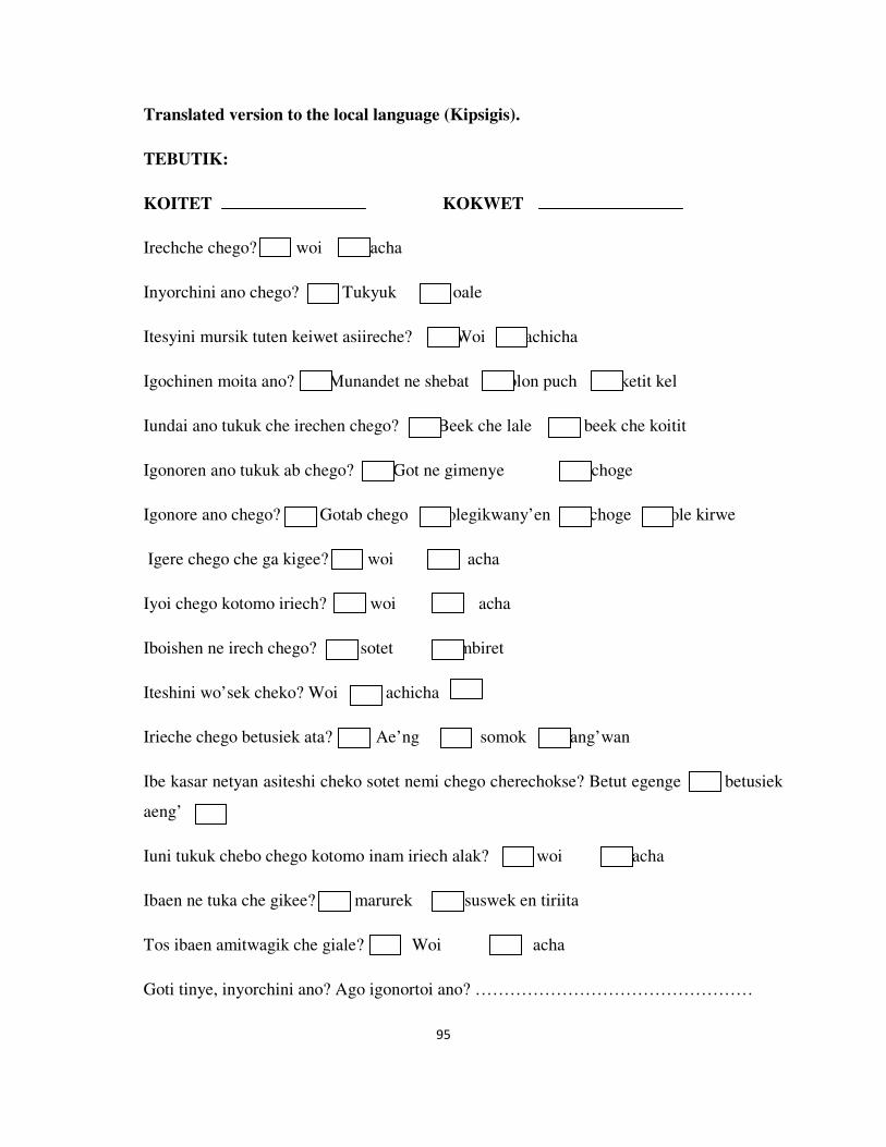
95
Translated version to the local language (Kipsigis).
TEBUTIK:
KOITET KOKWET
Irechche chego? woi acha
Inyorchini ano chego? Tukyuk oale
Itesyini mursik tuten keiwet asiireche? Woi achicha
Igochinen moita ano? Munandet ne shebat olon puch ketit kel
Iundai ano tukuk che irechen chego? Beek che lale beek che koitit
Igonoren ano tukuk ab chego? Got ne gimenye choge
Igonore ano chego? Gotab chego olegikwany’en choge ole kirwe
Igere chego che ga kigee? woi acha
Iyoi chego kotomo iriech? woi acha
Iboishen ne irech chego? sotet mbiret
Iteshini wo’sek cheko? Woi achicha
Irieche chego betusiek ata? Ae’ng somok ang’wan
Ibe kasar netyan asiteshi cheko sotet nemi chego cherechokse? Betut egenge betusiek
aeng’
Iuni tukuk chebo chego kotomo inam iriech alak? woi acha
Ibaen ne tuka che gikee? marurek suswek en tiriita
Tos ibaen amitwagik che giale? Woi acha
Goti tinye, inyorchini ano? Ago igonortoi ano? …………………………………………

96
Itinye tuten? woi acha
Amuche ayal tuten? Woi acha

97
APPENDIX II: INFORMED CONSENT DOCUMENT
PROJECT TITLE: Mycotoxin contamination and molecular charectarization of
mycotoxigenic fungi in traditionally fermented (mursik) milk in Soliat Location which
constitute Kongeren, Soliat, Kamasega and Motero Sub locations, Kericho County.
INVESTIGATORS:
1. Mr. Talaam Kiplangat Keith from Institute of Tropical Medicine and Infectious
Diseases/Jomo Kenyatta University Agriculture and Technology.
2. Dr. Bii C. Christine from Kenya Medical Research Institute (KEMRI).
3. Prof. Ng’ang’a Zipporah from Jomo Kenyatta University of Agriculture and
Technology.
INTRODUCTION:
My team and I (Talaam Kiplangat Keith, the lead investigator and a student) are
researchers from Institute of Tropical Medicine and Infectious Diseases, Kenya Medical
Research Institute and Jomo Kenyatta University of Agriculture and Technology involved
in research studies aimed at improving the health and wellbeing of Kenyans. We are
conducting a research on ‘Mycotoxin contamination and molecular charectarization of
mycotoxigenic fungi in traditionally fermented (mursik) milk in Soliat Location which
constitute Kongeren, Soliat, Kamasega and Motero Sub locations, Kericho County’.
This is a study or research whereby your participation is based on your willingness and no
one is going to force you to be part of the study. You are free to be part or refuse to be part
of the study.

98
PURPOSE OF THE RESEARCH
We intend to determine the fungi and their toxins in mursik. We will collect samples of
complete fermented milk (mursik) to check for fungi and mycotoxin contamination. This
will make it possible for us to determine the fate of mycotoxins during fermentation. We
will do it At Kenya Medical Research Institute. Anyone who is going to participate in the
study will be part of it for a period of three months.
PROCEDURES:
We request you to allow us to buy a cup (‘namba nane’) of your mursik. We will test your
samples for the presence of mycotoxins and fungi. For fungi tests, we will make them grow
by placing a small amount of mursik (100µl) on a medium where the fungi will grow. If the
fungi will grow, we will isolate and characterize them to know the types and if they are
capable of producing mycotoxins. We will determine the presence and levels mycotoxins
using test kits called Envirologix kits. Some other information will be asked through the
questionnaire whereby you will fill it accordingly. The test will run for three months.
BENEFITS
The study will tell us if the milk is contaminated with mycotoxins and if milk is actually
contaminated, we will get to advise you not to use or sell to other people and teaching
sessions will be done milk on how to handle milk and avoid the contamination.
RISKS:
There will be no risks in allowing us to test your mursik.
CONFIDENTIALITY:
In this study, all samples will be given code numbers and will not bear the names of the
fermenters. The findings of this study will be kept confidential on coded records that will
not be traced to individuals. In case a participant wants to have feedback his/her phone

99
numbers will be taken and the records containing the participant’s information will be only
accessed by the principal investigator.
COMPENSATION:
There will be no direct compensation for participation in the study. We will only buy a cup
of your fermented milk.
STORAGE, EXPORTATION OF SAMPLES AND FURTHER STUDIES:
Milk samples collected will be transported to Kenya Medical Research Institute and kept
refrigerators to wait for tests where fungal and mycotoxin contamination will be
determined. Small amounts of 100ml (aliquot) of the samples will be stored in case further
studies will be required and the rest will be autoclaved and discarded.
CONTACT OF THE PRINCIPAL INVESTIGATOR
For any questions you will like to ask, you can do so through the following contacts of the
Principal investigator: Talaam Kiplangat Keith. In Nairobi, P.O Box 19464-00202, Nairobi,
Kenya or at my home near cattle dip in Soliat location, Kongeren Sublocation, Chelogong
village P.O Box 11- 20208, Kiptugumo. Phone number: 0726 462 022 on days and night or
E-mail address: [email protected]
CONTACT OF KEMRI/ERC
For any questions concerning your right of participation, you are free to call the Kenya
Medical Research Institute’s Ethics review committee through; The Secretary, KEMRI
Ethics Review Committee, P.O Box 54840-00200, Nairobi. Telephone numbers: 020-
2722541, 0722205901, 0733400003; Email address: [email protected]
CONSENT AND SIGNATURE OPTIONS
I confirm that I understand the information provided to me for the above study and that I
have had the opportunity to ask questions. I understand that my participation is voluntary
and that I am free to be or not to be part of the study.

100
I …………………………………………………. agree to participate in the study and
allow my mursik to be sampled.
Yes, I would like to be contacted with the results of tests done on my mursik (tick
the check-box if yes)
Participant’s Signature/Thumb print …………………….. Date ………………………
Researcher’s Signature……………………………. Date ……………………….......
Researcher’s Name: TALAAM KIPLANGAT KEITH

101
Translated version of the Informed Consent Document to the local language
(Kipsigis).
KOGUYET AK NAET AB CHIGILISIONI.
METIT AB BOISIET: Chigilet ab ng’wanet ak kabesiaet ab bobek che gonu ng’wanet en
mursik en Soliat Location ne’iume kokwotunwek ang’wan choto go: Kongeren, Soliat,
Kamasega ak Motero, en jimboit ab Kericho.
CHIGILIK:
1. Mr. Talaam Kiplangat Keith goyab gotab kipsomaninik chebo barak nebo
muswagnotet ak kabotisiet, Jomo Kenyatta.
2. Dr. Bii C. Christine goyab gotab chililet ab kerichek nebo Kenya (KEMRI).
3. Prof. Ng’ang’a Zipporah goyab gotab kipsomaninik chebo barak nebo muswagnotet
ak kabotisiet, Jomo Kenyatta.
MWAET AB’GE EN NW’OGINDO:
Gibagenge nenyu ak ane (Talaam Kiplangat Keith, chigilindet ne-o ago kipsomaniat) go
chigilik goyab gotab kipsomaninik chebo barak nebo muswagnotet ak kabotisiet, Jomo
Kenyatta ak gotab chililet ab kerichek nebo Kenya (KEMRI) goyae chigilet chetogyi’nge
gotoror sabet ab kipkosabei en Kenya. Kiyae Chigilet ab ng’wanet ak kabesiaet ab bobek
che gonu ng’wanet en mursik en Soliat Location ne’iume kokwotunwek ang’wan choto go:
Kongeren, Soliat, Kamasega ak Motero, en jimboit ab Kericho. Niton ko chigilet ne mache
biik ko’gonge en chameny’wan ako iyanat koesio chi agetukul komat’kobota boisioni.
AMUNE NEBO CHIKILISIONI:
Gitakyinige gecheng’ bobek ak ngw’anet chegonu bobek en mursik. Kiyumi mursik tuten
asi kechigilik meng’isiet ab bobek ak ngw’anet en chego. Boisioni tugul keyae en gotab
chilisiet nebo kerichek nebo Kenya. Chi agetugul negonu’ge, koboto boisioni en kasartab
arawek somok.

102
OLEGIYAITO BOISIONI:
Kisome keyal mursi chebo gikomet ‘namba nane’. Ipgechigili gotkomi bobek ak nyw’anet
nyw’a. chigilet ab bobek, ko ba keibe mursik tuten ak kerangyi amitwogik che imuch koyai
bobek choto korut. Kotkorut, gi’cheru ak keger keboisien muswagnotet neimuche
koborwech kotgomuche bobekyuton kogon ngw’anet. En ngw’anet gechikili chang’indo
keboisien muswaknotet nebunu got ab Envirologix. Ara ng’olit alak gomi sogkyot age
nebendoti ak sokyondeni. Boisioni goibe kasar nebo arawek somok.
BOROTET:
Chigilisioni komawawech kotkomi nyw’anet ab bobek ak bobek che konu nyw’anet. Ko
kinetige kele many’olu keboisien anan kigichi chi agetugul anan gelada ak kanetisiet nebo
olegiribto chego amatiny bobek.
NG’OYONDIT:
Mami ng’oyondit agetugul en mursik che kichikili.
UNG’OTIET:
En chigilisioni, mujrsik chegiyumi koma kibartai en kaina nebo chi. Mursik kigochin
sirutik noton chenoyotin koba koitasiek. Walutik alaktugul kegonori en ung’ot ak ityitn
chigilinet ne-o. Chi agetugul nemache walutik gogonu oret ne kimuchi keityin ana kogon
koitosiek kwak cheba ‘simoinik’.
WALEIWET:
Mami waleiwet agetugul en chi agatugul nemi chigilishoni. Kiale mursik kityo chebo
kigombet ‘namba nane’.

103
KONORET, IBET AB MURSIK AK CHIKILISIET NEBO BETUSIEK
CHEBWONE:
Mursik kiyumi ak ke-ib koba gotab chigilisiet nebo kerichek (KEMRI) NEBO
KEGONORI EN MUSWAGNOTET NEIGOTITI KOGONYE CHIGILISIET. Kigonorri
tuten kogany chigilet nebo betusiek chebwone ak alak kebele.
ITY’INET AB CHIGILINDET NE-O:
En tebutik alaktugul che imuche iteb chi agetugul iteben chigilindet ne-o, imuche iyai
kobun posta: Talaam Kiplangat Keith. P.O Box 19464-00202, Nairobi, Kenya anan go en
gaa yenegityin ak dip nebo tuga en kokwet ab chelogong sublocation nebo Kongeren en
Soliat location P.O Box 11-20208, Kiptugumo. simoit: 0726 462 022 anan ko oret nebo E-
mail: [email protected]

104
ITY’INET AB BIIK CHE TONONCHIN IMANDAB KIPKOSABEI NEBO KEMRI:
En tebutik alaktugul che imuche iteb chi agetugul agobo imanda’ng’ung iteben che
tononchin imandab kipkosabei nebo kemri kobun sirindet ibune posta; The Secretary,
KEMRI Ethics Review Committee, P.O Box 54840-00200, Nairobi. simoit: 020-2722541,
0722205901, 0733400003; Email: [email protected]
KAYANET AK SI’YET
Ane ayani ale ng’alek che kagigonon en chigilisioni ko akuitosi ako kagekonon kasarian
ateb tebutik. Akuitosi ale ami en chigilisioni ko makisigyin chi ako atinye kasar koboton
chigilishoni.
Ane …………………………………………………. ayani goboton chigilisioni ak akonu
mursik.
Woi, amache keguron kemwoiwon walutik chebo mursik (inde si’yet)
Si’yet…………………….. Betut ………………………
Si’yet ab chigilindet………………. Betut ……………………….......
Chigilindet: TALAAM KIPLANGAT KEITH

105
APPENDIX III: ANALYTICAL PROFILE INDEX (API); YEAST FERMENTATION OF
SUGARS.
Analytical Profile Index (API) TESTS
Fungi Fermentation of sugars
Yeast spp. Glucose Maltose Galactose Sucrose Lactose
Geotricum
candidum
_ _ _ _ _
Sacharomyces
cerevisiae
+ + + + _
Candida
albicans
+ + + _ _
Candida
parapsilosis
+ _ _ _ _
Candida kefyr + _ + + +
Candida
glabrata
+ _ _ _ _
Candida
tropicalis
+ + +/_ + _
Candida krusei + _ _ _ _
Key; + showed that there was fermentation, +/_ no clear fermentation and – shows no
fermentation.

106
APPENDIX IV: AFLATOXIN QUANTIFIED LEVELS.
Aflatoxin Quantification
Toxin level (ppb) Frequency % of total
0 2 1.0
2.5 1 0.5
2.6 1 0.5
2.8 1 0.5
2.9 2 1.0
3 3 1.5
3.1 4 2.0
3.2 5 2.5
3.3 1 0.5
3.4 5 2.5
3.5 3 1.5
3.6 4 2.0
3.8 8 4.0
3.9 5 2.5
4 5 2.5
4.1 5 2.5
4.2 5 2.5
4.3 12 6.1
4.4 3 1.5
4.5 8 4.0
4.6 5 2.5
4.7 6 3.0
4.8 13 6.6
4.9 9 4.6
5 12 6.1
5.1 8 4.0

107
5.2 10 5.1
5.3 5 2.5
5.4 12 6.1
5.5 7 3.5
5.6 6 3.0
5.7 3 1.5
5.8 4 2.0
5.9 5 2.5
6 4 2.0
6.1 1 0.5
6.3 2 1.0
6.9 1 0.5
11 1 0.5
12 1 0.5
Total 198 100

108
APPENDIX V: INVESTIGATIVE VARIABLE RESULTS.
Investigation variables of milk fermentation
Variable Frequency % of total
Obtain milk from own cows 194 100
Milk fermenters 194 100
Use of calabash 194 100
Use of starter
cultures
yes 0 0
no 194 100
Milking cows in the
open
Yes 194 100
No 0
Washing calabashes
using
Hot water 155 80
Cold water 39 20
Storing calabash and
other milk containers
In maize
store
144 74
In living
rooms
50 26
Milk storage In maize store 144 74
In living
rooms
50 26
Covering freshly
milked milk.
Yes 19 10
No 175 90
Use of wo’sek Yes 194 100
No 0
Fermenting time 3-4 days 194 100
>4days 0
Time interval for
milk addition on
continuous cultures
1 day 194 100
2days 0
Cleaning calabashes Yes 194 100
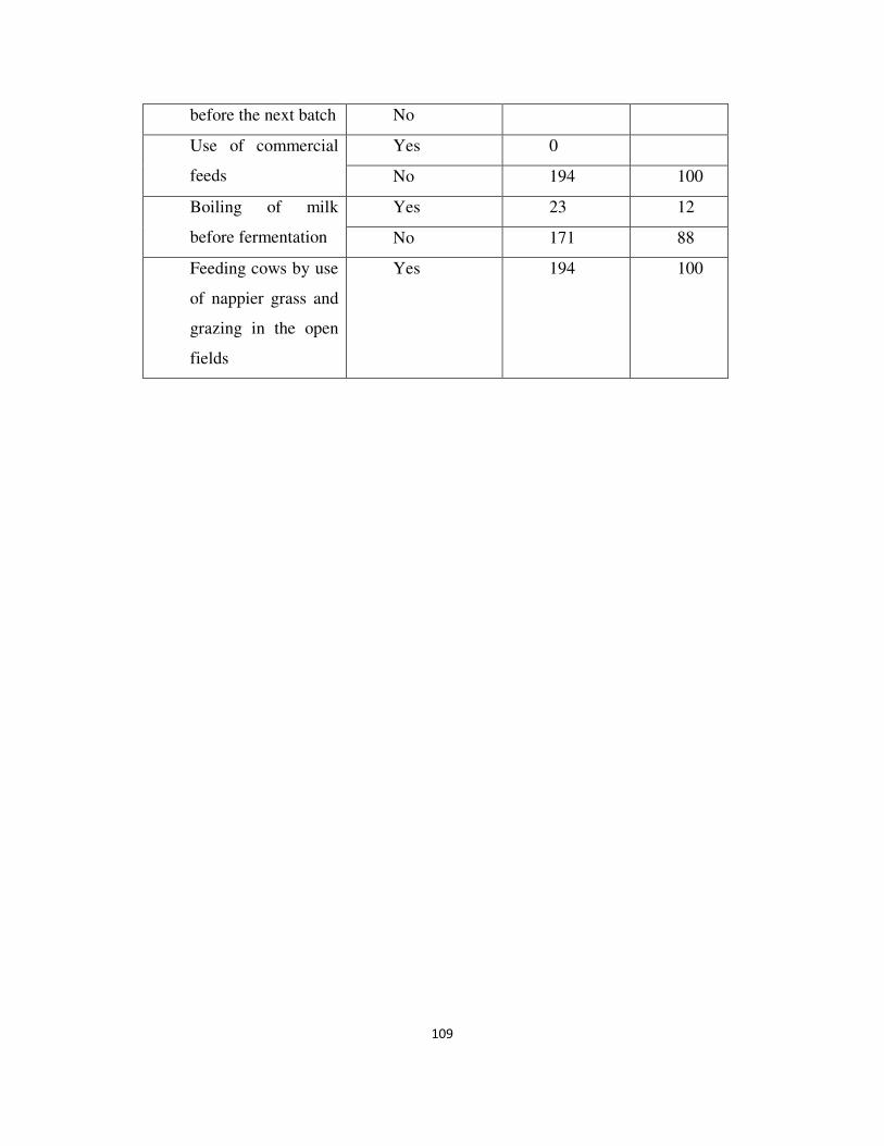
109
before the next batch No
Use of commercial
feeds
Yes 0
No 194 100
Boiling of milk
before fermentation
Yes 23 12
No 171 88
Feeding cows by use
of nappier grass and
grazing in the open
fields
Yes 194 100

110
APPENDIX VI: ETHICAL CLEARANCE