mycobacterium tuberculosis groel homologues unusually exist as
TRANSCRIPT

doi:10.1016/j.jmb.2004.07.066 J. Mol. Biol. (2004) 342, 605–617
Mycobacterium tuberculosis GroEL HomologuesUnusually Exist as Lower Oligomers and Retain theAbility to Suppress Aggregation of Substrate Proteins
Rohini Qamra1, Volety Srinivas2 and Shekhar C. Mande1*
1Laboratory of StructuralBiology, Centre for DNAFingerprinting and DiagnosticsECIL Road, NacharamHyderabad 500 076, India
2Centre for Cellular andMolecular Biology, Uppal RoadHyderabad 500 007, India
0022-2836/$ - see front matter q 2004 E
Abbreviations used: Cpn60s, chap1,1-bis (4-anilino) naphthalene-5,5 0-circular dichroism.E-mail address of the correspond
Chaperonin-60s are large double ring oligomeric proteins with a centralcavity where unfolded polypeptides undergo productive folding. Inconjunction with their co-chaperonin, Chaperonin-60s bind non-nativepolypeptides and facilitate their refolding in an ATP-dependent manner.The ATPase activity of Chaperonin-60 is tightly regulated by the 10 kDa co-chaperonin. In contrast to most other bacterial species, Mycobacteriumtuberculosis genome carries a duplicate set of cpn60 genes, one of whichoccurs on the groESL operon (cpn60.1), while the other is separatelyarranged on the chromosome (cpn60.2). Biophysical characterization of themycobacterial proteins showed that these proteins exist as lower oligomersand not tetradecamers, an unexpected property much different from theother known Chaperonin-60s. Failure of the M. tuberculosis chaperonins tooligomerize can be attributed to amino acid mutations at the oligomericinterface. Rates of ATP hydrolysis of the M. tuberculosis chaperoninsshowed that these proteins possess a very weak ATPase activity. Both theM. tuberculosis chaperonins were partially active in refolding substrateproteins. Interestingly, their refolding activity was seen to be independentof the co-chaperonin and ATP. We hypothesize that the ATP independentchaperones might offer benefit to the pathogen by promoting its existencein the latent phase of its life cycle.
q 2004 Elsevier Ltd. All rights reserved.
Keywords: chaperonin; protein folding; oligomer; aggregation;M. tuberculosis
*Corresponding authorIntroduction
Molecular chaperones are a diverse set ofproteins that mediate the correct folding, assembly,transport and degradation of other proteins in vivo.1
Chaperonins form a sub-group of molecular cha-perones that are found in all domains of life. GroupI chaperonins comprise a family of highly con-served proteins, approximately 60 kDa in molecularmass that form a cylindrical assembly of twoheptameric rings. Unfolded polypeptides bindand undergo productive folding within the largecentral cavity of this assembly.2 The Escherichia colichaperonin, GroEL, has provided a paradigm inunderstanding protein folding mechanisms
lsevier Ltd. All rights reserve
eronin-60s; bis-ANS,disulfonic acid; CD,
ing author:
mediated by the chaperonins.3 GroEL promotes denovo folding of w10–15% of all proteins in bacterialcytosol in co-ordination with the heptamericco-chaperonin, GroES.4 ATP-dependent confor-mational changes in GroEL have been shown to benecessary for proper chaperonin function in vivo.1,5
Chaperonins in all bacteria are encoded byessential groEL and groES genes (also called cpn60and cpn10), arranged on the bicistronic groESLoperon.6 The operon arrangement of the twogenes is highly conserved among all knownbacterial species. Interestingly, Mycobacterium tuber-culosis contains two copies of the cpn60 genes.7 Oneof these genes, cpn60.1, is organized on the operonwith cpn10, while the second copy, cpn60.2, isarranged separately on the genome.7 A similararrangement of cpn10 and cpn60 genes has beendescribed in Streptomyces spp.8 and Mycobacteriumleprae.9
The E. coli chaperonin, GroEL, is known to beessential at all temperatures of growth.10 Ubiqui-tous presence of chaperonins, and their high
d.

606 Unusual GroEL Homologues of M. tuberculosis
conservation suggests a similar indispensable rolethat these proteins play in all life forms. A highsequence similarity thatM. tuberculosisChaperonin-60s (Cpn60s) exhibit with E. coli GroEL, suggeststhat these might also perform a similar role in thisorganism. The existence of a duplicate set of cpn60genes inM. tuberculosis, however, has been perplex-ing. Plant chloroplasts also possess two copies ofCpn60s that are known to form a hetero-oligomericassembly.11 The two M. tuberculosis GroEL homo-logues may similarly be interdependent for theassembly of the protein into a functional hetero-tetradecamer. Duplication therefore might suggesteither an interdependence of the gene products oralternatively, the necessity for a redundant function.
While reasons for gene duplication remainunclear, both the Cpn60s of M. tuberculosis havebeen shown to be highly antigenic in nature,eliciting strong B-cell and T-cell immune responses.Cpn60s have also been proposed to play roles asvirulence determinants and as inducers of hostinflammatory responses.12 Moreover, both Cpn60.1and Cpn60.2 of M. tuberculosis have been shown tobe potent cytokine inducers.13 The Cpn60s mighttherefore represent important components ofM. tuberculosis, playing roles as immunomodula-tors, and perhaps also required for proper proteinfolding and transport.
Biochemical properties of M. tuberculosis Cpn60s,especially their role as molecular chaperones, havenot been characterized yet. Moreover, the presenceof two copies of these genes in the M. tuberculosisgenome is intriguing. To address these questions,we have expressed the M. tuberculosis cpn60 genesin E. coli in an attempt to elucidate their role inprotein folding.
Results
Cloning, expression and purification ofM. tuberculosis Cpn60.1 and Cpn60.2
The full length chaperonin cpn60.1 and cpn60.2clones were obtained and overexpressed in E. coli asdescribed in Materials and Methods. The twochaperonins were expressed in large quantitiesand exhibited a protomer molecular mass ofw60 kDa. As judged by SDS-PAGE, both theproteins were more than 98% pure after the metalaffinity purification.
Quaternary structure determination
A tetradecameric structure for wild-type Cpn60has been established for a number of chaperoninsfrom different species.14,15 In a few cases, however,Cpn60 homologues have also been reported to existas heptamers.16 The native molecular mass of thepurified M. tuberculosis proteins, Cpn60.1 andCpn60.2, was established by size exclusion chro-matography. Intriguingly, the purified Cpn60.1 andCpn60.2 eluted as sharp homogenous peaks at
volumes corresponding to molecular mass ofapproximately 110 kDawhen compared to standardmolecular weight markers (Figure 1). NeitherCpn60.1 nor Cpn60.2 eluted at the position whereE. coli GroEL and canonical Cpn60 14-mers wouldappear. The sharp peaks were suggestive of theglobular state of the purified proteins. Elutionprofile on the size exclusion chromatography ofthe two Cpn60s lacking the (His)6-tag was consist-ent with that of the (His)6-tagged proteins,suggesting that the presence of (His)6-tag did notinterfere in oligomerization (data not shown).
Osmolyte stabilizers, such as glycerol, are knownto promote stability of the oligomeric state ofproteins. Similarly, the presence of ATP and theco-chaperonin, Cpn10 may also be crucial to theoligomeric assembly of the mycobacterial Cpn60s.M. tuberculosis Cpn10 has already been demon-strated to exist as a heptamer in the presence ofdivalent cations.17 Purification of Cpn60.1 andCpn60.2 was performed in the presence of glycerol,or in the presence of ATP-g-S and M. tuberculosisCpn10. Their oligomeric state was then tested onNative-PAGE. Proteins purified under these con-ditions, however, did not exhibit oligomerization(Figure 2). Thus, purification of M. tuberculosisCpn60s, either in the presence of co-chaperonin orother osmolyte stabilizers did not seem to promotethe oligomeric assembly of these proteins. The sizeexclusion chromatography and Native-PAGE datahence suggest that the M. tuberculosis GroEL hom-ologues might intrinsically exist as lower oligomers.
Reconstitution of M. tuberculosis Cpn60s
In order to functionally reconstitute the chaper-onins in their canonical oligomeric forms, wesubjected the purified Cpn60.1 and Cpn60.2 to invitro reconstitution experiments under a variety ofconditions. It has previously been reported thaturea-dissociated E. coli GroEL 14-mers are capableof reassembling into the tetradecameric state uponremoval of the chaotrope.18 Reconstitution of theE. coli GroEL tetradecamers from the disassembledmonomers requires the presence of Mg2C andadenine nucleotides. Presence of ammonium sul-phate has also been reported to be essential for theoligomer reassembly.19 When subjected to similarrefolding conditions, neither of the two mycobac-terial GroEL homologues spontaneously assembledinto tetradecamers (Figure 3(a)). Reconstitution wasalso attempted in the presence of glycerol. How-ever, none of the various combinations of nucleo-tides, GroES, ammonium sulphate, glycerol, KC orMg2C, promoted oligomeric assembly of the myco-bacterial chaperonins (Figure 3(a)).
GroES has previously been shown to facilitate theoligomeric assembly of GroEL.20 Reconstitution ofCpn60s was thus performed in the presence of thecognate Cpn10. Presence of the M. tuberculosis co-chaperonin, Cpn10 in the reconstitution mix how-ever, had no effect on the oligomeric assembly of thepurified proteins (Figure 3(b)). Failure to

Figure 1. Gel filtration chromatograms of M. tuberculosis Cpn60s. Elution profile of the purified (a) Cpn60.1 and (b)Cpn60.2 on a Superdex-200 HR 10/30 column. Both Cpn60.1 and Cpn60.2 elute as 110 kDa species. (c) Elution of standardmolecular mass marker proteins shown as a function of logðMrÞ. Ve and Vo correspond to elution and void volumes,respectively.
Unusual GroEL Homologues of M. tuberculosis 607

Figure 2. Native gel electrophor-esis of purified M. tuberculosisCpn60s demonstrating their loweroligomeric state. Cpn60.1 andCpn60.2 were purified in the pre-sence of glycerol, ATP-g-S andM. tuberculosis Cpn10. The purifiedCpn60s migrate as lower oligo-mers, showing the proteins’inability to assemble into oligomersin the presence of these sup-plements.
Figure 3. Reconstitution ofM. tuberculosis Cpn60s demon-strating their lower oligomericstate. (a) In vitro reconstitution ofCpn60 oligomer was attempted inthe presence of ammonium sul-phate and glycerol as described inMaterials and Methods. Presenceof either ammonium sulphate orglycerol in the reconstitution mixdoes not alter the oligomeric stateof Cpn60.1 (lanes 1–4) or Cpn60.2(lanes 5–8) independent of thepresence of E. coli GroES. Co-existence of the two Cpn60s(lanes 9–12) also does not promotethe oligomeric assembly of themycobacterial Cpn60s. Lane 13indicates the oligomeric state ofthe wild-type E. coli GroEL. (b) Invitro reconstitution of Cpn60 oli-gomers was attempted in thepresence of the cognate co-chaper-onin, M. tuberculosis Cpn10. E. coliGroEL monomers subjected toreconstitution in the presence ofammonium sulphate (lane 1) andM. tuberculosis Cpn10 (lane 2)readily reassemble as tetradeca-mers. On the other hand,M. tuber-culosis Cpn60.1 and Cpn60.2,subjected to reconstitution in thepresence of ammonium sulphate(lanes 3 and 7), E. coliGroES (lanes4 and 6) andM. tuberculosis Cpn10(lanes 5 and 8), clearly show thatthe two proteins are unable toreconstitute as tetradecamers.
608 Unusual GroEL Homologues of M. tuberculosis

Unusual GroEL Homologues of M. tuberculosis 609
reconstitute theM. tuberculosis chaperonins in cano-nical oligomeric states thus suggested that theirexistence as lower oligomers might be an intrinsicproperty of the two chaperonins.
Plant chloroplast chaperonins are known tooligomerize as heterotetradecamers of two differentpolypeptide chains.11 In order to test the possibilitywhether the two M. tuberculosis chaperonins weresimilarly interdependent on each other for reas-sembly into tetradecameric state, reconstitution ofthe two proteins was attempted in the presence ofeach other. Incorporation of both Cpn60.1 andCpn60.2 in the reaction mix, however, did notalter the oligomeric state of the reconstitutedproteins (Figure 3(a)). Moreover, the oligomericstate remained unaltered irrespective of the pre-sence or absence of the nucleotides or GroES. Theseresults thus further corroborate that the M. tubercu-losis GroEL homologues are lower oligomericproteins, unlike their E. coli counterpart.
Circular dichroism measurements ofM. tuberculosis Cpn60s
The above data indicate that the two M. tubercu-losis GroEL homologues possess an unusual qua-ternary structure. The possibility that their loweroligomeric state might have arisen due to the loss ofsecondary and tertiary structure was tested bymeasuring the CD spectrum of the proteins. The farUV-CD spectrum of M. tuberculosis Cpn60.1 andCpn60.2 (Figure 4) is characteristic of highly helicalproteins with signature bands for helical structureat 208 nm and 222 nm.21 The data suggest that theM. tuberculosis chaperonins are in a significantly
Figure 4. Far UV-CD spectrum of E. coli GroEL, M. tuberculwavelength was measured at 25 8C in 10 mM Tris–Cl (pH 8.0and 222 nm, suggesting a well-formed secondary structurespectrum of E. coli GroEL, the dotted line represents that of C
folded conformation. The unusual quaternarystructure of the M. tuberculosis chaperonins thusdoes not appear to be due to the absence ofsecondary structure of the proteins.
Oligomeric state of M. tuberculosis Cpn60s invivo
As mentioned above, Cpn60s from other prokar-yotes exist as tetradecamers in their cellularenvironment and the same oligomeric state is alsoobserved in vitro. Our in vitro purification andreconstitution results show that the M. tuberculosisCpn60s exist as lower oligomers, a state surpris-ingly different from that observed for other Cpn60s.To discount the possibility that the unusual beha-viour of chaperonins as lower oligomers was aresult of their heterologous expression, we assessedthe oligomeric state of these proteins in theirhomologous cellular environment. This was testedby resolving the cellular fraction of M. tuberculosisproteins on Native-PAGE followed by Western blotanalysis (Figure 5). Upon immunoblotting, bothCpn60.1 and Cpn60.2 appeared as lower oligomers,and not tetradecamers, reconfirming our in vitrodata (Figure 5). In conclusion therefore, the absenceof the oligomeric state of theM. tuberculosis Cpn60s,in vivo and in vitro, indicates the natural tendency ofthese proteins to exist as lower oligomers.
Importance of Ala2 and Glu76 in oligomerization
Multiple sequence analysis of 43 Cpn60 proteinsequences suggested that residues at positions 2and 76, which occur at the inter-monomer interface
osis Cpn60.1 and Cpn60.2. Mean residue ellipticity versus). Signature bands for helical structure are seen at 208 nmfor the Cpn60s. The continuous line represents the CDpn60.1 and the broken line that of Cpn60.2.

Figure 5.Western blot analysis ofM. tuberculosis cultureproteins demonstrating lower oligomeric state of Cpn60s.Culture extracts of M. tuberculosis were subjected to 6%Native-PAGE and probed with monoclonal antibodies,IT-56 and mAb67-2, against Cpn60.1 (lane 1) and Cpn60.2(lane 2), respectively. Western blot analysis indicates thatCpn60s of M. tuberculosis exist as lower oligomers in vivo.Expected position of the oligomeric species is indicated.
Figure 6. Crucial side-chain–main-chain interaction atthe E. coli GroEL interface involving Glu76. Interactionbetween the side-chain carboxyl of E76 (chain A) andmain-chain amide of E386 (chain B) in E. coli GroEL hasbeen suggested to be important for the oligomerization ofthe protein. A natural occurrence of serine at position 76in M. tuberculosis Cpn60.1 may disrupt this interactionand is hypothesized to contribute to the loss of itsoligomeric structure. The two polypeptide chains havebeen coloured in different shades of grey. Other crucialinterface residues important for maintenance of theoligomeric state are indicated in the Supplementarydata. This Figure was generated using MOLSCRIPT.47
Figure 7. Effect of urea concentration on the dis-sociation of E. coli GroEL mutant. Urea-induced dis-sociation of the (a) wild-type and (b) A2S/E76S GroELmutant was analysed on a 6% Native-PAGE. (c) Ratio ofthe oligomeric to monomeric forms of GroEL plottedagainst urea concentration indicates that the mutantdissociates into monomers at 2.5 M urea concentration,in comparison to 3 M for the wild-type.
610 Unusual GroEL Homologues of M. tuberculosis
in the tetradecameric GroEL structure, are strictlyconserved as alanine and glutamate (Figure 6 andSupplementary data). These positions are, however,occupied by serine in M. tuberculosis Cpn60.1. Toassess the importance of these residues in mainten-ance of the oligomeric structure, we generated anA2S/E76S mutant of E. coli GroEL.
The monomer–tetradecamer equilibrium of thewild-type as well as that of the A2S/E76S mutantwas analysed as a function of urea concentration onNative-PAGE. In accordance with an earlier report,the wild-type protein dissociates into monomers ata urea concentration greater than 3 M (Figure 7).22
The mutant, however, disassociated into monomersat a much lower concentration of urea than the
Unusual GroEL Homologues of M. tuberculosis 611
wild-type GroEL (Figure 7). The results thusindicate that the thermodynamic stability of theGroEL oligomeric assembly is significantly reducedas a consequence of the mutations A2S and E76S. Anatural occurrence of serine at residue positions 2and 76 in Cpn60.1 might thus be responsible for thelower oligomeric state of this protein.
ATPase activity of the mycobacterial Cpn60s
E. coli GroEL possesses a weak KC stimulatedATPase activity, an essential component of thechaperonin-mediated protein folding reaction.23
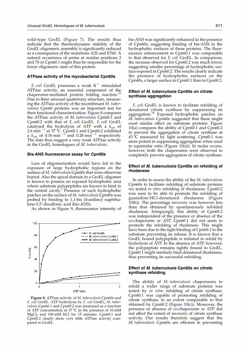
Due to their unusual quaternary structure, measur-ing the ATPase activity of the recombinantM. tuber-culosis Cpn60 proteins was an important test fortheir functional characterization. Figure 8 comparesthe ATPase activity of M. tuberculosis Cpn60.1 andCpn60.2 with that of E. coli GroEL. E. coli GroELcatalysed the hydrolysis of ATP with a kcat of2.6 minK1 at 37 8C. Cpn60.1 and Cpn60.2 exhibiteda kcat of 0.16 minK1 and 0.28 minK1 respectively.The data thus suggest a very weak ATPase activityin the GroEL homologues of M. tuberculosis.
Bis-ANS fluorescence assay for Cpn60s
Loss of oligomerization would have led to theexposure of large hydrophobic regions on thesurface ofM. tuberculosisCpn60s that were otherwiseburied. Also the apical domain in a GroEL oligomeris known to possess an exposed hydrophobic areawhere substrate polypeptides are known to bind inthe central cavity.3 Presence of such hydrophobicpatches on the surface ofM. tuberculosisCpn60s wasprobed by binding to 1,1-bis (4-anilino) naphtha-lene-5,5 0-disulfonic acid (bis-ANS).
As shown in Figure 9, fluorescence intensity of
Figure 8. ATPase activity of M. tuberculosis Cpn60s andE. coli GroEL. ATP hydrolysis by E. coli GroEL, M. tuber-culosis Cpn60.1 and Cpn60.2 was measured as a functionof ATP concentration at 37 8C in the presence of 10 mMMgCl2 and 100 mM KCl for 15 minutes. Cpn60.1 andCpn60.2 clearly show very little ATPase activity com-pared to GroEL.
bis-ANS was significantly enhanced in the presenceof Cpn60s, suggesting binding of bis-ANS to thehydrophobic surfaces of these proteins. The fluor-escence enhancement in Cpn60.1 was comparableto that observed for E. coli GroEL. In comparison,the increase observed for Cpn60.2 was much lower,suggesting smaller percentage of hydrophobic sur-face exposed in Cpn60.2. The results clearly indicatethe presence of hydrophobic surfaces on theCpn60s, a larger surface in Cpn60.1 than in Cpn60.2.
Effect of M. tuberculosis Cpn60s on citratesynthase aggregation
E. coli GroEL is known to facilitate refolding ofdenatured citrate synthase by suppressing itsaggregation.24 Exposed hydrophobic patches onM. tuberculosis Cpn60s suggested that these mightexert similar effect on substrate proteins. Figure10(a) compares the ability of Cpn60.1 and Cpn60.2to prevent the aggregation of citrate synthase at43 8C measured by light scattering. Cpn60.1 wasmore potent in suppressing aggregation when usedin equimolar ratio (Figure 10(a)). In molar excess,however, both the chaperonins were observed tocompletely prevent aggregation of citrate synthase.
Effect of M. tuberculosis Cpn60s on refolding ofrhodanese
In order to assess the ability of the M. tuberculosisCpn60s to facilitate refolding of substrate proteinswe tested in vitro refolding of rhodanese. Cpn60.2was seen to be able to promote the refolding ofguanidine-HCl-denatured rhodanese (Figure10(b)). The percentage recovery was however lessthan that obtained by spontaneously refoldedrhodanese. Intriguingly, this ability of Cpn60.2was independent of the presence or absence of theco-chaperonin or ATP. Cpn60.1 did not seem topromote the refolding of rhodanese. This mighthave been due to the tight binding of Cpn60.1 to thesubstrate preventing its release. It is known that aGroEL bound polypeptide is initiated to refold byhydrolysis of ATP. In the absence of ATP, however,the polypeptide remains tightly bound to GroEL.Cpn60.1 might similarly bind denatured rhodanese,thus preventing its successful refolding.
Effect of M. tuberculosis Cpn60s on citratesynthase refolding
The ability of M. tuberculosis chaperonins torefold a wider range of substrate proteins wastested by in vitro refolding of citrate synthase.Cpn60.1 was capable of promoting refolding ofcitrate synthase, to an extent comparable to thatobtained by Cpn60.2 (Figure 10(c)). Moreover, thepresence or absence of co-chaperonin or ATP didnot affect the extent of recovery of citrate synthaseactivity. Our results therefore suggest that theM. tuberculosis Cpn60s are efficient in preventing

Figure 9. Fluorescence intensityenhancement of bis-ANS uponbinding to M. tuberculosis Cpn60sThe fluorescence intensity of bis-ANS is observed to increase sig-nificantly in the presence of bothCpn60.1 (broken line) and Cpn60.2(dashed-double-dotted line),suggesting the presence of hydro-phobic surfaces on the proteins.The continuous line represents theenhancement seen for E. coli GroELin presence of bis-ANS. Fluor-escence intensity for the buffer(dotted line) and bis-ANS(dashed-dotted line) weremeasured as controls.
612 Unusual GroEL Homologues of M. tuberculosis
aggregation of denatured proteins, but do notpossess ATP-dependent chaperoning activity.
Discussion
GroEL is the major heat shock protein present inall forms of life guiding several essential stepsduring synthesis, folding, transport and degra-dation of proteins. In E. coli, groEL is known to bean essential gene for growth at all temperatures.10
Upon thermal stress, nearly 15% of the normalprotein mass of the cell consists of the GroEL andGroES proteins.25 Over 150 homologues of Cpn60sequences are currently available, with a pair-wisesimilarity extending from 40% to 100% at the aminoacid level.26 High conservation of Cpn60s acrossspecies suggests that they play an important role inthe physiology of all species.
Heat shock proteins, including Cpn60s, are notonly induced under thermal stress, but also showelevated expression levels under a variety of otherunnatural conditions. Not surprisingly therefore,heat shock proteins are induced within pathogenicorganisms upon invasion of host cells, presumablycontributing to their survival within the hosts.27
Study of regulation of heat-shock proteins inM. tuberculosis has shown enhanced expression ofboth the Cpn60s upon thermal shock to thebacteria,28 as well as upon phagocytosis by macro-phages.27 It is therefore reasonable to believe thatCpn60s of M. tuberculosis contribute to its defensiveresponse against external stress conditions.
Here we present interesting biochemical charac-teristics of the two Cpn60s of M. tuberculosis. Amajor surprising outcome of the present study hasbeen that the two Cpn60s are distinct from thecanonical GroEL homologues. The most distinctivefeature of M. tuberculosis Cpn60s is their oligomericnature, where unlike the known Cpn60s, they donot form a 14-meric assembly. The proteins rather
exist as lower oligomers irrespective of the presenceor absence of nucleotides. Several attempts toreconstitute the M. tuberculosis Cpn60.1 or Cpn60.2under a variety of conditions did not yield higheroligomers. The lower oligomeric state of M. tuber-culosis Cpn60s also appears to be their natural stateof existence as shown by our Western blot analysison Native-PAGE. Current understanding of thechaperonin function assumes a strict oligomericassembly of GroEL, a tetradecameric state, which isthe functional unit for protein refolding.29,30 Thus,the functional relevance of the lower oligomericstate of mycobacterial chaperonins is still not clear.
Interestingly, many of the highly conservedresidues, listed with “conservation index” betterthan 0.4 of Cpn60s are observed to be different inM. tuberculosis chaperonins.26 For example, a crucialglutamate at position 76, involved in an importantside-chain–main-chain interaction across twomonomers, is conserved in all Cpn60 sequences(Figure 6, see Supplementary data for the multiplesequence alignment). The conserved glutamate is,however, replaced by a serine in M. tuberculosisCpn60.1. Several other alterations have beenobserved to occur at the interface of Cpn60.1(Supplementary data). Similarly, the presence ofan alanine at position 2 has earlier been reported tobe crucial for maintenance of the oligomeric state ofGroEL. Mutation of alanine to serine at this positionwas shown to weaken the intersubunit interactionsin GroEL, destabilizing its oligomeric structure.31
Interestingly, this position is occupied by a serine inM. tuberculosis Cpn60.1. Thus, we hypothesize thatcrucial changes in the interface residues might haveresulted in the loss of oligomerization of theM. tuberculosis Cpn60.1. In an attempt to verify ourhypothesis, site-directed mutagenesis of theseresidues was performed in E. coli GroEL. Urea-induced unfolding transition show that themutation A2S/E76S indeed destabilizes the

Figure 10. Assessment of chaper-oning ability of Cpn60.1 andCpn60.2. (a) Aggregation of citratesynthase as a function of time:aggregation of citrate synthase at43 8C in the absence (C) and thepresence of equimolar ratios ofM. tuberculosis Cpn60.1 (7); andCpn60.2 (>), measured as a func-tion of light scattered at 465 nm.Increase in molar excess ratio ofCpn60.2 to twofold (B) or 14-fold(:) resulted in suppression ofaggregation of citrate synthase to abetter extent. Both the Cpn60s areclearly able to prevent light scatter-ing by citrate synthase when pre-sent in molar excess. Citratesynthase in the presence of BSA(&) was taken as a control. (b)Effect of M. tuberculosis Cpn60s onrefolding of chemically denaturedrhodanese. Refolding of denaturedrhodanese was monitored spon-taneously (C) or in the presenceof E. coli GroEL, GroES and ATP(;); GroEL, in the absence ofGroES or ATP (B); M. tuberculosisCpn60.1 in the presence (7) orabsence (,) of Cpn10 and ATPand M. tuberculosis Cpn60.2 in thepresence (&) or absence (%) ofCpn10 and ATP. Activity obtainedwhen rhodanese was refolded inthe presence of E. coli GroEL for 90minutes was considered to be100%. (c) Effect of M. tuberculosisCpn60s on refolding of chemicallydenatured citrate synthase. Refold-ing of denatured citrate synthasewas monitored in the presence ofE. coli GroEL, GroES and ATP (B);GroEL, in the absence of GroES orATP (C);M. tuberculosis Cpn60.1 inthe presence (7) or absence (;) ofCpn10 and ATP and M. tuberculosisCpn60.2 in the presence (,) orabsence (&) of Cpn10 and ATP.Activity obtained when denaturedcitrate synthase was refolded in thepresence of E. coli GroEL for 90minutes was considered as 100%.
Unusual GroEL Homologues of M. tuberculosis 613

614 Unusual GroEL Homologues of M. tuberculosis
oligomeric structure of GroEL. Similarly, otheridentified positions in Cpn60.1, which largelyoccur at the oligomeric interface in GroEL, mightfurther contribute to the loss of 14-meric state ofCpn60.1.
Another interesting observation in the presentstudy is the loss of ATPase activity ofM. tuberculosisCpn60s. The protein folding cycle of GroEL hasbeen reported to be largely dependent on theATPase activity of the protein. We present evidencethat the M. tuberculosis chaperonins have lost theirATPase activity. Interestingly however, despite theloss of canonical oligomeric state and ATPaseactivity, the M. tuberculosis Cpn60s retain theirability to suppress aggregation of substrate pro-teins. However, the extent to which the two Cpn60ssuppress aggregation is different, possibly due tovariation in the exposed hydrophobic surfaces ofthe two proteins. Our results indicate that Cpn60.1is more potent in preventing aggregation thanCpn60.2. For substrate proteins such as rhodanese,Cpn60.2 is able to bind these proteins reversibly,giving them a chance to fold to their nativeconformation. Cpn60.1, on the other hand, remainstightly bound to the substrate protein. The substrateprotein is thus prevented from aggregating but doesnot get refolded. Substrates such as citrate synthaseon the other hand, by reversible binding to Cpn60s,are refolded more efficiently.
The ability of M. tuberculosis Cpn60s to promoterefolding in the absence of ATP is similar to thatobserved for other known chaperones. For example,functionally active monomeric minichaperonescontaining part of the polypeptide binding domainof GroEL are known to be effective in vitro and invivo.32,33 Also monomeric Cpn60 from Thermusthermophilus has been shown to possess the abilityto suppress aggregation and promote proteinfolding with no requirement for ATP or GroES.34
Thus, M. tuberculosis Cpn60s in a similar fashionmight have evolved to function in lower oligomericstate with no ATP requirement.
A recent study surprisingly shows the presenceof the mycobacterial Cpn60s in the plasma mem-brane fraction although the major functional role ofthese proteins lies in the cytosol.35 Moreover,various other reports suggest alternative roles forthe GroEL homologues. For example, a GroELhomologue in Buchnera, an intracellular symbioticbacterium of aphids, is not only a molecularchaperone but also a phosphocarrier protein,suggesting that the protein plays a role in a signaltransducing system.36 The M. leprae Hsp65(Cpn60.2) has been shown to display proteolyticactivity that is catalytically related to the HslVUprotease.37 By similarity, theM. tuberculosis Cpn60.2too may have gained such a role.
Judicial utilization of energy sources in a cell isimportant to the organism for its survival. Theprotein folding cycle by the Cpn60s in all life formsis an ATP-dependent process utilizing seven ATPmolecules in each reaction cycle.38 Ability of anorganism to promote protein folding without
energy utilization would prove to be economical.We hypothesize that high demands of energyresulting from its extremely slow rate of metab-olism might have led M. tuberculosis to devise analtered route to protein folding, avoiding the usualenergy-dependent pathway. Cpn60s through such aroute thus continue to fold substrate proteins withinthe cytosol, sparing ATP for other crucial processesof the cell. We thus suggest that loss of anoligomeric state of Cpn60s, yet retaining theirchaperoning role, might have been designed inevolution to save the energy sources of thebacterium. It is pertinent to note in this contextthat other ATP-dependent enzymes, such as RecA,also have reduced ATPase activity in M. tuberculo-sis.39 Further analysis of the role of GroEL homo-logues would lead us to a better understanding ofthe importance of the presence of two Cpn60s inM. tuberculosis.
Materials and Methods
Cloning, expression and purification ofM. tuberculosis Cpn60s
The genes coding for the M. tuberculosis GroEL homo-logues, cpn60.1 (Rv3417c) and cpn60.2 (Rv0440) were PCRamplified from the M. tuberculosis H37Rv cosmid librarykindly provided by Stewart Cole.40 The two cpn60fragments were cloned into E. coli expression vectors,pET3a and pET28a (Novagen), respectively, through anintermediate sub-cloning step in pBluescript (SKC)(Stratagene). Primers used for amplification carried(His)6-tag, which was thus incorporated in the proteinsat the N-terminal and the C-terminal of the cpn60.1 andcpn60.2 gene products, respectively. E. coli BL21 (DE3)over-expressing the two genes were lysed by sonication.The resuspension buffer was supplemented with 10 mMsarkosyl during lysis of the Cpn60.1 expressing culture.The proteins were purified by affinity chromatographyover Ni-NTA agarose column. Purification of Cpn60s wasalso carried out in the presence of 10% (v/v) glycerol,1 mM ATP-g-S or 10 mM M. tuberculosis Cpn10. Thesesupplements were present in the buffers throughout thepurification procedure. The M. tuberculosis proteins werealso cloned in the expression vector without the (His)6-tag. The E. coli GroEL and GroES were overexpressed inE. coli harbouring the plasmid pKY206 (kindly providedby Dr K. Ito, Kyoto University) and purified using minormodifications of the published procedure.41 Purificationof M. tuberculosis Cpn10 was performed as described.17
Size exclusion chromatography
Size exclusion chromatography was performed at roomtemperature using the FPLC system (Pharmacia Amer-sham) equipped with Superdex-200 HR 10/30. Thecolumn was equilibrated with at least three bed volumesof 50 mM Tris–Cl (pH 8.0), 150 mM NaCl prior to eachrun. A typical flow rate of 0.35 ml/minute was main-tained. Absorbance at 280 nm was measured to monitorelution of proteins from the column.

Unusual GroEL Homologues of M. tuberculosis 615
Reconstitution of Cpn60 oligomers
Reconstitution was performed essentially as describedearlier with slight modifications.19 Briefly, 10 mM Cpn60(protomers) was incubated with 4 M urea on ice for 90minutes. Reconstitution was initiated by a rapid tenfolddilution of the monomeric Cpn60 in the buffer containing50 mM Tris–Cl (pH 8.0), 10 mM MgCl2, 5 mM ATP, 1 Mammonium sulphate or 10% glycerol and E. coli orM. tuberculosis Cpn10 in a 1 : 1 molar ratio. The recon-stitution mix was incubated at 25 8C for two hours andthen analysed on a 6% (w/v) Native-PAGE.
Circular dichroism measurement
Circular dichroism (CD) spectra of E. coli GroEL andM. tuberculosis Cpn60s were recorded using a Jasco J-715spectropolarimeter at room temperature. The proteins in10 mM Tris–Cl buffer (pH 8.0) were used at a concen-tration of 1 mg/ml. Far UV-CD spectrum was recordedusing a 0.01 cm path length cuvette.
Protein analysis by Native-PAGE and immunoblotting
Native proteins ofM. tuberculosiswere resolved on a 6%Native-PAGE. The proteins were then transferred ontoHybondC (Amersham) nitrocellulose sheet by themethod described by Towbin et al.42 Monoclonal anti-bodies, IT-56 (anti-Cpn60.1) and mAb67-2 (anti-Cpn60.2),kindly provided by John Belisle (Colorado State Univer-sity) and A. H. Kolk, respectively, were used for detectionof proteins. HRP-labelled goat anti-rabbit IgG (anti-anti-Cpn60.1) and goat anti-mouse IgG (anti-anti-Cpn60.2)were used at dilutions of 1 : 2000 and 1 : 10,000, respect-ively. The proteins were visualized using standardprotocols.
Site-directed mutagenesis of E. coli groEL
Point mutants of the E. coli groEL were generated usingthe Quik-Change site-directed mutagenesis kit (Strata-gene). The point mutant A2S was generated and used as atemplate for generation of the double mutant A2S/E76S.The forward and reverse primers for mutagenesis ofA2S were 5 0-TAAAGATAATGGCATCTAAAGACGTAAAATTC-3 0 and 5 0-GAATTTTACGTCTTTAGATGCCATTATCTTTA-3 0. For the E76S mutation, the forwardand reverse primers were 5 0-GCAGATGGTGAAATCAGTTGCCTCTAAA-3 0 and 5 0-TTTAGAGGCAACTGATTTCACCATCTGC-3 0, respectively. The underlinednucleotides indicate codons for the mutated residues. Themutant was purified using similar protocol as that for thewild-type GroEL.
Analysis of urea-promoted dissociation of the E. coliGroEL mutant
Urea-induced dissociation of E. coli GroEL and itsmutant was performed as reported.31 Briefly, 10 mlsamples containing 10.6 mg of the wild-type or mutantGroEL in 100 mM Tris–Cl (pH 8.0) supplemented with10 mM MgCl2, 100 mM KCl and 4.5 mM dithiothreitolwere pre-incubated for five minutes at 25 8C. This proteinwas then mixed with an equal volume of 0 to 9 M urea.The samples were incubated for 40 minutes at roomtemperature and then analysed on a 6% Native-PAGE.
ATP hydrolysis by Cpn60
The Cpn60 ATPase activity was quantified by acolorimetric assay performed in microtitre plates asdescribed.43 Briefly, the reaction buffer containing100 mM Tris–Cl (pH 8.0), 10 mM KCl, 10 mM MgCl2and 2.5 mM GroEL was incubated with varying concen-trations of ATP at 37 8C for 15 minutes. The enzymaticreaction was terminated by addition of 200 ml of the acidicsolution of malachite green, ammonium molybdate andpolyvinylalcohol. The activity was measured as theamount of inorganic phosphate (Pi) liberated at 655 nmin an ELISA plate reader. The values obtained werecorrected by subtracting the blank readings. A standardcurve with monobasic potassium phosphate was runconcurrently with each experiment and thus nanomolesof Pi released were calculated.
Bis-ANS fluorescence assay
Binding of bis-ANS to M. tuberculosis Cpn60s wasmonitored by exciting the probe at 395 nm and recordingthe emission spectra in the range of 400–600 nm. Theprotein and bis-ANS were used at a concentration of20 mM. Fluorescence intensity measurements were car-ried out at room temperature on a Varian Eclipsespectrofluorimeter. The fluorescence of buffer (100 mMTris–Cl, pH 8.0) and bis-ANS alone were measured ascontrols.
Aggregation of citrate synthase
Citrate synthase (0.015 mg/ml) was incubated at 43 8Cin 50 mM Hepes–KOH buffer (pH 7.5), in the presence orabsence of 14-molar excess ratios of M. tuberculosisCpn60.1 or Cpn60.2. Aggregation was monitored for 20minutes on a Hitachi F-4000 spectrofluorimeter withemission and excitation wavelengths set at 465 nm andcorresponding band passes set at 3.0 nm. Temperature ofthe sample was maintained using a Julabo circulatingwater-bath. Internal temperature of the cuvette wasmonitored using a Physitemp type T microcouple.
Chemical denaturation and refolding of citratesynthase
Denaturation and refolding of citrate synthase wascarried out as described.44 Briefly, 15 mM citrate synthasefrom pig heart was denatured by 6 M guanidine-HCl in100 mM Tris–Cl buffer (pH 8.0) containing 20 mM DTT.The enzyme was incubated in the denaturant for twohours at room temperature. Renaturation was carried by a100-fold dilution into 100 mM Tris–Cl buffer (pH 8.0),10 mM KCl, 10 mM MgCl2 containing the Cpn60, Cpn10and ATP to final concentrations of 1 mM, 2 mM and 2 mM,respectively. Aliquots were withdrawn at different timepoints and tested for the recovery of activity at roomtemperature.Citrate synthase activity was measured as described by
Srere.45 The reaction was monitored as a decrease inabsorbance at 233 nm due to cleavage of Acetyl-CoA andutilization of oxaloacetate. The reaction mix contained100 mM Tris–Cl (pH 8.0), 0.15 mM Acetyl CoA and0.1 mM oxaloacetate. The reaction was initiated byaddition of citrate synthase to a final concentration of3 nM. The activity assay was performed at 25 8C andthe absorbance recorded using a Unicam-UVspectrophotometer.

616 Unusual GroEL Homologues of M. tuberculosis
Chemical denaturation and refolding of rhodanese
Denaturation of rhodanese to a final concentration of9 mM was carried out in 100 mM Tris–Cl (pH 8.0)containing 6 M guanidine-HCl and 1 mM dithiothreitol(DTT) for two hours at room temperature. Refolding wasinitiated at 37 8C by a rapid dilution in 50 mMTris–Cl (pH8.0) supplemented with 20 mM MgCl2, 10 mM KCl,50 mM Na2S2O3 and 5 mM DTT in the presence orabsence of the different Cpn60s. The final concentrationof rhodanese in the refolding mix was 108 nM. Theprotomer concentrations of Cpn60s and Cpn10 weremaintained at 2.5 mMand 2.4 mM, respectively. 2 mMATPwas added to the refolding solution, containing thechaperonins, just before initiation of protein refolding.An aliquot from the refolding mix was withdrawn atdifferent time points and assayed for the recovery ofactivity as described.46 The activity was spectrophome-trically measured as the formation of ferrithiocyanatecomplex at 460 nm.
Acknowledgements
We are grateful to Douglas Young for stimulatingdiscussions. We thank K. Ito for providing theplasmid pKY206. We also thank John Belisle andColorado State University for providing the myco-bacterial extracts and the monoclonal antibody IT-56. We are grateful to A. H. Kolk for providing themonoclonal antibody, mAb 67-2. Help of B. Arunaand Nasreen Ehtesham in fluorescence measure-ments is gratefully acknowledged. R.Q. is a CSIRSenior Research Fellow. Financial support for thework was provided by the Department of Biotech-nology and the Wellcome Trust International SeniorResearch Fellowship to S.C.M.
Supplementary Data
Supplementary data associated with this articlecan be found, in the online version, at doi:10.1016/j.jmb.2004.07.066
References
1. Saibil, H. R. & Ranson, N. A. (2002). The chaperoninfolding machine. Trends Biochem. Sci. 27, 627–632.
2. Langer, T., Pfeifer, G., Martin, J., Baumeister, W. &Hartl, F. U. (1992). Chaperonin-mediated proteinfolding: GroES binds to one end of the GroELcylinder, which accommodates the protein substratewithin its central cavity. EMBO J. 11, 4757–4765.
3. Xu, Z., Horwich, A. L. & Sigler, P. B. (1997). The crystalstructure of the asymmetric GroEL-GroES-(ADP)7chaperonin complex. Nature, 388, 741–750.
4. Ewalt, K. L., Hendrick, J. P., Houry, W. A. & Hartl, F. U.(1997). In vivo observation of polypeptide flux throughthe bacterial chaperonin system. Cell, 90, 491–500.
5. Rye, H. S., Burston, S. G., Fenton,W. A., Beechem, J.M.,Xu, Z., Sigler, P. B. & Horwich, A. L. (1997). Distinctactions of cis and transATPwithin thedouble ringof thechaperonin GroEL.Nature, 388, 792–798.
6. Bachman, B. J. (1990). Linkage map of Escherichia coliK-12, Edition 8. Microbiol. Rev. 54, 130–197.
7. Kong, T. H., Coates, A. R. M., Butcher, P. D., Hickman,C. J. & Shinnick, T. M. (1993). Mycobacteriumtuberculosis expresses two chaperonin-60 homologues.Proc. Natl Acad. Sci. USA, 90, 2608–2612.
8. Mazodier, P., Guglielme, G., Davies, J. &Thompson, C. J. (1991). Characterization of thegroEL-like genes in Streptomyces albus. J. Bacteriol.173, 7382–7386.
9. Rinke de Wit, T. F., Bekelie, S., Osland, A., Miko, T. L.,Hermans, P. W. M., van Soolinger, D. et al. (1992).Mycobacteria contain two groEL genes: the secondMycobacterium leprae groEL gene is arranged in anoperon with groES. Mol. Microbiol. 6, 1995–2007.
10. Fayet, O., Ziegelhoffer, T. & Georgopoulos, C. (1989).The groES and groEL heat shock gene products ofEscherichia coli are essential for bacterial growth at alltemperatures. J. Bacteriol. 171, 1379–1385.
11. Dickson, R., Weiss, C., Howard, R. J., Alldrick, S. P.,Ellis, R. J., Lorimer, G. et al. (2000). Reconstitution ofhigher plant chloroplast chaperonin 60 tetradecamersactive in protein folding. J. Biol. Chem. 275,11829–11835.
12. Lewthwaite, Jo C., Skinner, A. &Henderson, B. (1998).Are molecular chaperones microbial virulencefactors? Trends Microbiol. 6, 426–428.
13. Lewthwaite, Jo C., Coates, A. R. M., Tormay, P., Singh,M., Mascagni, P., Poole, S. et al. (2001). Mycobacteriumtuberculosis chaperonin 60.1 is a more potent cytokinestimulator than chaperonin 60.2 (Hsp 65) andcontains a CD14-binding domain. Infect. Immun. 69,7349–7355.
14. Hubner, P., Dame, G., Sandmeier, U.,Vandekerckhove, J., Beyer, P. & Tadros, M. H. (1996).Molecular analysis of the Rhodobacter capsulatuschaperonin (groESL) operon: purification and charac-terization of Cpn60. Arch. Microbiol. 166, 193–203.
15. Fukami, T. A., Yohda, M., Taguchi, H., Yoshida, M. &Miki, K. (2001). Crystal structure of chaperonin-60from Paracoccus denitrificans. J. Mol. Biol. 312, 501–509.
16. Viitanen, P. V., Lorimer, G. H., Seetharam, R.,Gupta, R. S., Oppenheim, J., Thomas, J. O. & Cowan,N. J. (1992). Mammalian mitochondrial chaperonin 60functions as a single toroidal ring. J. Biol. Chem. 267,695–698.
17. Taneja, B. & Mande, S. C. (2001). Metal ions modulatethe plastic nature of Mycobacterium tuberculosischaperonin-10. Protein Eng. 14, 391–395.
18. Lissin, N. M., Venyaminov, S. Yu. & Girshovich, A. S.(1990). (Mg-ATP)-dependent self-assembly of mol-ecular chaperone GroEL. Nature, 348, 339–342.
19. Ybarra, J. & Horowitz, P. M. (1995). Refolding andreassembly of active chaperonin GroEL after dena-turation. J. Biol. Chem. 270, 22113–22115.
20. Seale, J. W., Gorovits, B. M., Ybarra, J. & Horowitz,P. M. (1996). Reversible oligomerization anddenaturation of the chaperonin GroES.Biochemistry, 35, 4079–4083.
21. Johnson, W. C., Jr. (1990). Protein secondary structureand circular dichroism: a practical guide. Proteins:Struct. Funct. Genet. 7, 205–214.
22. Arai, M., Inobe, T., Maki, K., Ikura, T., Kihara, H.,Amemiya, Y. & Kuwajima, K. (2003). Denaturationand reassembly of chaperonin GroEL studied bysolution X-ray scattering. Protein Sci. 12, 672–680.
23. Viitanen, P. V., Lubben, T. H., Reed, J., Goloubinoff, P.,

Unusual GroEL Homologues of M. tuberculosis 617
O’Keefe, D. P. & Lorimer, G. H. (1990). Mammalianmitochondrial chaperonin 60 functions as a singletoroidal ring. Biochemistry, 29, 5665–5671.
24. Buchner, J., Schmidt, M., Fuchs, M., Jaenicke, R.,Rudolph, R., Schmid, F. X. & Kiefhaber, T. (1991).GroE facilitates refolding of citrate synthase bysuppressing aggregation. Biochemistry, 30, 1586–1591.
25. Herendeen, S. L., VanBogelen, R. A. & Neidhardt, F. C.(1979). Levels ofmajorproteins ofEscherichia coliduringgrowth at different temperatures. J. Bacteriol. 139,185–194.
26. Brocchieri, L. & Karlin, S. (2000). Conservation amongHSP60 sequences in relation to structure, function,and evolution. Protein Sci. 9, 476–486.
27. Monahan, I., Betts, J., Banerjee, D. & Butcher, P. (2001).Differential expression of mycobacterial proteinsfollowing phagocytosis by macrophages. Micro-biology, 147, 459–471.
28. Stewart, G. R., Wernisch, L., Stabler, R., Mangan, J. A.,Hinds, J., Laing, K. G. et al. (2002). Dissection of theheat-shock response in Mycobacterium tuberculosisusing mutants and microarrays. Microbiology, 148,3129–3138.
29. White, Z. W., Fisher, K. E. & Eisenstein, E. (1995). Amonomeric variant of GroEL binds nucleotides but isinactive as a molecular chaperone. J. Biol. Chem. 270,20404–20409.
30. Weber, F., Keppel, F., Georgopoulos, C., Hayer-Hartl, M. K. & Hartl, F. U. (1998). The oligomericstructure of GroEL/GroES is required for biologi-cally significant chaperonin function in proteinfolding. Nature Struct. Biol. 5, 977–985.
31. Horovitz, A., Bochkareva, E. S., Kovalenko, O. &Girshovich, A. S. (1993). Mutation Ala2/Ser desta-bilizes intersubunit interactions in the molecularchaperone GroEL. J. Mol. Biol. 231, 58–64.
32. Zahn, R., Buckle, A. M., Perrett, S., Johnson, C. M.,Corrales, F. J., Golbik, R. & Fersht, A. R. (1996).Chaperone activity and structure of monomericpolypeptide binding domains of GroEL. Proc. NatlAcad. Sci. USA, 93, 15024–15029.
33. Chatellier, J., Hill, F., Lund, P. & Fersht, A. R. (1998).In vivo activities of GroEL minichaperones. Proc. NatlAcad. Sci. USA, 95, 9861–9866.
34. Taguchi, H., Makino, Y. & Yoshida, M. (1994).Monomeric chaperonin-60 and its 50-kDa fragmentpossess the ability to interact with non-nativeproteins, to suppress aggregation, and to promoteprotein folding. J. Biol. Chem. 269, 8529–8534.
35. Sinha, S., Arora, S., Kosalai, K., Namane, A., Pym, A. S.
& Cole, S. T. (2002). Proteome analysis of the plasmamembrane of Mycobacterium tuberculosis. Comp. Funct.Genom. 3, 470–483.
36. Matsumoto, K., Morioka, M. & Ishikawa, H. (1999).Phosphocarrier proteins in an intracellular symbioticbacterium of aphids. J. Biochem. (Tokyo), 126, 578–583.
37. Portaro, F. C. V., Hayashi, M. A. F., de Arauz, L. J.,Palma, M. S., Assakura, M. T., Silve, C. L. & deCamargo, A. C. M. (2002). The Mycobacterium lepraehsp65 displays proteolytic activity. Mutagenesisstudies indicate that the M. leprae hsp65 proteolyticactivity is catalytically related to the HslVU protease.Biochemistry, 41, 7400–7406.
38. Todd, M. J., Viitanen, P. V. & Lorimer, G. H. (1994).Dynamics of the chaperonin ATPase cycle: impli-cations for facilitated protein folding. Science, 265,659–666.
39. Datta, S., Prabu, M. M., Vaze, M. B., Ganesh, N.,Chandra, N. R., Muniyappa, K. & Vijayan, M. (2000).Crystal structures of Mycobacterium tuberculosis RecAand its complex with ADP-AlF4: implications fordecreased ATPase activity and molecular aggregation.Nucl. Acids Res. 28, 4964–4973.
40. Cole, S. T., Brosch, R., Parkhill, J., Garnier, T.,Churcher, C., Harris, D. et al. (1998). Deciphering thebiology of Mycobacterium tuberculosis from the com-plete genome sequence. Nature, 393, 537–544.
41. Clark, A. C., Ramanathan, R. & Frieden, C. (1998).Purification of GroEL with low fluorescence back-ground. Methods Enzymol. 290, 100–118.
42. Towbin, H., Staehelin, T. & Gordon, J. (1979).Electrophoretic transfer of proteins from polyacryl-amide gels to nitrocellulose sheets: procedure andsome applications. Proc. Natl Acad. Sci. USA, 76,4350–4354.
43. Henkel, R. D., Van de Berg, J. L. & Walsh, R. A. (1988).A microassay for ATPase. Anal. Biochem. 169, 312–318.
44. Zhi, W., Landry, S. J., Gierasch, L. M. & Srere, P. A.(1992). Renaturation of citrate synthase: influence ofdenaturant and folding assistants. Protein Sci. 7,522–529.
45. Srere, P. A. (1966). Citrate-condensing enzyme–oxa-lacetate binary complex. Studies on its physical andchemical properties. J. Biol. Chem. 241, 2157–2165.
46. Sorbo, B. H. (1953). Crystalline rhodanese. I. Purifi-cation and physiochemical examination. Acta Chem.Scand. 7, 1129–1136.
47. Kraulis, P. J. (1991). MOLSCRIPT: a program toproduce both detailed and schematic plots of proteinstructures. J. Appl. Crystallog. 24, 946–950.
Edited by A. R. Fersht
(Received 1 April 2004; received in revised form 18 July 2004; accepted 19 July 2004)