mutations in calphotin, the gene encoding a drosophila - genetics
TRANSCRIPT

Copyright 0 1994 by the Genetics Society of America
Mutations in calphotin, the Gene Encoding a Drosophila Photoreceptor Cell-Specific Calcium-Binding Protein, Reveal Roles in Cellular
Morphogenesis and Survival
Yingzi Yang” and Dennis Ballinger*9t”
*Graduate Program in Molecular Biology, Cornell University Graduate School of Medical Sciences, and tMolecular Biology Program, Sloan-Kettering Institute, New York, New York 10021
Manuscript received May 6, 1994 Accepted for publication July 8, 1994
ABSTRACT Calphotin is a Drosophila photoreceptor cell-specific protein expressed very early in eye development,
at the time when cell-type decisions are being made. Calphotin is a very hydrophobic and proline-rich protein which lacks obvious transmembrane domains. The cDNA encoding Calphotin was mapped to a region removed by a set of existing chromosomal deletions. Mutations that alter photoreceptor cell structure and development were isolated that fail to complement these deletions. These mutations fall into two classes. Class I mutations alter the structure of the rhabdomere, a photoreceptor cell organelle specialized for phototransduction. Class I1 mutations have rough eyes, due to misorientation of the rhabdomeres and photoreceptor cell death. Transformation rescue of these phenotypes in transgenic flies bearing calphotin genomic DNA indicates that both classes of mutations are in the calphotin gene. Analysis of these mutations suggest that Calphotin plays important roles in both rhabdomere development and in photoreceptor cell survival.
T HE photoreceptor cell is specialized for light a b sorption and for transmitting visual sensory infor-
mation on for further information processing. As the photoreceptor cells of the Drosophila compound eye begin to differentiate morphologically, they express pro- teins common to many neuronal cells, the pan-neuronal proteins (e .g . , molecules recognized by monoclonal an- tibody (mAb) 22C10 and anti-horseradish peroxidase (ZIPURSKY et al . 1984; TOMLINSON and R E A D Y 1987). At about the same time they also begin to express Calpho- tin, a photoreceptor cell-specific calcium binding pro- tein (BALLINGER et al . 1993). About 24 hr later in de- velopment, they express the photoreceptor cell-specific adhesion molecule Chaoptin (ZIPURSKY et al. 1984; REINKE et al . 1988; VAN VACTOR et al . 1988; KRANTZ and ZIPURSKY 1990). Mature Drosophila photoreceptor cells contain a number of other cell-specific proteins. Many of these function in the phototransduction process and are initially expressed during late pupal development (e .g . , opsin) (Zuker et al . 1985). Opsin expression be- gins about 5-6 days after the initial expression of Cal- photin and the pan-neural proteins.
Photoreceptor cell development in Drosophila appears to involve a number of progressive cell-type restrictions. First a precursor cell becomes a neuron, expressing pan-neural proteins, then it becomes a photoreceptor neuron, and finally is restricted to a particular photoreceptor cell subclass through interac- tions with its neighboring cells (ZIPURSKY et al. 1984;
Lake City, Utah 84108. Current address: Myriad Genetics, Inc., 421 Wakara Way, Suite 201, Salt
Genetics 138: 413-421 (October, 1994)
TOMLINSON and READY 1987; RENFIUNZ and BENZER 1989). The Calphotin protein is expressed very near the mor- phogenetic furrow in the developing eye imaginal disc, indicating that expression begins at about the same time as overt morphological differentiation of the photore- ceptor cells. Calphotin is expressed in all photoreceptor cells at the same time as the pan-neural proteins, when cell fate decisions are being made. However, unlike the pan- neural proteins, Calphotin is highly specific to photore- ceptor cells (BALLINGER et al. 1993; MARTIN et al. 1993).
Calphotin expression is not altered in glass mutant eye discs (BALLINGER et al. 1993). The Glass transcription factor is expressed in all cells of the eye imaginal disc, though its activity is restricted to photoreceptor cells. Glass activity appears to be specifically repressed in non- sensory cells to prevent them from adopting the pho- toreceptor cell fate (MOSES et al. 1989; MOSES and RUBIN 1991; ELLIS et al. 1993). The early expression of pan- neural antigens is not altered in glass mutant eye discs, while glass mutations block the later expression of other photoreceptor cell-specific antigens, including Chaop- tin (ZIPURSKY et al. 1984; RENFRANZ and BENZER 1989; ELLIS et al. 1993). This feature distinguishes calphotin from other known photoreceptor cell-specific genes, and may indicate a role for the gene in the determinative process or in photoreceptor cell differentiation, per- haps by either directly or indirectly influencing the pho- toreceptor cell-specific activation of the Glass transcrip tion factor.
To assess Calphotin function in photoreceptor cells, we describe the isolation and analysis of mutations in the
Dow
nloaded from https://academ
ic.oup.com/genetics/article/138/2/413/6012713 by guest on 10 February 2022

414 Y. Yang and D. Ballinger
gene. Mutations causing disruption of rhabdomere structure, misorientation of rhabdomeres and photore- ceptor cell death can be rescued by genomic DNA con- taining the calphotin gene. Thus, Calphotin appears to play important roles in rhabdomere development and photoreceptor cell survival.
MATERIALS AND METHODS
Preparation of DNA and RNA Genomic DNA was isolated from adult flies according to the procedure of MEYEROWITZ et al. (1980). Total Drosophila head RNA was prepared ac- cording to the procedure of CHIRGWIN et al. (1979). Radioac- tive probes were prepared from the nearly full length 2.8-kb calphotin cDNA (BALLINGER et al. 1993) subcloned in Blue- script (Statagene). Plasmid DNA was purified using the Qiagene plasmid purification column and labeled by random priming (Boehringer Mannheim) .
Southern and Northern blots: About 1 mg of genomic DNA was digested with different restriction endonucleases, sepa- rated on l% agarose gels and processed for Southern blot analysis. For Northern analysis, about 30 mg total head RNA were run on 1% agarose gels in 2.2 M formaldehyde. The RNA samples were incubated for 15 min at 65" before loading. Gels were run at 3 V/cm for 5 hr and then processed for Northern blot analysis according to the procedure suggested by the manufacturer of the transfer membrane (Amersham Corp.).
Mosaic analysis: To determine the possible function of Cal- photin, mosaic analysis was performed with both of the two small deficiencies that remove the calphotin gene [Df(3R)P29 and Df(3R)E-22%see Figure I]. Clones of cells homozygous for these deficiencies generated by -pray-induced mitotic re- combination in white mutant flies heterozygous for the defi- ciency and for a chromosome carrying the white' gene in- serted at 86C. Clones homozygous for the deficiency were recognized by their lack of pigment. Photoreceptor defects found in the mosaic clones resemble those shown for calphotin mutations (data not shown). These phenotypes were used to devise a screen for candidate calphotin mutations (see below). Similar mosaic analyses were performed with each preexisting lethal complementation group within Df(3R)P29 ( c k l O , ck12 and ck13, mutant alleles of c k l l are no longer available).
Isolation of candidate caZ$hotin mutants Over 15,000 progeny of males mutagenized with -prays (3,000 to 4,000 Rads) and heterozygous for Df(3RjP29were screened for de- fects in eye structure using the deep pseudopupil technique (FRANCESCHINI and KIRSCHFELD 1971). This technique allows for the inspection of photoreceptor cells in live anesthetized flies. Moreover, the deep pseudopupil image depends critically on the relative orientation of photoreceptors in a set of seven adjacent unit eyes, and thus provides a very sensitive assay for the overall structure of the compound eye, as well as for the integrity of the rhabdomeres of individual photoreceptor cells (FRANCESCHINI and KIRSCHFELD 1971).
Histology: Preparation of samples for transmission elec- tron microscopy was essentially as described in ROGGE et al. (1991). Thick sections (2 pm) of the Epon/Araldite embed- ded samples were stained with methylene blue and observed by light microscopy. For each genotype or staged sample, at least four heads were sectioned, and at least four thin sections per eye were analyzed by transmission electron microscopy.
Isolation and transformation of the caZ$hotin gene: Two cosmid clones containing the calphotin gene (each with an insert of about 30 kb) were obtained by screening a cosmid library (a gift from J. W. TAMKUN) of Drosophila genomic DNA cloned in the NotBamNot-CoSpeR cosmid vector (PIRROTTA
et al. 1985) using a calphotin cDNA probe (BALLINGER et al. 1993). An 8kb XhoI-XhoI fragment from one of the two cosmid clones was subcloned into the pW8 vector (KLEMENZ et al. 1987). Transgenic flies were isolated by injecting these genomic clones into y w ; Ki P[q+ A2-31 embryos, and se- lecting w+ transformants.
Behavior characterization: Phototactic behavior of the mu- tant flies were determined using a countercurrent apparatus as described (BENZER 1967; BALLINGER and BENZER 1988).
RESULTS
Genetic mapping of the calphotin cDNA The calpho- t i n cDNA maps by in si tu hybridization to the border between 87A and 87B on the right arm of chromosome 3 (BALLINGER and BENZER 1989; BALLINCER et al. 1993; MARTIN et al. 1993). Because of its close proximity to the hsp 70 gene at 87A7, this region of the third chromo- some has been well characterized genetically (GAUZ et al. 1981). A partial map of the region around 87A/B is shown in Figure 1. The deficiencies shown, as well as several others in the region, were checked for restriction fragment length polymorphisms (RFLPs) by genomic Southern analysis, and two contained RFLPs when probed with the calphotin cDNA (those with adjacent asterisks in Figure 1, data not shown). These data indi- cate that the cDNA lies at least partially within the region bounded by these two deficiencies. There are two small deficiencies that each remove this region, Df(3R)P29 and Df(3R)E-229 (the top two deficiencies depicted in Figure 1).
Isolation of candidate calphotin mutations: To assess the possible phenotype of mutations in the calphotin gene, genetic mosaic experiments were performed. Ge- netic mosaics can indicate the function in a particular cell type of a mutation that is lethal to the whole or- ganism by creating patches of cells that are homozygous for the mutation in an otherwise heterozygous animal. Mosaic patches were made that were homozygous for each of the two small deficiencies [Df(3R)P29 and Df(3R)E-229], and the three lethal complementation groups they contain (see MATERIALS AND METHODS for details).
These mosaic experiments indicated that the two de- ficiencies tested remove a gene required for the normal structure of photoreceptor cells. Given that at least part of the calphotin gene is removed by these deficiencies, and that it is specifically expressed in photoreceptor cells, it was reasonable to assume that calphotin was this gene. None of the lethal complementation groups tested show significant phenotypic effects in mosaic pho- toreceptor cells, indicating that the gene within the de- ficiencies required for photoreceptor cell development is unlikely to correspond to an existing lethal comple- mentation group (see MATERIALS AND METHODS for details).
The phenotype observed in mosaic patches suggested a possible screen for mutations in the gene responsible
Dow
nloaded from https://academ
ic.oup.com/genetics/article/138/2/413/6012713 by guest on 10 February 2022

Mutational Analysis of calphotin 415
14 15 1- Df(3R)P29 -1 1- Df(3R)E-229
10
Df(3R)T47 * 12
l 3 1- Df(3R)kar E271 16
IO 12 l 3 I-, Df(3R)kar[Sz30]
* Df(3R)kar/Sz-I2] b
IO I2 13 Df(SR)kar(DI] b
FIGURE 1.-Physical mapping of the calphotin gene. Lethal complementation groups ( c k ) and deficiencies around 87A/B on chromosome 3R are shown. Regions removed by the deficiencies are indicated by solid lines. The numbers above these lines indicate lethal mutations for which we repeated complementation tests. ms indicates male sterility associated with flies of the genotype Df(3R)E-229/Df(3R)[email protected]. sup indicates that ck16 corresponds to the sevenup gene (MLODZIK et al. 1990). Southern blots probedwith the calphotin cDNA reveal restriction fragment length polymorphisms in Df(3R) T-4 7and Df(3R) k ~ 7 s " " ~ (marked by asterisks, data not shown). Df(3R)P29 and Df(3R)E-229 at least partially uncover the calphotin gene (see text for details).
for the photoreceptor phenotype. The deep pseudopu- pi1 technique allows for the direct observation of photoreceptor cell structure in the Drosophila com- pound eye. Moreover, the deep pseudopupil image depends critically on the relative orientations of photoreceptors in a set of seven adjacent unit eyes, and thus provides a very sensitive assay for the overall struc- ture of the compound eye, as well as for the integrity of the rhabdomeres of individual photoreceptor cells (FRANCESCHINI and KIRSCHFELD 1971).
Over 15,000 progeny of males mutagenized with yrays were screened for defects in eye structure using the deep pseudopupil technique. Nine mutations were isolated (in an isogenic ry506 mutant background) that failed to complement Df(3R)P29. These mutations fall into two general classes (Table 1 ) . Because of the trans- formation rescue by the calphotin gene of these muta- tions (see below), they are designated cup (culphotin) mutations. Both mutant classes have defects revealed by the deep pseudopupil technique, while class I1 muta- tions also have a semidominant roughened eye surface.
All of the mutations appear to fall into a single complementation group. That is, the disorganized deep pseudopupil phenotype of each class I allele was not complemented when heterozygous with either Df(3R)P29, Df(3R)E229 or a class I1 allele. Thus, it is likely that the two classes of mutations represent differ- ent types of defects in a single gene. None of the lethal complementation groupswithin Df(3R)P29 (ckl0, ck12 or ck13) appear to be allelic to cup mutations because all tested painvise combinations show full complemen- tation of recessive cap mutant phenotypes.
TABLE 1
Isolation of calphotin mutations
Mutation Mutant phenotype,
class
I1 I I I1 I I1 I1 I I
Mutations were isolated that fail to complement Df(3R)P29, a deletion that removes at least part of the calphotin gene (Figure 1) . These mutations fail into two classes (see text for details). Both class I and class I1 mutants have rhabdomere defects. Class I1 mutants also have rough eyes.
~~~
Class I alleles appear to be hypomorphic, their phe- notype is worse over a deficiency than when homozy- gous. Class I1 mutations may be semidominant antimor- phic alleles of the culphotin gene because flies heterozygous for class I1 mutations and Df(3R)P29 have less severely rough eyes than homozygous class I1 mu- tations, and extra copies of the normal culphotin gene restore a more normal phenotype. Flies homozygous for strong class I1 mutations ( ry jo6 cap6'/ry506 cap65) have very rough eyes and lack pigment in part of the eye, which is similar to the glass mutant phenotype (MOSES et al. 1989). Phenotypic analysis of candidate culphotin muta-
tions: Class I cup mutations have a partially penetrant defect in rhabdomere structure (compare Figure 2, A
Dow
nloaded from https://academ
ic.oup.com/genetics/article/138/2/413/6012713 by guest on 10 February 2022

416 Y. Yang and D. Ballinger
FIGURE 2.-Phenotypic characterization of cap"". (A and B) Light microscopic analysis of thick tangential sections; (C and D) electron microscopic analysis of thin tangential sections. (A and C) Wild type (0-R). The ommatidia are arranged in a regular trapezoid. Rhabdomeres of seven photorcceptor cells are visible: six outer, and the inner R7 rhabdomere. The rhah domere of the RS photoreceptor lies more proximal in the retina, and is not visible in these sections. The photoreceptor cells are sur- rounded by a regular lattice of pigment cells. (I3 and D) ty5"Cap"X/l)j3R)P29. Ninety-eight per- cent of the photoreceptor cells lack intact rhah domeres (arrows in B point to cells that retain rhabdomeres). All photoreceptor cell bodies are present and morphologically normal except the disorganization of rhabdomere microvilli (ar- rowheads in D). The pigment cell lattice is un- affected. Scale bars = 1 0 pm (in A and B) and 1 p n (in C and D). Anterior to the top in A, to the right in B. C and D.
and B). The rhabdomere of an insect photoreceptor cell is a tightly packed array of microvilli, specialized for the phototransduction process (WADDINGTON and PERRY 1960; VAN VACTOR el al. 1988). The rhabdomeres of a p proximately 70% of photoreceptors are disorganized in flies homozygous for the class I cap6* mutation. How- ever, when cap6' is heterozygous over Df(3R)P29, the rhabdomeres of about 98% of photoreceptor cells are disorganized. This indicates that cap6* is a hypomorphic allele. The higher magnification micrographs include a pair of normal or mutant unit eyes (Figure 2, C and D). All eight photoreceptor cells are clearly visible in each cap6* unit eye, though most of them have disorganized rhabdomeres. The normal tightly packed microvillar ar- ray is replaced by disorganized and unarrayed microvilli, a phenotype very similar to null mutations of the pho- toreceptor cell-specific adhesion molecule encoded by chaoptin (VAN VACTOR et al. 1988). These rhabdomere defects are not the result of light-induced rhabdomere degeneration because cup6' mutant flies raised in dark also have disorganized rhabdomeres (data not shown).
Micrographs of compound eyes from flies hemizygous for one of the class 11 semidominant rough mutations [ry'06cap65/Df(3R)P29] are shown in Figure 3. Class I1 cap mutations are plieotropic. Some photoreceptor cells entirely lack rhabdomeres, while others project their rhabdomeres toward the pigment cells that surround the unit eye rather than toward its center (the arrow- heads in Figure 3D point to two photoreceptor cellswith misoriented rhabdomeres). Other photoreceptor cells have more than one rhabdomere with at least one of them oriented in the wrong direction. This type of rhabdomere misorientation has not been previously re- ported and may represent a relatively subtle develop
mental phenotype in which the photoreceptor cell is unable to orient itself within the developing unit eye, resulting in a misdirection of its rhabdomere. Alterna- tively, the process of directed membrane trafficking dur- ing rhabdomere development or maintenance may be disrupted.
In addition, some unit eyes of class I1 mutants have fewer than 8 photoreceptor cells. In sections from pupal eyes, most ommatidia have the correct photoreceptor cell number, but some photoreceptor cells appear elec- tron dense, with characteristics of apoptotic cell death (WYLLIE et al. 1980; KERR et al. 1987; CLARKE 1990). Such dying photoreceptor cells were observed in the eyes of late pupae and adults of class I1 mutant flies (Figures 3 and 4). Homozygous q ~ " ~ c a p ~ ' mutant eye discs are nearly normal. About one in six homozygous mutant discs have a single area of apparent pattern disruption (monitored by staining with mAb 22C10). This pattern disruption is often associated with a cell or cells which appear to be dying (highly refractive cells observed by differential interference contrast optics-data not shown). Thus, while rare early defects are seen in eye discs, most pupal ommatidia have the normal cellular composition. However, pupal photoreceptor cells in cup65 homozygotes show all defects associated with adult mutant flies, in approximately the same proportions.
The phenotypes of severe class I1 calphotin mutants indicate that impairment of calphotin function can cause defects in rhabdomere orientation and photore- ceptor cell death. The two classes of mutant calphotin alleles may represent defects in two separable functions of the culphotin gene. For example, the rhabdomere defect$ of class I mutations may represent a structural requirement for cnlphotin function in rhabdomeres,
Dow
nloaded from https://academ
ic.oup.com/genetics/article/138/2/413/6012713 by guest on 10 February 2022
Mutational Analysis of mfpholin 417
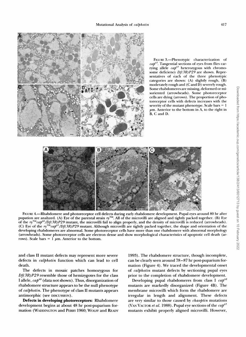
FIGURE 3.-Phenotypic characterization of cap"'. Tangential sections of eves from flies car- rying allele cap6' heterozygous with chromo- some deficiency Df(3R)P29 are shown. Repre- sentatives of each of the three phenotypic categories are shown: (A) slightly rough, (R) moderately rough and (C and 11) severely rough. Some rhabdomeres are missing, deformed or m i - soriented (arrowheads). Some photoreceptor cells are dying (arrows). The proportion of p h e toreceptor cells with defects increases with the severity of the mutant phenotype. Scale bars = 1 pm. Anterior to the bottom in A, to the right in R, C and D.
FIGURE 4.-Rhal~tlomere and photoreceptor cell defects during e;wly rhabdomere devclopmcnt. Pupal eves around 80 hr after pupation are analymd. (A) Eye of the parental strain ,"". A l l of the microvilli are aligned and tightly packed together. (R) Eye of the ry5"6cap6X/f)f(3R)P29 mutant, the microvilli fail to align properly. and the density of microvilli is reduced (arrowheads). ( C ) Eye of the 3,~"6cnp"'/Df(3R)P29 mutant. Although microvilli arc tightly packed together, the shape and orientation of the developing rhabdomeres are abnormal. Some photoreceptor cells have more than one rhabdomere with abnormal morphology (arrowheads). Some photoreceptor cells are electron dense and show morphological characteristics of apoptotic cell death (ar- rows). Scale bars = 1 pn . Anterior to the bottom.
and class I1 mutant defects may represent more severe defects in cnlphotin function which can lead to cell death.
The defects in mosaic patches homozygous for Df(3R)P29 resemble those of hemizygotes for the class I allele, cap6R (data not shown). Thus, disorganization of rhabdomere structure appears to be the null phenotype of cnlphotin. The phenotype of class I1 mutants appears antimorphic (see DISCUSSION).
Defects in developing photoreceptors: Rhabdomere development begins at about 48 hr post-puparium for- mation (WADDINGTON and PERRY 1960; WOLF and RFADY
1993). The rhabdomere structure, though incomplete, can be clearly seen around 78-87 hr post-puparium for- mation (Figure 4). We traced the developmental onset of cnlphotin mutant defects by sectioning pupal eyes prior to the completion of rhabdomere development.
Developing pupal rhabdomeres from class I cuphR mutants are markedly disorganized (Figure 4B). The membrane microvilli which form the rhabdomere are irregular in length and alignment. These defects are very similar to those caused by chnoptin mutations (VAV VACTOR et nl. 1988). Pupal eye sections of the cuph5 mutants exhibit properly aligned microvilli. However,
Dow
nloaded from https://academ
ic.oup.com/genetics/article/138/2/413/6012713 by guest on 10 February 2022

418 Y. Yang and D. Rallinger
- 30 Kb PL x - x x x x PL P I I I I In
I I 1 I I I I N R R R R R R R N
No. 5
No. 10 - 31 Kb
x x x - x I I I
I I I N R R R R N x t x -
I Kb
FIGURE 5 . P e n o m i c clones of the cnlphofin gene. Two cosmid clones containing genomic DNA around the cnlpitolin gene are diagrammed. An 8-kh Xlrol-Xhol genomic fragment contained hv hoth cosmid clones has both the ris-elernents sufficient for photoreceptor cell-specific expression ofa IncZreporter gene (N. XCE and D. RAIJJXGER, manuscript in prepara- tion), and the transcribed region of the cn/pholin gene. This fragmcnt was subcloned into the transformation vector pM'8. The resulting pM'KXcap and the two cosmid clones were u.sctl to generate transgenic flies.
both the photoreceptor cells and their rhabdomeres have abnormal shapes (Figure 4C). Thus, one calphotin function, defective in cap"' mutants, is required for the orderly alignment of the rhabdomeric microvilli from the beginning of rhabdomere development. Another as- pect of calphotin function appears to be involved in the organization of the photoreceptor cell as a whole. The latter function, altered by caph5 mutations, can result in defective morphology of both photoreceptor cells and their rhabdomeres, and in photoreceptor cell death.
Behavioral characterization: Phototaxis was meas- ured in a countercurrent apparatus (BENZER 1967). The test measures phototactic preference during an agitated- state escape response. The visible light used in the test was strong enough to dominate other stimuli in the com- plex test environment (HEISENRERG and Gon 1975). When given six consecutive trials of phototaxis toward visible light, normal flies and ~y~"~cap"*/Qf(3R)P29mu- tant flies choose light over darkness an average of 5 or 6 times, while ~y~"~cap"/Df(3R)P29rnutant flies choose light an average of 4 or 5 times. Thus, the pathway trig- gered by light and leading to phototaxis is capable of functioning, even though the light reception organelle, the rhabdomere is disorganized in cap6' and caph5 mutants.
Transformation rescue of calfihotin mutations: To prove that the mutationswe isolated are mutations in the calphotin gene, we carried out transformation rescue experiments. Two cosmid genomic clones containing calphotin were isolated (Figure 5). An 8 k b XhoI-XhoI fragment contained in both cosmids was subcloned into the pW8 vector (KLEMENZ et al. 1987), and the resulting plasmid is called pWtkap (Figure 5). A I 1 three con- struct. were used to generate transgenic flies (see
MATERIALS AND METHODS for details). A I 1 three construct.. can rescue both class I and class I1 calphotin mutations (Figure 6). Some transgenic lines did not rescue as well as others, presumably due to chromosomal position ef- fects of the insertion site on the level of calphotin gene expression. In addition, the degree of rescue depended on the copy number of the transgene. One of the pWS cap transgenic lines, XSl was most effective in rescuing calphofin mutations (Table 2 and Figure 7), more than 95% of the ommatidia in rescued capb8 and cap6' flies had normal photoreceptors. The lack of complete res- cue may indicate that the transgene is not expressed at normal levels. It may also be a reflection of the semi- dominant nature of cap". Additionally, cap6* heterozy- gous flies have a small number of defective ommatidia. Thus, the XS1 transgene may be expressed at fairly nor- mal levels. Northern blot analysis of head RNA indicates that the Skb Xhol-XhoI fragment contains only the cal- photin gene (data not shown). Thus, both class I and class I1 mutations are mutations in the calphotin gene.
DISCUSSION
The calphotin gene was initially identified by the p h e toreceptor cell-specific staining of monoclonal antibod- ies (BALLINGER ct al. 1993; MARTIN et al. 1993). Calphotin protein expression begins right after the morphoge- netic furrow, at about the same time as overt morpho- logical differentiation of the photoreceptor cells, and about 24 hr earlier than the earliest photoreceptor cell- specific protein that has been previously characterized, Chaoptin (VAN VACTOR et a1. 1988). In a previous report (RAILINGER et al. 1993). we studied the gene structure and biochemical properties of the calphotin gene. Here
Dow
nloaded from https://academ
ic.oup.com/genetics/article/138/2/413/6012713 by guest on 10 February 2022

Mutational Analysis of calphotin 419
FIGURE 6.-A normal calphotin gene can rescue the mutant phenotypes of cap6' and cap", (A and B) Light microscopic analysis of thick sections. (C and D) Electron micrographic analysis of thin sec- tions. Arrowheads indicate abnormal unit eyes; in C one cell has no rhabdomere (*), in D one photoreceptor cell is missing. (A and C) Allele cap", heterozygous with chromosome deficiency D/(3R)P29, with two copies of the P[cap+, w+JX-8 transgene, made with construct pW8Xcap (Fig- ure 5). 95% of the photoreceptor cells have normal rhabdomeres. (B and D) Allele cap", heterozygous with chromosome deficiency Df(3R)P29, with one copy of the P[cnp', 7u']X-8 transgene. Almost all flies have slightly rough eyes (see legend of Figure 2), and 95% of the unit eyes are normal. Scale bars = 10 pm (in A and B), 1 pm (in C and D). Anterior to the lower right in A and D, to the right in B and C.
TABLE 2
Wild-type calphotin transgene partially rescues the deep pseudo pupil defects of the class I mutant, cap@
Transgenic line
None" X8-1 xs-1 5.1-2(2)' 5.1-3(3)." 10,200-4(2)'
Transgene copies 0 2 1 1 1 1 Percent rescud 0 95% (19/20) 76.3% (45/59) 85.9% (55/64) 60% (27/45) 85.7% (30/35)
" No transgene, flies of the genotype ry5"'ccap6'/D/(3R)P29. Several hundred flies have been observed, none with a normal deep pseudopupil. This mutant genotype was used in all studies included in this table.
X8-1, a transgenic fly line generated with pW8-Xcap (Figure 5). ',."Two transgenic fly lines generated with cosmid no. 5 (Figure 5). 'A transgenic fly line generated with cosmid no. 10 (Figure 5). 'Percent rescue indicates the percentage of flies which have normal looking deep pseudo pupil images.
we describe the isolation of mutations in the calphotin gene, and its function in rhabdomere development and photoreceptor cell survival.
Calphotin has a number of structural domains, in- cluding an N-terminal domain involved in calcium binding, and an Cterminal leucine zipper that may be important for interactions with other proteins (BALLINGER et al. 1993; MARTIN et al. 1993). In addi- tion, the protein is very hydrophobic and proline rich (overall hydrophobic content of 67%, and proline content of 21%), and has a 200-amino acid stretch at its amino terminus that is 82% hydrophobic. Calpho- tin contains several regions of continuous hydropho- bic character sufficiently long to permit membrane spanning; however, each of them is interrupted by two or more proline residues which may prevent their use as such (BALLINCER et al. 1993; MARTIN et al. 1993). Calphotin is localized within the photoreceptor cell in a poorly defined structure that underlies the rhab domere and may be bounded by membranes (MARTIN
et al. 1993). However, we could not detect any specific
membrane association of the Calphotin protein when it was expressed in Drosophila tissue culture cells or in rabbit reticulocyte lysates in the presence of dog pan- creas membranes (our unpublished data).
We isolated several mutations in the calphotin gene, as indicated by their rescue in calphotin transgenic flies. The mutations fall into two phenotypic classes. Recessive class I ( cap6') mutations cause deformed rhabdomeres while semidominant class I1 (cap") mutations result in misoriented rhabdomeres and photoreceptor cell death. Flies hemizygous for the class I mutations (class I mutations over deficiency) have stronger phenotypes than homozygous mutant flies, thus these alleles represent partial loss of function calphotin mutations. Mosaic patches homozygous for Df(3R)P29 resemble ryso6cap6'/Df(3R)P29 eyes, indicating that the severe class I mutant phenotype is likely to be the null pheno- type of calphotin. Class I1 mutations are antimorphic because they have a more severe phenotype as homozy- gotes than hemizygotes, and additional wild-type calpho- t in copies can restore them to a more normal pheno-
Dow
nloaded from https://academ
ic.oup.com/genetics/article/138/2/413/6012713 by guest on 10 February 2022

420 Y. Yang and D. Ballinger
1 0 0 90 80 70 60 50 40 30 20 10 0 I,
I Lightlv rough 1 Moderately rough I Severelvrough I I $06 cap65/ Df(3R)P29
I P[cap+, w+fl8-1 I + ; $06 cap65 / Df(3R)P29
rZa P[cap+, w+]5.1-2(2) I + ; $os cap65/ Df(3R)P29
P[cap+, w+]5.1-3(3) I + ; ry5@5cap65/Df(3R)P29
Bp P[cap+, w+]10.92-1(2) I+ ; $06 cap65/Df3R)P29 FIGURE 7.-Rescue of the class I1 calpholin mutation, cup65.
For comparison, flies were separated into three classes based on overall eye roughness (see legend to Figure 3). Flies hem- izygous for the cup65 mutation [ry5"6cup65/Df(3R~P29] are a p proximately equally divided between these three categories. Transgenic flies carrying two of the three genomic clones and also hemizygous for cupe5 are rescued; most of the eyes are only weakly rough, indicating that the roughness is due to a mu- tation in the calphotin gene.
type. Perhaps the class I1 mutations alter the interaction of Calphotin with another protein. One possible inter- acting protein co-immunopurifies with Calphotin (BALLINGER et al. 1993). Western blot analysis shows that the functional abnormality of both classes of calphotin mutations is not due to detectable changes in the level of expression or gel mobility of Calphotin (our unpub- lished data).
Light activation of rhodopsin, the rhabdomere- localized photopigment can cause retinal degeneration when a Ca2+ binding serine/threonine protein phos- phatase (rdgC) is mutant (STEELE et al. 1992). Since Cal- photin can bind Ca2+ in vitro (BALLINGER et al. 1993; MARTIN et al. 1993), and lightdependent fluctuations in Ca2' concentrations in part mediate phototransduction (SMITH et al. 1991), we tested the possibility that the calphotin mutant phenotype was lightdependent. How- ever, cap6* had a similar phenotype whether reared in light or dark, indicating that rhabdomere disruptions were not caused by light-induced degeneration. Indeed, the rhabdomeres of cap6* mutant flies are abnormal when they begin to develop at the mid pupal stage (Fig- ure 4). Thus, it appears that the rhabdomere defects of class I cap mutants reflect a developmental function of the calphotin gene.
The developmental function of Calphotin suggests a possible structural role, or a defect in the process of membrane trafficking during rhabdomere develop ment. Rhabdomere development begins during mid pu- pal stage, which is initially characterized by disorganized membrane ruffling (WADDINGTON and PERRY 1960). Rhabdomeres of wild-type flies are constructed as closely packed and hexagonally arranged stacks of microvilli. Because the rhabdomere of each photoreceptor cell has
its own characteristic shape and direction, rhabdomere growth requires directed, highly polarized membrane trafficking (WOLFF and READY 1993). Calphotin may play a critical role in regulating directed membrane traffick- ing during rhabdomere development. This role may in- volve specific protein-protein or protein-membrane in- teractions via the leucine zipper and hydrophobic domains of Calphotin, or its modulation of intracellular calcium level.
Mutations in the calphotin gene affect both the regu- larity and the direction of rhabdomere growth. Devel- oping cup6* rhabdomeres have fewer microvilli and they fail to align properly. This could result from an insuf- ficient level of membrane trafficking in class I calphotin mutant flies. This interpretation is in consistent with the genetic data that cap6" is a partial loss of function allele of calphotin. Photoreceptor cells with rhabdomeres pointing toward the pigment cells instead of toward the center of the unit eye and cells with two rhabdomeres pointing in different directions are seen in semidomi- nant class I1 calphotin mutant flies. These defects may result from a misdirection of membrane trafficking in cap6' mutant. The incomplete penetrance of both cap6" and cap6* mutants may reflect a partial redundancy of function. Molecular characterization of calphotin mu- tations may shed more light on possible Calphotin func- tions in the membrane transport process.
In class I1 calphotin mutant flies, some photoreceptor cells undergo cell death. This could result from the se- verely impaired development or function of the dying photoreceptor cells. The observed rhabdomere abnor- mality may be only one visible indication of a more gen- eral impairment of photoreceptor cell function. Alter- natively, the class I1 mutations appear to be antimorphic, and may interfere with the function of another protein that interacts with Calphotin. If so, cell death may reflect the normal function of this interacting protein.
While rare dying cells are seen in rySo6 capb5 homozy- gous discs, the bulk of calphotin mutant phenotypes are not manifest until the beginning of rhabdomere devel- opment. This is about 4-5 days after the onset of Cal- photin expression, This phenotypic delay is similar to chaoptin mutations (VAN VACTOR et al. 1988). It may represent fortuitous early expression of Calphotin or functional redundancy at early developmental stages.
Calphotin may also alter photoreceptor development indirectly by modulating Glass protein activity. Calpho- tin expression is not altered by glass mutations (BALLINGER et al. 1993). Glass is a zinc-finger containing transcription factor whose activity is restricted to the photoreceptor cells (ELLIS et al. 1993). It is possible that some Glass target genes may function in the membrane trafficking pathway. Glass is highly modified (ELLIS et dl. 1993) suggesting that its activity could be regulated by post-translational modification in response to differen- tiation signals. The intriguing early expression of Cal- photin, 24 hr prior to Chaoptin expression and 3-4 days
Dow
nloaded from https://academ
ic.oup.com/genetics/article/138/2/413/6012713 by guest on 10 February 2022

Mutational Analysis of calphotin 42 1
prior to the development of rhabdomere, may suggest the involvement of Calphotin in the modification of Glass, or some other regulatory protein, at an early stage of photoreceptor cell development. In glass mutants, when the photoreceptors are prevented from attaining their photoreceptor identity, they die (MOSES et al. 1989). Indeed, some ommatidia in the severe class I1 caf5 mutant appear similar to those in glass mutants. Future genetic and biochemical dissection of the path- way which leads to the modification of Glass, and an analysis of Glass protein in calphotin mutants may in- dicate a link between Calphotin and Glass modification or function.
We thank Y. GAUZ and K. MATTHEWS (of the Indiana Drosophila Stock Center) for many of the mutations and deficiencies used in these studies, S. L. ZIPURSKY for vectors for tissue culture expression, K. HARSHMAN for help with injections and for valuable discussions, M. WIEDMANN for help with in vitro transfections and N. LAMPEN for help with the electron microscopy. We also thank T. SCHCJPBACH and the anonymous reviewers for helpful comments on the manuscript. This work was supported in part hy National Institutes of Health Grant ~3o.c~-0874a27.
LITERATURE CITED
BALLINCER, D. G., and S. BENZER, 1988 Photophobe (Ppb), aDrosophila mutant with a reversed sign of phototaxis; the mutation shows an allele-specific interaction with sevenless. Proc. Natl. Acad Sci. USA
BALLINCER, D. G., and S. BENZER, 1989 Targeted gene mutations in Drosophila. Proc. Natl. Acad. Sci. USA 86: 9402-2406.
BALLINCER, D. G., N. XUE and K. D. HARSHMAN, 1993 A Drosophila photoreceptor cell-specific protein, Calphotin, binds calcium and contains a leucine zipper. Proc. Natl. Acad. Sci. USA 90:
BENZER, S., 1967 Behavioral mutants of Drosophila isolated by coun- tercurrent distribution. Proc. Natl. Acad. Sci. USA 58: 11 12-1 119.
CHIRCWIN, J. M., A. E. P ~ Y L A , R. J. MACDONALD and W. J. RUTTER, 1979 Isolation of biologically active ribonucleic acid from sources enriched in ribonuclease. Biochemistry 18: 5294-5299.
CLARKE, P. G. H., 1990 Developmental cell death: morphological di- versity and multiple mechanisms. h a t . Embyrol. 181: 195-213.
ELLIS, M. C., E. M. O’NEILL and G. M. RUBIN, 1993 Expression of Drosophila Glass protein and evidence for negative regulation of its activity in non-neuronal cells by another DNA-binding protein. Development 119: 855-865.
FRANCESCHINI, N., and K KIRSCHFELD, 1971 Pseudopupil phenomena in the compound eye of Drosophila. Kybernetik 9: 159-182.
GAUZ, J., H. GWRKOWCS, G. BENCZE, A. A. M. AWAD, J. J. HOLDEN et al., 1981 Genetic characterization of the region between 86F1.2 and 87B15 on chromosome 3 of Drosophila melanogaster. Genetics
HEISENBERG, M., and K. G. GOTZ, 1975 The use of mutations for the partial degradation ofvision in Drosophila melanogaster. J. Comp. Physiol. 98: 217-244.
~ R R , J. F. R., J. SEARLE, B. V. HERMON and C. J. BISHOP, 1987 Apoptosis, pp. 93-128 in Perspectives on Mammalian Cell Death, edited by c . s. POTTEN. Oxford University Press, Oxford.
85: 3960-3964.
1536-1540.
98: 775-789.
KLEMENZ, R., U. WEBER and W. J. GEHRING, 1987 The white gene as marker in a new P element vector for gene transfer in Drosophila. Nucleic Acids Res. 15 3947-3959.
KRANTZ, D. E., and S. L. ZIPURSKY, 1990 Drosophila Chaoptin, a member of the leucine-rich repeat family, is a photoreceptor cell- specific adhesion molecule. EMBO J. 9 1969-1977.
MARTIN, J. H., S. BENZER, M. RUDNICKA and C. A. MILLER, 1993 Calphotin: a Drosophila photoreceptor cell calcium- binding protein. Proc. Natl. Acad. Sci. USA 90: 1531-1535.
MEYEROWITZ, E. M., G. M. GUID, L. S. PRETIDCE and D. S. HOCNES, 1980 A new high capacity cosmid vector and its use. Gene 11:
MLODZIK, M., Y. HIROMI, U. WEBER, C. S. GOODMAN and G. M. RUBIN, 1990 The Drosophila seven-up gene, a member of the steroid receptor gene superfamily, controls photoreceptor cell fates. Cell
MOSES, K., M. C. ELLIS and G. M. RUBIN, 1989 The glass gene encodes a zinc-finger protein required by Drosophila photoreceptor cells. Nature 340: 531-536.
MOSES, R, and G. M. RUBIN, 1991 Glass encodes a sitespecific DNA- binding protein that is regulated in response to positional signals in the developing Drosophila eye. Genes Dev. 5: 583-593.
PIRROTTA, V., H. STELLER and M. P. BOZZETTI, 1985 Multiple upstream regulatory elements control the expression of the Drosophila white gene. EMBO J. 4: 3501-3508.
REINKE, R., D. E. KRANTZ, D. YEN and S. L. ZIPURSKY, 1988 Chaoptin, a cell surface glycoprotein required for Drosophila photoreceptor cell morphogenesis, contains a repeat motif found in yeast and human. Cell 52: 291-301.
RENFRANZ, P. J., and S. BENZER, 1989 Monoclonal antibody probes discriminate early and late mutant defects in development of the Drosophila retina. Dev. Biol. 136: 411-429.
ROCGE, R. D., C. A. KARLOWCH and U. BANERJEE, 1991 Genetic dissection of a neurodevelopmental pathway: Son of sevenless functions downstream of the sevenless and EGF receptor tyrosine kinases. Cell 6 4 39-48.
SMITH, D. P., M. A. STAMNES and C. S. ZUKER, 1991 Signal transduction in the visual system of Drosophila. Annu. Rev. Cell Biol. 7:
STEELE, F. R., T. WASHBURN, R. RIECER and J. E. O’Tous~, 1992 Dro- sophila retinal degeneration C ( rdgC) encodes a novel serine/ threonine protein phosphatase. Cell 69: 669-679.
TOMLINSON, A., and D. F. READY, 1987 Neuronal differentiation in the Drosophila ommatidium. Dev. Biol. 120: 366-376.
VAN VACTOR, D. L. J., D. E. ~ T Z , R. REINKE and S. L. ZIPURSKY, 1988 Analysis of mutants in Chaoptin, a photoreceptor cell- specific glycoprotein in Drosophila, reveals its role in cellular morphogenesis. Cell 5 2 281-90.
WADDINGTON, C., and M. PERRY, 1960 The ultrastructure of the de- veloping eye of Drosophila. Proc. R. SOC. Lond. Ser. B 153
WOLFF, T., and D. F. READY, 1993 Pattern formation in the Drosophila Retina, pp. 1277-1325 in The Development of Drosophila mela- nogaster, edited by M. BATE and A. M. ARIAS. Cold Spring Harbor Laboratory, Cold Spring Harbor, N.Y.
WILLIE, A. H., J. F. R. KERR and A. R. CURRIE, 1980 Cell death: the significance of apoptosis. Int. Rev. Cytol. 6 8 251306,
ZIPURSKY, S. L., T. R. VENKATESH, D. B. TEPLOW and S. BENZER, 1984 Neuronal development in the Drosophila retina: mono- clonal antibodies as molecular probes. Cell 36: 15-26.
ZUKER, c . S., A. F. COWMAN and G. M. RUBIN, 1985 Isolation and struc- ture of a rhodopsin gene from D . melanogaster. Cell 40: 851-8.
Communicating editor: T. SCHUPBACH
271-282.
60: 21 1-224
161-190.
155-178.
Dow
nloaded from https://academ
ic.oup.com/genetics/article/138/2/413/6012713 by guest on 10 February 2022