muscles & muscle tissue - gore's anatomy &...
TRANSCRIPT

1
Muscles &
Muscle TissueChapter 6
I. Overview of
Muscle

2
A. MUSCLE TYPES
� SKELETAL: striated, voluntary
� CARDIAC: only in heart
• involuntary
• striated
� SMOOTH: walls of organs
• involuntary
• nonstriated
All Muscle Cells are
� Elongated
� Can Shorten & Contract
� Prefixes
• Myo- & Mys – muscle
• Sarco - flesh

3
1. Skeletal Muscle
� Skeletal muscle fibers: packaged into organs called skeletal muscles that attach to skeleton
� Fibers are cigar-shaped, multinucleate cells, up to 1ft in length
� Striated: fibers appear to be striped
� Voluntary: conscious control
� Can be activated by reflexes
� Tires easily
� Held together by connective tissue

4
a. Gross Anatomy� Muscle: organ (fig. 6.1 p.164)
• consists of hundreds to thousands
of muscle cells
• covered by epimysium (CT)
–binds muscles into functional groups
–Blend into strong, cordlike tendons or into sheetlike sponeuroses
• blood vessels & nerve fibers
� fascicle: portion of muscle
• bundle of muscle cells
• surrounded by perimysium (CT)

5
2. Smooth Muscle
� No striations, involuntary
� Found in walls of hollow organs
� Spindle shaped, single nucleus
� Arranged in two sheets or layers
� Figure 6.2a p. 165
� Alternately contract & relax
� Food through digestive system

6
3. Cardiac Muscle
� Found only in heart
� Striated, involuntary
� Arranged in spiral or figure 8-shaped bundes
� Fig. 6.2b p. 165
� Branching cells joined by intercalated discs that allow for communication
B. Muscle Functions

7
1. Producing Movement
� Mobility of body
� Forces fluids & other substances
through internal body channels
2. Maintaining Posture
� Skeletal muscles maintain an erect or
seated posture despite gravity

8
3. Stabilizing Joints
� As skeletal muscles pull on bones to
cause movement, they also stabilize
joints of skeleton
4. Generating Heat
� By-product of muscle activity
� ATP powers muscle contraction, ¾ of
energy is lost as heat
� Vital in maintaining normal body
temperature - 40%

9
II. Microscopic Anatomy Of
Skeletal Muscle
� Sarcolemma: muscle fiber plasma
membrane
� myofibril: complex organelle
composed of bundles of
myofilaments
• banded
� sarcomere: contractile unit
• composed of myofilaments made
of contractile proteins
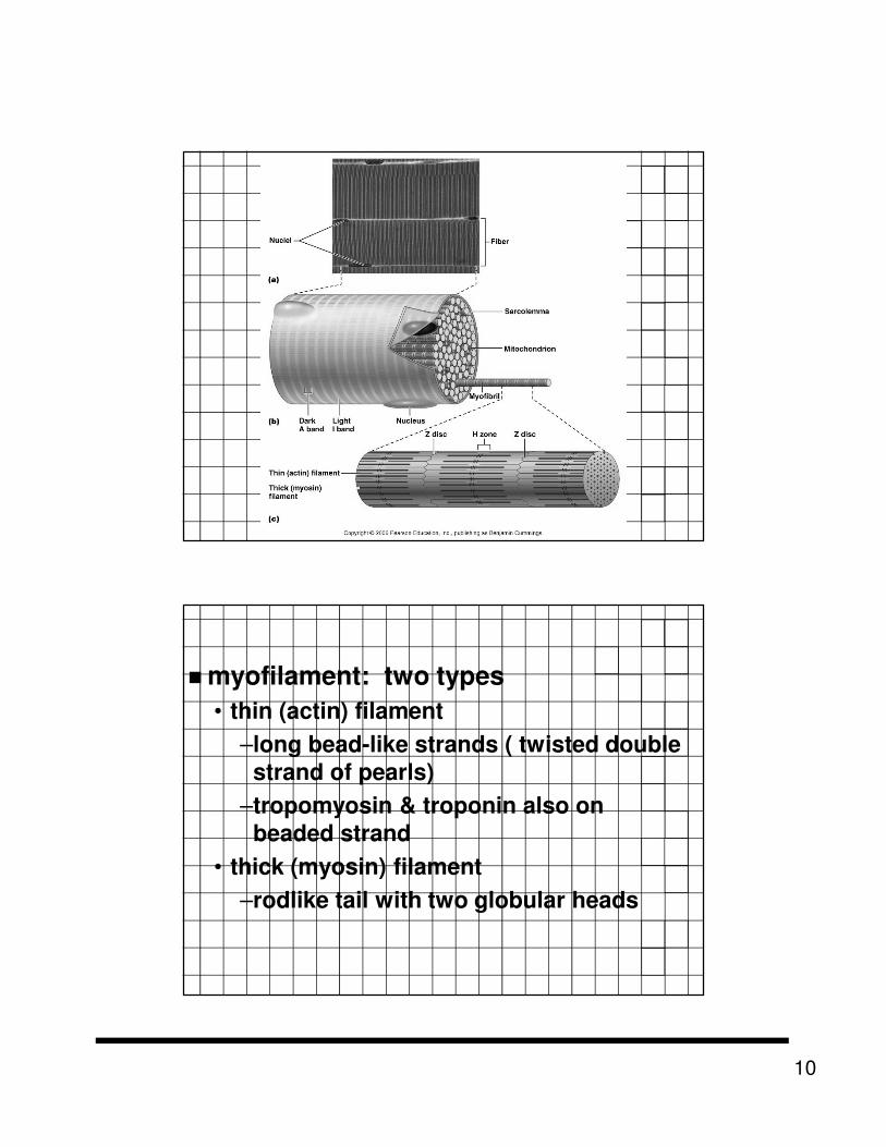
10
� myofilament: two types
• thin (actin) filament
–long bead-like strands ( twisted double
strand of pearls)
–tropomyosin & troponin also on
beaded strand
• thick (myosin) filament
–rodlike tail with two globular heads

11
� p. 167 fig. 6.3
� A bands
• Contain actin & myosin
�1 sarcomere
• extends from Z line to next Z line
• contain both actin & myosin
� I bands
• contain actin

12
� Sarcoplasmic reticulum (SR)
• Interconnecting tubules that surround each myofirbril
• Store calcium

13
III. Skeletal Muscle Activity
A. Stimulation & Contraction of Single Skeletal Muscle Cells
� Special functional properties
• Irritability: ability to receive & respond to a stimulus
• Contractility: ability to shorten when an adequate stimulus received
• Extensibility – ability to lengthen
• Elasticity – ability to stretch and return to normal lengths

14
1. Nerve Stimulus & Action Potential
Motor Unit
� motor neuron & all the muscle fibers it supplies
� fine control: fingers & eyes
• < 150 muscle fibers / motor neuron
� less precise: hips & legs
• > 150 muscle fibers / motor neuron

15
Neuromuscular Junctions
� Where axonal terminals forms
junctions with sarcolemma
� Synaptic cleft: gap between axonal
terminal & sarcolemma
• Filled with interstitial fluid
� Nerve impulse reaches axonal terminals
� Neurotransmitter acetylcholine (Ach) released
� Diffuses across synaptic cleft & attaches to receptors on sarcolemma
� Membrane become permeable to Na+
which rush into muscle cell
� Upsets normal balance & generates electrical current called action potential

16
Contraction of Fiber
� sarcomeres shorten myofibrils shorten .......
� sliding filament theory of contraction
1. Cross bridge attachment: activated myosin heads are strongly attracted to exposed binding sites on actin & cross bridge binding occurs

17
2. Power Stroke: as myosin head binds, it changes from high-energy configuration to its bent, low-energy shape, which causes head to pull on thin filament, sliding it toward center of sarcomere
• ADP & Pi generated during prior contraction cycle are released from myosin head
3. Cross bridge detachment
� as new ATP molecule binds to myosin head, myosin cross bridge is released from actin

18
4. Cocking of myosin head
� hydrolysis of ATP to ADP and Pi
provides energy needed to return myosin head to its high-energy or cocked position, which gives it potential energy
needed for next attachment
� figure 6.8 p. 171

19
Role of Ca+2 in contraction
� low Ca+2 concentrations, blocks
binding sites on actin & prevents
cross bridge attachment - relaxed
muscle
� high Ca+2 concentrations, binding
site open
• myosin head binds - contraction
• warm-up for athletes increases
Ca+2 levels

20
Rigor mortis
� death rigor
� illustrates that cross bridge
detachment is ATP-driven
� muscles stiffen 3-4 hours after death
� rigidity peaks at 12 hours then
gradually decreases next 48-60 hours
due to breakdown of biological
molecules
B. Contraction of a Skeletal
Muscle as a Whole

21
1. Graded Responses
� All-or-none law: muscle cell will contract to fullest when stimulated never partially
� Skeletal muscles react to stimuli with graded response• Change frequency of muscle
stimulation
• Change # of muscle cells being stimulated
22
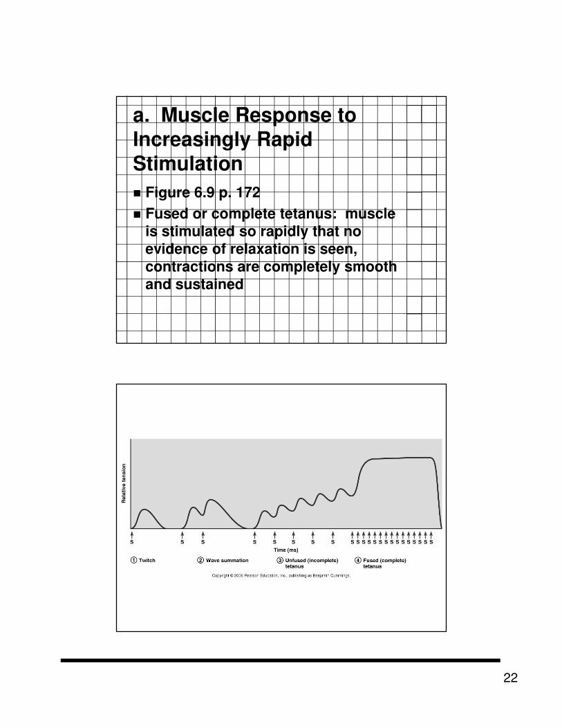
a. Muscle Response to
Increasingly Rapid
Stimulation
� Figure 6.9 p. 172
� Fused or complete tetanus: muscle
is stimulated so rapidly that no
evidence of relaxation is seen,
contractions are completely smooth
and sustained

23
b. Muscle Response to
Stronger Stimuli
� Force of muscle contraction depends
on how many cells are stimulated

24
2. Providing Energy for
Muscle Contraction
� Bonds of ATP are hydrolyzed to
release energy
� Muscle only store 4-6 sec of energy
� ATP must be regenerated
continuously
a. Phosphorylatin of ADP
by creatine phosphate
� Figure 6.10a
� Creatine phosphate found in muscle
cells but not other cells

25
b. Aerobic respiration
� Figure 6.10c
� Produces 95% of ATP at rest & light
exercise
� Mitochondria
� 36 ATP per 1 glucose molecule
� Requires oxygen
c. Anaerobic glycolysis
� Figure 6.10b
� Only 5% as much ATP from each glucose molecule as aerobic respiration
� 2.5 time faster
� Can provide most of ATP for 30-60 s
� Uses huge amounts of glucose for small ATP harvest
� Accumulates lactic acid that promotes muscles fatigue & soreness

26
3. Muscle Fatigue & Oxygen
Debt
� Strenuous & prolonged activity
causes fatigue due to accumulation
of lactic acid in muscle & decrease in
ATP
� After exercise oxygen debt is repaid
by rapid deep breathing

27
4. Types of Muscle
Contractions
� Isotonic: muscle shortens &
movement occurs
� Isometric: muscle tries to shorten
but cannot
• Try to push over a brick wall

28
5. Muscle Tone
� Not consciously controlled
� Some fibers are always contracted
while other relax
6. Effect of Exercise on
Muscles
� Use it or lose it

29
IV. Muscle Movements,
Types, & Names

30
A. Types of Body
Movements
� Origin: attached to immovable or
less movable bone
� Insertion: attached to movable bone
� Figure 6.13 p. 177-178
B. Types of Muscles
� Prime mover: major responsibility for movement
� Antagonists: oppose or reverse a movement
� Synergists: help prime movers by producing same movement or by reducing undesirable movements
� Fixators: hold a bone still or stabilize origin of prime mover

31
C. NAMING OF SKELETAL MUSCLES
1. Location of muscle
� indicate the bone or body region where muscle in located

32
2. Shape of muscle
� deltoid: triangular
� trapezius: form trapezoid
3. Size of muscle
� maximus: largest
� minimus: smallest
� longus: long

33
4. Direction of muscle
fibers
� rectus: fibers run parallel
� transverse: horizontally
� oblique: diagonally
5. Number of origins
� biceps: two origins
� triceps: three origins
� quadriceps: four origins

34
6. Location origin /
insertion
� name according to attachment point
� sternocleidomastoid
• sterno- origin on sternum
• cleido- origin on clavicle
• mastoid inserts on mastoid
process of temporal bone
7. Action of muscle
� flexor
� extensor
� abductor
� adductor

35
V. MUSCLE LIST
KNOW THOSE FROM
WORKSHEET