muscles. 40-45 % of body mass only system converting chemical energy into mechanical 2 types...
TRANSCRIPT
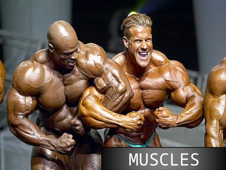
MUSCLES
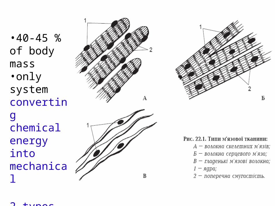
•40-45 % of body mass•only system converting chemical energy into mechanical
2 types-skeletal (striated)-smooth
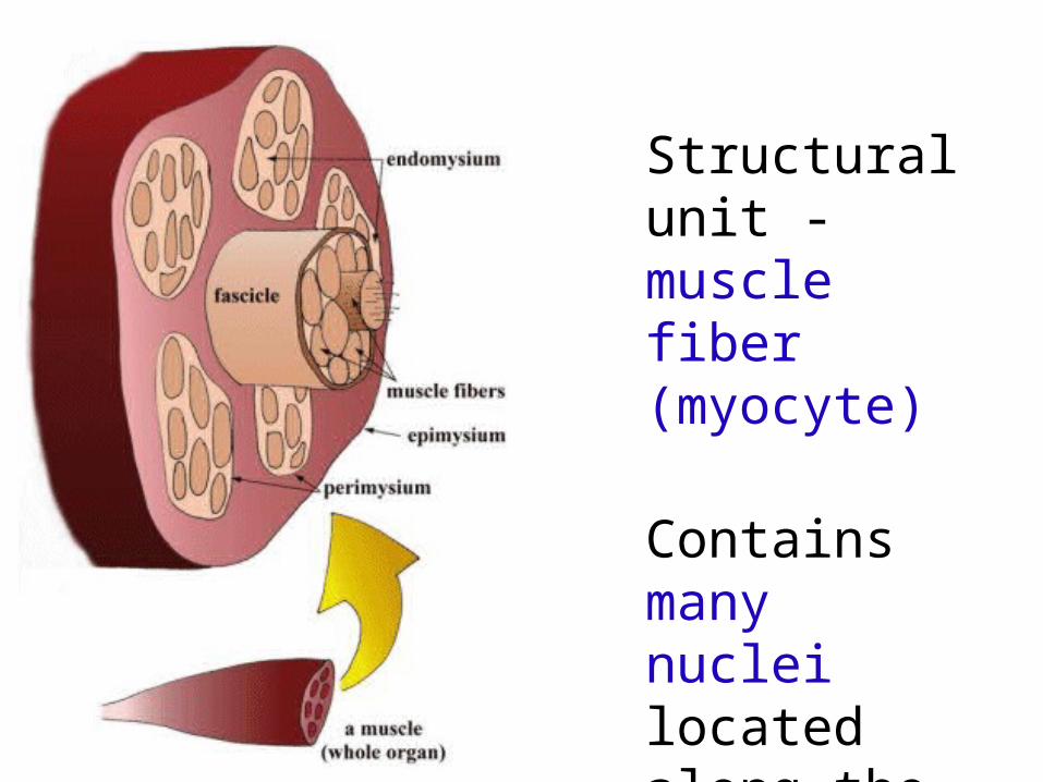
Structural unit - muscle fiber (myocyte)
Contains many nuclei located along the cell
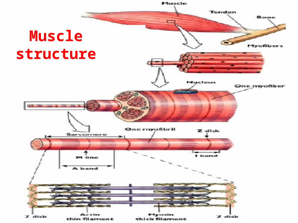
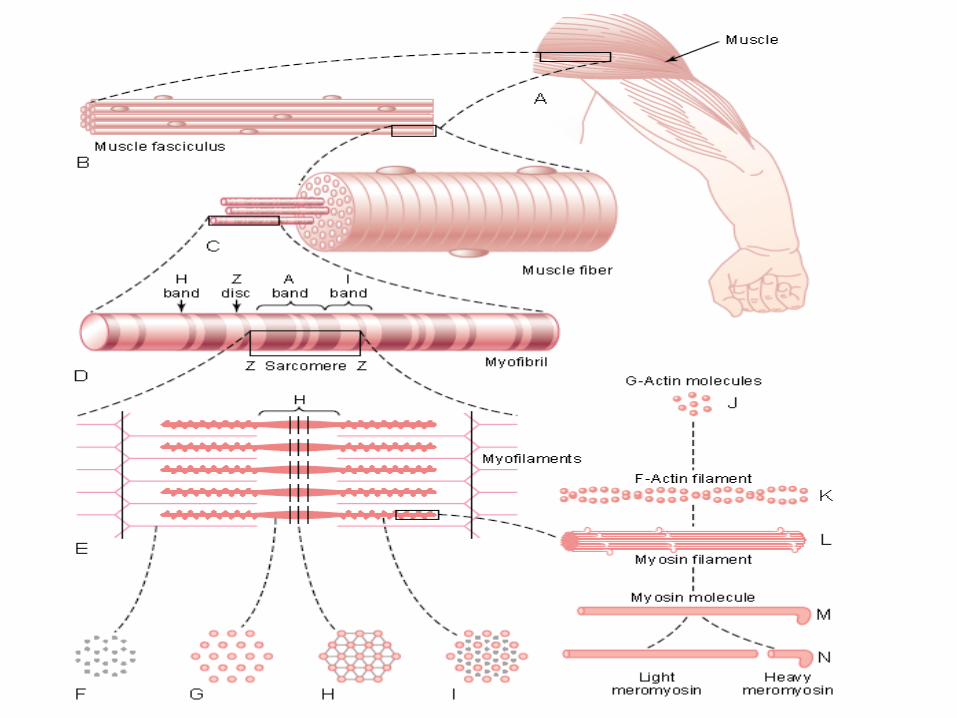
Muscle structure
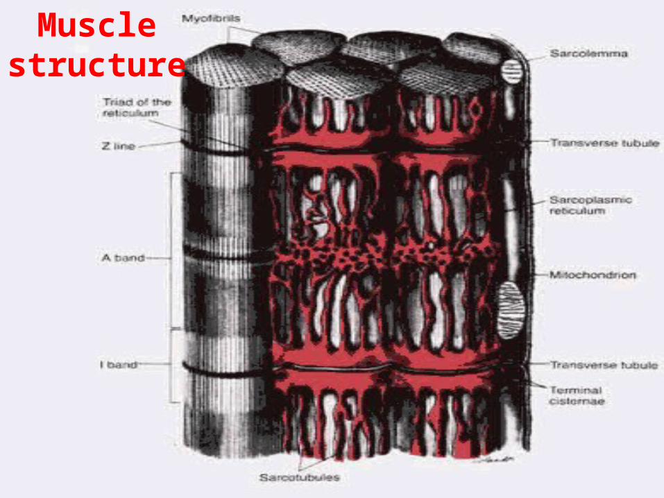
Muscle structure

Types of Muscle
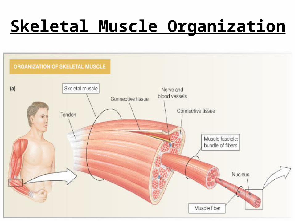
Skeletal Muscle Organization
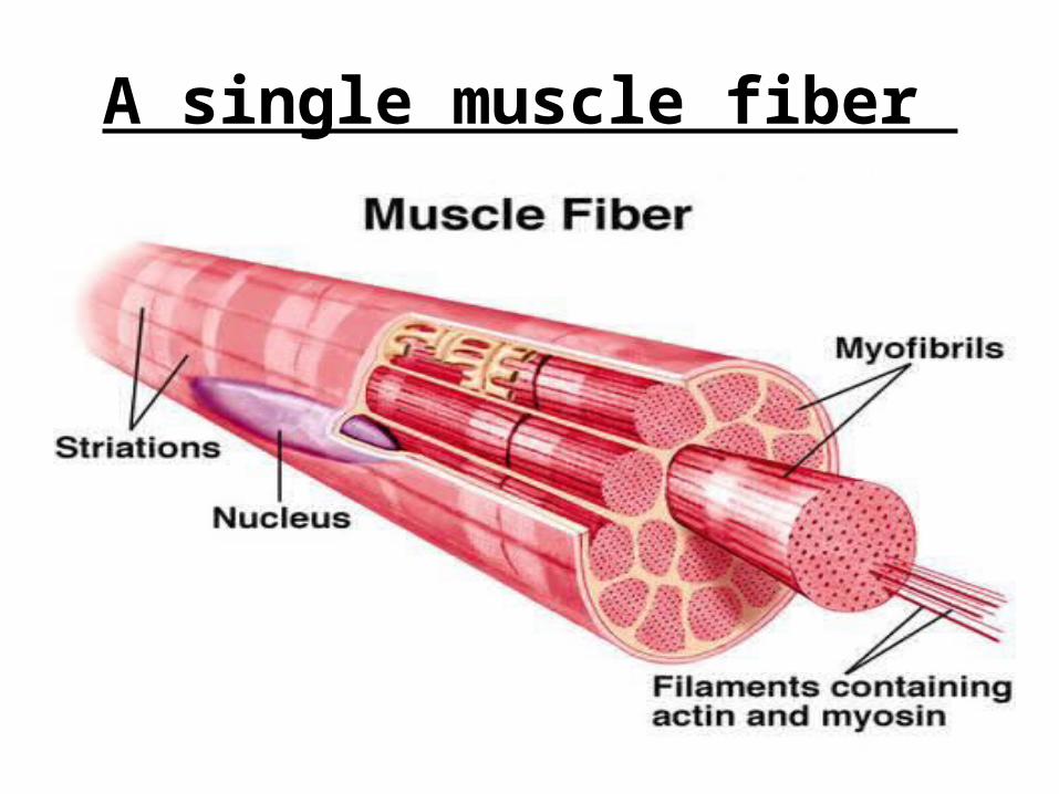
A single muscle fiber
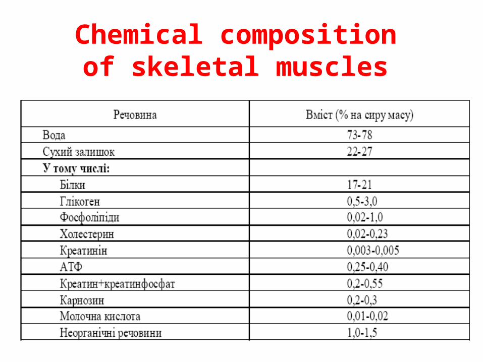
Chemical composition of
skeletal muscles
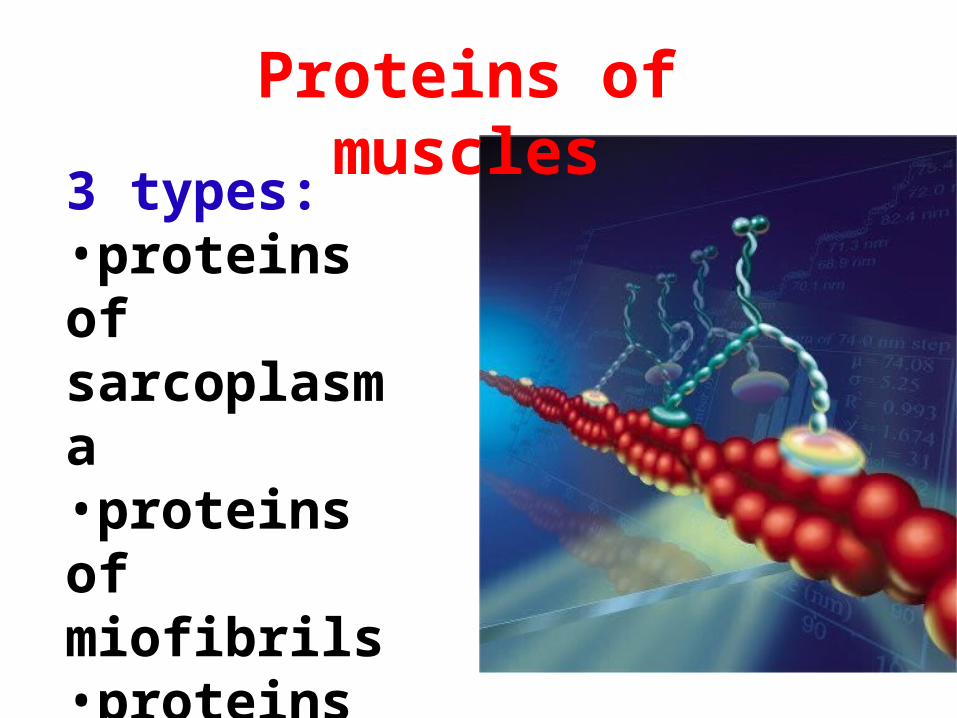
Proteins of muscles
3 types:•proteins of sarcoplasma•proteins of miofibrils•proteins of stroma
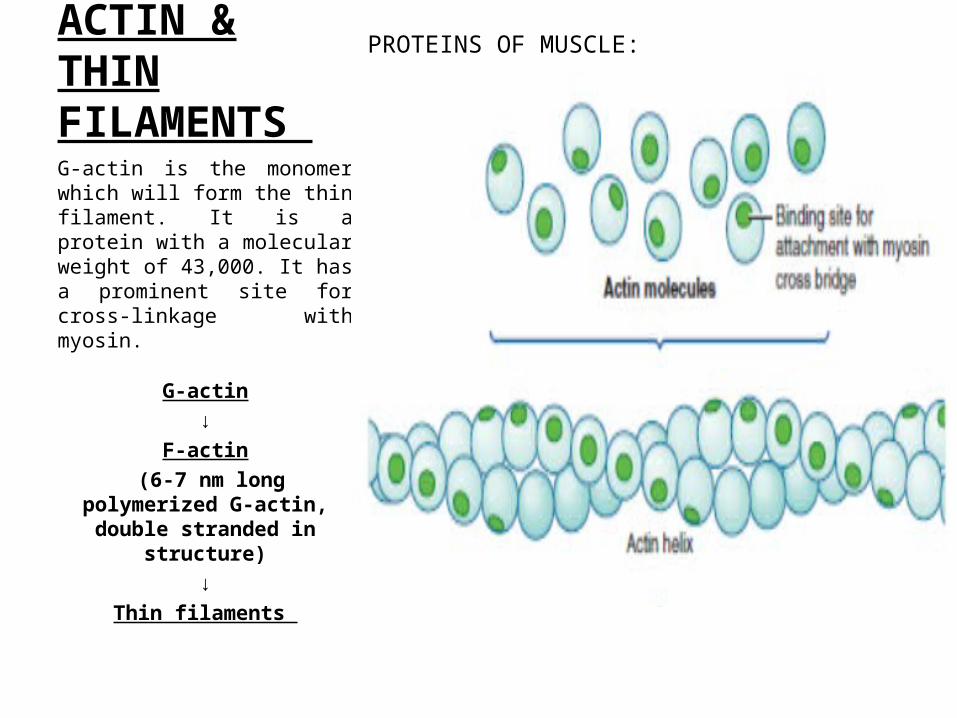
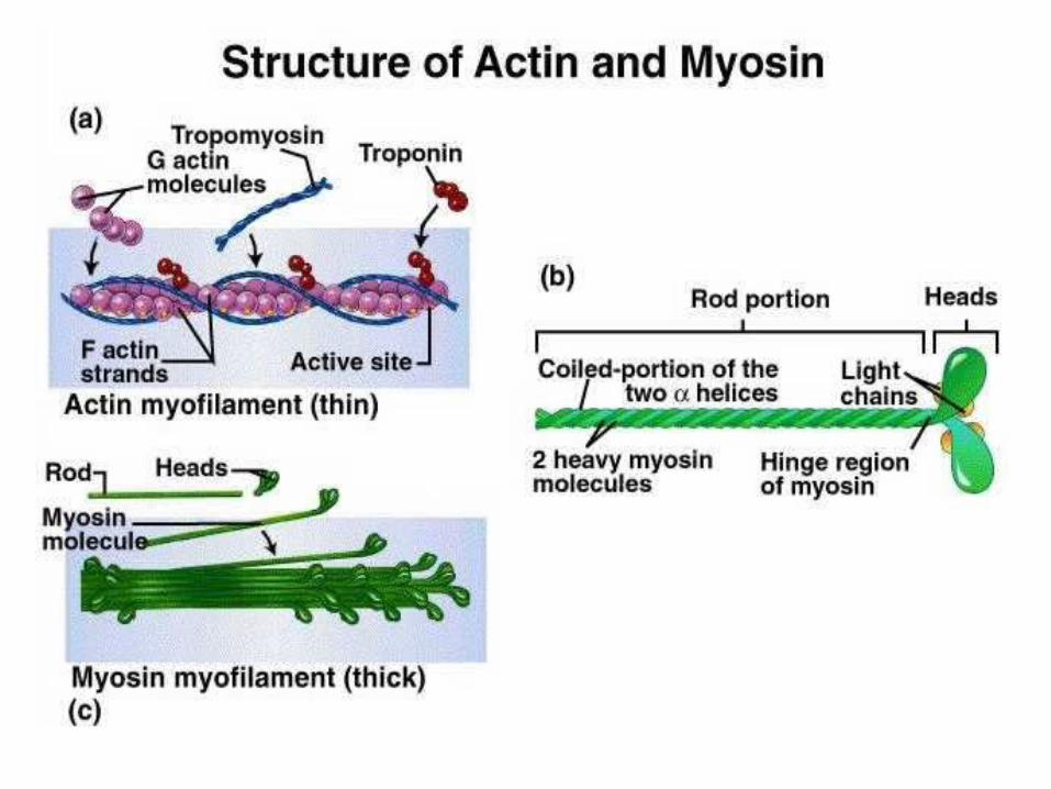
ACTIN & THIN FILAMENTS G-actin is the monomer which will form the thin filament. It is a protein with a molecular weight of 43,000. It has a prominent site for cross-linkage with myosin.
G-actin↓
F-actin (6-7 nm long polymerized G-
actin, double stranded in structure)
↓Thin filaments
PROTEINS OF MUSCLE:
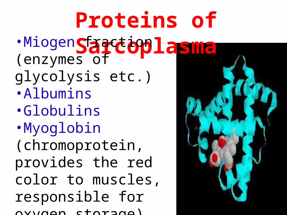
Proteins of Sarcoplasma•Miogen fraction
(enzymes of glycolysis etc.)•Albumins•Globulins•Myoglobin (chromoprotein, provides the red color to muscles, responsible for oxygen storage)
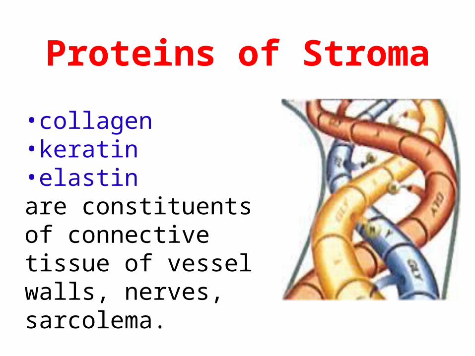
Proteins of Stroma
•collagen•keratin•elastinare constituents of connective tissue of vessel walls, nerves, sarcolema.
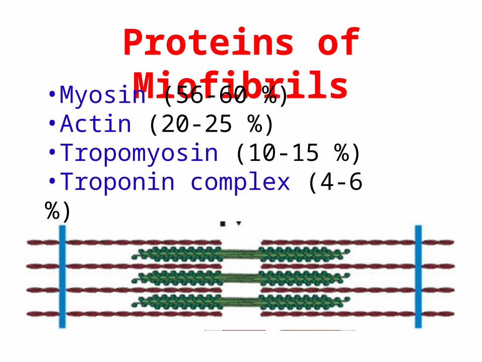
Proteins of Miofibrils•Myosin (56-60 %)•Actin (20-25 %)•Tropomyosin (10-15 %)•Troponin complex (4-6 %)
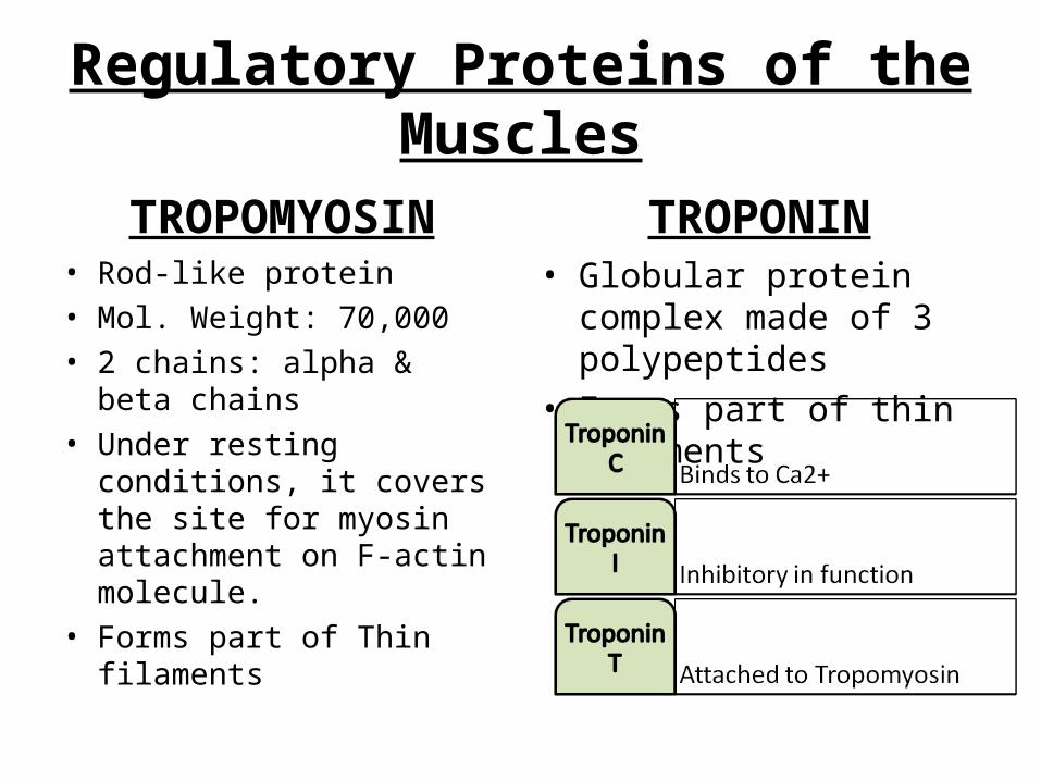
Regulatory Proteins of the Muscles
TROPOMYOSIN• Rod-like protein• Mol. Weight: 70,000• 2 chains: alpha & beta
chains• Under resting conditions, it
covers the site for myosin attachment on F-actin molecule.
• Forms part of Thin filaments
TROPONIN• Globular protein complex
made of 3 polypeptides • Forms part of thin filaments
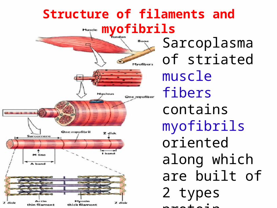
Structure of filaments and myofibrils
Sarcoplasma of striated muscle fibers contains myofibrils oriented along which are built of 2 types protein filaments: thick and thin
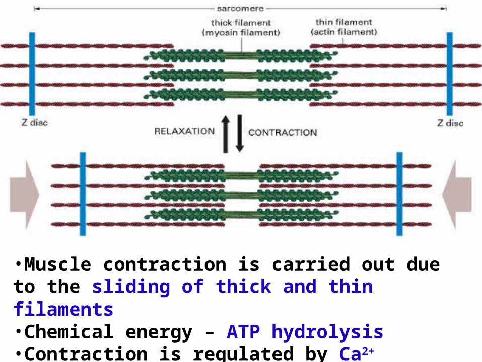
•Muscle contraction is carried out due to the sliding of thick and thin filaments•Chemical energy – ATP hydrolysis•Contraction is regulated by Ca2+ concentration
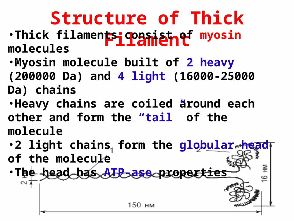
Structure of Thick Filament•Thick filaments consist of myosin
molecules•Myosin molecule built of 2 heavy (200000 Da) and 4 light (16000-25000 Da) chains•Heavy chains are coiled around each other and form the “tail” of the molecule•2 light chains form the globular head of the molecule•The head has ATP-ase properties
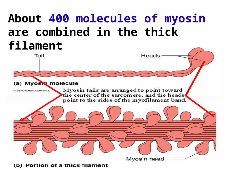
About 400 molecules of myosin are combined in the thick filament
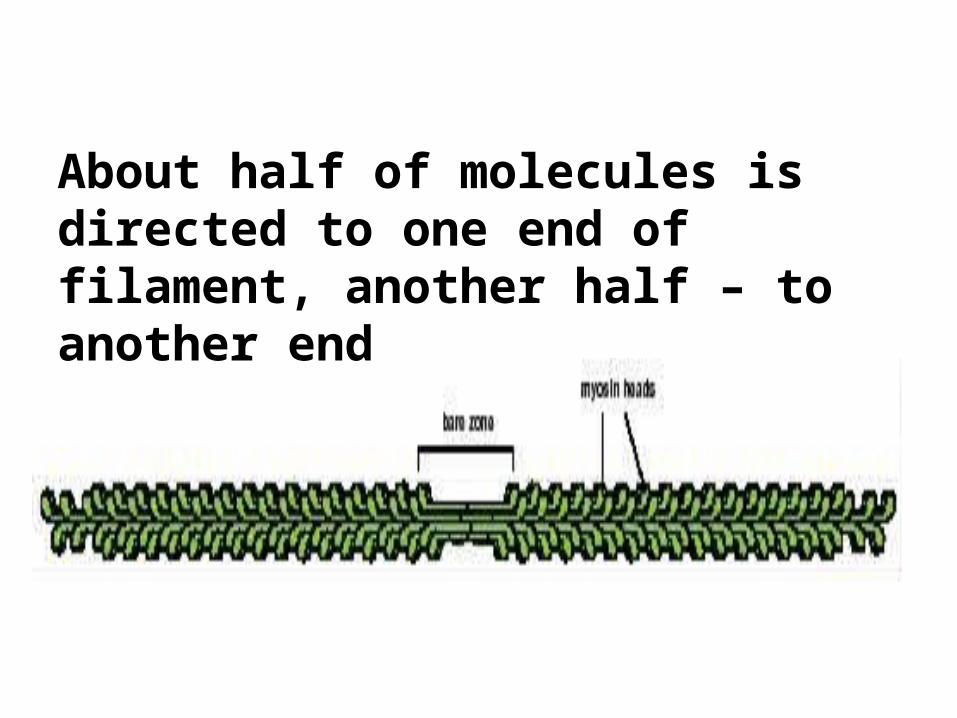
About half of molecules is directed to one end of filament, another half – to another end
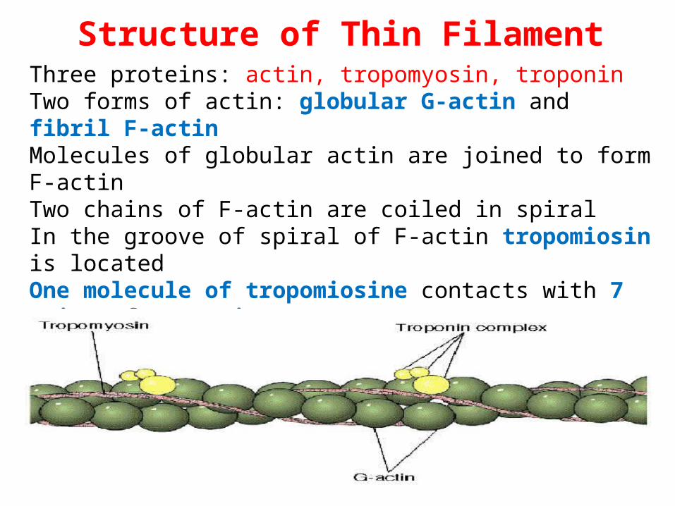
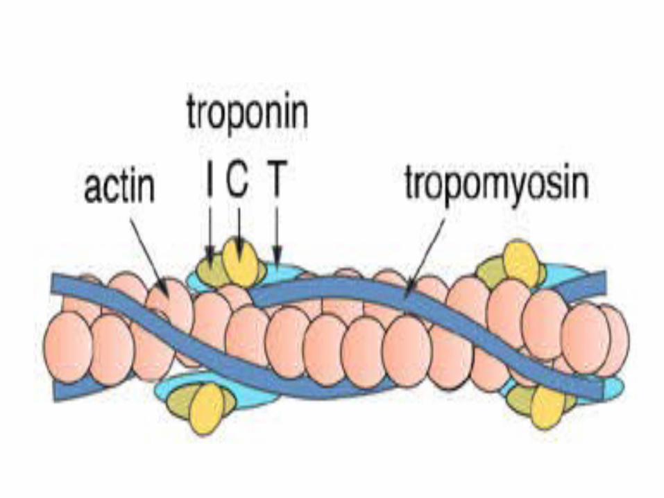
Structure of Thin FilamentThree proteins: actin, tropomyosin, troponinTwo forms of actin: globular G-actin and fibril F-actin Molecules of globular actin are joined to form F-actin Two chains of F-actin are coiled in spiralIn the groove of spiral of F-actin tropomiosin is located One molecule of tropomiosine contacts with 7 pairs of G-actin 1 molecule of troponin drops on 1 molecule of tropomiosinThere are three subunits of troponin
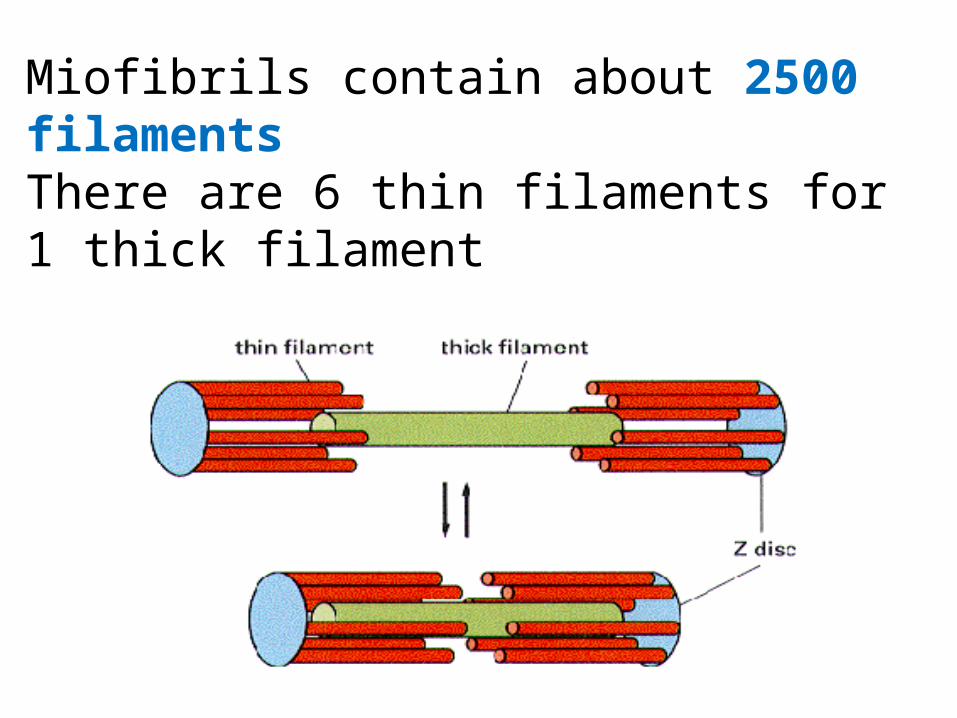
Miofibrils contain about 2500 filaments There are 6 thin filaments for 1 thick filament
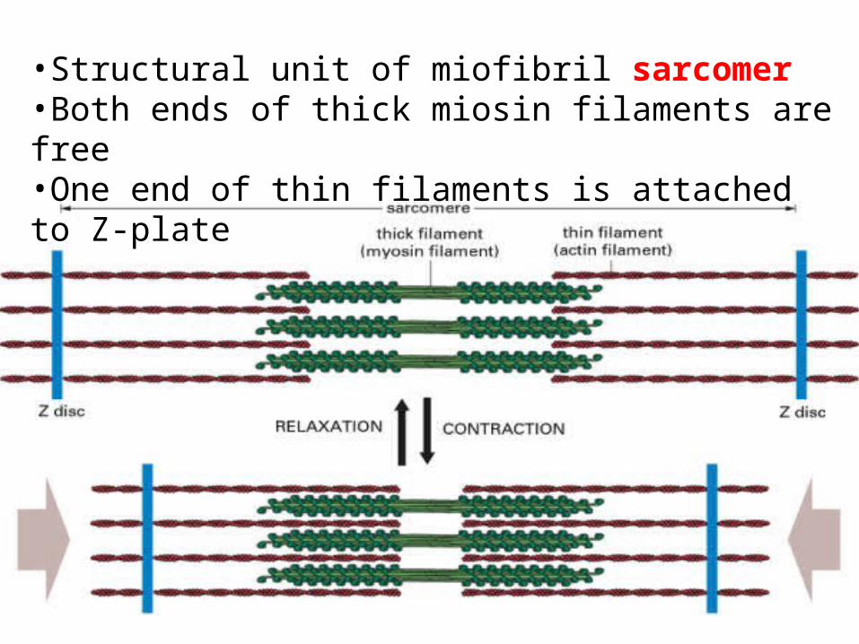
•Structural unit of miofibril sarcomer•Both ends of thick miosin filaments are free•One end of thin filaments is attached to Z-plate
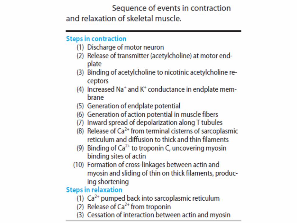
BIOMECHANISM OF MUSCLE CONTRACTION
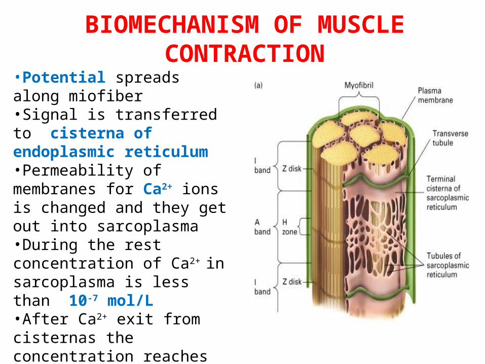
•Potential spreads along miofiber•Signal is transferred to cisterna of endoplasmic reticulum •Permeability of membranes for Ca2+ ions is changed and they get out into sarcoplasma •During the rest concentration of Ca2+ in sarcoplasma is less than 10-7 mol/L •After Ca2+ exit from cisternas the concentration reaches 10-5 mol/L
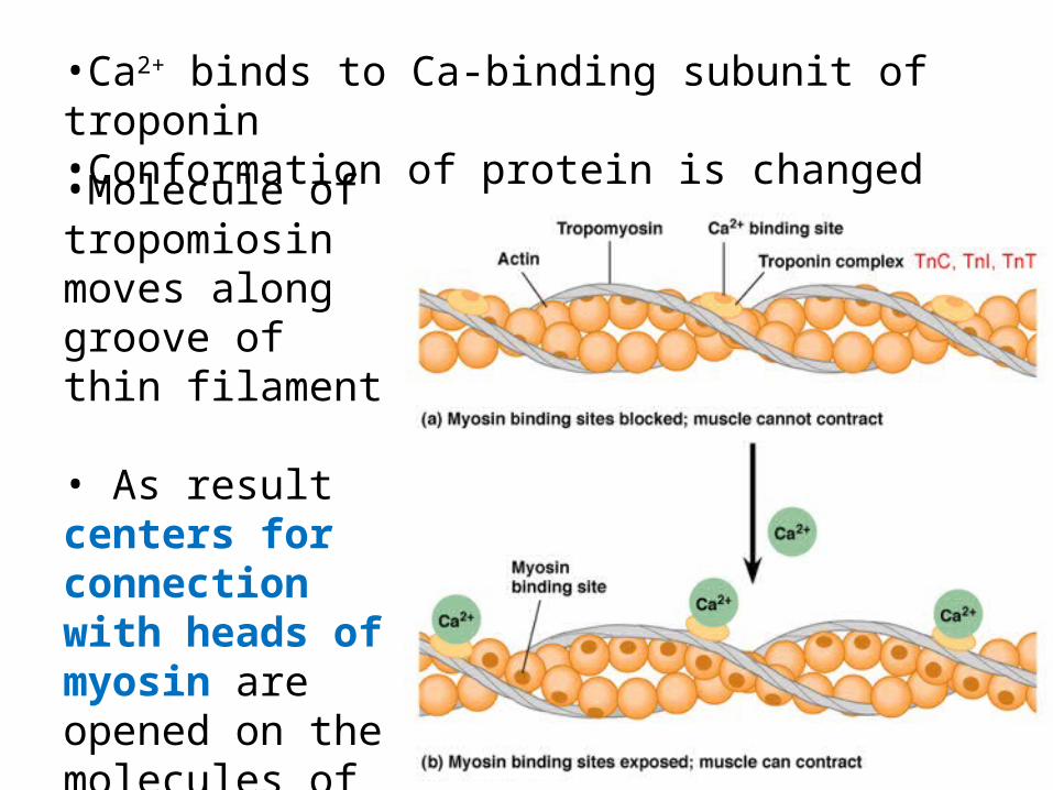
•Molecule of tropomiosin moves along groove of thin filament • As result centers for connection with heads of myosin are opened on the molecules of G-actin
•Ca2+ binds to Ca-binding subunit of troponin•Conformation of protein is changed
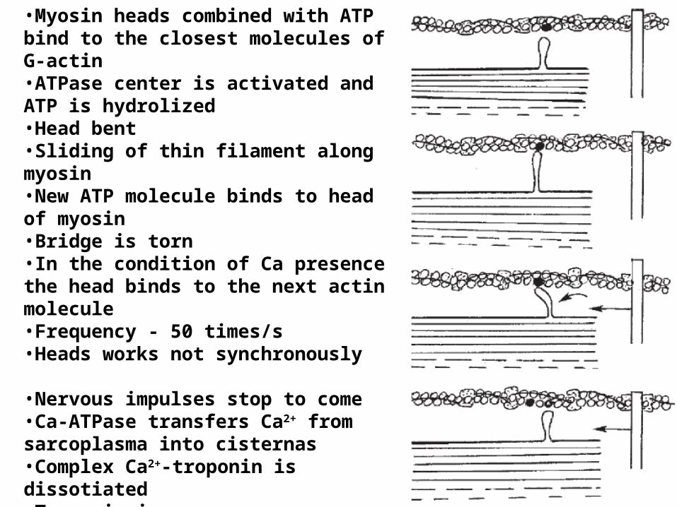
•Myosin heads combined with ATP bind to the closest molecules of G-actin•ATPase center is activated and ATP is hydrolized•Head bent•Sliding of thin filament along myosin•New ATP molecule binds to head of myosin •Bridge is torn •In the condition of Ca presence the head binds to the next actin molecule•Frequency - 50 times/s •Heads works not synchronously
•Nervous impulses stop to come•Ca-ATPase transfers Ca2+ from sarcoplasma into cisternas•Complex Ca2+-troponin is dissotiated•Tropomiosin moves •Molecules of actin are blocked•Bridges are torn•Muscle relaxation
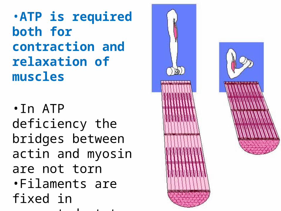
•ATP is required both for contraction and relaxation of muscles
•In ATP deficiency the bridges between actin and myosin are not torn •Filaments are fixed in connected state – muscle contraction (cadaver rigidity)
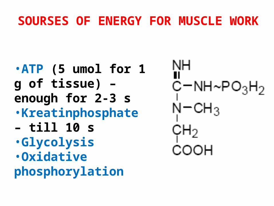
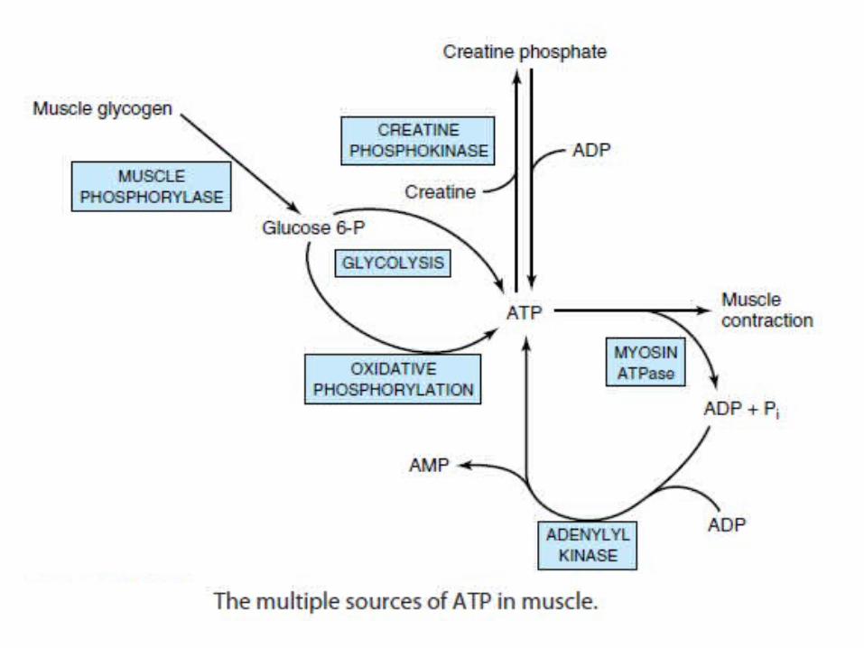
SOURSES OF ENERGY FOR MUSCLE WORK
•ATP (5 umol for 1 g of tissue) – enough for 2-3 s•Kreatinphosphate – till 10 s•Glycolysis•Oxidative phosphorylation
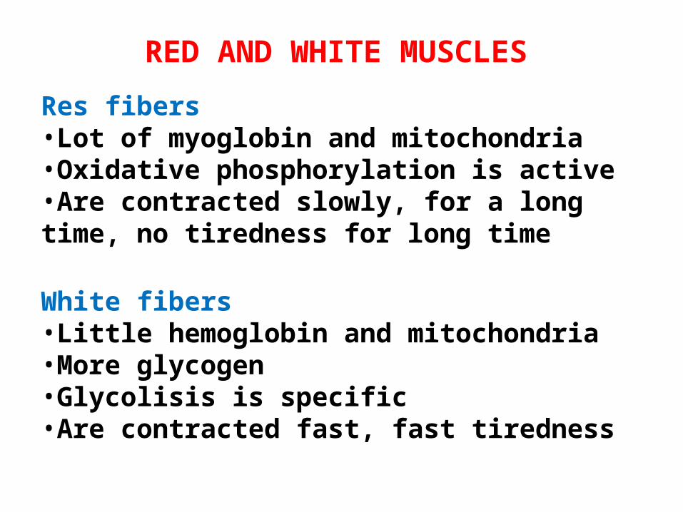
RED AND WHITE MUSCLES
Res fibers•Lot of myoglobin and mitochondria•Oxidative phosphorylation is active•Are contracted slowly, for a long time, no tiredness for long time
White fibers•Little hemoglobin and mitochondria•More glycogen •Glycolisis is specific•Are contracted fast, fast tiredness

• Skeletal muscle functions under both aerobic (resting) and anaerobic (eg, sprinting) conditions, so both aerobic and anaerobic glycolysis operate, depending on conditions.
• Skeletal muscle contains myoglobin as a reservoir of oxygen.
• Skeletal muscle contains different types of fibers primarily suited to anaerobic (fast twitch fibers) or aerobic (slow twitch fibers) conditions.
• Actin, myosin, tropomyosin, troponin complex (TpT, Tpl, and TpC), ATP, and Ca2+ are key constituents in relation to contraction.
• The Ca2+ ATPase, the Ca2+ release channel, and calsequestrinare proteins involved in various aspects of Ca2+ metabolism in muscle.
Summary of major features of the biochemistry of skeletal muscle related to its metabolism

• Insulin acts on skeletal muscle to increase uptake of glucose.
• In the fed state, most glucose is used to synthesize glycogen, which acts as a store of glucose for use in exercise; “preloading” with glucose is used by some long-distance athletes to build up stores of glycogen.
• Epinephrine stimulates glycogenolysis in skeletal muscle, whereas glucagon does not because of absence of its receptors.
• Skeletal muscle cannot contribute directly to blood glucose because it does not contain glucose-6-phosphatase.
• Lactate produced by anaerobic metabolism in skeletal musclepasses to liver, which uses it to synthesize glucose, which can then return to muscle (the Cori cycle).
Summary of major features of the biochemistry of skeletal muscle related to its metabolism
• Skeletal muscle contains phosphocreatine, which acts as an energy store for short-term (seconds) demands.
• Free fatty acids in plasma are a major source of energy, particularly under marathon conditions and in prolonged starvation.
• Skeletal muscle can utilize ketone bodies during starvation.
• Skeletal muscle is the principal site of metabolism of branched-chain amino acids, which are used as an energy source.
• Proteolysis of muscle during starvation supplies amino acids for gluconeogenesis.
• Major amino acids emanating from muscle are alanine (destinedmainly for gluconeogenesis in liver and forming part of the glucose-alanine cycle) and glutamine (destined mainly for the gut and kidneys).
Summary of major features of the biochemistry of skeletal muscle related to its metabolism