muscle tissue slides - hershey bear 121/integrated/muscle/muscle... · facial nerve (vii) bell’s...
TRANSCRIPT
1
Chapter 10Muscle Tissue
Alternating contraction and relaxation of cells
Chemical energy changed into mechanical energy
11-2
11-3
11-4
11-5
11-6
11-7
11-8
11-9
11-10
11
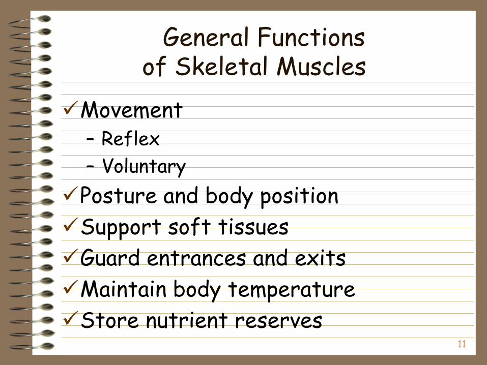
General Functionsof Skeletal Muscles
Movement– Reflex
– Voluntary
Posture and body position
Support soft tissues
Guard entrances and exits
Maintain body temperature
Store nutrient reserves
12
Skeletal Muscle
Anatomy
11-13
The Muscular System
Skeletal muscle major groupings
How movements occur at specific joints
Learn the origin, insertion, function and innervation of all major muscles
Important to allied health care and physical rehabilitation students
11-14
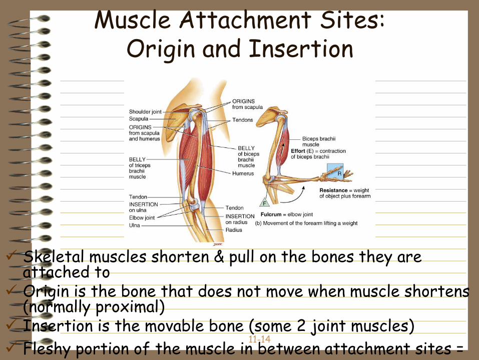
Muscle Attachment Sites:Origin and Insertion
Skeletal muscles shorten & pull on the bones they are attached to
Origin is the bone that does not move when muscle shortens (normally proximal)
Insertion is the movable bone (some 2 joint muscles)
Fleshy portion of the muscle in between attachment sites = belly
11-15
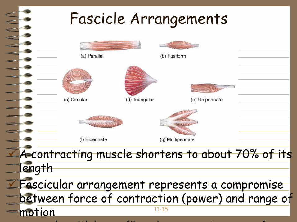
Fascicle Arrangements
A contracting muscle shortens to about 70% of its length
Fascicular arrangement represents a compromise between force of contraction (power) and range of motion– muscles with longer fibers have a greater range of
11-16
Coordination Within Muscle Groups
Most movement is the result of several muscle working at the same time
Most muscles are arranged in opposing pairs at joints– prime mover or agonist contracts to cause the
desired action
– antagonist stretches and yields to prime mover
– synergists contract to stabilize nearby joints
– fixators stabilize the origin of the prime mover• scapula held steady so deltoid can raise arm
11-17
How Skeletal Muscle are Named
Direction the muscle fibers run
Size, shape, action, number of origins or locations
Examples from Table 11.2– triceps brachii -- 3 sites of origin
– quadratus femoris -- square shape
– serratus anterior -- saw-toothed edge
11-18
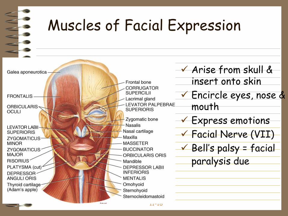
Muscles of Facial Expression
Arise from skull & insert onto skin
Encircle eyes, nose & mouth
Express emotions
Facial Nerve (VII)
Bell’s palsy = facial paralysis due
11-19
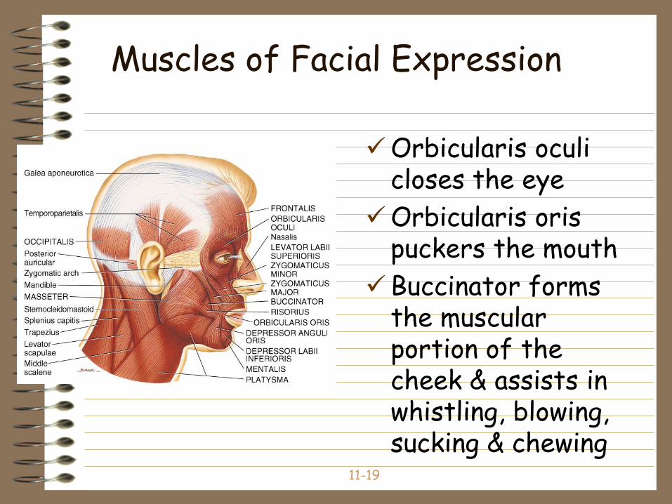
Muscles of Facial Expression
Orbicularis oculi closes the eye
Orbicularis oris puckers the mouth
Buccinator forms the muscular portion of the cheek & assists in whistling, blowing, sucking & chewing
11-20
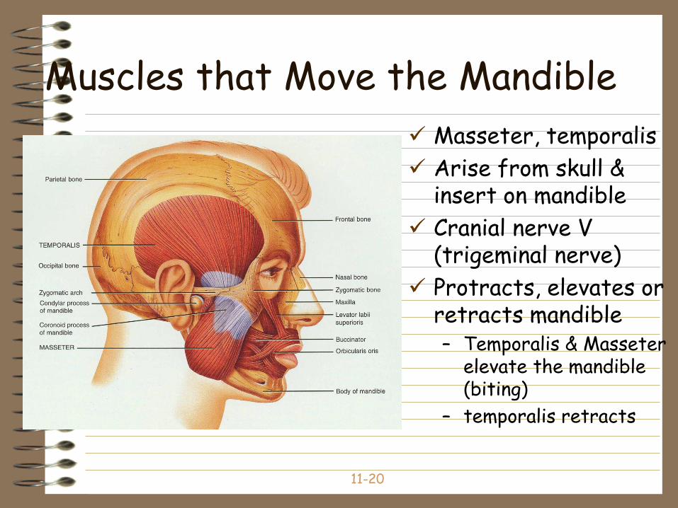
Muscles that Move the Mandible
Masseter, temporalis
Arise from skull & insert on mandible
Cranial nerve V (trigeminal nerve)
Protracts, elevates or retracts mandible– Temporalis & Masseter
elevate the mandible (biting)
– temporalis retracts
11-21
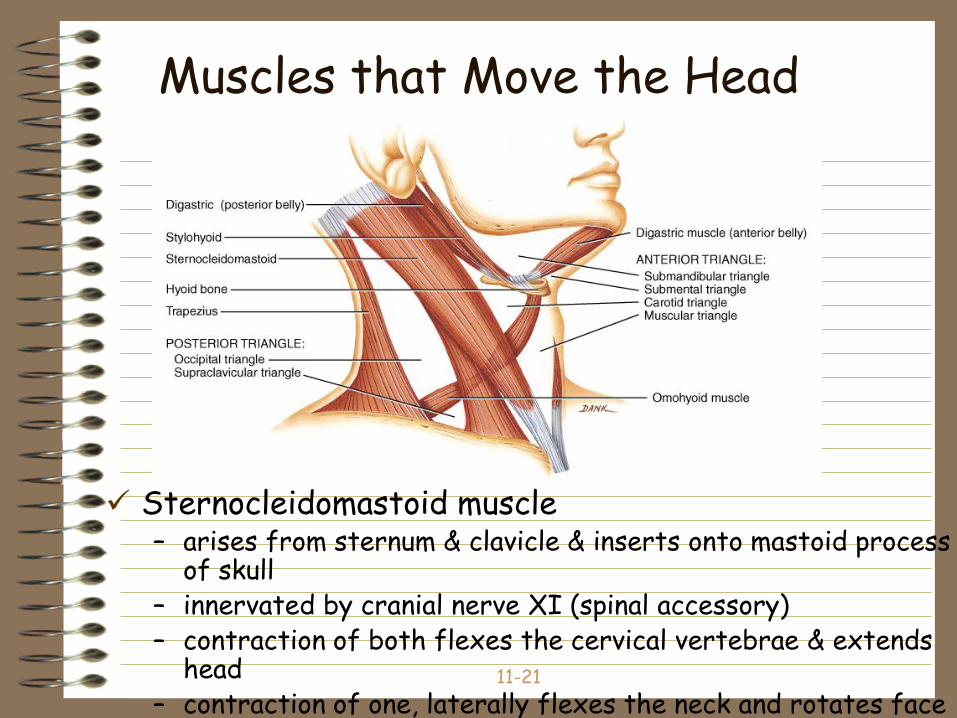
Muscles that Move the Head
Sternocleidomastoid muscle– arises from sternum & clavicle & inserts onto mastoid process
of skull– innervated by cranial nerve XI (spinal accessory)– contraction of both flexes the cervical vertebrae & extends
head – contraction of one, laterally flexes the neck and rotates face
in opposite direction
11-22
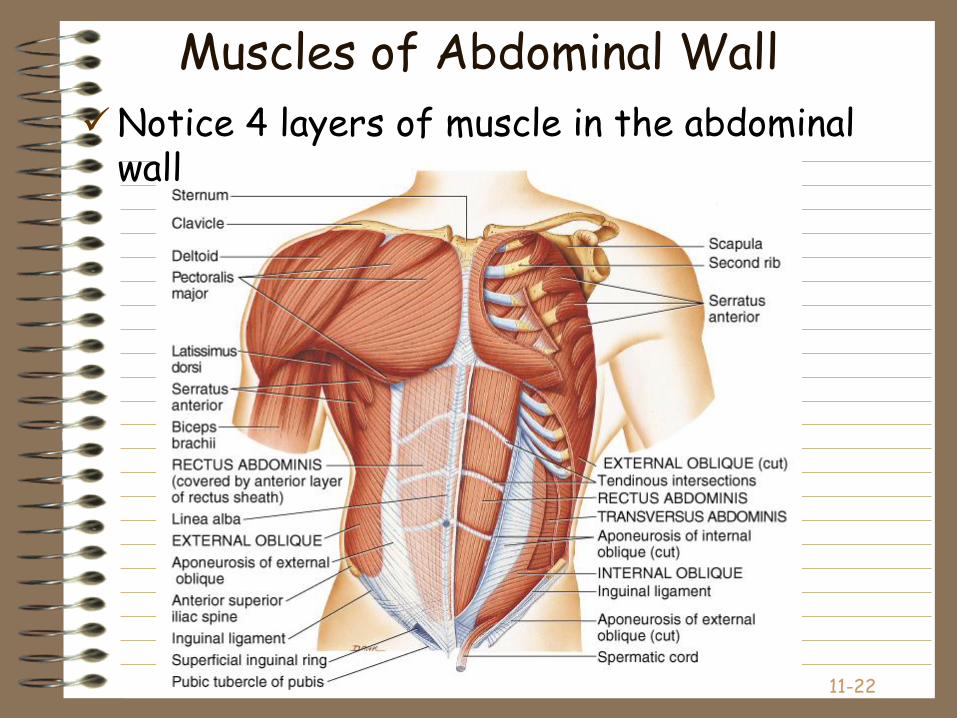
Muscles of Abdominal Wall
Notice 4 layers of muscle in the abdominal wall
11-23
Muscles of Abdominal Wall
4 pairs of sheetlike muscles– rectus abdominis = vertically oriented
– external & internal obliques and transverses abdominis• wrap around body to form anterior body wall
• form rectus sheath and linea alba
11-24
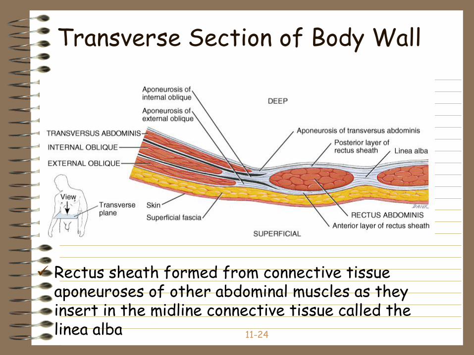
Transverse Section of Body Wall
Rectus sheath formed from connective tissue aponeuroses of other abdominal muscles as they insert in the midline connective tissue called the linea alba
11-25
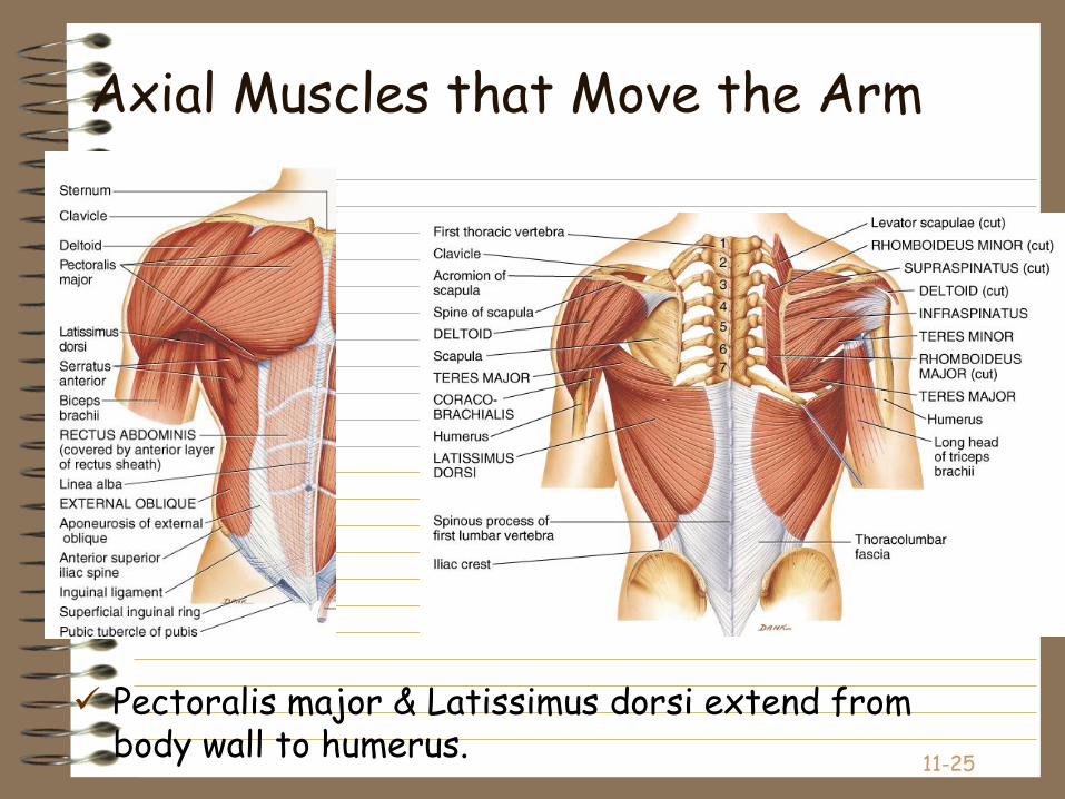
Axial Muscles that Move the Arm
Pectoralis major & Latissimus dorsi extend from body wall to humerus.
11-26
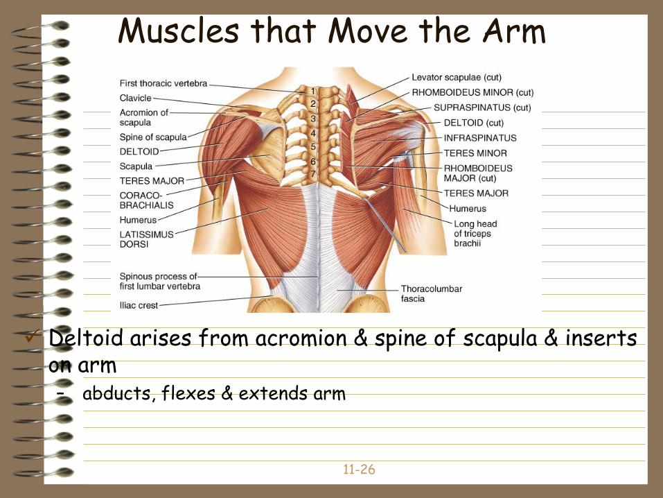
Muscles that Move the Arm
Deltoid arises from acromion & spine of scapula & inserts on arm – abducts, flexes & extends arm
11-27
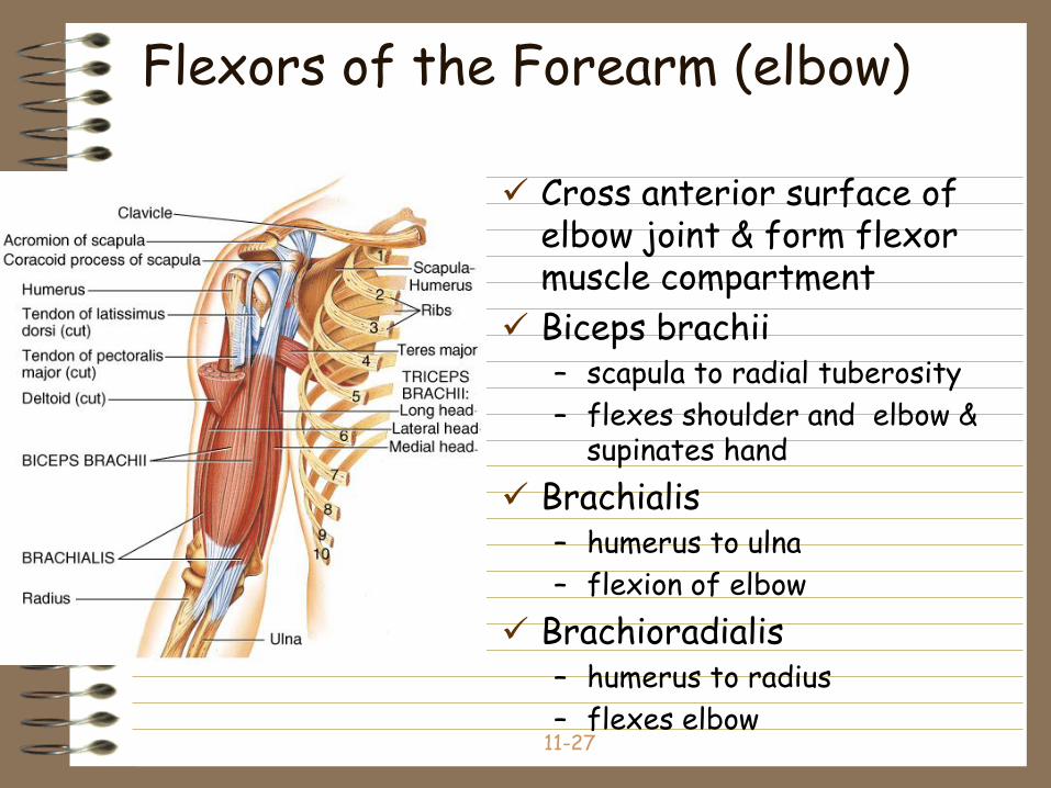
Flexors of the Forearm (elbow)
Cross anterior surface of elbow joint & form flexor muscle compartment
Biceps brachii– scapula to radial tuberosity
– flexes shoulder and elbow & supinates hand
Brachialis– humerus to ulna
– flexion of elbow
Brachioradialis– humerus to radius
– flexes elbow
11-28
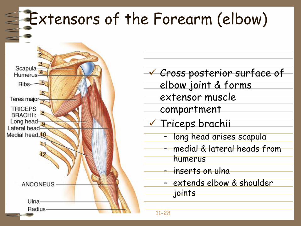
Extensors of the Forearm (elbow)
Cross posterior surface of elbow joint & forms extensor muscle compartment
Triceps brachii– long head arises scapula
– medial & lateral heads from humerus
– inserts on ulna
– extends elbow & shoulder joints
11-29
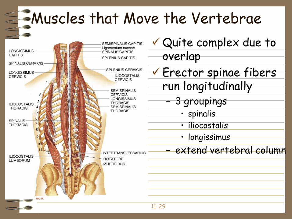
Muscles that Move the Vertebrae
Quite complex due to overlap
Erector spinae fibers run longitudinally– 3 groupings
• spinalis
• iliocostalis
• longissimus
– extend vertebral column
11-30
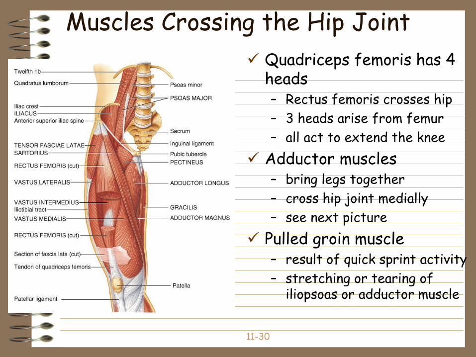
Muscles Crossing the Hip Joint
Quadriceps femoris has 4 heads– Rectus femoris crosses hip
– 3 heads arise from femur
– all act to extend the knee
Adductor muscles – bring legs together
– cross hip joint medially
– see next picture
Pulled groin muscle– result of quick sprint activity
– stretching or tearing of iliopsoas or adductor muscle
11-31
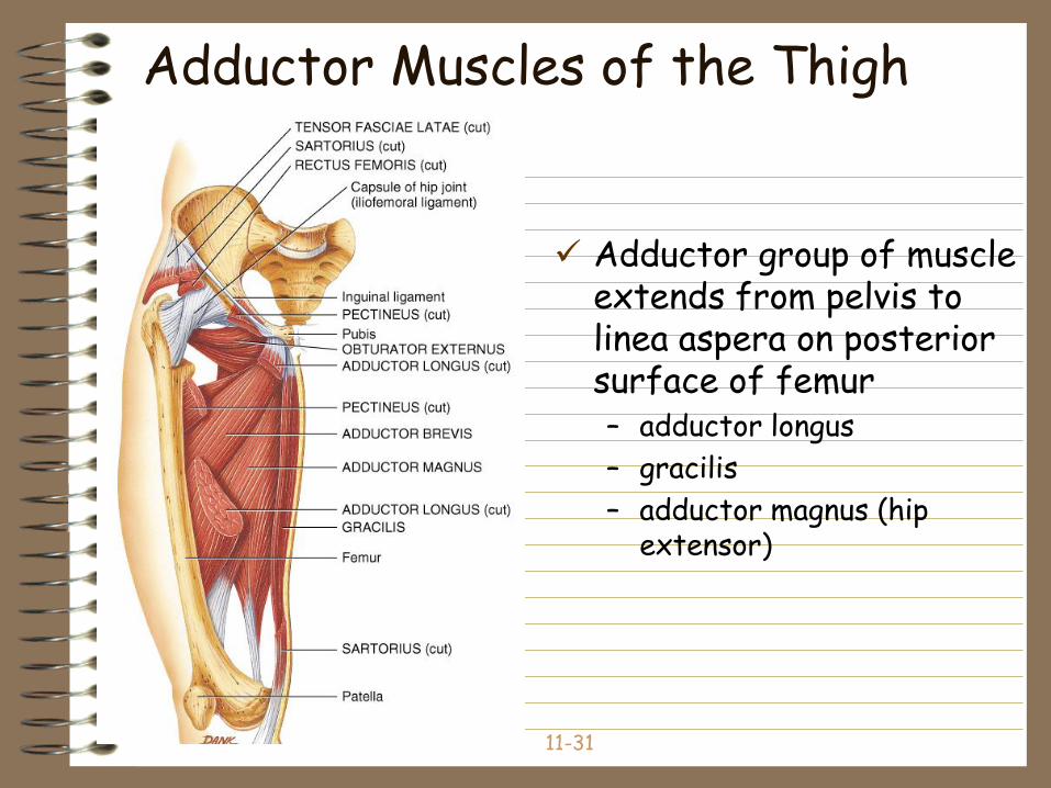
Adductor Muscles of the Thigh
Adductor group of muscle extends from pelvis to linea aspera on posterior surface of femur– adductor longus
– gracilis
– adductor magnus (hip extensor)
11-32
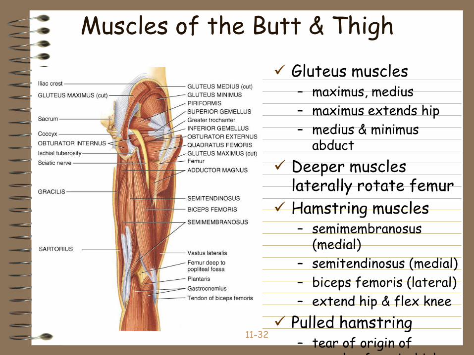
Muscles of the Butt & Thigh
Gluteus muscles– maximus, medius
– maximus extends hip
– medius & minimus abduct
Deeper muscles laterally rotate femur
Hamstring muscles– semimembranosus
(medial)
– semitendinosus (medial)
– biceps femoris (lateral)
– extend hip & flex knee
Pulled hamstring– tear of origin of
muscles from ischial
11-33
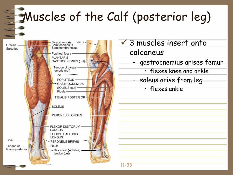
Muscles of the Calf (posterior leg)
3 muscles insert onto calcaneus– gastrocnemius arises femur
• flexes knee and ankle
– soleus arise from leg• flexes ankle
11-34
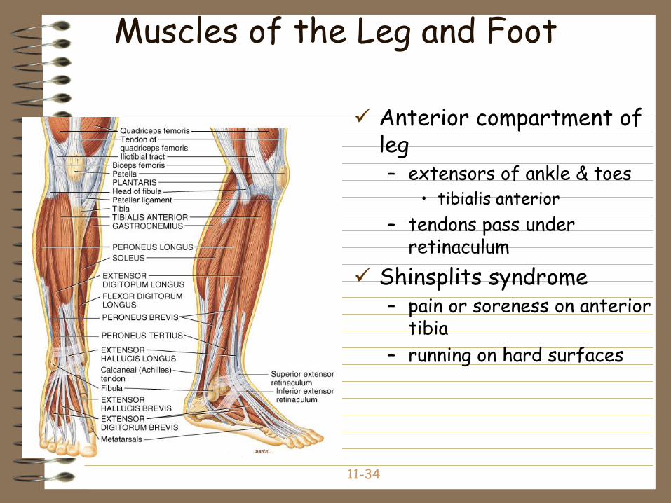
Muscles of the Leg and Foot
Anterior compartment of leg– extensors of ankle & toes
• tibialis anterior
– tendons pass under retinaculum
Shinsplits syndrome– pain or soreness on anterior
tibia
– running on hard surfaces
35
Functional Anatomy
Connective tissue components
Muscle fibers (cells)
Nerves: – (Containing motor neurons) convey impulses for
muscular contraction
Blood supply: – Provides nutrients and oxygen for contraction
36
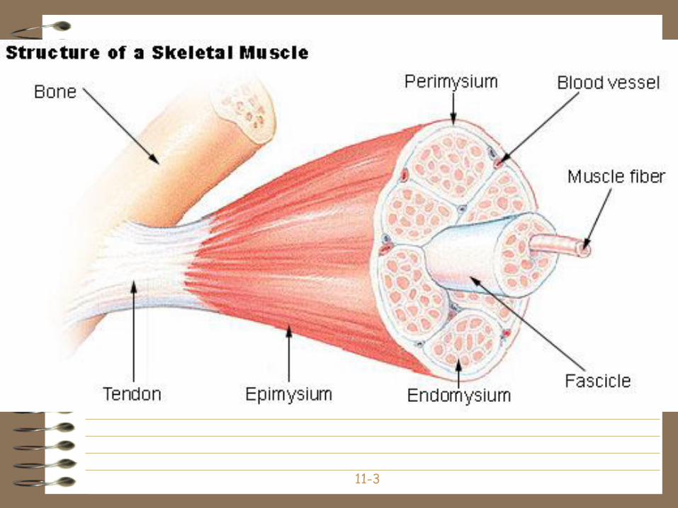
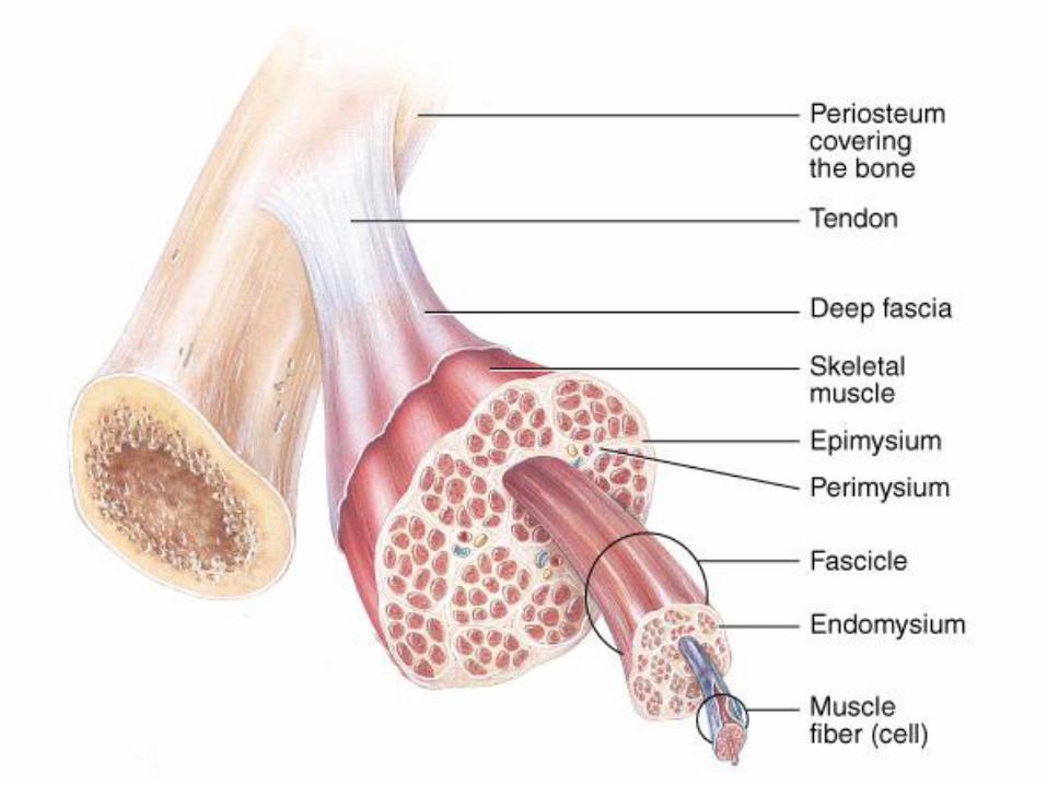
Functional Anatomy Connective Tissue
Superficial fascia: loose CT & fat underlying the skin
Deep fascia: dense irregular CT around muscle
Connective tissue components of muscle include– Epimysium: surrounds the whole muscle
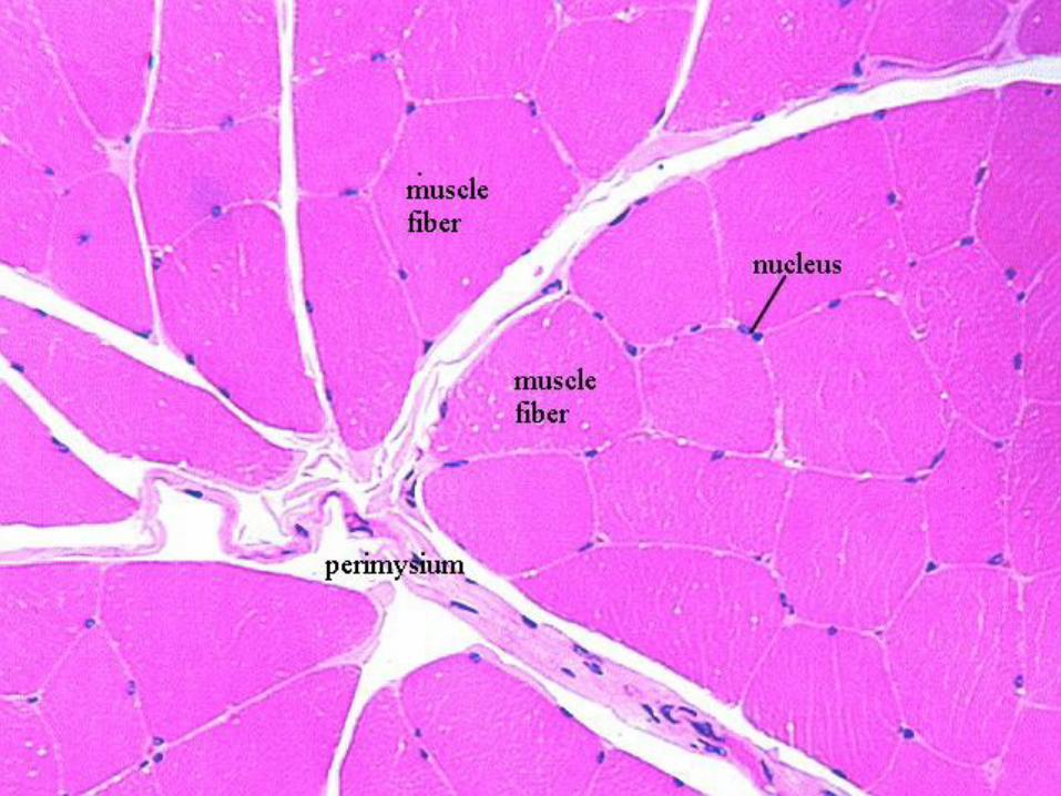
– Perimysium: surrounds bundles (fascicles) of 10-100 muscle cells
• Blood vessels
• nerves
– Endomysium: separates individual muscle cells• Capillary network
• Satellite cells
• Nerve fibers
37
Functional Anatomy Connective Tissue
All the CT layers extend beyond the muscle belly to form:– Tendon: strong tough cord of connective tissue
that extends from muscle to bone
– Aponeurosis: a strong sheath of connective tissue that extends from muscle to muscle

39
Skeletal Muscle Fibers
Muscle Fibers: highly specialized skeletal muscle cells – Myoblasts
– Multinucleated
– Fascicles: groups of skeletal muscle fibers
40
Skeletal Muscle Fibers
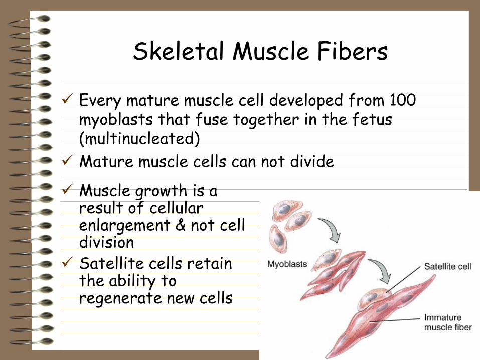
Every mature muscle cell developed from 100 myoblasts that fuse together in the fetus (multinucleated)
Mature muscle cells can not divide
Muscle growth is a result of cellular enlargement & not cell division
Satellite cells retain the ability to regenerate new cells
41
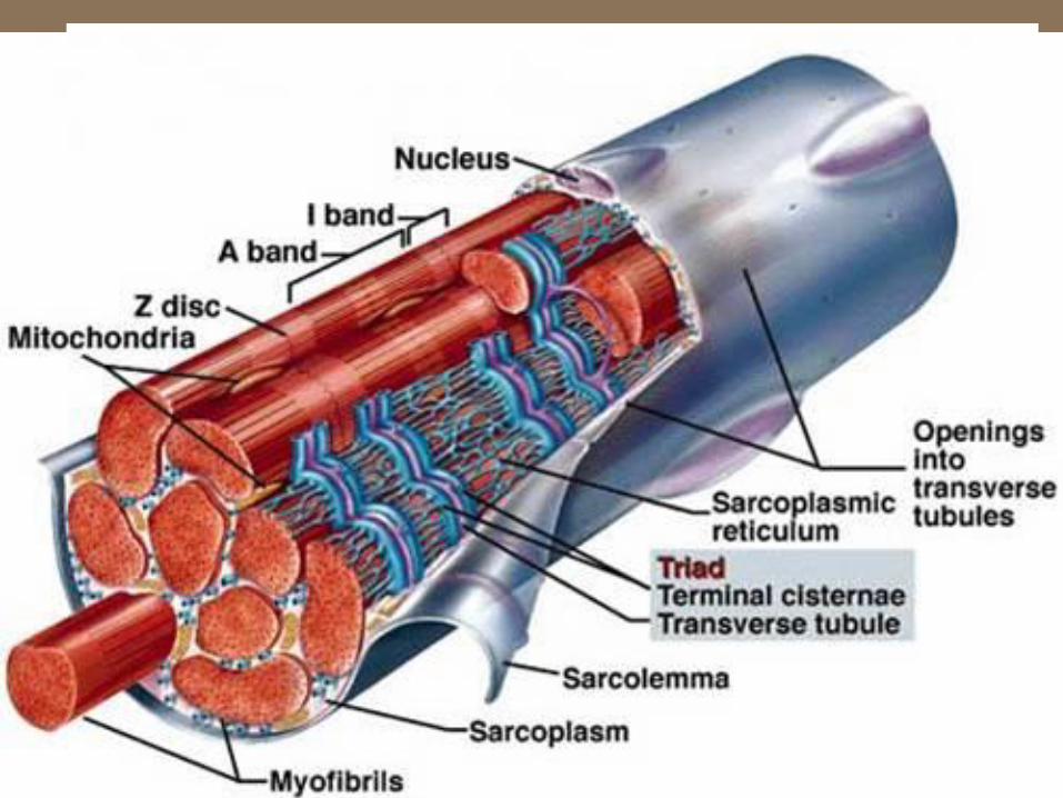
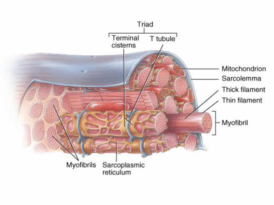
Skeletal Muscle Fibers
Sarcolemma = plasma membrane
Sarcoplasm = cytoplasm– Myofibrils, myofilaments
– Myoglobin: red-colored, oxygen-binding protein
Transverse tubules: invaginations of the sarcolemma into the center of the cell– filled with extracellular fluid
– carry muscle action potentials down into cell
Mitochondria lie in rows throughout the cell– near the muscle proteins that use ATP during contraction
Sarcoplasmic reticulum: storage site for calcium
Terminal cisternae
43
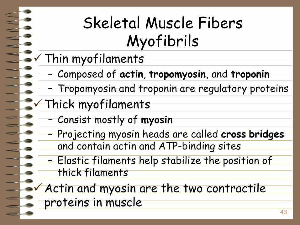
Skeletal Muscle FibersMyofibrils
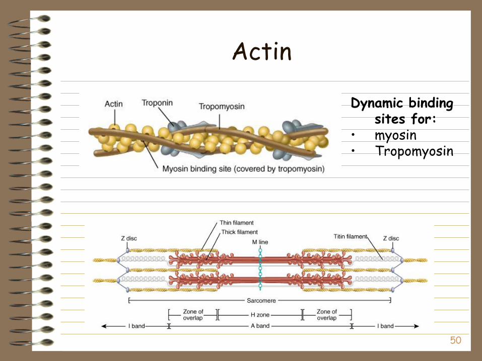
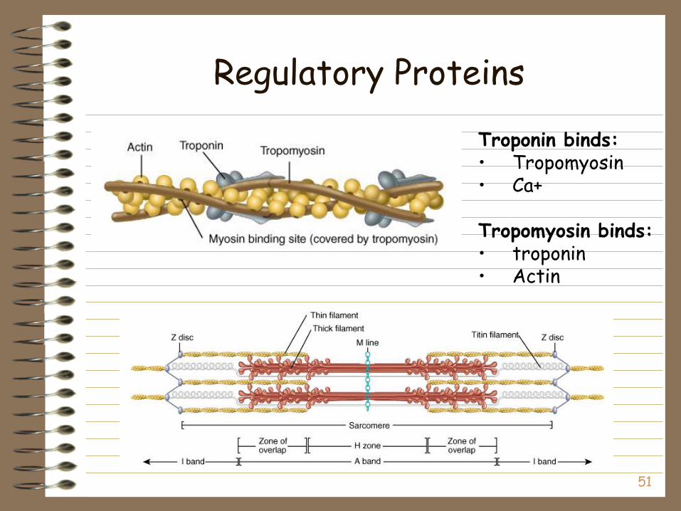
Thin myofilaments – Composed of actin, tropomyosin, and troponin
– Tropomyosin and troponin are regulatory proteins
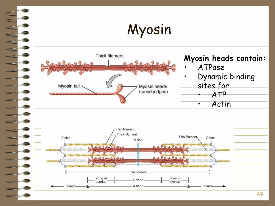
Thick myofilaments – Consist mostly of myosin
– Projecting myosin heads are called cross bridgesand contain actin and ATP-binding sites
– Elastic filaments help stabilize the position of thick filaments
Actin and myosin are the two contractile proteins in muscle
44
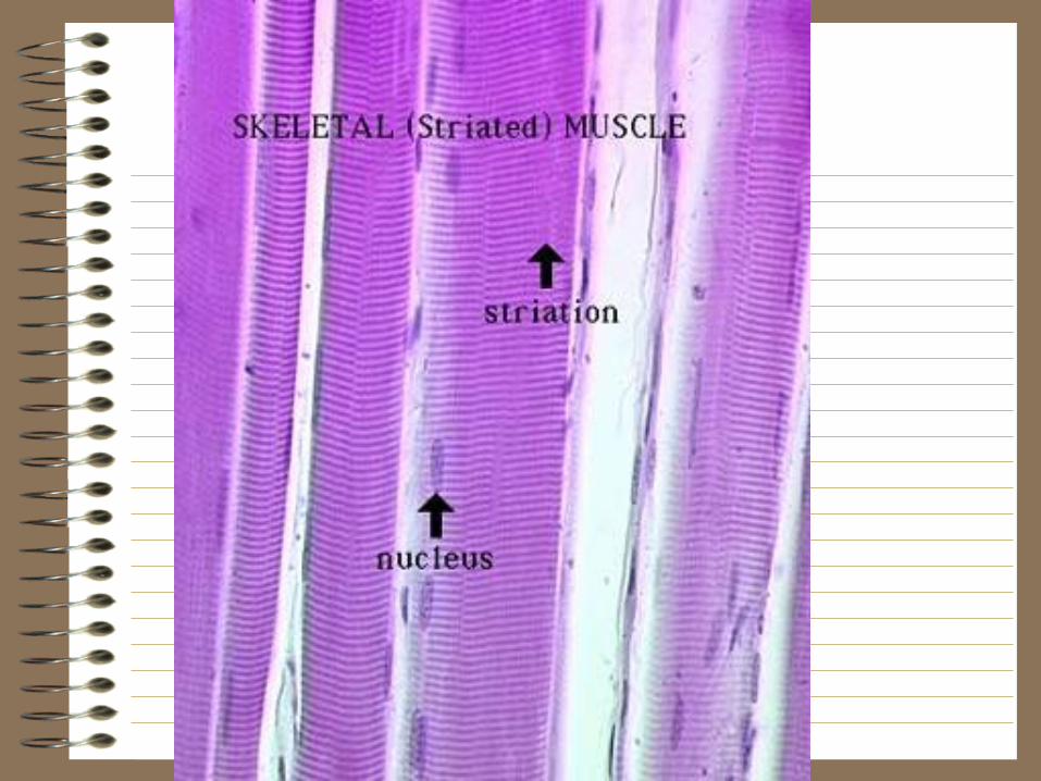
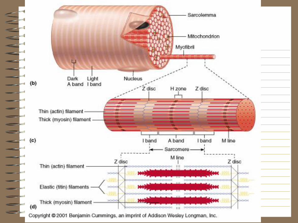
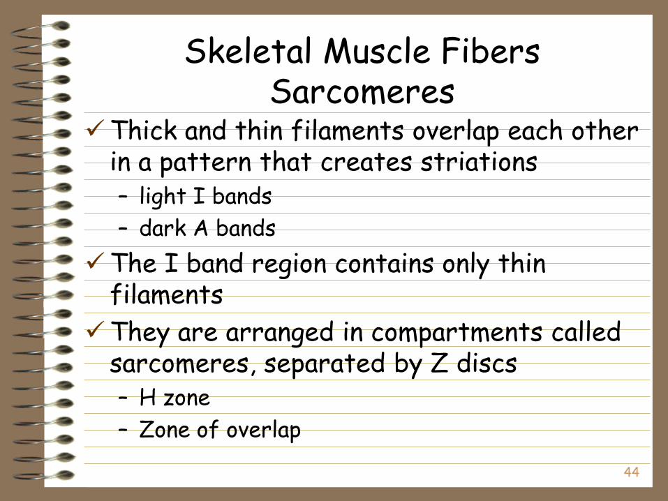
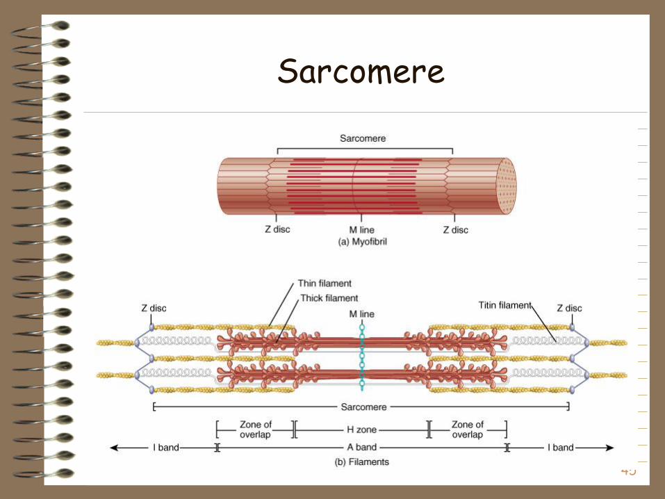
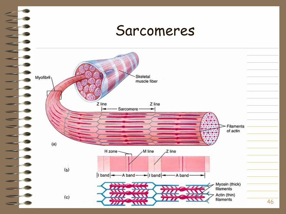
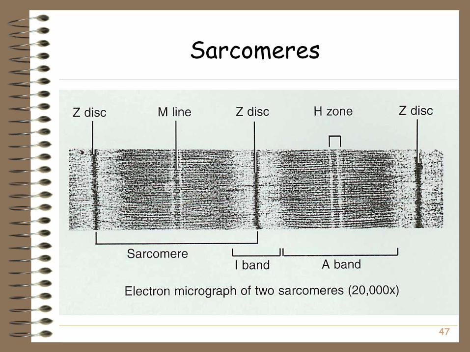
Skeletal Muscle FibersSarcomeres
Thick and thin filaments overlap each other in a pattern that creates striations – light I bands
– dark A bands
The I band region contains only thin filaments
They are arranged in compartments called sarcomeres, separated by Z discs– H zone
– Zone of overlap
45
Sarcomere
46
Sarcomeres
47
Sarcomeres

48
The Proteins of Muscle
Myofibrils are built of 3 kinds of protein– contractile proteins
• myosin and actin
– regulatory proteins which turn contraction on & off• troponin and tropomyosin
– structural proteins which provide proper alignment, elasticity and extensibility• titin, myomesin, nebulin and dystrophin
49
Myosin
Myosin heads contain:• ATPase• Dynamic binding
sites for• ATP• Actin
50
Actin
Dynamic binding sites for:
• myosin• Tropomyosin
51
Regulatory Proteins
Troponin binds:• Tropomyosin• Ca+
Tropomyosin binds:• troponin• Actin
52
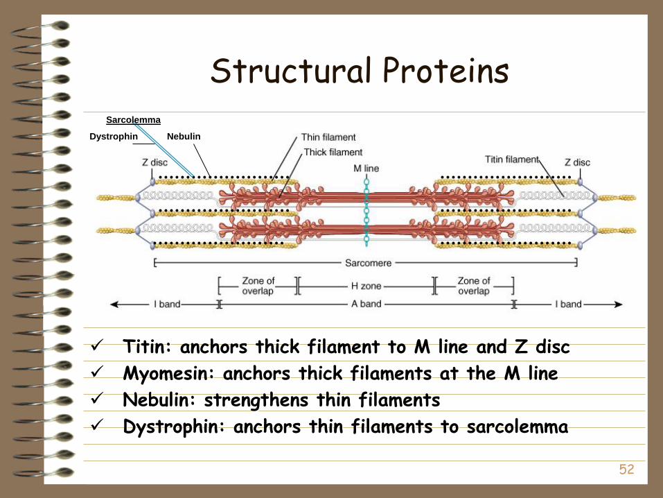
Structural Proteins
Titin: anchors thick filament to M line and Z disc
Myomesin: anchors thick filaments at the M line
Nebulin: strengthens thin filaments
Dystrophin: anchors thin filaments to sarcolemma
NebulinDystrophin
Sarcolemma
53
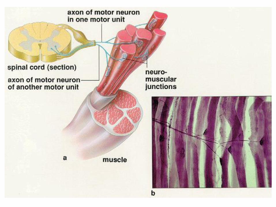
Control of Skeletal Muscle Activity
A motor neuron transmits a nerve impulse (action potential) to skeletal muscle where the nerve impulse serves as a stimulus for contraction
Motor unit: a motor neuron and the muscle fibers it stimulates– A single motor unit may innervate as few as 10 or as many as 2000
muscle fibers, with an average of 150 fibers being innervated by each motor neuron
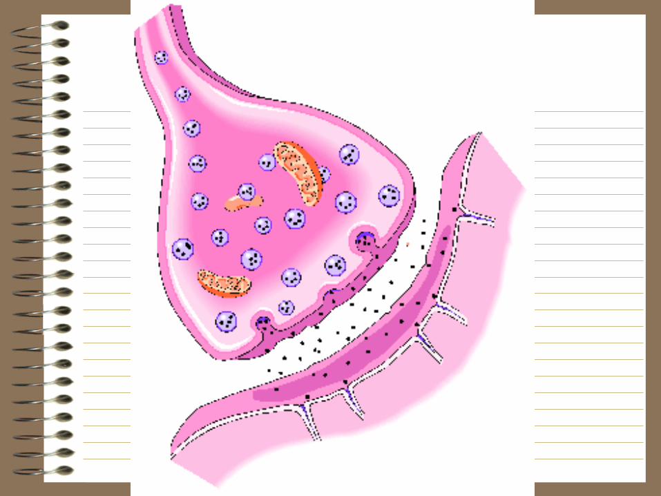
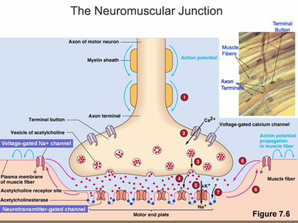
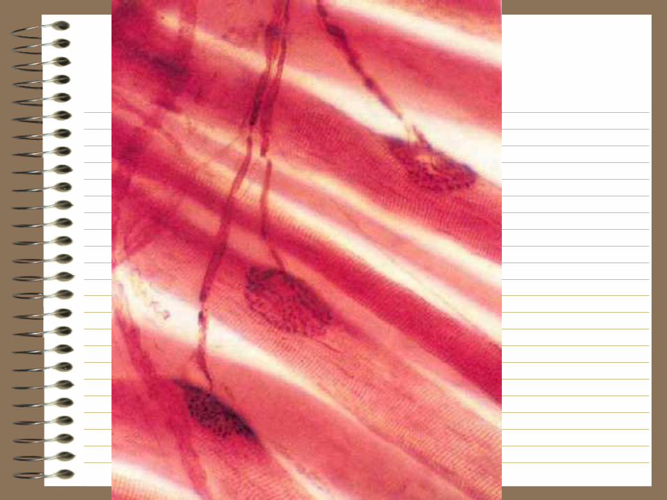
Neuromuscular junction (synapse): site where axon terminal of a motor neuron meets the muscle fiber sarcolemma (plasma membrane)– Separated by a gap called the neuromuscular cleft– Motor end plate: pocket formed around motor neuron by
sarcolemma– Acetylcholine is released from synaptic vesicles of the motor
neuron• Triggers a muscle action potential
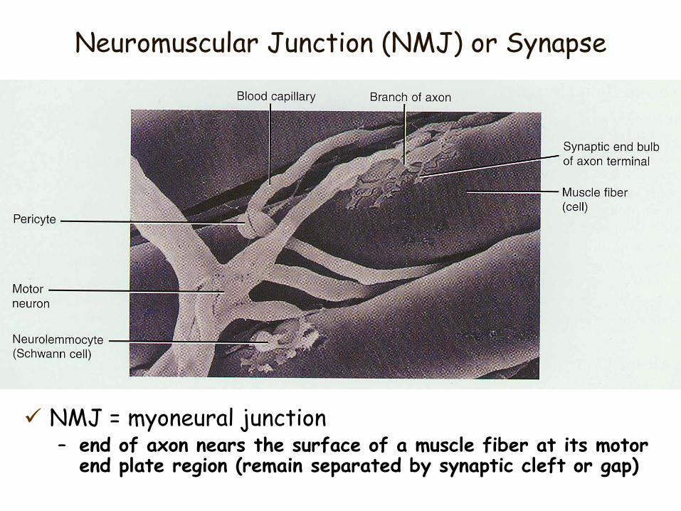
Neuromuscular Junction (NMJ) or Synapse
NMJ = myoneural junction– end of axon nears the surface of a muscle fiber at its motor
end plate region (remain separated by synaptic cleft or gap)
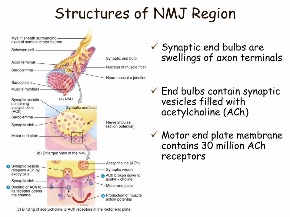
Structures of NMJ Region
Synaptic end bulbs are swellings of axon terminals
End bulbs contain synaptic vesicles filled with acetylcholine (ACh)
Motor end plate membrane contains 30 million ACh receptors
57
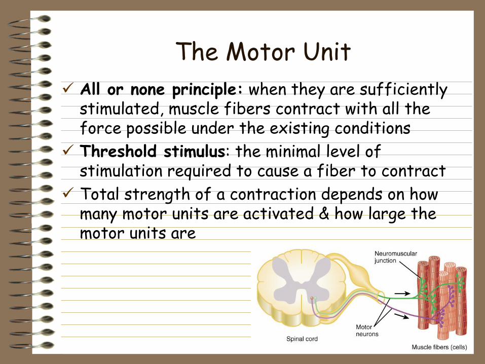
The Motor Unit
All or none principle: when they are sufficiently stimulated, muscle fibers contract with all the force possible under the existing conditions
Threshold stimulus: the minimal level of stimulation required to cause a fiber to contract
Total strength of a contraction depends on how many motor units are activated & how large the motor units are
58
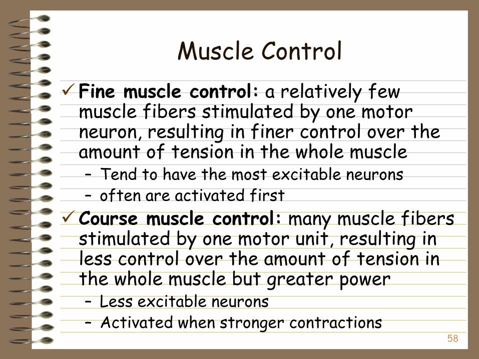
Muscle Control
Fine muscle control: a relatively few muscle fibers stimulated by one motor neuron, resulting in finer control over the amount of tension in the whole muscle– Tend to have the most excitable neurons– often are activated first
Course muscle control: many muscle fibers stimulated by one motor unit, resulting in less control over the amount of tension in the whole muscle but greater power– Less excitable neurons– Activated when stronger contractions
59
Skeletal Muscle
Physiology
60
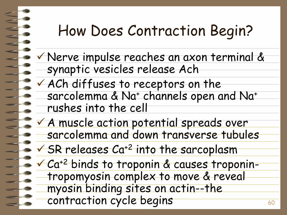
How Does Contraction Begin?
Nerve impulse reaches an axon terminal & synaptic vesicles release Ach
ACh diffuses to receptors on the sarcolemma & Na+ channels open and Na+
rushes into the cellA muscle action potential spreads over
sarcolemma and down transverse tubulesSR releases Ca+2 into the sarcoplasmCa+2 binds to troponin & causes troponin-
tropomyosin complex to move & reveal myosin binding sites on actin--the contraction cycle begins
61
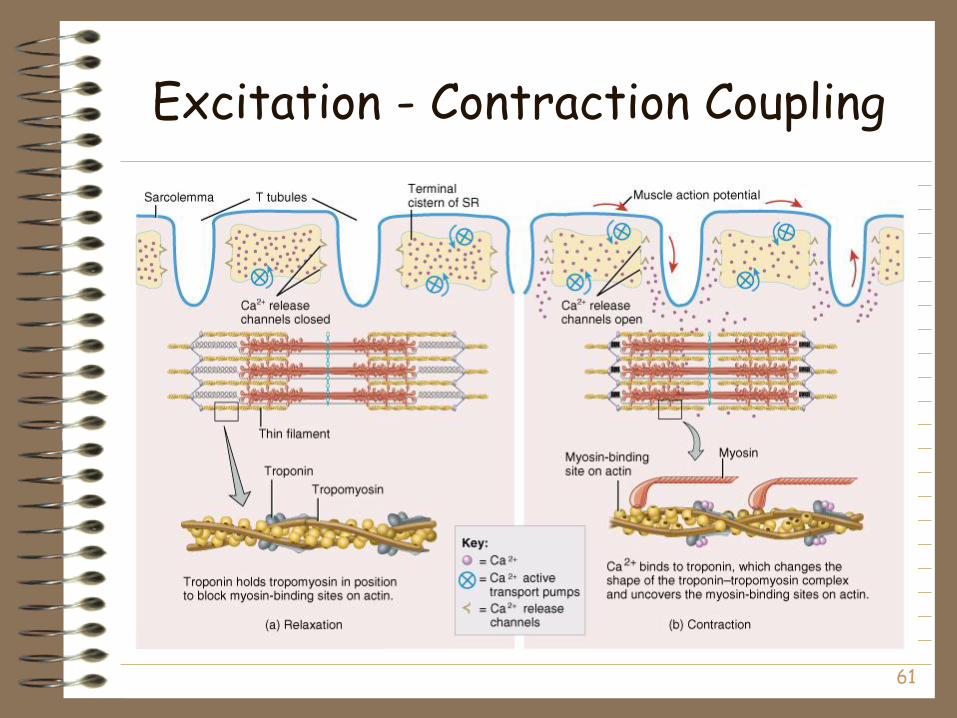
Excitation - Contraction Coupling
62
Contraction Cycle
Sliding Filament Model: Repeating sequence of events that cause the thick & thin filaments to move past each other
4 steps to contraction cycle– ATP hydrolysis– attachment of myosin to actin to form
crossbridges– power stroke– detachment of myosin from actin
Cycle keeps repeating as long as there is ATP available & high Ca+2 level near thin filament
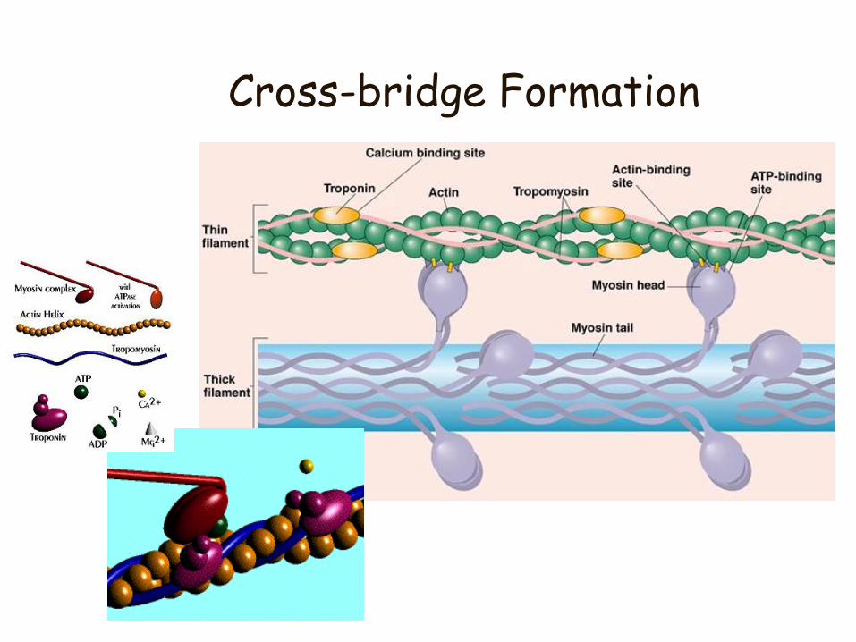
Cross-bridge Formation
64
Relaxation
Acetylcholinesterase (AChE) breaks down ACh within the synaptic cleft– Sodium potassium pumps remove sodium
Ca+2 release channels closeActive transport pumps Ca+2 back into
storage in the sarcoplasmic reticulumCalcium-binding protein (calsequestrin)
helps hold Ca+2 in SR – Ca+2 concentration 10,000 times higher than in
cytosol
Tropomyosin-troponin complex covers binding site on the actin
65
Rigor Mortis
Rigor mortis is a state of muscular rigidity that begins 3-4 hours after death and lasts about 24 hours
After death, Ca+2 ions leak out of the SR and allow myosin heads to bind to actin
Since ATP synthesis has ceased, crossbridges cannot detach from actin until proteolytic enzymes begin to digest the decomposing cells
66
Tension Production
Contraction: a shortening or increase in tension– Tension: a stretching or pulling force
Active tension (internal): tension generated by contractile elements (thick and thin filaments)– All or none response
Passive tension (external): tension generated by elastic elements. It is not related to muscular contraction– Elastic elements: elastic filaments (titin), connective
tissue coverings, and tendons– Elastic elements stretch slightly before they relay the
tension generated by the sliding filaments to the whole muscle
67
Tension Produced by Muscle FibersLength Tension Relationship
Optimal overlap of thick & thin filaments– produces greatest number of crossbridges and the
greatest amount of tension
As stretch muscle (past optimal length)– fewer cross bridges exist & less force is produced
If muscle is overly shortened (less than optimal)– fewer cross bridges exist & less force is produced
– thick filaments crumpled by Z discs
Normally– resting muscle length remains between 70 to 130% of the
optimum
68
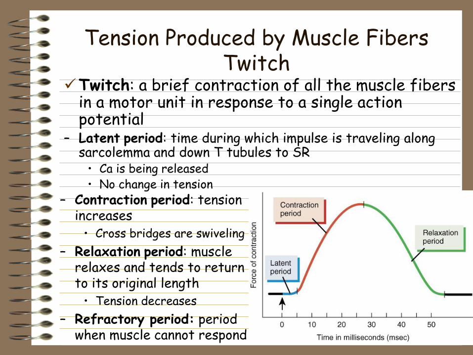
Tension Produced by Muscle Fibers Twitch
Twitch: a brief contraction of all the muscle fibers in a motor unit in response to a single action potential
– Latent period: time during which impulse is traveling along sarcolemma and down T tubules to SR
• Ca is being released• No change in tension
– Contraction period: tension increases
• Cross bridges are swiveling
– Relaxation period: muscle relaxes and tends to return to its original length
• Tension decreases
– Refractory period: period when muscle cannot respond
69
Tension Produced by Muscle Fibers
Wave Summation: the increased strength of a contraction resulting from the application of a second stimulus before the muscle has completely relaxed
– Tetanus: when a muscle fiber is stimulated so rapidly it does not relax between stimuli
• Incomplete (unfused) tetanus: a sustained muscle contraction that permits partial relaxation between stimuli
• Complete (fused) tetanus: a sustained contraction that lacks even partial relaxation between stimuli
70
Tension Produced by Skeletal MusclesMotor Units
Recruitment: process of increasing the number of active motor units– low levels of stimulation -- relatively small
number of motor neurons are activated, so relatively few motor units are stimulated
– higher levels of stimulation -- more motor neurons stimulated resulting in the recruitment of more motor units
– A muscle is contracting at maximal intensity when all motor units are activated simultaneously
Asynchronous motor unit summation: motor units are activated on a rotating basis– Some are resting while others are contracting– Prevents fatigue
71
Tension Produced by Skeletal Muscles Muscle Tone
Involuntary contraction of a small number of motor units (alternately active and inactive in a constantly shifting pattern)– keeps muscles firm even though relaxed
– does not produce movement
Hypotonia: decreased or lost muscle tone– Such muscles are said to be flaccid
Hypertonia: increased muscle tone and may be expressed as either spasticity(stiffness) or rigidity
72
Tension Produced by Skeletal Muscles Contractions
Load: the weight of an object to be moved
Isotonic: tension or force generated by muscle is greater than the load and the muscle changes length– Concentric contraction– Eccentric contraction
Isometric: load is greater than the tension or force generated by the muscle and the muscle does not change length
73
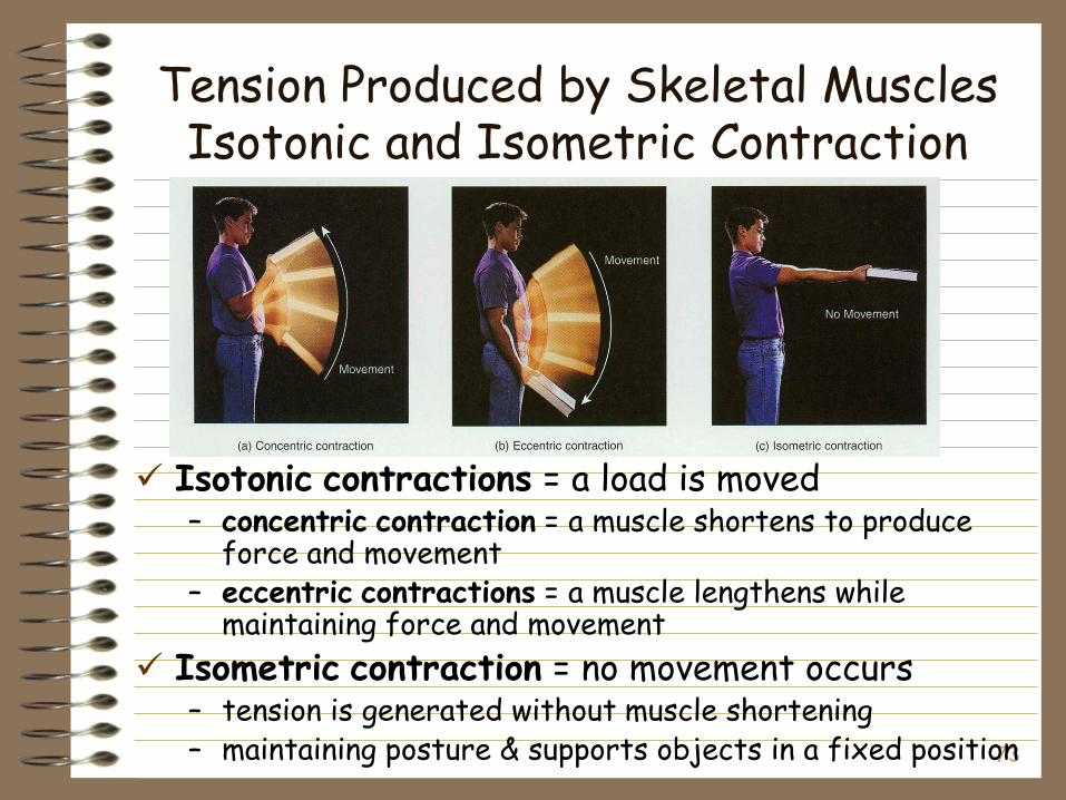
Tension Produced by Skeletal Muscles Isotonic and Isometric Contraction
Isotonic contractions = a load is moved – concentric contraction = a muscle shortens to produce
force and movement– eccentric contractions = a muscle lengthens while
maintaining force and movement
Isometric contraction = no movement occurs– tension is generated without muscle shortening– maintaining posture & supports objects in a fixed position
74
Energy Use and Muscular ActivityProduction of ATP in Muscle Fibers
Muscle uses ATP at a great rate when activeEnergy Reserves
– Stored ATP in cell• Sarcoplasmic ATP only lasts for few seconds
– Creatine phosphate– Stored Glycogen
Generation of ATP– Glycolysis (anaerobic cellular respiration)– Aerobic cellular respiration

75
Energy Use and Muscular ActivityGlycolysis
ATP produced from glucose breakdown into pyruvic acid during glycolysis – if no O2 present
• pyruvic converted to lactic acid which diffuses into the blood
Glycolysis can continue anaerobically to provide ATP for 30 to 40 seconds of maximal activity
76
Energy Use and Muscular ActivityAerobic Cellular Respiration
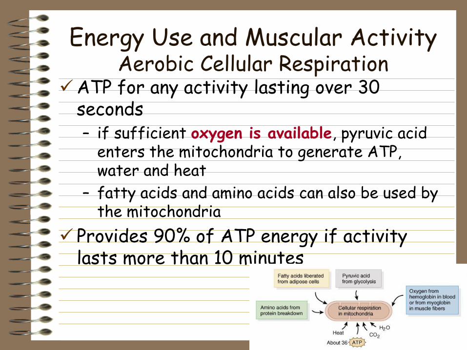
ATP for any activity lasting over 30 seconds – if sufficient oxygen is available, pyruvic acid
enters the mitochondria to generate ATP, water and heat
– fatty acids and amino acids can also be used by the mitochondria
Provides 90% of ATP energy if activity lasts more than 10 minutes
77
Energy Use and Level of Activity Muscle Fatigue
Muscle fatigue: physiological inability to contract– Glycogen stores are exhausted and ATP
regeneration does not match ATP use– Levels of lactic acid increase and pH
drops adversely affecting enzymes• Thereby slowing reactions
Contractures: state of continuous contraction
78
Energy Use and Level of ActivityRecovery Period
Muscle tissue has two sources of oxygen.– diffuses in from the blood– released by myoglobin inside muscle fibers
Aerobic system requires O2 to produce ATP needed for prolonged activity– increased breathing effort during exercise
Recovery oxygen uptake– Oxygen Debt: elevated oxygen use after
exercise– lactic acid is converted back to pyruvic acid
elevated body temperature
79
Muscle PerformanceVariations in Skeletal Muscle Fibers
Myoglobin, mitochondria and capillaries– red muscle fibers
• more myoglobin, an oxygen-storing reddish pigment • more capillaries and mitochondria
– white muscle fibers• less myoglobin and less capillaries give fibers their
pale color
Contraction and relaxation speeds vary– how fast myosin ATPase hydrolyzes ATP
Resistance to fatigue– different metabolic reactions used to generate
ATP
80
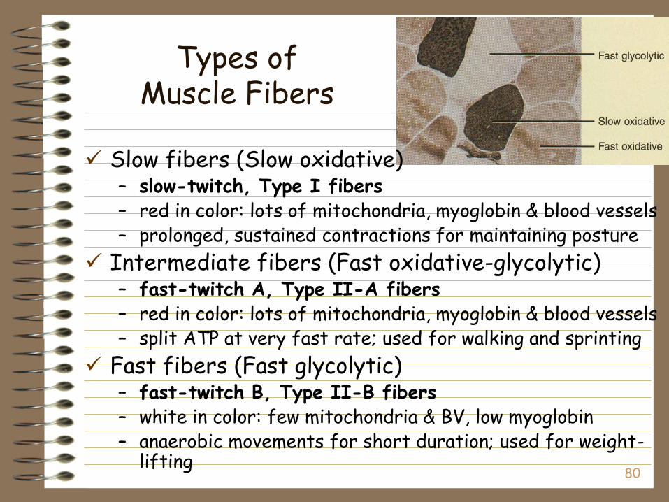
Slow fibers (Slow oxidative) – slow-twitch, Type I fibers– red in color: lots of mitochondria, myoglobin & blood vessels– prolonged, sustained contractions for maintaining posture
Intermediate fibers (Fast oxidative-glycolytic)– fast-twitch A, Type II-A fibers– red in color: lots of mitochondria, myoglobin & blood vessels– split ATP at very fast rate; used for walking and sprinting
Fast fibers (Fast glycolytic) – fast-twitch B, Type II-B fibers– white in color: few mitochondria & BV, low myoglobin– anaerobic movements for short duration; used for weight-
lifting
Types of Muscle Fibers
81
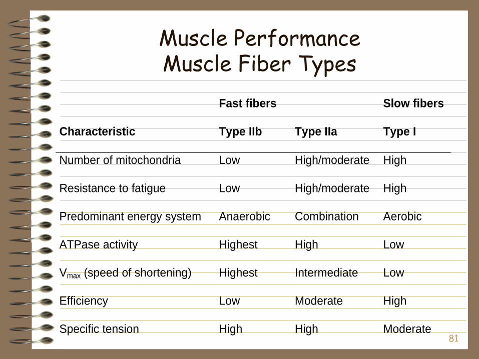
Muscle PerformanceMuscle Fiber Types
Fast fibers Slow fibers
Characteristic Type IIb Type IIa Type I
Number of mitochondria Low High/moderate High
Resistance to fatigue Low High/moderate High
Predominant energy system Anaerobic Combination Aerobic
ATPase activity Highest High Low
Vmax (speed of shortening) Highest Intermediate Low
Efficiency Low Moderate High
Specific tension High High Moderate
82
Fiber Types and Performance
Power athletes – Sprinters– Possess high percentage of fast fibers
Endurance athletes – Distance runners– Have high percentage of slow fibers
Others– Weight lifters and nonathletes– Have about 50% slow and 50% fast fibers
83
Muscle Performance Atrophy and Hypertrophy
Atrophy– wasting away of muscles– caused by disuse (disuse atrophy) or severing of
the nerve supply (denervation atrophy)– the transition to connective tissue can not be
reversed
Hypertrophy– increase in the diameter of muscle fibers – resulting from very forceful, repetitive
muscular activity and an increase in myofibrils, SR & mitochondria
84
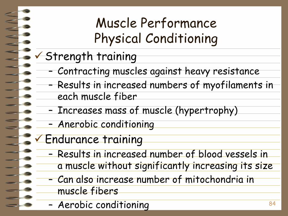
Muscle PerformancePhysical Conditioning
Strength training– Contracting muscles against heavy resistance
– Results in increased numbers of myofilaments in each muscle fiber
– Increases mass of muscle (hypertrophy)
– Anerobic conditioning
Endurance training– Results in increased number of blood vessels in
a muscle without significantly increasing its size
– Can also increase number of mitochondria in muscle fibers
– Aerobic conditioning

85
Cardiac Muscle
86
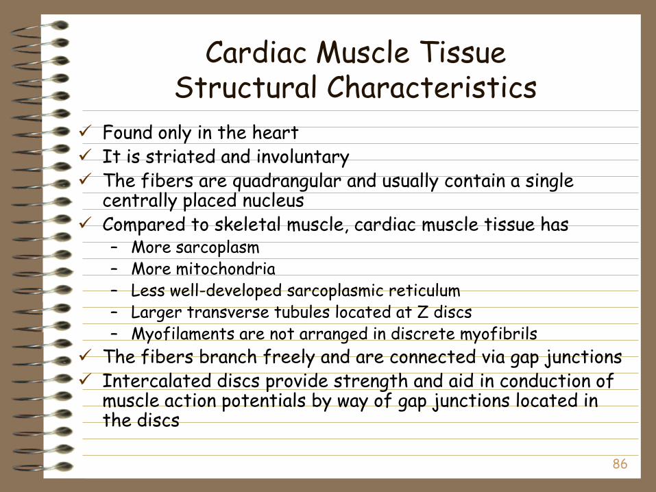
Cardiac Muscle TissueStructural Characteristics
Found only in the heart It is striated and involuntary The fibers are quadrangular and usually contain a single
centrally placed nucleus Compared to skeletal muscle, cardiac muscle tissue has
– More sarcoplasm– More mitochondria– Less well-developed sarcoplasmic reticulum– Larger transverse tubules located at Z discs– Myofilaments are not arranged in discrete myofibrils
The fibers branch freely and are connected via gap junctions Intercalated discs provide strength and aid in conduction of
muscle action potentials by way of gap junctions located in the discs
87
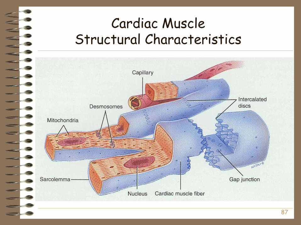
Cardiac MuscleStructural Characteristics
89
Cardiac Muscle TissueFunctional Characteristics
Unlike skeletal muscle tissue, cardiac muscle tissue contracts and relaxes rapidly, continuously, and rhythmically– ATP is generated aerobically in large, numerous
mitochondria
Cardiac muscle can contract without extrinsic (outside) stimulation and can remain contracted longer than skeletal muscle tissue
Cardiac muscle has a long refractory period that allows time for the heart to relax between beats and which prevents tetanus
90
Smooth Muscle
91
Smooth Muscle TissueStructural Characteristics
Smooth muscle tissue is non-striated and involuntary
Smooth muscle fibers are considerably smaller than skeletal muscle fibers and are thickest at the center, tapering at the ends– Each fiber contains a single, oval, centrally located
nucleus and thick and thin myofilaments, although the arrangement of the myofilaments is not in orderly sarcomeres as in skeletal muscle tissue
Smooth muscle fibers contain intermediate filaments and dense bodies (which function as Z discs)
92
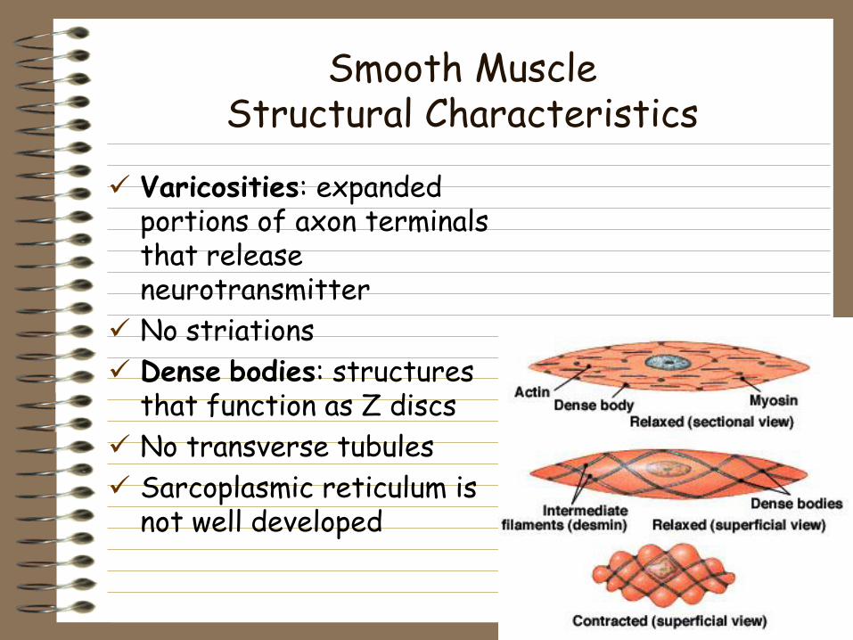
Smooth MuscleStructural Characteristics
Varicosities: expanded portions of axon terminals that release neurotransmitter
No striations
Dense bodies: structures that function as Z discs
No transverse tubules
Sarcoplasmic reticulum is not well developed
93
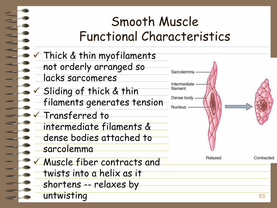
Smooth MuscleFunctional Characteristics
Thick & thin myofilaments not orderly arranged so lacks sarcomeres
Sliding of thick & thin filaments generates tension
Transferred to intermediate filaments & dense bodies attached to sarcolemma
Muscle fiber contracts and twists into a helix as it shortens -- relaxes by untwisting
94
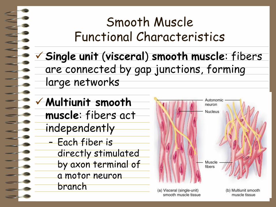
Smooth MuscleFunctional Characteristics
Single unit (visceral) smooth muscle: fibers are connected by gap junctions, forming large networks
Multiunit smooth muscle: fibers act independently– Each fiber is
directly stimulated by axon terminal of a motor neuron branch
95
Smooth MuscleFunctional Characteristics
Contraction starts slowly & lasts longer– no transverse tubules & very little SR
– Ca+2 must flows in from outside
Calmodulin replaces troponin– Ca+2 binds to calmodulin turning on an enzyme
(myosin light chain kinase) that phosphorylates the myosin head so that contraction can occur
– enzyme works slowly, slowing contraction
96
Smooth Muscle Tone
Ca+2 moves slowly out of the cell– delaying relaxation and providing for
state of continued partial contraction
– sustained long-term
Useful for maintaining blood pressure or a steady pressure on the contents of GI tract

97
Regulation of Contraction
Regulation of contraction due to – nerve signals from autonomic nervous system– changes in local conditions (pH, O2, CO2,
temperature & ionic concentrations)– hormones (epinephrine -- relaxes muscle in
airways & some blood vessels)
Stress-relaxation response– when stretched, initially contracts & then
tension decreases to what is needed– stretch hollow organs as they fill & yet
pressure remains fairly constant– when empties, muscle rebounds & walls firm up
99
Regeneration of Muscle Tissue
Skeletal muscle fibers cannot divide and have limited powers of regeneration– Growth after the first year is due to
enlargement of existing cells, rather than an increase in the number of fibers (although new individual cells may be derived from satellite cells)
Cardiac muscle fibers cannot divide or regenerate
Smooth muscle fibers have limited capacity for division and regeneration
100
Aging and Muscle Tissue
Aging– Beginning at about 30 years of age there is a progressive
loss of skeletal muscle, which is replaced by fat
– There is also a decrease in maximal strength and a slowing of muscle reflexes
Developmental anatomy of the muscular system– With few exceptions, muscle develop from mesoderm
– Skeletal muscles of the head and extremities develop from general mesoderm; The remainder of the skeletal muscles develop from the mesoderm of somites
101
Abnormal Contractions
Spasm: involuntary contraction of single muscle Cramp: a painful spasm Tic: involuntary twitching of muscles normally
under voluntary control--eyelid or facial muscles
Tremor: rhythmic, involuntary contraction of opposing muscle groups
Fasciculation: involuntary, brief twitch of a motor unit visible under the skin
Paralysis: loss of voluntary movement– Flaccid paralysis: paralysis with loss of muscle tone– Spastic paralysis: paralysis with rigidity