muscle fibers muscle tissue and muscles• thick filament • thin filament thin filaments • thin...
TRANSCRIPT

1
Muscle Tissue and Muscles
Anatomy
Muscle Fibers• Each fiber is surrounded by connective tissue called
endomysium • Fibers are closely associated with blood vessels
(capillaries), lymph vessels, and nerves• Each fiber has at least one neuromuscular junction • Contraction is monitored by proprioceptors (such as
Golgi tendon organs and muscle spindles)• “All-or-none” principle applies to contraction• Fiber recruitment (or the lack of) allows for changes
in degree of tension produced by the whole muscle
Muscle Fiber Anatomy Identify• A-band• Axon• Axon terminals• I-band• Motor end-plate• Myofibrils• Neuromuscular junction• Sarcolemma• Sarcomere• Z disc (line)
Fiber Structure
Identify• Horizontal tubules
of SR• Myofibrils• T-tubule• Terminal cisterna
(lateral sacs) of SR• Triad
2
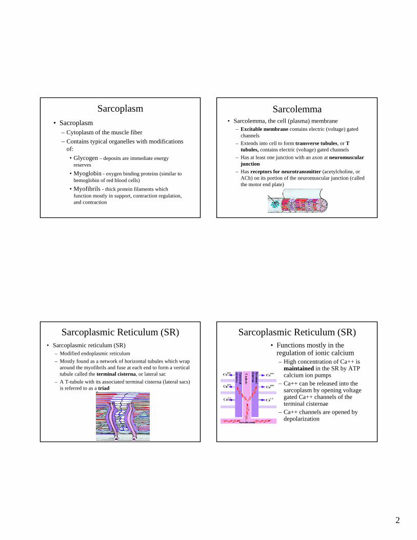
Sarcoplasm• Sacroplasm
– Cytoplasm of the muscle fiber– Contains typical organelles with modifications
of:• Glycogen – deposits are immediate energy
reserves
• Myoglobin - oxygen binding proteins (similar to hemoglobin of red blood cells)
• Myofibrils - thick protein filaments which function mostly in support, contraction regulation, and contraction
Sarcolemma• Sarcolemma, the cell (plasma) membrane
– Excitable membrane contains electric (voltage) gated channels
– Extends into cell to form transverse tubules, or T tubules, contains electric (voltage) gated channels
– Has at least one junction with an axon at neuromuscular junction
– Has receptors for neurotransmitter (acetylcholine, or ACh) on its portion of the neuromuscular junction (called the motor end plate)
Sarcoplasmic Reticulum (SR)• Sarcoplasmic reticulum (SR)
– Modified endoplasmic reticulum– Mostly found as a network of horizontal tubules which wrap
around the myofibrils and fuse at each end to form a vertical tubule called the terminal cisterna, or lateral sac
– A T-tubule with its associated terminal cisterna (lateral sacs) is referred to as a triad
Sarcoplasmic Reticulum (SR)• Functions mostly in the
regulation of ionic calcium– High concentration of Ca++ is
maintained in the SR by ATP calcium ion pumps
– Ca++ can be released into the sarcoplasm by opening voltage gated Ca++ channels of the terminal cisternae
– Ca++ channels are opened by depolarization
3
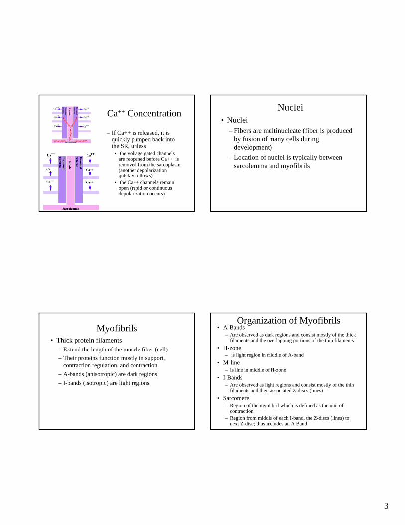
Ca++ Concentration
– If Ca++ is released, it is quickly pumped back into the SR, unless
• the voltage gated channels are reopened before Ca++ is removed from the sarcoplasm(another depolarization quickly follows)
• the Ca++ channels remain open (rapid or continuous depolarization occurs)
Nuclei• Nuclei
– Fibers are multinucleate (fiber is produced by fusion of many cells during development)
– Location of nuclei is typically between sarcolemma and myofibrils
Myofibrils• Thick protein filaments
– Extend the length of the muscle fiber (cell)– Their proteins function mostly in support,
contraction regulation, and contraction– A-bands (anisotropic) are dark regions– I-bands (isotropic) are light regions
Organization of Myofibrils• A-Bands
– Are observed as dark regions and consist mostly of the thick filaments and the overlapping portions of the thin filaments
• H-zone– is light region in middle of A-band
• M-line– Is line in middle of H-zone
• I-Bands– Are observed as light regions and consist mostly of the thin
filaments and their associated Z-discs (lines)• Sarcomere
– Region of the myofibril which is defined as the unit of contraction
– Region from middle of each I-band, the Z-discs (lines) to next Z-disc; thus includes an A Band
4
Fiber Structure
Identify• A-band• H-zone• I-band• M-line• Sarcomere• Thick filament• Thin filament
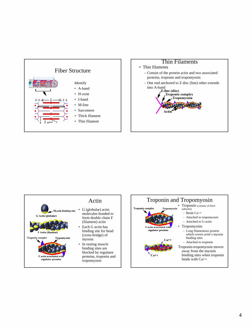
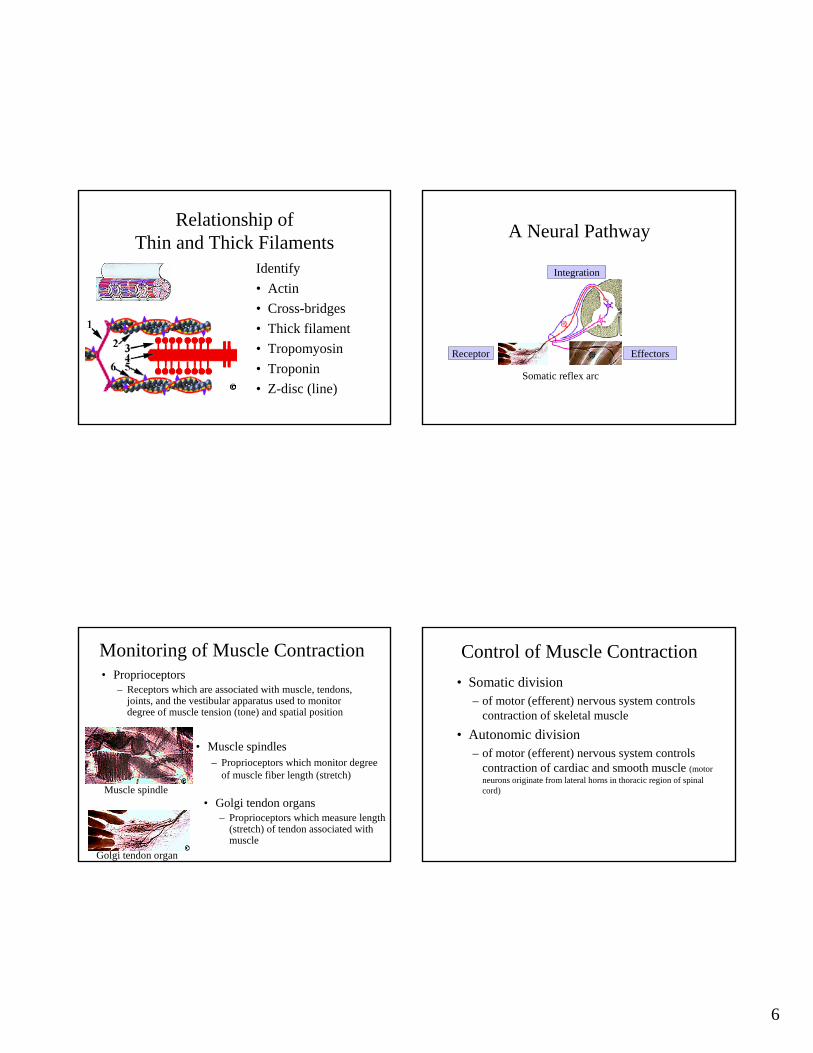
Thin Filaments• Thin filaments
– Consist of the protein actin and two associated proteins, troponin and tropomyosin
– One end anchored to Z disc (line) other extends into A-band
Actin• G (globular) actin
molecules bonded to form double chain F (filament) actin
• Each G actin has binding site for head (cross-bridge) of myosin
• In resting muscle binding sites are blocked by regulator proteins, troponin and tropomyosin
Troponin and Tropomyosin• Troponin (consists of three
subunits)– Binds Ca++– Attached to tropomyosin– Attached to G-actin
• Tropomyosin– Long filamentous protein
which covers actin’s myosin binding sites
– Attached to troponinTroponin-tropomyosin moves
away from the myosin binding sites when troponin binds with Ca++
5
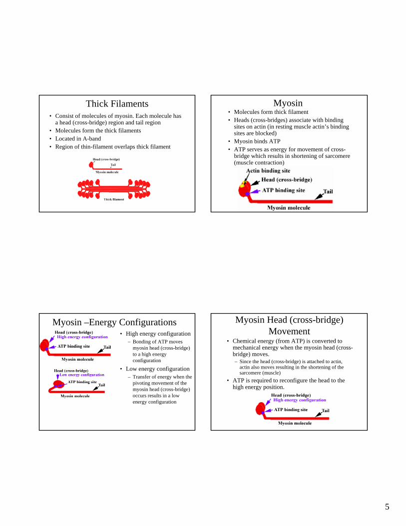
Thick Filaments • Consist of molecules of myosin. Each molecule has
a head (cross-bridge) region and tail region• Molecules form the thick filaments• Located in A-band• Region of thin-filament overlaps thick filament
Myosin• Molecules form thick filament• Heads (cross-bridges) associate with binding
sites on actin (in resting muscle actin’s binding sites are blocked)
• Myosin binds ATP• ATP serves as energy for movement of cross-
bridge which results in shortening of sarcomere (muscle contraction)
Myosin –Energy Configurations• High energy configuration
– Bonding of ATP moves myosin head (cross-bridge) to a high energy configuration
• Low energy configuration– Transfer of energy when the
pivoting movement of the myosin head (cross-bridge) occurs results in a low energy configuration
Myosin Head (cross-bridge) Movement
• Chemical energy (from ATP) is converted to mechanical energy when the myosin head (cross-bridge) moves.– Since the head (cross-bridge) is attached to actin,
actin also moves resulting in the shortening of the sarcomere (muscle)
• ATP is required to reconfigure the head to the high energy position.
6
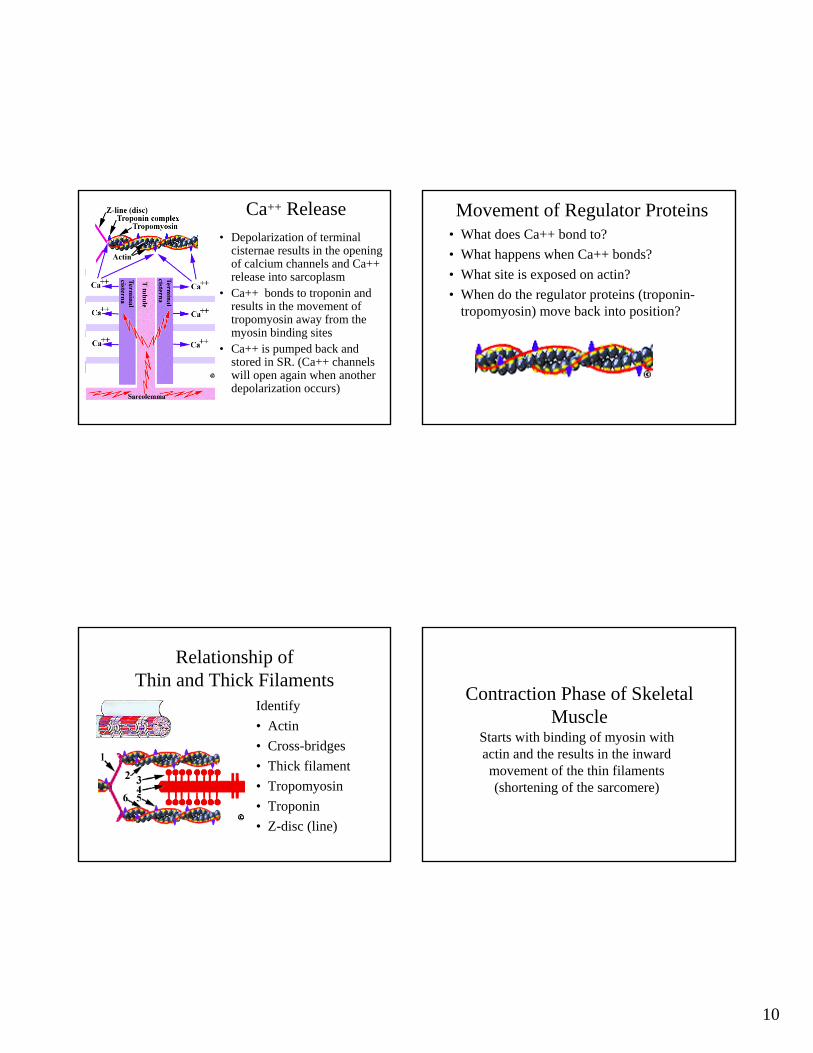
Relationship of Thin and Thick Filaments
Identify• Actin• Cross-bridges • Thick filament• Tropomyosin• Troponin• Z-disc (line)
A Neural Pathway
Somatic reflex arc
Receptor
Integration
Effectors
Monitoring of Muscle Contraction
• Muscle spindles– Proprioceptors which monitor degree
of muscle fiber length (stretch)Muscle spindle
Golgi tendon organ
• Proprioceptors– Receptors which are associated with muscle, tendons,
joints, and the vestibular apparatus used to monitor degree of muscle tension (tone) and spatial position
• Golgi tendon organs – Proprioceptors which measure length
(stretch) of tendon associated with muscle
Control of Muscle Contraction• Somatic division
– of motor (efferent) nervous system controls contraction of skeletal muscle
• Autonomic division – of motor (efferent) nervous system controls
contraction of cardiac and smooth muscle (motor neurons originate from lateral horns in thoracic region of spinal cord)
7
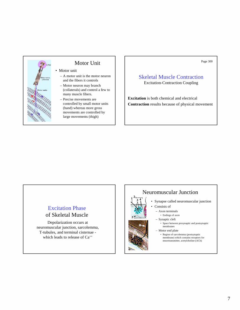
Motor Unit• Motor unit
– A motor unit is the motor neuron and the fibers it controls
– Motor neuron may branch (collaterals) and control a few to many muscle fibers.
– Precise movements are controlled by small motor units (hand) whereas more gross movements are controlled by large movements (thigh)
Skeletal Muscle ContractionExcitation-Contraction Coupling
Excitation is both chemical and electricalContraction results because of physical movement
Page 300
Excitation Phaseof Skeletal MuscleDepolarization occurs at
neuromuscular junction, sarcolemma, T-tubules, and terminal cisternae -
which leads to release of Ca++
Neuromuscular Junction• Synapse called neuromuscular junction• Consists of
– Axon terminals• Endings of axon
– Synaptic cleft• Space between presynaptic and postsynaptic
membranes
– Motor end plate• Region of sarcolemma (postsynaptic
membrane) which contains receptors for neurotransmitter, acetylcholine (ACh)

8
Neural Synapse (EPSP)1. Action potentialarrives
2. Calcium ionchannels open
3. Calcium ions promote exocytosis of neurotransmitter acetylcholine (ACh)from presynaptic membrane. Calcium
ions are quickly removed
4. Acetylcholine (ACh) binds to its receptors on postsynaptic membrane
5. Receptors promote openingof associated Na+ channelsNa+ moves inward.
6. Inward movement of Na+ atpostsynaptic membrane producesdepolarization (EPSP)
7. Acetylcholine (ACh) isdeactivated by enzyme acetylcholinesterase (AChE)
Threshold and Action Potentials• Threshold
– Point of depolarization (stimulation) which initiates an effect (action potential)
– In this case the electrically-gated Na+ channels open, (which are adjacent to the active mechanically-gated channels).
– The mechanically gated Na+ channels become inactive
• Action potential– Not local; travels great distance– Involves electrically-gated channels– Propagated along fiber (axon)
Repolarization as K+ Moves Out• Local current opens
adjacent electrically-gated K+ channels
• K+ moves outward and repolarization occurs
• Local currents open adjacent Na+ channels
• Action potential is propagated to adjacent forward section
9
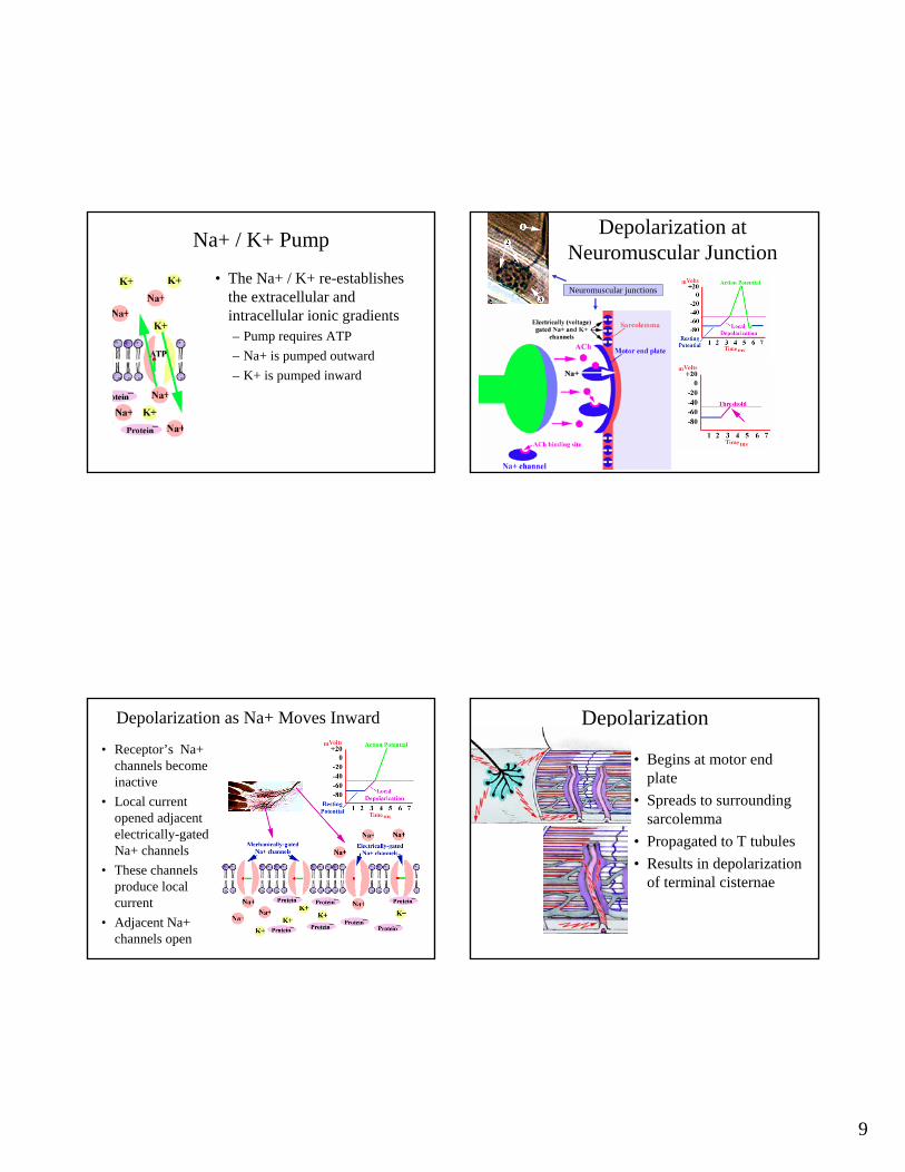
Na+ / K+ Pump
• The Na+ / K+ re-establishes the extracellular and intracellular ionic gradients – Pump requires ATP– Na+ is pumped outward– K+ is pumped inward
Neuromuscular junctions
Depolarization at Neuromuscular Junction
Depolarization as Na+ Moves Inward
• Receptor’s Na+ channels become inactive
• Local current opened adjacent electrically-gated Na+ channels
• These channels produce local current
• Adjacent Na+ channels open
Depolarization
• Begins at motor end plate
• Spreads to surroundingsarcolemma
• Propagated to T tubules• Results in depolarization
of terminal cisternae
10
Ca++ Release• Depolarization of terminal
cisternae results in the opening of calcium channels and Ca++ release into sarcoplasm
• Ca++ bonds to troponin and results in the movement of tropomyosin away from the myosin binding sites
• Ca++ is pumped back and stored in SR. (Ca++ channels will open again when another depolarization occurs)
Movement of Regulator Proteins• What does Ca++ bond to?• What happens when Ca++ bonds?• What site is exposed on actin?• When do the regulator proteins (troponin-
tropomyosin) move back into position?
Relationship of Thin and Thick Filaments
Identify• Actin• Cross-bridges • Thick filament• Tropomyosin• Troponin• Z-disc (line)
Contraction Phase of Skeletal Muscle
Starts with binding of myosin with actin and the results in the inward movement of the thin filaments (shortening of the sarcomere)
11
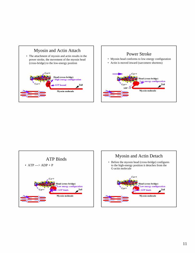
Myosin and Actin Attach• The attachment of myosin and actin results in the
power stroke, the movement of the myosin head (cross-bridge) to the low-energy position
Power Stroke• Myosin head conforms to low energy configuration• Actin is moved inward (sarcomere shortens)
ATP Binds• ATP ---> ADP + P
Myosin and Actin Detach• Before the myosin head (cross-bridge) configures
to the high-energy position it detaches from the G-actin molecule

12
Myosin Configures to High-Energy Position
• Binding of ATP produces a change in the shape of the myosin head (cross-bridge)
Myosin and Actin Attach• Myosin can now attach to the next G-actin molecule• Attachment will produce the next power stroke
Contraction Continues
• For as long as– Ca++ concentration is high and troponin-tropomyosin
complex does not block myosin binding sites– ATP is supplied to permit
• Detachment of myosin from G-actin• Energize myosin head
– Fatigue does not occur• pH change (lactic acid)• Decrease in ATP• Ionic imbalance• Nutritional imbalance
Contraction Ends• If another rapid
depolarization does not occur– Ca++ pumped into SR– Troponin-tropomyosin
complex moves back into blocking position
Thus, myosin can not bind with actin

13
Resting Fiber• Myosin can not interact with actin until Ca++
bonds to troponin resulting in removal of troponin-tropomyosin blockade
Review Animations
Myosin Binds with Actin• The tension (force of contraction) which the
fiber (cell) develops depends upon ?• What causes the movement of the head (cross-
bridge)?
Myosin Releases from Actin• When does myosin release from actin?• When does myosin configure to its high
energy position?

14
Sliding of Thin Filament• List the steps involved in the “ratcheting”
inward of thin filament:
Muscle Contraction
Myograms