murdoch research repositoryresearchrepository.murdoch.edu.au/id/eprint/13061/1/hind...macropus,...
TRANSCRIPT

MURDOCH RESEARCH REPOSITORY
This is the author’s final version of the work, as accepted for publication following peer review but without the publisher’s layout or pagination.
The definitive version is available at http://dx.doi.org/10.1071/ZO12059
Warburton, N.M., Yakovleff, M. and Malric, A. (2012) Anatomical adaptations of the hind limb musculature of tree-kangaroos for arboreal locomotion (Marsupialia : Macropodinae). Australian
Journal of Zoology, 60 (4). pp. 246-258.
http://researchrepository.murdoch.edu.au/13061/
Copyright: © 2012 CSIRO
It is posted here for your personal use. No further distribution is permitted.
1
Short running page heading: Tree-kangaroo hind limb muscular anatomy
Title: Anatomical adaptations of the hind limb musculature of tree-kangaroos for
arboreal locomotion (Marsupialia: Macropodinae)
Natalie M. WARBURTON1, 2*
, Maud YAKOVLEFF3 and Auréline MALRIC
3
1School of Veterinary and Biomedical Science, Murdoch University, South Street, Murdoch,
Western Australia 6150.
2Department of Earth and Planetary Sciences, Western Australian Museum.
3L’Ecole Nationale Veterinaire de Toulouse, France.
* Corresponding author: [email protected]
Telephone: +61 8 9360 7658
Fax: +61 8 9360 4144
Pages: 36
Figures: 8
2
Abstract
Tree-kangaroos (Dendrolagini) are Australasian marsupials that inhabit tropical forests of far
northeastern Queensland and New Guinea. The secondary adaptation of tree-kangaroos to an
arboreal lifestyle from a terrestrial heritage offers an excellent opportunity to study the
adaptation of the musculoskeletal system for arboreal locomotion, particularly from a
template well-adapted to terrestrial bipedal saltation. . We present a detailed descriptive study
of the hind limb musculature of Lumholtz’s tree-kangaroo (D. lumholtzi) in comparison to
other macropodines to test whether the hind limb musculature of tree-kangaroos is
functionally adapted to the different mechanical demands of locomotion in the uneven three-
dimensional arboreal environment. The hind limb musculature of Lumholtz’s tree-kangaroo
(Dendrolagus lumholtzi), the western brush wallaby (Macropus irma), the western grey
kangaroo (Macropus fuliginosus) and the quokka (Setonix brachyurus) are described. The
hind limb anatomy of D. lumholtzi differed from that of the terrestrial macropodines in that
the muscles had a greater degree of internal differentiation, relatively longer fleshy bellies and
very short, stout tendons of insertion. There was also a modified arrangement of muscle
origins and insertions that enhance mechanical advantage. Differences in the relative
proportions of the hind limb muscle mass between tree-kangaroos and terrestrial
macropodines reflect adaptation of the limb musculature of tree-kangaroos for arboreal
locomotion. The hind limb musculature of Setonix was different to both Dendrolagus and
Macropus, possibly reflecting its more basal phylogenetic position within the Macropodinae.
Key words: Dendrolagus, marsupial evolution, myology, Setonix
Introduction
Adaptation to an arboreal lifestyle has taken place in a number of mammalian groups
including Primates, Carnivora, ‘Insectivora’, Rodentia, Xenarthra and various families of
Marsupialia (Dublin 1903). The uneven and discontinuous nature of the arboreal realm
imposes strong selective pressure on animals towards maintaining contact with the substrate,

3
maintaining balance, and providing propulsion on flexible, discontinuous and inclined branch
substrates (Grand 1990). Small arboreal mammals (<3kg), including primates, have similarly
flexed limb posture and movements which effectively lower the centre of mass for improved
balance (Schmidt 2005a). Larger canopy-dwelling primates are often recognisable as being
particularly acrobatic, and many demonstrate highly specialised use of the forelimbs for
suspensory behaviours (Fleagle 1999). It has been noted, however, that primate morphology
and locomotion are fundamentally different to other mammals and reflects forelimb
adaptations in basal primates for locomotion on fine-branches (Jungers 1984; Schmitt and
Lemelin 2002). Other large arboreal mammals, for examplesloth (Bradypus spp. and
Choloepus spp.) binturong (Arctictis binturong) and koala (Phascolarctos cinereus), tend to
move relatively slowly and deliberately, and employ prehensile digits and/or large, grasping
claws to provide increased resistance to slipping and to help maintain contact with the
substrate (Dublin 1903; Cartmill 1979; Grand 1990a; Argot 2001). A long, muscular, non-
prehensile tail may be employed as a counter balance; or a prehensile tail may function as a
grasping organ (Organ, Teaford et al. 2009; Organ 2010). On the whole, climbers are less
muscular than runners, reflecting the reduced value of propulsive force as a function of
branch compliance (Grand 1983; Grand 1990b). Instead, adaptations of the musculoskeletal
system typically reflect enhanced flexibility of the joints for enhanced reaching and stepping
movement, and improved mechanical advantage of limb adductor and digital flexor muscles
to facilitate grasping of tree trunks or branches (Argot 2001). Further, a greater balance
between pushing and pulling motions results in a trend towards a more even distribution of
muscle mass between the fore- and hind-limbs in arboreal groups when compared to their
terrestrial counterparts (Grand 1990b). Morphological variation between related species
reflects the degree to which species inhabit the canopy (Dublin 1903).
Taxonomically, tree-kangaroos are placed within the marsupial subfamily Macropodinae,
which includes kangaroos, wallabies and related taxa including the quokka (Setonix
brachyurus). Tree-kangaroos evolved during the Late Miocene from a common ancestor with

4
rock-wallabies (Flannery 1989; Prideaux and Warburton 2010). Extinct plesiomorphic tree-
kangaroos Bohra spp. have been described from a number of Pliocene and Pleistocene fossil
deposits in Australia (Flannery and Szalay 1982; Dawson 2004; Hocknull 2005; Prideaux and
Warburton 2008; Prideaux and Warburton 2009). There are ten species of extant tree-
kangaroos (Dendrolagus) that are classified into two groups on the basis of morphology. The
‘long-footed group’ includes the two Australian species (D. bennettianus and D. lumholtzi),
together with D. inustus (of New Guinea); the remaining seven New Guinean species
comprise the ‘short-footed group’ (Flannery 1989). The long-footed group of tree-kangaroos
is apparently plesiomorphic, and thus likely to more closely approximate the hypothetical
ground-dwelling ancestor (Grand 1990a; Flannery, Martin et al. 1996). Morphological
variation within these groups (Warburton and Prideaux 2010) is likely to be tied to both
phylogenetic and behavioural patterns, and for the short-footed group may reflect endemic
species in highly localised regions of New Guinea (Flannery 1990). More detailed analysis of
osteological differences between living and extinct species of tree-kangaroos will rely on
investigations of the muscular anatomy of these species.
The musculoskeletal anatomy of Doria’s tree-kangaroo (D. dorianus) was documented in an
early monograph (Carlsson 1914). More recently, observations of captive and, occasionally,
wild animals have highlighted specialised behaviours in comparison to terrestrial kangaroos.
Tree-kangaroos utilise a wide variety of behaviours and locomotor patterns (Windsor and
Dagg 1971; Procter-Gray and Ganslosser 1986), and have variable adaptations for manual
dexterity and manipulation (Procter-Gray and Ganslosser 1986; Iwaniuk, Nelson et al. 1998).
Grand (1990) summarised the principal adaptations of tree-kangaroos for improved stability
and propulsion suited to arboreality. Tree-kangaroos have more equal length and muscle
strength between limbs and limb segments than terrestrial kangaroos (Grand 1990a). Their
feet are short and broad, more plantigrade in posture, and have roughened palmar and volar
surfaces and enlarged, powerful claws for enhanced frictional resistance (Carlsson 1914).
They also have a reduced lumbar length and decreased flare of the iliac crest. When moving

5
along branches, tree-kangaroos employ a modified low-speed, alternating gait (Windsor and
Dagg 1971). Dendrolagus has a reduced proportion of muscle mass to total body weight (less
than three-quarters of the muscle weight of kangaroos), reduced epaxial musculature, and
more equal ratios of forelimb to hind limb muscle and bone in comparison to terrestrial
macropods (Grand 1990). These adaptations to the muscular system reflect changes to the
centre of gravity, strength-to-weight ratio and muscular actions through the body and are
convergent with other slow-moving arboreal climbers (Grand 1990). Grand (1990) also noted
that mobility of the shoulder and hip joints was enhanced in Dendrolagus spp., and that
forearms, calves, paws, and feet were relatively heavier, although no empirical data were
presented.
Previous studies have alluded to likely adaptations of the muscular system of tree-kangaroos
for an arboreal lifestyle. While the overall distribution of muscle mass within the body is
suggestive (Grand 1990), it does not allow interpretation of differences in muscular system
function. We might expect, for example, differences in the muscular attachments to improve
mechanical advantage or manoeuvrability for locomotion in an arboreal setting. Here we
present a detailed descriptive study of the hind limb musculature of Lumholtz’s tree-kangaroo
(D. lumholtzi) in comparison to other macropods to test whether the hind limb musculature of
tree-kangaroos is functionally adapted to the different mechanical demands of locomotion in
the uneven three-dimensional arboreal environment. To this end, we undertook to answer the
following questions: 1) what is the anatomical arrangement of the hind limb muscles of
Lumholtz’s tree-kangaroo, 2) how does arrangement of the muscles compare with that of
terrestrial taxa of the subfamily Macropodinae with respect to the bony attachments and
distribution of muscle mass within functional muscle groups, and 3) to what extent do the
differences observed between species reflect functional adaptations for climbing in tree-
kangaroos? This information was then used to elucidate some of the functional patterns
observed in the osteology of living and extinct tree-kangaroos.

6
Materials and Methods
An adult male road-killed specimen of Lumholtz’s tree-kangaroo Dendrolagus lumholtzi from
the Atherton Tableland region was made available for dissection by the Environmental
Protection Authority, Queensland. Although this species is reportedly common in suitable
habitat (Winter, Burnett et al. 2008)few specimens are collected and made available for
anatomical dissection. Thus, this study is based on the dissection of only one specimen.
However, in our experience, differences between species are generally greater than the
variation observed within (e.g., Harvey and Warburton, 2010). The specimen was skinned,
eviscerated and embalmed in 10% formalin 4% glycerol solution for one week, before being
stored in 70% ethanol. Standard dissecting techniques were used. Muscles were identified and
their attachments recorded. Muscle actions were determined by observation of the
arrangement of the muscle fibres in relation to their position of origin and insertion relative to
the bony articulation across which their pass.
Descriptions of macropod musculature found in the published literature were supplemented
with our own new dissections and descriptions of the western grey kangaroo (Macropus
fuliginosus), the western brush wallaby (M. irma) and the quokka (Setonix brachyurus). One
specimen of each species was obtained for dissection: M. fuliginous purchased from King
River International abattoir, Canning Vale WA; M. irma and S. brachyurus were collected as
victims of road-incidents and donated for study by Western Australian Department of
Environment and Conservation. These specimens were dissected fresh, following thawing and
the muscle attachments were recorded.
The following published accounts of macropod musculature reviewed for comparative
interpretation: Aepyprymnus rufescens (Lodder 1991), Dendrolagus dorianus (Carlsson
1914), Hypsiprymnodon moschatus (Carlsson 1915), Macropus spp. (Windle and Parsons
1898; Elftman 1929; Badoux 1965; Hopwood and Butterfield 1976; Hopwood and Butterfield
1990; Lodder 1991; Bishop 1997) and Petrogale xanthopus (Parsons 1896).

7
Results
Extrinsic muscles of the pelvic limb
M. iliacus arose from the iliac fossa on the entire ventral aspect of the body of the ilium (Figs
1A, 2B). M. psoas major originated from the ventral surface of the body and the transverse
processes of the last three lumbar vertebrae. The two muscles joined at the level of the
acetabulum to insert together to the lesser trochanter of the femur and medial aspect of the
neck of the femur (Fig 3C). Together these muscles act to flex the hip joint. The bony
attachments of the iliopsoas appear similar throughout the macropods we observed (M. irma,
M. fuliginosus, S. brachyurus) and also in the literature (Hopwood & Butterfield 1976).
Gluteal region
M. gluteus superficialis (m. gluteus maximus) originated from the thick lumbar fascia
adjacent to the dorso-medial border of the ilium and inserted to the caudal aspect of the
greater trochanter, and proximal fascia of the biceps (Figs 2A, 3B, C). A small, additional
portion inserted into the proximo-caudal fascia of the M. vastus lateralis. This muscle would
act as an extensor and abductor of the hip joint. In M. irma the origin included a portion from
the crest of the ilium, together with the gluteofemoralis, in addition to the thoracolumbar
fascia, and the insertion included a strong aponeurotic connection to the fascia of the vastus
lateralis at the position of the caudo-lateral aspect of the greater trochanter of the femur. As in
M. giganteus (Hopwood and Butterfield 1976), fibres of the gluteofemoralis blended with the
superficial gluteal. In S. brachyurus, the origin was from the thoracolumbar fascia only.
M. gluteofemoralis (m. caudofemoralis, m. femorococcygeus, m. agitator caudae)
M. gluteofemoralis comprised three distinct portions; a large superficial portion, had the most
caudal origin from the transverse processes of the third and fourth caudal vertebrae; the
middle portion (incompletely separable into two heads) from the transverse processes of the

8
third caudal vertebrae and from the deep caudolumbar fascia; and the third small, deep
portion from the deep lumbar fascia together with the superficial gluteal muscle. Each fleshy
belly gave rise to a tendon halfway along the length of the femur which converged to insert
laterally to the capsule of the knee and proximo-lateral aspect of the patelloid (Fig 2A). The
small, deep most cranial portion lay on the deep surface of the middle portion and had a
common tendon. The gluteofemoralis functions to abduct femur and extend the hip joint.
The gluteofemoralis was more differentiated in D. lumholtzi with three distinct portions rather
than the usual two. In M. irma the cranial portion had a broad origin from the crest of the
ilium and the thoraco-lumbar fascia, together with the superficial gluteal. The caudal portion
was very long from the caudolumbar fascia and the transverse process of the caudal vertebrae
III, IV. The tendons united two-thirds of the length of the femur and insert together to
patelloid. The insertion extends more distally in D. lumholtzi than in other macropodine taxa.
In M. fuliginosus caudal head originated from the caudal vertebral transverse processes III, IV
and V; the cranial head was very difficult to separate from the superficial gluteus at the origin.
A small, incompletely separable portion was observed lying between the cranial and caudal
heads, originating with the cranial head from the crest of the ilium. Lodder (1991) described
cranial and caudal heads in Aepyprymnus and M. giganteus, as did Hopwood and Butterfield
(1976). The cranial head appeared relatively larger in M. fuliginosus than in M. irma. In S.
brachyurus there was no origin from the ilium. Carlsson (1915) describes two distinct
portions (m. femorococcygeus and m. caudofemoralis) which apparently have a more
proximal origin in H. moschatus .
M. gluteus medius
M. gluteus medius had two large and distinct portions (Figs 1, 2A). The superior, caudal
portion extended along lateral border of the ilium from the iliac crest to the caudal dorsal iliac
spine. The inferior, cranial portion originated from the crest of the ilium and gluteal fascia.
The cranial head inserted to the cranial aspect of the greater trochanter; the caudal portion

9
inserted to the caudolateral aspect of the greater trochanter by a strong, tendinous fibres (Figs
3A, B, C). This muscle acts to extend the hip joint.
In D. lumholtzi the two portions of the gluteus medius were very distinct; the large portion of
the deep gluteal muscle laid between the two portions of the gluteus medius (Figs 1A, B). In
M. irma two portions were present; a large, thick portion from the tuberosity of the ilium and
a thinner caudal portion from along the dorsolateral crest of the ilium. Both portions inserted
over the proximal aspect of the greater trochanter of the femur. Two portions have also been
described for M. giganteus (Hopwood & Butterfield 1976) and Petrogale (Parsons 1896).
Carlsson (1915) suggested that the middle gluteal was often arranged in two layers within
marsupials, but not in H. moschatus. In S. brachyurus, the two portions were not separable
from one another.
M. gluteus profundus
M. gluteus profundus was a very large muscle from the large gluteal fossa covering the lateral
aspect of the wing of the ilium (Figs 1A, B). It had a strong insertion with superficial
tendinous fibres and deep fleshy fibres to the proximal crest greater trochanter (Figs 3A, B,
C). This muscle acts to extend the hip joint.
In S. brachyurus the origin was similarly from most of the elongate gluteal fossa. In M. irma
the origin was more lateral along the wing of the ilium, and this insertion more medial on the
greater trochanter. M. fuliginosus had an additional insertion with fleshy fibres to the medial
aspect of the greater trochanter.
M. gluteus minimus
M. gluteus minimus was a small muscle from a triangular section at the base gluteal fossa of
the ilium, in the quarter proximal to the acetabulum (Figs 1A, B). Insertion to the craniolateral
base of the greater trochanter (Fig 3A). This muscle contributes to extension of the hip joint.

10
In M. irma, M. fuliginosus and S. brachyurus the origin was much more extensive, from the
base of the ilium and along the entire lateral edge, along side the gluteal fossa. In each species
the insertion was longer than in D. lumholtzi, along an arc from the craniomedial aspect and
along the craniolateral line of the greater trochanter. The gluteal muscles as a group are
apparently less specialised in H. moschatus (Carlsson 1915).
M. piriformis
M. piriformis arose from the transverse process of the first and second caudal vertebrae (Fig.
2B) and inserted proximally into the trochanteric fossa (Fig 3C). This muscle acts to extend
and abduct the femur at the hip joint.
In M. fuliginosus and M. irma, the insertion was also to the crest of the greater trochanter,
immediately proximal to the intertrochanteric fossa. We observed an additional very small
slip of muscle adjacent to the piriformis from the transverse process of the second caudal
vertebra which inserted to the mid-caudal tubercle of the femur, however, we are unable to
attribute a formal name to this portion.
M. obturatorius internus
M. obturatorius internus originated from the dorsal portion of the medial face of the ischium
and inserted into the middle portion of the trochanteric fossa of the femur together with the m.
gemelli (Figs 2B, 3C). This muscle laterally rotates the femur and may also contribute to
abduction at the hip joint.
The origin does not completely surround the obturator foramen in D. lumholtzi. The muscle
appeared to be relatively small. The broad, flat tendon lies on the surface of the cranial
gemellus; it was not separable from the cranial gemellus. A similar description of the
obturator internus was given for Petrogale (Parsons 1896) and M. giganteus (Hopwood &

11
Butterfield 1976); the latter authors summarised that this muscle is generally reduced in size
in marsupials, including macropods, in comparison to other mammals.
Mm. gemelli
Mm. gemelli comprised two adjacent portions from a large area on the lateral aspect of the
ischium, between the acetabulum and the ischiatic tuberosity (Fig 1A). The insertion was to
the trochanteric fossa of the femur, distal to the insertion of the m. piriformis; immediately
proximal to the insertion of the external obturator muscle (Fig 3C). This muscle medially
rotates and abducts the femur at the hip joint.
In M. irma the ilium was relatively longer, thus the muscle appeared larger. In S. brachyurus,
the caudal belly extended onto the medial aspect of the ischiatic table.
M. quadratus femoris (m. ischiofemoralis)
M. quadratus femoris had a long, thin origin along the caudal aspect of the ischium, adjacent
to the border of the ischium, from the lateral angle of the ischiatic tuberosity and along the
ischiatic arch toward the pelvic symphysis (Fig 1A). The muscle inserted to the mid-caudal
tubercle on the femoral shaft (Fig 3C). This muscle functions as a strong extensor of the hip
joint and appeared to be relatively much larger in D. lumholtzi in comparison to the other
species examined.
In M. irma and S. brachyurus the origin was triangular in shape, filling much of the lateral
aspect of the ischiatic table; a triangular, raised fossa was observed on the bone. Hopwood
and Butterfield (1976) reported that this muscle was mostly tendinous in M. giganteus to
prevent hyperextension of the hip joint. The insertion is more proximal in H. moschatus to the
greater trochanter (Carlsson 1915).
Cranial thigh
12
M. sartorius
M. sartorius originated from the cranioventral iliac spine (tuber coxae) (Figs 1A, 2A, B). The
insertion was to the cranial aspect of the fascia of the knee and patelloid (Figs 2A, B). This
muscle flexes the hip and extends the knee.
This strap muscle, with triangular section was incompletely separable into two portions, was
similar in all macropod species examined.
Mm. quadriceps femoris
The four muscle of the quadriceps group function to extend the knee; the m. rectus femoris
also acts to flex the hip. M. rectus femoris arose from the enlarged rectus tubercle on the
lateral aspect of the ilium, cranial to the acetabulum (Figs 1A, 2B) and inserted to the cranial
border of the patelloid. M. vastus lateralis originated from the cranial, lateral and caudal
aspects of the base of the greater trochanter and proximal shaft of the femur (Figs 2A, B, 3A,
B, C) and inserted to the lateral aspect of the proximal border of the patelloid. M. vastus
medialis originated from the cranial aspect of the neck of the femur (Fig 3A) and inserted to
the medial border of the patelloid. M. vastus intermedius was a very deep muscle along the
surface of the femur, not clearly separable from the medial vastus. Its origin was from the
craniomedial, cranial and craniolateral surface of the mid three-fifths of the shaft of the femur
(Fig 3A) and it inserted to the craniolateral border of the patelloid.
All four bellies of the quadriceps femoris are closely attached; the three vasti share
aponeuroses along their adjacent borders. In S. brachyurus, the origin of the vastus lateralis
was thinner along the cranial, lateral and caudal aspects of the epiphyseal line of the greater
trochanter. Origin of the vastus lateralis in M. irma was from the caudal aspect of the greater
tubercle only. M. fuliginosus had a larger origin from the lateral surface of the greater
trochanter. In M. fuliginosus the rectus femoris was very thick and incompletely separable
into two portions by the presence of a very strong internal aponeurosis. A single head was

13
observed in M. giganteus (Hopwood & Butterfield 1976); Parsons (1896) reported two heads
for this muscle in Petrogale. Overall, the quadriceps muscle group, and especially the vastus
lateralis muscle appeared to be relatively smaller in D. lumholtzi than in the species of
Macropus dissected here.
M. tensor of the fasciae latae
M. tensor of the fascia latae was a short, thick, triangular muscle belly from the
thoracolumbar fascia (Figs 2B, 4). It inserted to the deep, cranio-medial fascia of the
quadriceps. This muscle is a tensor of the deep cranial fascia of the thigh, and thus contributes
to extension of the knee.
In M. irma, M. fuligniosus and S. brachyurus origin extended along to the ilium, from the iliac
spine to the proximal to the rectus tubercle, and inserted onto the fascia of the vastus medialis.
Medial thigh
M. pectineus
M. pectineus arose from the iliopubic eminence and triangular area of the pubic ramus medial
to acetabulum (Fig 1A). The thin insertion was along medial aspect of middle half of femur
(Fig 3A).Pectineus contributes to adduction and lateral rotation of the femur at the hip joint.
In M. irma, the origin was smaller in area, from the more distinct and protuberant iliopubic
eminence. The extent of the insertion along the caudal aspect of the femur varied. In M. irma
to the medial edge of the mid-caudal rugosity and distally along the medial shaft. In M.
fuliginosus the insertion is to the proximal third of the medial shaft of the femur. In M.
giganteus (Hopwood & Butterfield 1976) there was both a fleshy attachment on the proximal
third of the femur and an aponeurosis for the two distal thirds. In S. brachyurus the insertion
was to the proximal one-fifth of the medial femur, immediately distal to the third trochanter.

14
M. gracilis
M. gracilis took an aponeurotic origin along the length of the pelvic symphysis, and fleshy
fibres from the caudal symphyseal tubercle (Fig 1A). The flat, aponeurotic insertion was to
the medial surface superficial fascia of the crus at the level of the tibial crest (Fig 4). The
gracilis muscle acts as an adductor of the thigh.
The gracilis was relatively thick in D. lumholtzi. In M. irma and S. brachyurus the origin
extended laterally along the ischiatic arch. Division into two heads was observed in M.
fuliginosus, as previously described for M. giganteus (Hopwood & Butterfield 1976). We did
not observe an origin from the epipubic bone, as described for M. giganteus (Hopwood &
Butterfield 1976). The origin is apparently more caudally restricted in H. moschatus (Carlsson
1915).
Mm. adductores (M. adductor longus, M. adductor brevis, M. adductor magnus)
Mm. adductores arose in two portions from caudal border of the ischium and from along the
pubic symphysis (m. adductor magnus et brevis) (Figs 1A, 4). An additional small triangular
portion (m. adductor longus) had an aponeurotic origin from the pubis together with the pubic
portion above. The insertion was in three portions to the caudal aspect of the femur (Figs 3B,
C). The adductor longus inserted immediately distal the trochanteric fossa; the pubic portion
inserted to middle half of the caudal aspect of the femur; ischiatic portion to the distal fifth of
distal medial aspect of the femur, immediately proximal to the intercondylar groove. This
muscle group strongly adducts the femur at the hip joint.
In M. irma the origin was from the ventral surface of the pubis from the iliopectineal
eminence to the ilium and ventral surface of two thirds of the greater ischiatic arch and
including the mid-pubic tubercle. In M. fuliginosus the combined adductor mass (difficult to
completely separate into portions) inserts to the entire medial surface of the femoral shaft.
The insertion of the pectineus muscle separated the two portions of the adductor in M. irma

15
but not in M. fuliginosus. S. brachyurus had a similar arrangement to M. irma. During
dissection, the adductor muscle mass appeared to be relatively large in D. lumholtzi in
comparison to M. fuliginosus and M. irma.
M. obturatorius externus
M. obturatorius externus was a very large, thick muscle which encircled the ventral aspect of
the obturator foramen, and extended over a large surface of the caudolateral ischium and
pubis. It inserted deeply into the distal trochanteric fossa of the femur. The external obturator
is a lateral rotator of the femur at the hip joint. This muscle appeared to be relatively very
large in the tree-kangaroo. The insertion was closely associated with the tendons of the mm.
gemelli.
Caudal thigh
M. biceps femoris
M. biceps femoris passes from the lateral tip of the ischiatic tuberosity and the fascia covering
the semitendinosus to insert to the lateral fascia of the knee and crus covering the proximal
third of the tibia (Figs 1A, 2A). The biceps femoris functions to abduct and extend the femur
at the hip joint and extend the knee during the supporting phase (when the limb is weight-
bearing). During the swing phase (non-weight-bearing) this muscle contributes to flexion of
the knee.
In D. lumholtzi the two heads of the biceps femoris were incompletely separable. In M. irma
the large, thick cranial head arose dorsally from the ischiatic tuberosity, the sacrotuberous
ligament and fascia lata; the caudal head arose laterally from the ischial tuberosity. The
cranial portion was inserted laterally to patelloid and crural fascia and the caudal portion
inserted to crural fascia over gastrocnemius; the fascia continued distally to the common
calcaneal tendon. The two portions were contiguous along their margin and were very
strongly attached to the semitendinosus at their origin. The bony attachments were similar in

16
S. brachyurus. In M. fuliginosus the attachment between the biceps and the semitendinosus
was not as strong as in M. irma. In M. giganteus the origin was from the tuber ischii
(Hopwood & Butterfield 1976). Parsons (1896) noted additional origins from the sacrum and
caudal vertebrae in Petrogale.
M. abductor cruris caudalis (m. tenuissimus)
M. abductor cruris caudalis arose from the proximal third lateral fascia of the semitendinosus.
The insertion was to the lateral fascia of the crus, over the gastrocnemius, immediately distal
to the insertion of the m. biceps femoris. The caudal crural abductor contributes to extend the
thigh at the hip joint; it may also flex the knee joint.
M. semitendinosus
M. semitendinosus originated from the caudo-ventral aspect of the ischiatic tuberosity (Figs
1A, B, 2A, B, 4). The muscle crossed the caudal superficial aspect of the thigh and inserted
into the deep medial crural fascia, from the level of the distal end of the tibial crest and
covering a distance approximately one fifth of the length of the tibia. The fascia continued
distally to contribute to the common calcaneal tuberosity. The semitendinosus functions for
extension of the hip and knee joints, and, in non-weight bearing limb, flexion of the knee
joint.
M. semimembranosus
M. semimembranosus arose from the middle half of the ischiatic arch (Figs 1A, 2B). The
muscle was incompletely separable into two portions. The cranial head was inserted to the
medial condyle of the tibia. The caudal head was inserted to the proximo-medial aspect of the
superficial fascia of the crus and proximal end of the medial border of the tibia, at the level of
the tibial crest. The semimembranosus contributes to extension of the hip and knee joints.

17
The slightly thicker cranial portion of the semimembranosus lies along the caudal aspect of
the adductor, and is very difficult to separate. In the terrestrial macropodine taxa the origin
was limited to the middle third of the ischiatic arch. In M. fuliginosus an additional insertion
was observed to the medial epicondyle of the femur and the medial face of the tibia
immediately below the condyle of the tibia.
Cranial leg
M. tibialis cranialis
M. tibialis cranialis arose from the cranial tibial crest and proximal lateral fossa of the tibia
immediately distal to the lateral tibial condyle; proximal third of the cranial tibial shaft (Figs
5A, B, 6). The muscle passes across the cranial aspect of the ankle to insert to the plantar-
medial tubercule of the base of metatarsal II (Fig 7) and acts to dorsiflex and invert of the pes.
Reflecting the relative length of the tibia, the origin is relatively shorter in M. irma (proximal
quarter of the tibia shaft) and M. fuliginosus (proximal one fifth of tibial shaft). Tendon
passed obliquely over the lower ankle joint and inserts onto the fascia covering the medial
cuneiform (tarsal I) and the proximomedial aspect of the fourth metatarsal. An insertion to
metatarsal IV was also noted for M. giganteus (Hopwood & Butterfield 1990), though more
distally to the head of that bone, as well as to the base of metatarsal II. In S. brachyurus the
origin was restricted to the proximal third of the tibial fossa (proximal one eighth of the tibia).
M. extensor digitorum longus (EDL)
M. EDL of digits II and III was a small, superficial muscle from the proximolateral aspect of
the tibia, immediately below the epiphyseal line (Figs 5A, 6). A single tendon from the
proximal third of the leg passed through a retinaculum deep to the other digital extensors to
insert to digits II and III. The thin tendon divided at the level of the middle phalanges, one for
each toe, and inserted to the dorsal aspect of the base of the distal phalanx. The tendon to the
II and III digits has an especially deep retinaculum, separate from the two larger tendons. The

18
arrangement of the EDL is similar in S. brachyurus. This muscle is very small in the M. irma,
with the tendon from proximal fifth of the tibia only.
M. EDL of digit IV arose from the cranial aspect of the head of the fibula and proximal two-
thirds of tibia (Figs 5A, 6). The aponeurotic fibres blended into the dorsal fascia of the
proximal phalanx, together EDL IV and V, to form strong insertions to the bases of the
proximal and distal phalanges of digit IV. The fleshy belly of this muscle extends along the
proximal two-thirds of tibia in D. lumholtzi; in M. irma the fleshy portion is only on the first
third of the tibia. In S. brachyurus the origin was from the caudo-lateral aspect of the lateral
tibial condyle rather than the fibular head. A femoral origin has been reported for Petrogale
(Parsons 1896) and M. rufus (Windle & Parsons 1898), but not in M. giganteus (Hopwood &
Butterfield 1990).
M. EDL of digits IV and V arose from the cranial aspect of the head and proximal fifth of the
cranial border of the fibula, and the interosseous membrane (Fig 5A). The tendon split to
insert to the distal phalanx V and a tendon to the dorsal fascia of digit IV. Origin in M. irma
from proximal half of fibula. Origin in M. fuliginosus origin from the cranial fibula, and small
portion of the tibia, immediately distal to the extensor of digit IV; insertion traced to the distal
phalanx of digit V only. In S. brachyurus the origin was from the caudo-lateral aspect of the
tibia below the lateral condyle rather than from the fibula.
As a group, these muscles extend the phalanges and dorsiflex the foot. A strong retinaculum
holds the tendons of the extensor muscles on the dorsal aspect at the level of the lower ankle
joint. In S. brachyurus the tendons of insertion do not merge as observed in Macopus; the
EDL IV inserted to the medial side of the toe, and the EDL IV and V inserted to the lateral
side.
Lateral leg

19
M. fibularis [peroneus] longus et brevis
M. fibularis longus et brevis originated from the lateral aspect of the head and shaft of the
fibula (Figs 5A, B). Its tendon passed under the tendon of the lateral digital extensor to insert
deeply to the dorso-lateral aspect of the base of the fifth metatarsal (Fig 6). It acts to dorsiflex
and evert the pes.
The tendon was relatively thick in D. lumholtzi in comparison to other macropods. The fleshy
belly extended along the proximal third of the crus in D. lumholtzi; but was limited to the
proximal quarter of the crus in M. irma and the proximal tenth in S. brachyurus. In M.
fuliginosus the origin was from the head of the fibula only. Carlsson (1915) described four
muscles of the peroneus group in H. moschatus. Hopwood and Butterfield (1990) described
three muscles in M. giganteus, with a common origin from the head and proximal surface of
the fibula, with insertions to tarsal I, digit IV and digit V.
M. extensor digitorum lateralis
M. extensor digitorum lateralis originates from the lateral aspect of the mid fibula (Figs 5B,
C). The tendon passed under extensor retinaculum and behind the lateral malleolus to insert to
the lateral aspect of the base of the middle phalanx V (Fig 6). The muscle acts to extend and
abduct digit V and also dorsiflexes the talocrural joint.
The origin of this muscle in M. irma and S. brachyurus was from the proximal third of the
lateral fibular shaft. In M. fuliginosus the origin was from the proximal quarter of the lateral
aspect of the fibula. This muscle seems to correspond to the m. peroneus digit V described for
M. giganteus (Hopwood & Butterfield 1990).
An additional very tiny muscle with long tendon was found on lateral aspect of the EDL in D.
lumholtzi; we were unable to trace it to its insertion. It may have been the M. extensor hallucis
brevis mentioned in D. dorianus (Carlsson 1915).

20
Caudal leg
M. triceps surae
M. gastrocnemius medial head arose from the medial supracondylar fossa of the femur (Figs
3C, 7). The lateral head arose via thick aponeurotic fibres from the lateral epicondyle and the
fascia of the knee and patelloid (Figs 3B, 6).
M. soleus lay on the deep surface of the lateral gastrocneumius; arising from the ligament of
the sesamoid of the superficial digital flexor. The three portions insert to the calcaneal
tuberosity via the common calcaneal tendon and act to plantarflex the pes.
In D. lumholtzi the fleshy portion of the gastrocnemius was on the proximal two-thirds of the
length of the tibia and the tendon was relatively short and inserted to the lateral aspect of the
calcaneal tuberosity. In M. irma the origin of the lateral head was from the lateral
supracondylar ridge; medial head from the distal aspect of the medial supracondylar fossa. In
M. giganteus, the lateral head is attached to the lateral epicondyle of the femur (Lodder 1991).
In M. fuliginosus the origin was more obviously from the fascia of the patelloid than the
epicondyle of the femur. In S. brachyurus we observed an aponeurotic connection to the
medial aspect of the crest of the tibia. In Aepyprymnus, the lateral head is attached to the
patelloid the tibia and fibula and to the lateral epicondyle (Lodder 1991). The soleus was
more completely separable from gastrocnemius in D. lumholtzi; conjoined at origin from
sesamoid, but distinct belly. In M. irma the soleus was incompletely separable from the lateral
gastrocnemius; distinct origin, but conjoined distally. Hopwood and Butterfield (1990) note a
possible remnant of the soleus in M. giganteus in association with the lateral head of the
gastrocnemius. The muscle was described as absent in Petrogale (Parsons 1896).
M. flexor digitorum superficialis (m. plantaris)

21
M. flexor digitorum superficialis took its origin from the lateral epicondyle of the femur via a
thick tendon with an internal sesamoid bone (lateral fabella) (Figs 3B, C). The broad, thick
tendon passes over the calcaneal tuberosity and along the plantar aspect of the pes (Figs 6, 7).
The tendon split at the level of the calcaneal-cuboid articulation to insert to digits IV and V.
Digit IV had a large tendon to the base of the middle phalanx and a smaller tendon to the
lateral aspect of the base of the middle phalanx of digit V. The tendon to digit IV was
perforated by the deep flexor tendon; the tendon to digit V was not perforated, but passed
laterally to the deep flexor tendon. A thick retinaculum covered the tendon over the proximal
half of the base of the proximal phalanx. This muscle acts to plantarflex the digits and
talocrural joint.
In D. lumholtzi the fleshy belly extended almost the entire length of the tibia, converging to a
short, thick tendon immediately before its attachment to the calcaneal tuberosity. In M. irma
and M. fuliginosus the fleshy belly gave rise to the tendon in the proximal half of the crus.
The sole of the foot was very fatty; the tendons of the superficial digital flexor were
embedded within the fatty pad. It was interesting that while the superficial flexor tendon to
the fourth toe was perforated by the deep digital flexor tendon, as is typical in other mammals,
the tendon to the fifth toe was not. Rather it inserted laterally on the fifth toe only. This
arrangement was noted in all the specimens we observed.
M. flexor digitorum profundus (m. flexor digitorum longus)
M. flexor digitorum profundus originated from the head of the fibula, proximal third of the
caudal aspect of the fibula, the interosseous membrane, mid-caudal shaft of tibia, below m.
popliteus and a small portion from the lateral condyle of the tibia (Figs 5B, C). The short,
thick tendon passed along the medial aspect of the calcaneus, through the plantar groove of
the cuboid, to insert to the base of the distal phalanx of each digit (II, III, IV, V) (Figs 7, 8).
The thick tendon separated into two at the calcaneal-cuboid joint on the sole of the foot; one
tendon to digits II and III; the other to digits IV and V. The very thin tendons for digits II and

22
III remained combined until immediately before their insertion. The tendons to digits IV and
V separated mid-length along the metatarsals. A retinaculum held each tendon in place at the
level of the metatarsal head. The relative thickness of the tendons reflected the relative size of
the digits.
The muscle covered the distal half of the tibia and was fleshy for almost the entire length of
the crus. The proximal origin was very tightly bound to the popliteus. In M. irma the
aponeurotic and fleshy origin was from the caudal aspect of the proximal half of the fibula
and tibia (flexor fossa), from the fibrous interosseous membrane, and from the fascia of the
popliteus. In M. fuliginosus the origin was from the proximal two-fifths of caudal tibia (flexor
fossa), fibula and interosseous membrane, and the lateral aspect of the head of the fibula. S.
brachyurus was similar to M. irma.
M. tibialis caudalis
M. tibialis caudalis was a small, short muscle belly from the surface of the popliteus. Its
insertion was to the proximoventral tubercle of the medial trochlear crest of the talus. This
muscle acts as a plantarflexor of the talocrural joint.
This muscle was not observed as a distinct portion in the other macropodines we dissected.
This muscle was not mentioned in the description of D. dorianus (Carlsson 1914) and was not
recorded in M. giganteus (Hopwood and Butterfield 1990).
M. popliteus
M. popliteus arose from the intercondylar fossa of the femur and inserted to the proximal two-
fifths of the caudolateral aspect of the tibia including the tibial condyles, proximal two-fifths
of fibula including the head of the fibula, and the interosseous membrane (Figs 3C, 5B, C, 7).
The muscle fills deep caudal depression between the tibia and fibula and is triangular in

23
section. The popliteus would contribute to flexion of the knee and medial rotation of the tibia
relative to the femur.
In M. irma the origin extended onto the disto-lateral fossa on the lateral epicondyle of the
femur; the insertion was to proximal fifth of the caudal aspect of the tibia and proximal
medial sixth of the fibula. In M. fuliginosus there was also an attachment to the medial aspect
of the head of the fibula. Varied descriptions of this muscle, including the distinction of m.
rotator fibulae have been recorded including Petrogale (Parsons 1896), M. rufus (Windle &
Parsons 1898) and H. moschatus (Carlsson 1915).
Plantar pes
M. flexor digiti IV brevis
M. flexor digiti IV brevis was identified as a short fleshy muscle from the tendon of the deep
digital flexor at the level of the mid-calcaneus gave a thin tendon to the fourth toe, deep to the
superficial flexor tendon (Fig 8). The short tendon passed to insert into the deep ventral fascia
of the fourth toe.
Mm. lumbricales
Two large and flattened superficial muscles of the pes observed after the removal of the
superficial digital flexor where classified as mm. lumbricales (Fig 8). A medial lumbrical
originated from the deep flexor tendon at the separation between the tendon of the digits II
and III and the tendon of digit IV; distally at the level of the calcaneus and passing to the
metatarsophalangeal joint. The lateral lumbrical originated from the deep flexor tendon at the
split between the tendons of digits IV and V. The thin tendons passed in the fascia at the base
of the fourth digit. Hopwood and Butterfield (1990) describe these muscles as lumbricals in
M. giganteus.
Mm. flexores and adductores digiti

24
Deep to the tendon of the flexor digitorum profundis there was a dense mass of muscles;
however the state of preservation of our specimen did not allow clear separation (Fig 8). The
superficial layer of muscles appeared to arise from the plantar ligaments. Carlsson (1915)
reported that typically two or three lumbricals are retained in the Macropodidae, although
they were missing in H. moschatus. The deep layer comprised a number of interconnected
short muscle fibres that passed to the phalanges (mm. flexores and adductors digiti) and filled
the uneven spaces between the metatarsals (mm. interossei). This muscle mass included an
abductor of the fifth toe which inserted to the medial aspect of the proximal phalanx of the
fifth digit. A portion also appeared to originate from caudal plantar sesamoid bone at the level
of the base of the fourth metatarsal. This group of muscles was much larger and more obvious
in D. lumholtzi than in the other macropods examined.
Discussion
The hind limb musculature anatomy of the tree-kangaroos differed from that of terrestrial
macropodines in a number of ways that appeared to be functionally related to locomotion
within an arboreal environment. Firstly, the muscles of the hind limb of D. lumholtzi had a
greater degree of differentiation, in that they were either more clearly separable from
associated muscles or more highly subdivided internally. This was true of individual muscles
and entire muscle groups. The m. gluteofemoralis comprised three distinct portions rather
than the usual two. The two bellies of the gluteus medius and portions of the adductor
muscles both had two distinct and separate origins from the pelvis. Within the crus, the digital
extensor muscles and lateral muscles of the leg were much more readily separable into
individual muscles, and a distinct m. tibialis caudalis was identified. A much greater number
of intrinsic muscles were observed within the pes. Anapol and Barry (1996) similarly found
greater internal differentiation in arboreal versus semi-terrestrial guenons (Cercopithecus
spp.), particularly in the thigh, and took this to indicate reduced division of labour for
terrestrial gaits. Thus, the greater differentiation of muscles within the hind limb of
25
Dendrolagus suggests a much greater range and dexterity in the movements within the
individual joints and regions of the hind limb. This in turn may reflect the greater diversity of
movements and manoeuvrability of the hind limb in tree-kangaroos, and correspond with the
more variable and flexible nature of the arboreal habitat in comparison to the relatively
constant terrestrial environment.
Secondly, within the tree-kangaroo hind limb, and particularly within the crus, many muscles
had relatively longer fleshy bellies, with longer, more distally extending origins and very
short, stout tendons of insertion. This was true of all of the muscles of the leg which typically
(in other macropodines) insert with relatively very long tendons: the triceps surae (mm.
gastrocnemii and soleus), the pedal flexors and extensors and the fibularis muscle group. As
noted by Hopwood and Butterfield (1990), the bellies of the crural muscles of M. giganteus
are restricted to the proximal half of the crus, with long tendons enabling the insertion to the
pes. In contrast, in D. lumholtzi, the bellies of the crural muscles all reached greater than half
the length of the crus and in most cases over three-quarters the length of the crus. Indeed, the
superficial and deep digital flexor muscles converged to a tendon only immediately before
entering the pes. Typically, the long tendons of gastrocnemius and superficial digital flexor
scale with positive allometry with increased size in macropodoids (McGowan, Skinner et al.
2008). Functionally the long tendons of these muscles are important for locomotor efficiency
by acting to store and then return elastic energy during continuous bipedal bounding
(Alexander and Vernon 1975). Our observations suggest that the tendons of these muscles in
tree-kangaroos would not scale with the terrestrial macropodoids examined by McGowan et
al. (2008). The greatly reduced length of tendons in tree-kangaroos would lessen the role of
tendon functioning for elastic energy storage. Instead, these muscles in tree-kangaroos are
likely to function for a much greater degree of joint control which would be highly
advantageous for movement along uneven and flexible branches.

26
Thirdly, in D. lumholtzi, some of the muscles that are important for providing stability or
propulsion had a modified arrangement of their bony attachments that enhance mechanical
advantage. In particular, the m. pectineus and m. gluteofemoralis had more distally positioned
insertions to the femur. The more distally placed insertion functionally would result in an
improved mechanical advantage by increasing the length of the in-lever arm. The relatively
shorter limb segments in tree-kangaroos and, in particular, the relatively reduced length of the
pes, would provide a further mechanical advantage through a reduced length of the out-lever
arm in comparison to terrestrial macropodines. The improved mechanical advantage of the m.
pectineus would support the function of the enlarged adductor mass for strong adduction of
the hind limb during climbing. The improved mechanical advantage of the m. gluteofemoralis
would contribute to extension of the hip.
Fourthly, the relative size and development of the deep muscles of the hip in D. lumholtzi was
different from that of the terrestrial macropods. The bony attachments of the gluteus minimus,
external obturator, gemelli and quadratus femoris muscles were somewhat differentin D.
lumholtzi. It seems likely that this may reflect an important role of the deep rotator muscles of
the hip for improved manoeuvrability. The origin of the gluteus minimus muscle was more
compact, arising from the base of the ilium in D. lumholtzi, in comparison to M. irma, M.
fuliginosus and S. brachyurus where the origin extended along the entire lateral edge of the
ilium, alongside the gluteal fossa. The insertion in each terrestrial species was also longer than
in D. lumholtzi, along an arc from the craniomedial aspect and along the craniolateral line of
the greater trochanter. The elongate origin of the gluteus minimus in the terrestrial
macropodines would contribute to greater mechanical advantage for hip extension. The more
localised bony attachments in D. lumholtzi may also allow this muscle more discrete actions
on a finer scale of movement in tree-kangaroos, rather than contributing to broad actions of
large hip extensor groups in terrestrial macropodines. The gluteus minimus muscle of D.
lumholtzi was better arranged to have a discrete action of medial rotation of the femur at the

27
hip joint, and thus enhance dexterity of the hind limb, in comparison to that of terrestrial
macropodines.
Finally, while detailed analysis of comparative quantitative muscle parameters was not
undertaken in this study, there were apparent differences in the relative size of hind limb
muscles between tree-kangaroos and terrestrial macropodines that are suggestive of possible
physiological adaptation of the limb musculature of tree-kangaroos for arboreality. The mm.
adductores appeared to be relatively large which seems likely to reflect strong adduction of
the hindlimbs against tree trunks or branches during climbing. In D. lumholtzi, the m.
quadratus femoris (which does not cross the knee) appeared relatively large while the m.
biceps femoris appeared relatively small. This diversion from the proportions more typically
seen in terrestrial macropodines suggest that hip extension in tree-kangaroos occurs with less
emphasis on simultaneous extension of the knee. This would correlate with known differences
in the gait of tree-kangaroos, at least some of which utilise a shuffling, alternate use of the
flexed hind limbs, in comparison to terrestrial macropodines where limb extension for leaping
is more important (Windsor and Dagg 1971). In contrast, it has been suggested that hopping
in terrestrial macropodines relies on a greater emphasis on the extensor muscles in the pelvic
limb, particularly that act across both the hip and knee joints (Hopwood & Butterfield 1990).
The mm. quadriceps group (in particular the m. vastus lateralis) appeared relatively smaller in
D. lumholtzi in comparison to M. irma, which is further suggestive of the maintenance of a
much more flexed knee during locomotion in tree-kangaroos. This observation agrees with
findings in other arboreal groups, for example primates (Schmitt 2003; Schmidt 2005b),
where a more flexed limb posture has been shown to result in greater stability during climbing
by lowering the centre of mass (Cartmill 1985).
It is of interest to note that S. brachyurus was different in some respects to both M. irma, M.
fuliginosus and D. lumholtzi. When moving at slower speeds, S. brachyurus employs a
quadrupedal bounding gait (Windsor and Dagg 1971), similar to that observed in some
28
Dendrolagus spp., while at faster speeds quokkas utilise the more typical macropodine
bipedal hop. Anatomical differences were observed in the arrangement of the m.
gluteofemoralis, mm. gemelli, m. gluteus medius, and the m. extensor digitorum longus. In
some instances, similarities between S. brachyurus and D. lumholtzi may reflect the slower
speed and more flexed posture of the knee during locomotion in S. brachyurus. It is also
possible that the anatomy of S. brachyurus reflects a more plesiomorphic condition for this
group. S. brachyurus apparently diverged from the macropodine lineage earlier than the other
genera investigated here (Prideaux and Warburton 2010); however, additional data are
required from more basal groups of macropods to test this hypothesis.
Overall, the pattern of observed differences between D. lumholtzi and terrestrial
macropodines, exemplified by the species of Macropus, are essentially comparable to the
muscular variation that has been described for other groups of terrestrial versus arboreal
mammals. The differences between the species in this study can be related to generalised
principles that reflect the constraints imposed by the different physical and mechanical
properties of arboreal versus terrestrial substrates. However, the differences between tree-
kangaroos and terrestrial macropodines are less extreme in nature than would be generally
expected between arboreal and terrestrial members of a group. Tree-kangaroos, uniquely,
evolved from bipedal, saltatorial ancestors. This evidently imposed much greater adaptive
constraint on the morphology of tree-kangaroos than has occurred in many other arboreal
mammal groups.
Acknowledgements
This work was undertaken with financial and technical support from Murdoch University and
specimens provided through the Western Australian Department of Environment and
Conservation and the Western Australian Museum. The authors would like to thank D. Nottle,
J. Hong and L. Boston for technical assistance. Sincere thanks to W. Bancroft for helpful
comments during the preparation of the manuscript and to the editor P. Cooper and two

29
anonymous reviewers for their thorough and detailed comments and suggestions which
greatly improved the focus and clarity of this work.
References
Alexander, M.R., and Vernon, A. (1975) Mechanics of hopping by kangaroos
(Macropodidae). Journal of Zoology, London 17, 265-303.
Anapol, F., and Barry, K. (1996) Fiber architecture of the extensors of the hindlimb in
semiterrestrial and arboreal guenons. American Journal of Physical Anthropology
99(3), 429-447.
Argot, C. (2001) Functional-adaptive anatomy of the forelimb in the Didelphidae, and
the paleobiology of the Paleocene marsupials Mayulestes ferox and Pucadelphys
andinus. Journal of Morphology 247, 51-79.
Badoux, D.M. (1965) Some notes on the functional anatomy of Macropus giganteus
Zimm. with general remarks on the mechanics of bipedal leaping. Acta Anatomica 62,
418-433.
Bishop, N. (1997) Functional anatomy of the macropodid pes. Proceedings of the
Linnean Society of New South Wales 117, 17-50.
Carlsson, A. (1914) Uber Dendrolagus dorianus. Zoologische Jahrbuecher. Abteilung
fuer Systematic Oekologie und Geographie der Tiere 36, 547-617.
Carlsson, A. (1915) Zur Morphologie des Hypsiprymnodon moschatus. Kungelige
Svenska Vetenskapsakademiens Handlingar , 52, 1-51.
Cartmill, M. (1979) The volar skin of primates: Its frictional characteristics and their
functional significance. American Journal of Physical Anthropology 50(4), 497-509.
Cartmill, M. (1985) Climbing. In 'Functional Vertebrate Morphology.' (Eds. M
Hildebrand, DM Bramble, KF Liem and DB Wake) pp. 73–88. (Belknap Press:
Cambridge)
Dawson, L. (2004) A new Pliocene tree kangaroo species (Marsupialia:
Macropodinae) from the Chinchilla Local Fauna, southeastern Queensland.
Alcheringa 28, 267-273.
Dublin, L.I. (1903) Adaptations to aquatic, arboreal, fossorial and cursorial habits in
mammals, II. Arboreal adaptations. The American Naturalist 37(443), 731-736.
Elftman, H.O. (1929) Functional adaptations of the pelvis in marsupials. Bulletin
American Museum of Natural History LVIII, 189-232.

30
Flannery, T. (1989) Phylogeny of the macropodoidea: a study in convergence. In
'Kangaroos, wallabies and rat-kangaroos.' (Ed. PJaIH G. Grigg) pp. 1-46. (Surrey
Beatty and Sons Pty Ltd: Sydney)
Flannery, T.F. (1990) 'Mammals of New Guinea.' (Robert Brown and Associates:
Brisbane) 440
Flannery, T.F., Martin, R.D., and Szalay, A. (1996) 'Tree Kangaroos: A Curious
Natural History.' (Reed Books: Australia)
Flannery, T.F., and Szalay, F. (1982) Bohra paulae, a new giant fossil tree kangaroo
(Marsupialia: Macropodidae) from New South Wales, Australia. Australian
Mammalogy 5, 83-94.
Fleagle, J.G. (1999) 'Primate evolution and adaptations.' Second edn. (Academic
Press: San Diago, USA) 596
Grand, T.I. (1983) Body weight: its relationship to tissue composition, segmental
distribution of mass, and motor function III. The Didelphidae of French Guyana.
Australian Journal of Zoology 31, 299-312.
Grand, T.I. (1990a) Body composition and the evolution of the Macropodidae
(Potorous, Dendrolagus and Macropus). Anatomy and Embryology 182, 85-92.
Grand, T.I. (1990b) The functional anatomy of body mass. In 'Body Size in
Mammalian Paleobiology: Estimation and Biological Implications.' (Ed. BJM J.
Damuth) pp. 39-48. (Cambridge University Press: Cambridge)
Hocknull, S.A. (2005) Additional specimens of Bohra (Marsupialia: Macropodidae)
from the Pliocene of Queensland. Memoirs of the Queensland Museum 51(1), 26.
Hopwood, P.R., and Butterfield, R.M. (1976) The musculature of the proximal pelvic
limb of the Eastern Grey Kangaroo Macropus major (Shaw) Macropus giganteus
(Zimm). Journal of Anatomy 121(2), 259-277.
Hopwood, P.R., and Butterfield, R.M. (1990) The locomotor apparatus of the crus and
pes of the eastern grey kangaroo, Macropus giganteus. Australian Journal of Zoology
38, 397-413.
Iwaniuk, A.N., Nelson, J.E., Ivanco, T.L., Pellis, S.M., and Whishaw, I.Q. (1998)
Reaching, grasping and manipulation of food objects by two tree kangaroo species,
Dendrolagus lumholtzi and Dendrolagus matschiei. Australian Journal of Zoology 46,
235-248.
Jungers, W.L. (1984) Aspects of size and scaling in primate biology with special
reference to the locomotor skeleton. American Journal of Physical Anthropology
27(S5), 73-97.
Lodder, M. (1991) Functional morphology of the hindleg in two kangaroos Macropus
giganteus and Aepyprymnus rufescens. European Journal of Morphology 29, 5-30.

31
McGowan, C.P., Skinner, J., and Biewener, A.A. (2008) Hind limb scaling of
kangaroos and wallabies (superfamily Macropodoidea): implications for hopping
performance, safety factor and elastic savings. Journal of Anatomy 212(2), 153-163.
Organ, J., Teaford, M., and Taylor, A. (2009) Functional Correlates of Fiber
Architecture of the Lateral Caudal Musculature in Prehensile and Nonprehensile Tails
of the Platyrrhini (Primates) and Procyonidae (Carnivora). The Anatomical Record
292(6), 827-841.
Organ, J.M. (2010) Structure and Function of Platyrrhine Caudal Vertebrae. The
Anatomical Record: Advances in Integrative Anatomy and Evolutionary Biology
293(4), 730-745.
Parsons, F.G. (1896) On the anatomy of Petrogale xanthopus, compared with that of
other kangaroos. Proceedings of the Zoological Society, London, 683-714.
Prideaux, G.J., and Warburton, N.M. (2008) A new fossil tree-kangaroo
(Diprotodontia: Macropodidae) from the Nullarbor Plain of South-Central Australia.
Journal of Vertebrate Paleontology 28(2), 463-478.
Prideaux, G.J., and Warburton, N.M. (2009) Bohra nullarbora sp. nov., a second tree-
kangaroo (Marsupialia: Macropodidae) from the Pleistocene of the Nullarbor Plain,
Western Australia. Records of the Western Australian Museum 25, 165-179.
Prideaux, G.J., and Warburton, N.M. (2010) An osteology-based appraisal of the
phylogeny and evolution of kangaroos and wallabies (Macropodidae: Marsupialia).
Zoological Journal of the Linnean Society 159, 954-987.
Procter-Gray, E., and Ganslosser, U. (1986) The Individual Behaviors of Lumholtz’s
Tree-kangaroo: Repertoire and Taxonomic Implications. Journal of Mammology
67(2), 343-352.
Schmidt, M. (2005a) Hind limb proportions and kinematics: are small primates
different from other small mammals? Journal of Experimental Biology 208(17), 3367-
3383.
Schmidt, M. (2005b) Quadrupedal locomotion in squirrel monkeys (Cebidae: Saimiri
sciureus): A cineradiographic study of limb kinematics and related substrate reaction
forces. American Journal of Physical Anthropology 128(2), 359-370.
Schmitt, D. (2003) Substrate size and primate forelimb mechanics: implications for
understanding the evolution of primate locomotion. International Journal of
Primatology 24(5), 1023-1036.
Schmitt, D., and Lemelin, P. (2002) Origins of primate locomotion: Gait mechanics of
the woolly opossum. American Journal of Physical Anthropology 118(3), 231-238.
Warburton, N.M., and Prideaux, G.J. (2010) Functional pedal morphology of the
extinct tree-kangaroo Bohra (Diprotodontia: Macropodidae). In 'Macropods: The

32
Biology of Kangaroos, Wallabies and Rat-kangaroos.' (Eds. G Coulson and MDB
Eldridge) pp. 137-151. (CSIRO Publishing)
Windle, B.C.A., and Parsons, F.G. (1898) On the anatomy of Macropus rufus.
Journal of Anatomy and Physiology 32, 119-134.
Windsor, D.E., and Dagg, A.I. (1971) The gaits of the Macropodinae (Marsupialia).
Journal of Zoology, London 163, 165-175.
Winter, J., Burnett, S., and Martin, R. (2008) Dendrolagus lumholtzi. In 'IUCN Red
List of Threatened Species. Version 2012.1.')

33
Figure captions
Figure 1 - Left os coxa (innominate) of Dendrolagus lumholtzi showing areas of muscle
origin and insertion. A, ventrolateral view; B, dorsolateral view.

34
Figure 2 - Muscles of the lateral hip and thigh of Dendrolagus lumholtzi (left), A superficial
muscles, B deep muscles (after removal of gluteal muscle group and biceps femoris).

35
Figure 3 – Left femur of Dendrolagus lumholtzi showing areas of muscle origin and insertion.
A, cranial view; B, lateral view; C, caudal view.

36
Figure 4 - Superficial muscles of the medial thigh of Dendrolagus lumholtzi (left).

37
Figure 5 - Left tibia and fibula of Dendrolagus lumholtzi showing areas of muscle origin and
insertion. A, cranial view; B, lateral view; C, caudal view.
38
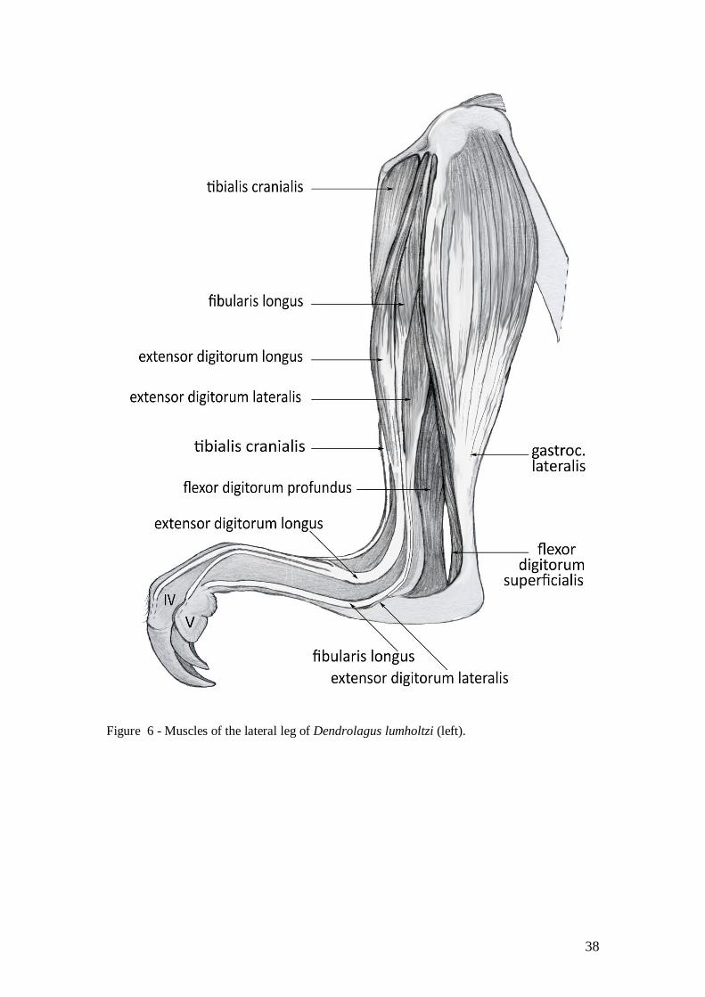
Figure 6 - Muscles of the lateral leg of Dendrolagus lumholtzi (left).

39
Figure 7 - Muscles of the medial leg of Dendrolagus lumholtzi (left).

40
Figure 8 - Intrinsic muscles of the plantar aspect of the pes of Dendrolagus lumholtzi (left;
tendons of the m. flexor digitorum superficialis have been removed).