multiplex analysis of urinary cytokine levels in rat model of cyclophosphamide-induced cystitis
TRANSCRIPT

MCCMB
O
M
R
C
Itcsfiae
TaaI
RP
Pu
©
Basic and Translational Science
ultiplex Analysis of Urinaryytokine Levels in Rat Model ofyclophosphamide-induced Cystitis
arc C. Smaldone, Yoram Vodovotz, Vikas Tyagi, Derek Barclay,rian J. Philips, Naoki Yoshimura, Michael B. Chancellor, and Pradeep Tyagi
BJECTIVES The urinary proteome is a potential easily accessible source of biomarkers for inflammatorybladder diseases, including interstitial cystitis. In the present study, we subjected rat urine tomultiplex cytokine analysis in an attempt to identify an inflammatory signature of the temporalcourse of cyclophosphamide (CYP)-induced cystitis.
ETHODS Rat urine was collected for 12 hours after CYP injection (150 mg/kg) for multiplex analysis of 14cytokines by a multiple antigen bead assay (Luminex 100 IS). Urine from each void wascollected, and the voiding frequency was determined. The bladder tissue was analyzed forcytokines levels and histologic evidence of inflammation.
ESULTS Significant changes were noted in the urine levels of all cytokines with respect to baseline at 2,4, 6, and 10 hours after CYP injection. Elevation was noted at all times for most cytokines, exceptfor monocyte chemotactic protein-1, which had a 5-fold decrease at 2 hours. The urine and tissuelevels of interleukin (IL)-1�, IL-4, and growth-related oncogene/keratinocyte-derived chemo-kine correlated significantly, with a positive Spearman correlation also noted for granulocyte-macrophage colony-stimulating factor, monocyte chemotactic protein-1-1, IL-18, and inter-feron-�. The tissue levels for most cytokines, except for IL-2, and urinary frequency weresignificantly elevated in the CYP-treated rats compared with the control vehicle-treated rats.The hints of severe inflammation in the bladder indicated by the urinary cytokines wereconfirmed by bladder histologic examination and the tissue cytokine levels at necropsy.
ONCLUSIONS The progression of CYP-induced cystitis was clearly reflected in the urine matrix by thetemporal and quantitative changes in the cytokine levels. Additional delineation of urineand bladder tissue cytokine expression might yield biomarkers for cystitis. UROLOGY 73:
421– 426, 2009. © 2009 Published by Elsevier Inc.ci
dbcpottosa
piufp
nterstitial cystitis (IC) is a chronic inflammatory dis-ease of unknown etiology characterized by urinaryfrequency, urgency, and suprapubic pain.1 The Na-
ional Institutes of Health have established diagnosticriteria for IC based on the presence of irritative voidingymptoms in the absence of other identifiable pathologicndings.2 In the present study, we hypothesized that thenalysis of urinary cytokines might allow for the discov-ry of disease-specific targets and/or biomarkers that
his study was supported in part by National Institute of Arthritis, Diabetes, Digestivend Kidney Diseases grant DK 066138 (to P. Tyagi), National Institute of Disabilitynd Rehabilitation Research grant H133E070024 (to Y. Vodovotz), and Nationalnstitute of General Medical Sciences grant P50-GM-53789-09.
From the Departments of Urology and Surgery, and Center for Inflammation andegenerative Modeling, McGowan Institute for Regenerative Medicine, University ofittsburgh, Pittsburgh, PennsylvaniaReprint requests: Pradeep Tyagi, Ph.D., Department of Urology, University of
ittsburgh, 3471 Fifth Avenue, Suite 700, Pittsburgh, PA 15213. E-mail: tyagip@
Spmc.eduSubmitted: February 18, 2008, accepted (with revisions): July 7, 2008
2009 Published by Elsevier Inc.
ould be of use as diagnostic and prognostic markers fornflammatory bladder disease.3
The urine is one of the ideal biologic samples for theiscovery of noninvasive biomarkers for human diseases,ecause it is available from almost all patients, and itsollection is simple and does not require any invasiverocedures. Recent studies have suggested that cytokinesr chemokines contribute to lower urinary tract dysfunc-ion. Therefore, cytokines might serve as direct therapeu-ic targets or as potential biomarkers for the developmentf targeted therapy designed to prevent the long-termequelae of chronic bladder inflammatory conditions suchs IC.4
Systemic or intraperitoneal injection with cyclophos-hamide5 induces a reproducible dose-dependant chem-cal cystitis in both mice and rats and therefore has beensed as an experimental model of IC.6 Increased voidingrequency, decrease urine volume per void, and increasedermeability of the bladder wall is seen in this model.5,7
imilar studies reported previously only examined cyto-
0090-4295/09/$34.00 421doi:10.1016/j.urology.2008.07.031

kirstodb
c[I[icfdw
M
AAd
vmpUwbbt
UFieSm(ttemumt
Ftralhiaia n �
4
ine expression in rat bladder tissue using enzyme-linkedmmunosorbent assay and not in the urine.8 Saban et al.9
eported the upregulation of cytokines in pooled urineamples from an inflammatory mouse model using a mul-iplex suspension array. The multiplex analysis (xMAP)f urinary proteins after cyclophosphamide (CYP)-in-uced cystitis in rat using Luminex technology has noteen previously reported.
The present study was designed to examine the acutehanges in urinary levels of 14 cytokines (interleukin-1�IL-1�], IL-1�, IL-2, IL-4, IL-5, IL-6, IL-10, IL-12p70,L-18, granulocyte-macrophage colony-stimulating factorGM-CSF], monocyte chemotactic protein-1 [MCP-1],nterferon-� [IFN-�], growth-related oncogene/keratino-yte-derived chemokine [GRO/KC], and tumor necrosisactor-� [TNF-�]) after CYP-induced cystitis. The blad-er inflammation revealed by the urinary cytokine levelsas verified by tissue analysis and histologic examination.
ATERIAL AND METHODS
nimalsll animal experimentation described was performed in accor-
igure 1. Temporal profile of urinary cytokines after cyclopho creatinine excretion in urine at baseline and 2, 4, 6, anelative to baseline in levels of growth-related oncogene/kernd granulocyte-macrophage colony-stimulating factor (GM-
evels at same point (*P � .05). Monocyte chemotactic prours relative to baseline (*P � .05; Fig. A, middle). (C,D) Tn IL-5 and IL-1� levels at 4 hours (*P � .05). Middle panelst 4 and 6 hours, respectively (*P � .05). (Bottom) IFN-� le
ncrease of tumor necrosis factor (TNF)-� at 4 hours did not rnd considered significant relative to baseline at P � .05 (
ance with institutional guidelines and approval from the Uni- w
22
ersity of Pittsburgh Institutional Animal Care and Use Com-ittee. Intraperitoneal CYP injections (150 mg/kg) were
erformed in female Sprague-Dawley rats (weight 276-292 g).10
rine specimens obtained from rats kept in metabolic cagesere frozen immediately in liquid nitrogen and stored at �80°Cefore analysis. Baseline urine samples were obtained 24 hoursefore CYP injection and from vehicle-treated rats. Bladderissue was harvested from CYP-treated and vehicle-treated rats.
rine and Bladder Tissue Cytokine Expressionrozen urine samples from each point and bladder tissue proteinsolates from the end of the study were thawed, and 50 �L fromach sample was analyzed in on the Luminex 100 IS (MiraiBio,outh San Francisco, CA) using a LINCOplex cytokine/che-okine Luminex Bead immunoassay Kit 14-Plex bead set
LINCO Research, St. Charles, MO). The cytokine concentra-ions provided by Luminex for each point were normalized tohe creatinine concentration in urine for that point and arexpressed as the amount of cytokine in picograms excreted perilligram of creatinine. The creatinine in urine was measured
sing the previously published high-performance liquid chro-atography method.11 Cytokine concentrations in the tissue at
he end of the study were normalized to the respective bladder
amide (CYP) injection. Urine levels of cytokines normalizedhours after CYP injection. (A,B) Five- to sixfold increase
cyte-derived chemokine (GRO/KC), interleukin (IL)-6, IL-1�,seen at 4 hours compared with 10-fold increase in IL-18(MCP)-1 levels showed significant decrease of 8-fold at 2
anels show 15-fold significant increase relative to baseline3-fold increase relative to baseline in IL-10 and IL-4 levels
showed significant 4-fold by 10 hours; in contrast, 60-foldstatistical significance. Values expressed as mean � SEM8).
osphd 10atinoCSF)oteinop pshowvelseach
eight of each rat.
UROLOGY 73 (2), 2009

HTbTssp
SDbocc
sV
R
TCAdrdTt
pcwIM6KiFhc2.sbllpwrIh4b
FI(cueo
U
istologic Analyseshe bladders were fixed in 4% buffered formaldehyde, followedy cryopreservation and serial sectioning into 20-�m sections.he sections were stained with hematoxylin-eosin.12,13 The
tained sections were visualized for leukocyte infiltration, de-truction of urothelium, and edema. Toluidine blue staining waserformed for mast cells.
tatistical Analysisata comparison was done between the mean cytokine levels ataseline and 2, 4, 6, and 10 hours after CYP treatment usingne-way analysis of variance followed by Dunnett’s multipleomparison test for statistical significance. Differences wereonsidered significant at P � .05.
In addition, the cytokine values in the last voided urineamples were compared with the bladder tissue expression.alues are expressed as the mean � SEM.
ESULTS
emporal Quantitative Changes in Urinaryytokine Levels After CYP-induced Cystitiscute disease progression was profiled by the CYP-in-
uced time-dependent changes in urinary cytokine levelselative to baseline values. All 15 cytokines assayed wereetected in the urine of CYP-treated rats (Figs. 1 and 2).he urinary cytokine levels at baseline and after CYP
Baseline 2h 4h 6h 10h0
10
20
30
40
50
60
70
Baseline 2h 4h 6h 10h0
10
20
30
40
50
60
70
Baseline 2h 4h 6h 10h0
10
20
30
40
50
60
70
* *
** *IL-17
IL-2
IL-12p70
Uri
nar
y C
yto
kin
e L
evel
s in
pg
/mg
of
Cre
atin
ine
A
igure 2. (A) Temporal profile of urinary cytokines after cycloL-12p70, and IL-2 normalized to creatinine excretion in urinTop) IL-17 and (Middle) IL-12p70 levels tripled significantlontrast, levels only doubled for IL-2 without statistical signrinary frequency after CYP injection. Peak urinary frequexpression at 4-6 hours and preceded peak IL-6 elevation. Vf one void every hour or every second hour.
reatment were normally distributed, justifying the use of 4
ROLOGY 73 (2), 2009
arametric tests for statistical significance. Significanthanges compared with baseline values were notedithin 4 hours of CYP-injection for IL-1�, IL-1�, IL-5,
L-6, IL-10, IL-18, and GM-CSF (P � .05; Fig. 1A-D).ost robust changes were seen at 4 hours with a 5- to
-fold increase relative to baseline in the levels of GRO/C, IL-6, IL-1�, and GM-CSF compared with a 10-fold
ncrease in the levels of IL-18 at the same point (P � .05;ig. 1A,B). A 7-fold increase in the GRO/KC level at 6ours after CYP administration was statistically signifi-ant (Fig. 1a, top panel). The IL-6 levels had doubled byhours and were 6-fold higher at 4 and 10 hours (P �
05; Fig. 1B, top panel). The urinary levels of MCP-1howed a significant 8-fold decrease at 2 hours relative toaseline (Fig. 1A, middle panel). The IL-5 and IL-1�evels increased sharply to nearly 15-fold relative to base-ine at 4 hours (P � .05; Fig. 1C,D, top panels). Com-aratively, a modest 3-fold elevation relative to baselineas seen in the IL-10 and IL-4 levels at 4 and 6 hours,
espectively (P � .05; Fig. 1C,D, middle panels). TheFN-� levels showed a significant 4-fold increase by 10ours (P � .05); however, a 60-fold increase in TNF-� athours did not reach statistical significance (Fig. 1C,D,
ottom panels).The IL-17 and IL-12p70 levels tripled significantly by
0.0 2.5 5.0 7.5 10.0.0
.5
.0
.5 CYP 150mg/kgVehicle Treated Rats
Time After Injection
B
sphamide (CYP) injection. Urine levels of interleukin (IL)-17,baseline and at 2, 4, 6, and 10 hours after CYP injection.
4 hours after CYP injection (*P � .05; n � 4). (Bottom) Ince (n � 8) at same point. (B) Time-dependent changes in
after CYP injection concurrent with peak urinary cytokinele-treated rats showed relatively constant voiding frequency
0
2
5
7
Nu
mb
er o
f V
oid
ing
Ep
iso
des
per
Ho
ur
phoe aty byificanncyehic
hours after CYP injection (P � .05; Fig. 2A, top and
423

mw2ni6iw
CPAc2tetinMccfc(
HHesssp
bt(cw
CTanctacfcf
oatflssSci
pIa
).
4
iddle panels); however, the IL-2 levels only doubled,ithout statistical significance, at the same point (Fig.A, bottom panel). Urinary frequency was defined as theumber of voiding episodes hourly for each rat after CYP
njection (Fig. 2B). A mean peak urinary frequency of.1 � 2.8 voids/h was observed at 4-6 hours after CYPnjection (n � 8). Vehicle-treated rats (n � 8) voidedith a relatively constant frequency.
hanges in Tissue Cytokinerotein Levels After CYP-induced Cystitist 24 hours after CYP injection, the tissue levels for most
ytokines in the CYP-treated rats were elevated at least-fold compared with the vehicle-treated control bladderissue levels. The IL-6 levels showed the most drasticlevations, nearly 24-fold compared with the sham-reated rats, and similarly a 10-fold elevation was notedn the tissue levels of GRO/KC and TNF-� (Table 1). Aearly 3- to 4-fold increase was seen in the IL-1� andCP-1 levels. In contrast, the tissue levels of IL-2 de-
reased by one half after CYP treatment. A positiveorrelation between the urine and tissue levels was notedor GM-CSF, MCP-1, IL-18, and IFN-�, with a signifi-ant Spearman correlation for IL-1�, IL-4, and GRO/KCP � .05).
istologic Analysisistologic evaluation of the bladder with hematoxylin-
osin staining 24 hours after CYP treatment revealedigns of CYP-induced hemorrhagic cystitis of varyingeverity, marked by severe edema and inflammation, ero-ion, ulceration of the urothelium, and hemorrhage com-
Table 1. Correlation between cytokine levels in bladder at
Cytokine
Last Voided Urine(pg/mg
creatinine)
Tissue Cytokine LeExpressed Per Blad
(ng)
GM-CSF 5.2 � 1.07 27.8 � 2.1IL-1� 26.3 � 11.5 47.1 � 13.1MCP-1 121.6 � 47.1 213.2 � 58.8IL-4 64.9 � 48.2 35 � 3.28IL-1� 13.9 � 8.7 76.4 � 18.4IL-2 2.4 � 1.7 33.6 � 10.4IL-6 130.2 � 62.9 438.7 � 184.IL-10 13.7 � 4.3 28.3 � 15.3IL-12p70 16.7 � 5.2 25.7 � 11.3IL-5 0 34.8 � 7.5IFN-� 44.9 � 37.9 16.4 � 0.6IL-18 10.3 � 3.7 1285. 2 � 297.GRO/KC 184.3 � 100.2 522.7 � 265.TNF-� 0 23.9 � 7.7IL-17 16.6 � 2.4 17.5 � 1.5
Abbreviations: CYP, cyclophosphamide; GM-CSF, granulocyte-machemotactic protein-1; IFN-�, interferon-�; GRO/KC, growth-relatefactor-�.
Cytokine concentrations in tissue were normalized to respectivtissue levels noted for GM-CSF, MCP-1, IL-18, and IFN-�, with sig
Measured Spearman correlation coefficient between levels insignificant differences from 0 by setting 2-tailed P � .05 (n � 4-8
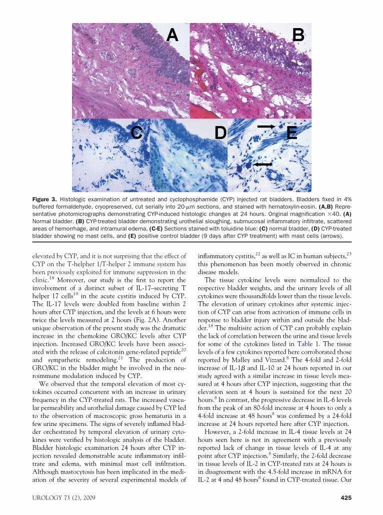
ared with untreated specimens (Fig. 3A,B). Toluidine d
24
lue staining failed to reveal the presence of mast cells inhe control bladders or CYP treated bladders at 24 hoursFig. 3C,D) but did demonstrate the presence of mastells in bladders of chronic CYP treated rats (9 days) thatere used as positive control (Fig. 3E).
OMMENThe present report describes the application of multiplexnalysis of inflammatory cytokines using Luminex tech-ology to analyze microliter quantities of urine specimensollected at varying points after chemically induced cys-itis in rats. Our results suggest that urine multiplexnalysis might be a promising approach for the identifi-ation of IC biomarkers, aiming at the detection of ICrom a single voided urine specimen. In contrast, mosturrent urine proteome testing requires urine collectionor a 24-hour period.14
Clinical studies have reported elevated urinary levelsf IL-614-16 in patients with IC and demonstrated anssociation between IL-6 expression and clinical symp-om severity.14 CYP is well known to induce frank in-ammation in the bladder,17 and the results from thesetudies have demonstrated that inflammation at the tis-ue level is reflected by varying urinary cytokines levels.imilar published studies on the urine measurement ofytokines in animal models have not reported the inter-ndividual variability of cytokines between animals.9
Significant increases in urinary cytokines specificallyroduced by T-helper 1 cells (IL-2, IL-18, IL-12, andFN-�), as well as by T-helper 2 cells (IL-4, IL-5, IL-6,nd IL-10), were observed. GM-CSF and TNF-� pro-
al death and in last voided urine specimen before death
Spearman’s CorrelationCoefficient
x-Fold Change in CYP-treated Bladder vs
Vehicle-treatedBladder
0.87 10.5 2.50.8 3.50.97* 20.97* 4
�0.05 0.50.5 24
�0.6 2.5�0.5 2
0 20.66 20.80 20.9* 100 10
�0.5 2
age colony-stimulating factor; IL, interleukin; MCP-1, monocytecogene/keratinocyte-derived chemokine; TNF-�, tumor necrosis
dder weight of each rat; positive correlation between urine andnt Spearman correlation for IL-1�, IL-4, and GRO/KC.
and last voided urine sample for each cytokine and tested for
anim
velsder
3
01
crophd on
e blanificatissue
uced by both T-helpher 1 and T-helper 2 cells were also
UROLOGY 73 (2), 2009
eCbcihThtuiiaaGr
tfltfdkBjtAa
itd
rcTtrdtflrissehf4i
hrpii
FbsNab dder
U
levated by CYP, and it is not surprising that the effect ofYP on the T-helpher 1/T-helper 2 immune system haseen previously exploited for immune suppression in thelinic.18 Moreover, our study is the first to report thenvolvement of a distinct subset of IL-17–secreting Telper 17 cells19 in the acute cystitis induced by CYP.he IL-17 levels were doubled from baseline within 2ours after CYP injection, and the levels at 6 hours werewice the levels measured at 2 hours (Fig. 2A). Anothernique observation of the present study was the dramaticncrease in the chemokine GRO/KC levels after CYPnjection. Increased GRO/KC levels have been associ-ted with the release of calcitonin gene-related peptide20
nd sympathetic remodeling.21 The production ofRO/KC in the bladder might be involved in the neu-
oimmune modulation induced by CYP.We observed that the temporal elevation of most cy-
okines occurred concurrent with an increase in urinaryrequency in the CYP-treated rats. The increased vascu-ar permeability and urothelial damage caused by CYP ledo the observation of macroscopic gross hematuria in aew urine specimens. The signs of severely inflamed blad-er orchestrated by temporal elevation of urinary cyto-ines were verified by histologic analysis of the bladder.ladder histologic examination 24 hours after CYP in-
ection revealed demonstrable acute inflammatory infil-rate and edema, with minimal mast cell infiltration.lthough mastocytosis has been implicated in the medi-
igure 3. Histologic examination of untreated and cyclophuffered formaldehyde, cryopreserved, cut serially into 20-�entative photomicrographs demonstrating CYP-induced hiormal bladder. (B) CYP-treated bladder demonstrating uroreas of hemorrhage, and intramural edema. (C-E) Sectionsladder showing no mast cells, and (E) positive control bla
tion of the severity of several experimental models of I
ROLOGY 73 (2), 2009
nflammatory cystitis,22 as well as IC in human subjects,23
his phenomenon has been mostly observed in chronicisease models.The tissue cytokine levels were normalized to the
espective bladder weights, and the urinary levels of allytokines were thousandfolds lower than the tissue levels.he elevation of urinary cytokines after systemic injec-
ion of CYP can arise from activation of immune cells inesponse to bladder injury within and outside the blad-er.18 The multisite action of CYP can probably explainhe lack of correlation between the urine and tissue levelsor some of the cytokines listed in Table 1. The tissueevels of a few cytokines reported here corroborated thoseeported by Malley and Vizzard.8 The 4-fold and 2-foldncrease of IL-1� and IL-10 at 24 hours reported in ourtudy agreed with a similar increase in tissue levels mea-ured at 4 hours after CYP injection, suggesting that thelevation seen at 4 hours is sustained for the next 20ours.8 In contrast, the progressive decrease in IL-6 levels
rom the peak of an 80-fold increase at 4 hours to only a-fold increase at 48 hours8 was confirmed by a 24-foldncrease at 24 hours reported here after CYP injection.
However, a 2-fold increase in IL-4 tissue levels at 24ours seen here is not in agreement with a previouslyeported lack of change in tissue levels of IL-4 at anyoint after CYP injection.8 Similarly, the 2-fold decreasen tissue levels of IL-2 in CYP-treated rats at 24 hours isn disagreement with the 4.5-fold increase in mRNA for
hamide (CYP) injected rat bladders. Bladders fixed in 4%ections, and stained with hematoxylin-eosin. (A,B) Repre-gic changes at 24 hours. Original magnification �40. (A)al sloughing, submucosal inflammatory infiltrate, scattereded with toluidine blue: (C) normal bladder, (D) CYP-treated(9 days after CYP treatment) with mast cells (arrows).
ospm s
stolothelistain
L-2 at 4 and 48 hours8 found in CYP-treated tissue. Our
425

rctpfi
COatopb
R
1
1
1
1
1
1
1
1
1
1
2
2
2
2
4
esults suggest that multiplex analysis of urine cytokinesan accurately and noninvasively discriminates CYP-reated rats from controls. Future studies on the urine ofatients with IC will be undertaken to validate thesendings.
ONCLUSIONSur findings report on a profile of inflammation-associ-
ted cytokines in the urine and bladder tissue of CYP-reated rats. These results have demonstrated the utilityf the multiplex analysis for urinary proteomics and itsotential in the noninvasive assessment of inflammatoryladder disease.
eferences1. Parsons CL. The role of the urinary epithelium in the pathogenesis
of interstitial cystitis/prostatitis/urethritis. Urology. 2007;69:9-16.2. Hanno PM, Landis JR, Matthews-Cook Y, et al. The diagnosis of
interstitial cystitis revisited: lessons learned from the NationalInstitutes of Health Interstitial Cystitis database study. J Urol.1999;161:553-557.
3. Keay S, Zhang CO, Chai T, et al. Antiproliferative factor, heparin-binding epidermal growth factor-like growth factor, and epidermalgrowth factor in men with interstitial cystitis versus chronic pelvicpain syndrome. Urology. 2004;63:22-26.
4. Erickson DR, Tomaszewski JE, Kunselman AR, et al. Do theNational Institute of Diabetes and Digestive and Kidney Diseasescystoscopic criteria associate with other clinical and objectivefeatures of interstitial cystitis? J Urol. 2005;173:93-97.
5. Eichel L, Scheidweiler K, Kost J, et al. Assessment of murinebladder permeability with fluorescein: Validation with cyclophos-phamide and protamine. Urology. 2001;58:113-118.
6. Lanteri-Minet M, Bon K, de Pommery J, et al. Cyclophosphamidecystitis as a model of visceral pain in rats: Model elaboration andspinal structures involved as revealed by the expression of c-Fos andKrox-24 proteins. Exp Brain Res. 1995;105:220-232.
7. Wood R, Eichel L, Messing EM, et al. Automated noninvasivemeasurement of cyclophosphamide-induced changes in murinevoiding frequency and volume. J Urol. 2001;165:653-659.
8. Malley SE, Vizzard MA. Changes in urinary bladder cytokinemRNA and protein after cyclophosphamide-induced cystitis.
Physiol Genom. 2002;9:5-13.26
9. Saban MR, Simpson C, Davis C, et al. Discriminators of mousebladder response to intravesical bacillus Calmette-Guérin (BCG).BMC Immunol. 2007;8:6.
0. Batler RA, Sengupta S, Forrestal SG, et al. Mast cell activationtriggers a urothelial inflammatory response mediated by tumornecrosis factor-alpha. J Urol. 2002;168:819-825.
1. George SK, Dipu MT, Mehra UR, et al. Improved HPLC methodfor the simultaneous determination of allantoin, uric acid andcreatinine in cattle urine. J Chromatogr B Anal Technol Biomed LifeSci. 2006;832:134-137.
2. Davidson RA, McCloskey KD. Morphology and localization ofinterstitial cells in the guinea pig bladder: Structural relationshipswith smooth muscle and neurons. J Urol. 2005;173:1385-1390.
3. Tyagi P, Chancellor MB, Li Z, et al. Urodynamic and immunohis-tochemical evaluation of intravesical capsaicin delivery using ther-mosensitive hydrogel and liposomes. J Urol. 2004;171:483-489.
4. Erickson DR, Xie SX, Bhavanandan VP, et al. A comparison ofmultiple urine markers for interstitial cystitis. J Urol. 2002;167:2461-2469.
5. Erickson DR, Belchis DA, Dabbs DJ. Inflammatory cell types andclinical features of interstitial cystitis. J Urol. 1997;158:790-793.
6. Lotz M, Villiger P, Hugli T, et al. Interleukin-6 and interstitialcystitis. J Urol. 1994;152:869-873.
7. Westropp JL, Buffington CA. In vivo models of interstitial cystitis.J Urol. 2002;167:694-702.
8. Perini P, Calabrese M, Rinaldi L, et al. The safety profile ofcyclophosphamide in multiple sclerosis therapy. Expert Opin DrugSaf. 2007;6:183-190.
9. Chang H, Hanawa H, Yoshida T, et al. Alteration of IL-17 relatedprotein expressions in experimental autoimmune myocarditis andinhibition of IL-17 by IL-10-Ig fusion gene transfer. Circul J. 2008;72:813-819.
0. Qin X, Wan Y, Wang X. CCL2 and CXCL1 trigger calcitoningene-related peptide release by exciting primary nociceptive neu-rons. J Neurosci Res. 2005;82:51-62.
1. Xie WR, Deng H, Li H, et al. Robust increase of cutaneoussensitivity, cytokine production and sympathetic sprouting in ratswith localized inflammatory irritation of the spinal ganglia. Neuro-sciences. 2006;142:809-822.
2. Bjorling DE, Jerde TJ, Zine MJ, et al. Mast cells mediate theseverity of experimental cystitis in mice. J Urol. 1999;162:231-236.
3. Theoharides TC, Kempuraj D, Sant GR. Mast cell involvement ininterstitial cystitis: A review of human and experimental evidence.
Urology. 2001;57:47-55.UROLOGY 73 (2), 2009