multiple pregnancies: determining chorionicity and amnionicity
TRANSCRIPT
Thomas Jefferson UniversityJefferson Digital Commons
Department of Radiologic Sciences Faculty Papers Department of Radiologic Sciences
January 2006
Multiple Pregnancies: Determining Chorionicityand AmnionicityTraci B. FoxThomas Jefferson University Hospital, [email protected]
Let us know how access to this document benefits youFollow this and additional works at: http://jdc.jefferson.edu/rsfp
Part of the Obstetrics and Gynecology Commons
This Article is brought to you for free and open access by the Jefferson Digital Commons. The Jefferson Digital Commons is a service of ThomasJefferson University's Center for Teaching and Learning (CTL). The Commons is a showcase for Jefferson books and journals, peer-reviewed scholarlypublications, unique historical collections from the University archives, and teaching tools. The Jefferson Digital Commons allows researchers andinterested readers anywhere in the world to learn about and keep up to date with Jefferson scholarship. This article has been accepted for inclusion inDepartment of Radiologic Sciences Faculty Papers by an authorized administrator of the Jefferson Digital Commons. For more information, pleasecontact: [email protected].
Recommended CitationFox, Traci B., "Multiple Pregnancies: Determining Chorionicity and Amnionicity" (2006).Department of Radiologic Sciences Faculty Papers. Paper 1.http://jdc.jefferson.edu/rsfp/1
Energizing Education
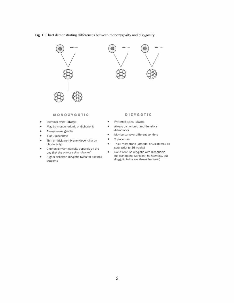
Multiple Pregnancies: Determining Chorionicity and Amnionicity TRACI B. FOX, BS, RT(R), RDMS, RVT The determination of chorionicity and amnionicity in multiple gestations is one that confounds many in the medical field. The importance of diagnosing the type of multiple gestation cannot be overstated. This is intended to be an introductory primer and refresher to chorionicity and amnionicity. We will first examine what determines mono- versus dichorionic twinning including zygosity and cleavage of the zygote. Keywords: Twins, Multiple Gestations, Zygosity, Chorionicity, Amnionicity, Dizygotic, Dichorionic, Monochorionic, Monozygotic, Monoamniotic, Monochorionic Introduction The identification and classification of twin pregnancies is of primary importance to the sonographer and clinician. Twins occur spontaneously in approximately one out of every 80 livebirths.1 The American Institute of Ultrasound in Medicine (AIUM) in its AIUM Practice Guideline for the Performance of Antepartum Obstetric Ultrasound Examination state that both fetal number and, when possible, amnionicity and chorionicity be documented.2 The rise in assisted reproduction due to infertility has increased the rate of dizygotic twinning. Many singleton pregnancies start off as twins, but one of the twins is resorbed. This is called a “vanishing twin.”1 The incidence of multiple pregnancies has been on the rise over the last 20 years. In addition to the number of the multiple gestations related to increasing use of infertility treatment, multiples (especially twins) are also increased with delayed child bearing.3 Twins have a higher risk of prenatal complications. While only 2% of singletons are born at a gestational age <33 weeks, 14% of twins are premature. There is also an association with NICU admissions and increasing hospital stay length with multiple pregnancies compared to singletons.3 It can clearly be seen that multiple pregnancies should not be taken lightly. Zygosity vs. Chorionicity and Amnionicity Monozygotic twins: 1 egg, 1 sperm. They meet and join to form a zygote. This becomes a singleton pregnancy. However, if that zygote splits into two equal zygotes, you have twins. Because the same set of chromosomes is in each zygote, you have identical twins. These are called monozygotic twins. One zygote splits to form two. Dizygotic twins: 2 sperm meet 2 eggs. Two zygotes are formed. Each zygote has their own individual set of chromosomes. These are known as fraternal, or non-identical twins (although they may be the same gender). Spontaneous dizygotic twins can be familial, and it is common to hear from patients that other members of their families are twins. Spontaneous dizygotic twinning is also thought to occur as a result from increased levels of follicle stimulating hormone (FSH). Whereas increased levels of FSH can be used to explain dizygotic twinning, there are no such explanations for monozygotic twins. 1 Figure 1 demonstrates how zygosity is determined. Monozygotic Twins Monozygotic twins are nearly* always identical. However, monozygosity does not determine chorionicity. If two separate zygotes implant, there will usually* be two chorions, and therefore a dichorionic pregnancy. (*There are rare instances of fusion described in the literature, but these will not be discussed here). The chorionicity and amnionicity of monozygotic twins is determined by the time at which the zygote splits, or cleaves. If the cleavage occurs by day 3, you will have two separate blastocysts, and therefore, two sites of implantation, resulting in dichorionic-diamniotic (di-di) twins. Di-di monozygotic twins occur about 25-
2
30%1 of the time. It is a difficult concept for many that di-di twins can be identical, but remember that they both may come from the same zygote. In the other 75% of monozygotic twins, the cleavage occurs after day 3, when the blastocyst has already formed. This results in a monochorionic pregnancy. A monochorionic pregnancy has one placenta, shared by both twins. As long as the cleavage occurs between days 4 and 8 (which is usually the case), each twin will form an amnion. Therefore, each twin will have their own inner sac (amnion), but be surrounded by one outer sac (chorion). This is monochorionic-diamniotic twinning (mono-di). However, if cleavage occurs between days 8 and 13, it’s too late for the amnion to form separately for each twin. This results in twins having one outer sac (chorion), and one inner sac (amnion). This is a monochorionic-monoamniotic pregnancy, or mono-mono twins. Fortunately, this only occurs in 2% of monozygotic twins, and is a very high-risk situation with a high mortality rate for both twins due to umbilical cord entanglement . In rare instances, cleavage occurs after day 13. This produces twins that are mono-mono, but the embryos themselves have not had time to completely separate, and therefore produces conjoined twins.4 (Note: using the term “mono-mono” is redundant. If a sac is monoamniotic, it has to be monochorionic.) Dizygotic twins Roughly two-thirds of all twins are dizygotic4. Dizygotic twins are always dichorionic-diamniotic, with two placentas. The placentas may fuse early on, giving the appearance of one placenta, but there are no vascular connections between the two. Dichorionic twins arise from two separate ova fertilized by two separate sperm, and therefore are as genetically different as any other non-twin sibling.1 Note that if a sac is dichorionic, it has to be diamniotic. (Note: saying “dichorionic” is the same as saying “di-di.” You do not need to specify “diamniotic;” it is implied if the twins are dichorionic). Figure 2 uses apartments as an analogy to describe the different types of twinning. How to tell chorionicity/amnionicity in the first trimester Dichorionic-Diamniotic: In the first trimester there are two distinct sacs in the uterus. (Remember - these may still be monozygotic twins. The zygote may have split so early after conception that there are two distinct sacs. Do not assume that they are necessarily “fraternal” twins). Each sac contains an embryo with a yolk sac. Each sac has a chorion and an amnion. It clearly looks like two separate sacs. (Fig. 3). A thick membrane is seen between the twins. Monochorionic-Diamniotic: Note that there is no thick membrane between the twins. It looks like one sac with two embryos in the uterus. There is only one outer sac, the chorion. Each twin is inside its own inner sac, or amnion. (Fig. 4) Each embryo has a yolk sac. (Two amnions = two yolk sacs). It is safe to tell the patient these are identical twins, as nearly all monochorionic twins are monozygotic, and therefore identical. The membrane may be difficult to see even with a high resolution transducer. It cannot be overstated that it is imperative a membrane be found if one is present. Transvaginal sonography, switching to a higher frequency transducer, and changing patient position may all be attempted to try and find a separating membrane. B-color mode may also be used to enhance the differences in amniotic fluid echogenicity. Monochorionic-Monoamniotic: There is only one chorion and one amnion, with both embryos inside the same amnion. (Fig. 5) This is a very high risk situation with approximately 50% mortality.4 There should only be one yolk sac since there is only one amnion.5 Monoamniotic twins are treated differently in the clinical setting due to their higher risk, so confirmation of the absence of a membrane is crucial. If monoamniotic twins are suspected, careful assessment should be made to exclude conjoined twinning, especially if the twins appear to be facing each other. It is important to identify monochorionic twins because of the three- to five-fold rate of increased morbidity and mortality compared to twins that are dichorionic. The risk is increased due to the diagnosis and management of twin transfusion, anomalies, and the death of 1 co-twin.6 Rare types of twinning, such as conjoined twins and the acardiac twins are present only in monoamniotic twins. The rate of congenital
3
anomalies in singletons is 2-3% while there is a two- to three-fold increase in monozygotic twins.1 Determination of chorionicity also aids in cases where selective termination is desired. With singletons and dichorionic twins it is possible to use intracardiac agents to stop the fetal heart. Monochorionic twins, however, have the possibility of having shared vascular connections or anastomoses, which may result in the unintended demise of the co-twin.7 How to tell chorionicity/amnionicity in the second trimester Dichorionic-Diamniotic: Dichorionic twins may have two discrete placentas or one fused placenta. If two placentas fuse early on, it may be difficult to determine chorionicity. There are some signs that would lead more to a diagnosis of dichorionicity, however. Where the two placentas fuse is where the membrane is attached to the placenta. In the early second trimester it may be possible to see a “lambda,” or chorionic peak sign (Fig. 6). The lambda sign is virtually diagnostic of dichorionicity. Note that the lambda sign may disappear after 16 weeks. Another hallmark of dichorionic pregnancies is a thick membrane. (Fig. 7) Since there are 4 layers in a dichorionic membrane it is usually not difficult to visualize, especially when compared to monochorionic membranes. When a patient presents for the first sonographic evaluation in the late third trimester, it may be impossible to determine chorionicity or amnionicity until after delivery. Monochorionic-Diamniotic: A lambda sign will not be seen with monochorionic twins. The membrane implants flat on the placenta, like a T. The membrane is thin and may be very difficult to find. (Fig. 8) There will only be one placenta. Even though there is a membrane separating the twins, they are at higher risk for adverse outcome than dichorionic twins because there is always some degree of shared vascular connections in the single placenta of the monochorionic twins. This places monochorionic twins at risk for anomalies, abnormal growth or twin transfusion syndrome, which can result in a poor outcome. Monochorionic-Monoamniotic: Second trimester findings include intertwined fetal parts (arms, legs) and intertwined cords. Color Doppler may be used to demonstrate intertwining of the cords (Fig. 9). Spectral Doppler may demonstrate the two different heart rates of the twins in one clump of intertwined cord (Fig. 10). Conclusion With the increasing use of fertility drugs and delayed childbearing, twins are becoming more common.3 In order to manage their higher risk, it is important to be able to determine chorionicity and amnionicity at the earliest age possible. Chorionicity and amnionicity determination in the first trimester is almost 100% accurate. While more difficult in the second trimester, it is usually possible to determine chorionicity using membrane thickness and presence or absence of a lambda sign. Figure 11 is a flowchart that can be used to aid in the determination of chorionicity and amnionicity throughout pregnancy. Special thanks to Dr. Stuart Weiner for his guidance and support.

4
References 1 Hall J: Twinning. The Lancet 2003; 362: 735-743. 2 AIUM practice guideline for the performance of an antepartum obstetric ultrasound examination. J Ultrasound Med 2003; 22: 1116-1125. 3 Strong C: Too many twins, triplets, quadruplets, and so on: a call for new priorities. J Law Med Ethics
2003; 31:272-282. 4 Callen PW: Ultrasonography in Obstetrics and Gynecology. 3rd Ed. Philadelphia, Pennsylvania, W.B. Saunders Company, 1994. 5 Benacerraf, Ultrasound of Fetal Syndromes, Philadelphia, Pennsylvania, Churchill Livingstone, 1998. 6 Stenhouse E, Hardwick C, Maharaj S, Webb J, Kelly T, Mackenzie FM: Chorionicity determination in twin pregnancies: how accurate are we? Ultrasound Obstet Gynecol 2002; 19:350-352. 7 Machin GA: Why is it important to diagnose chorionicity and how do we do it? Best Practice & Research Clinical Obstetrics and Gynaecology 2004; 4:515-530.
5
Fig. 1. Chart demonstrating differences between monozygosity and dizygosity

6
Fig. 2. The “apartment analogy” for use in determining chorionicity and amnionicity
7
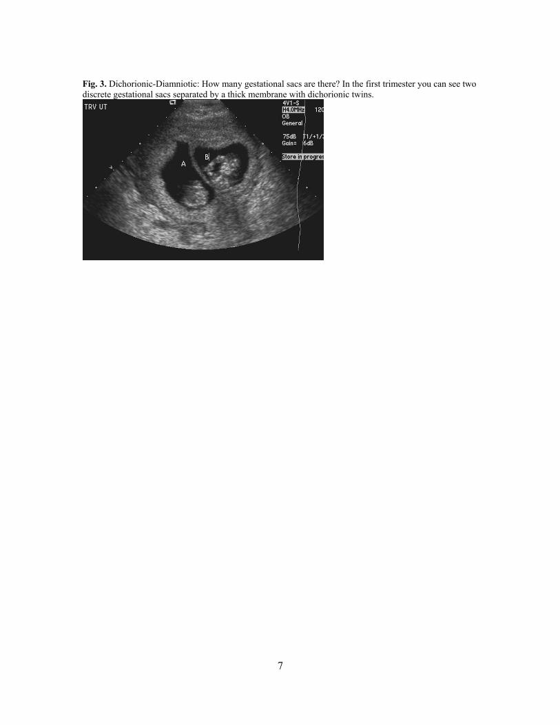
Fig. 3. Dichorionic-Diamniotic: How many gestational sacs are there? In the first trimester you can see two discrete gestational sacs separated by a thick membrane with dichorionic twins.
8
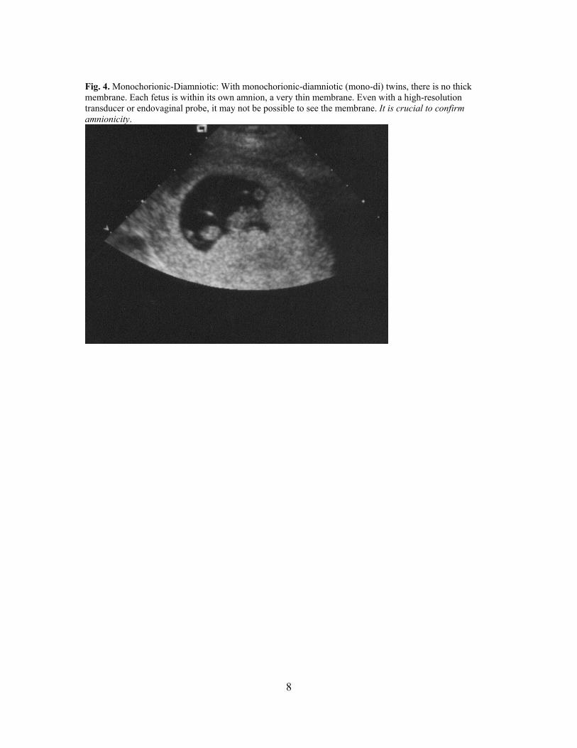
Fig. 4. Monochorionic-Diamniotic: With monochorionic-diamniotic (mono-di) twins, there is no thick membrane. Each fetus is within its own amnion, a very thin membrane. Even with a high-resolution transducer or endovaginal probe, it may not be possible to see the membrane. It is crucial to confirm amnionicity.
9
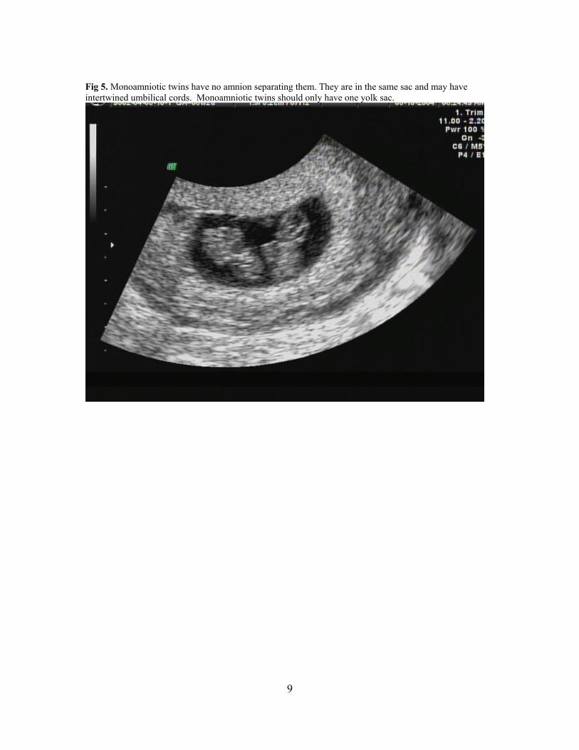
Fig 5. Monoamniotic twins have no amnion separating them. They are in the same sac and may have intertwined umbilical cords. Monoamniotic twins should only have one yolk sac.
10
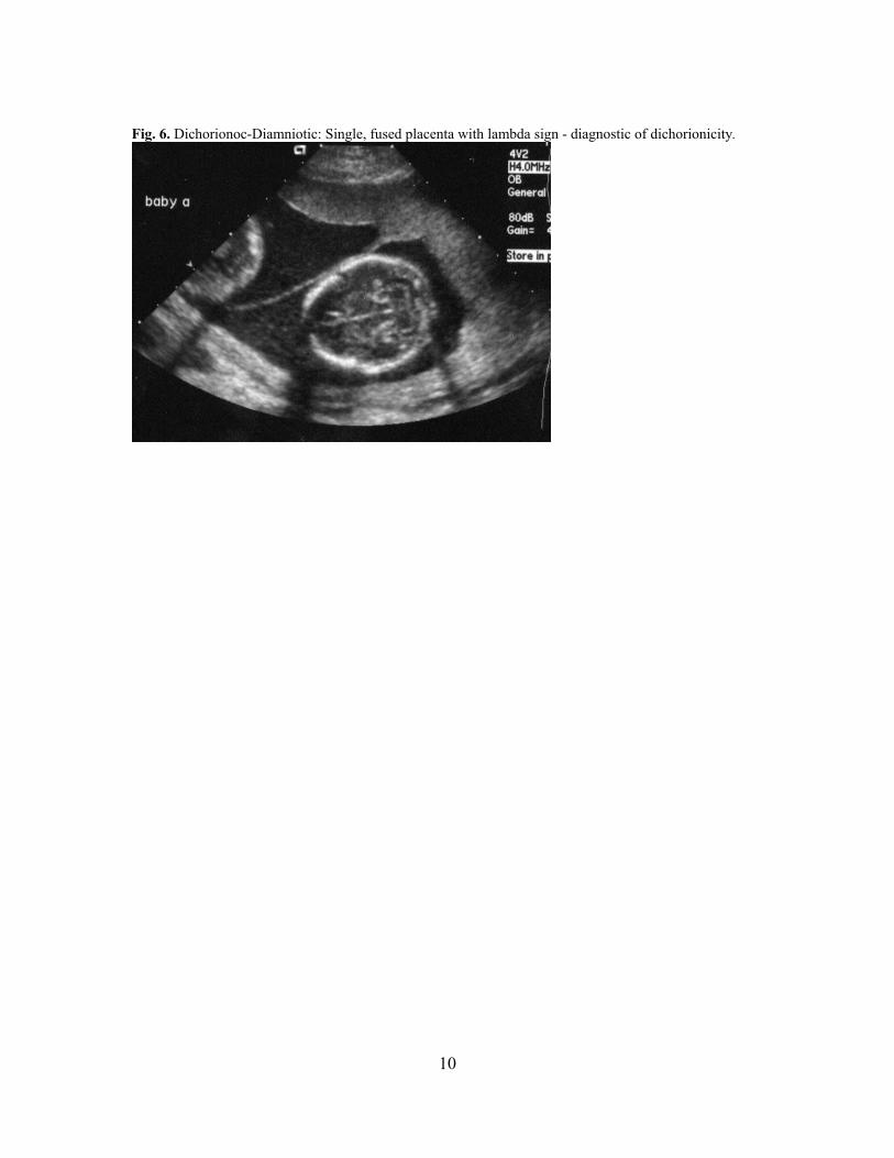
Fig. 6. Dichorionoc-Diamniotic: Single, fused placenta with lambda sign - diagnostic of dichorionicity.

11
Fig. 7. Dichorionic-Diamniotic: Two placentas with a thick membrane
12
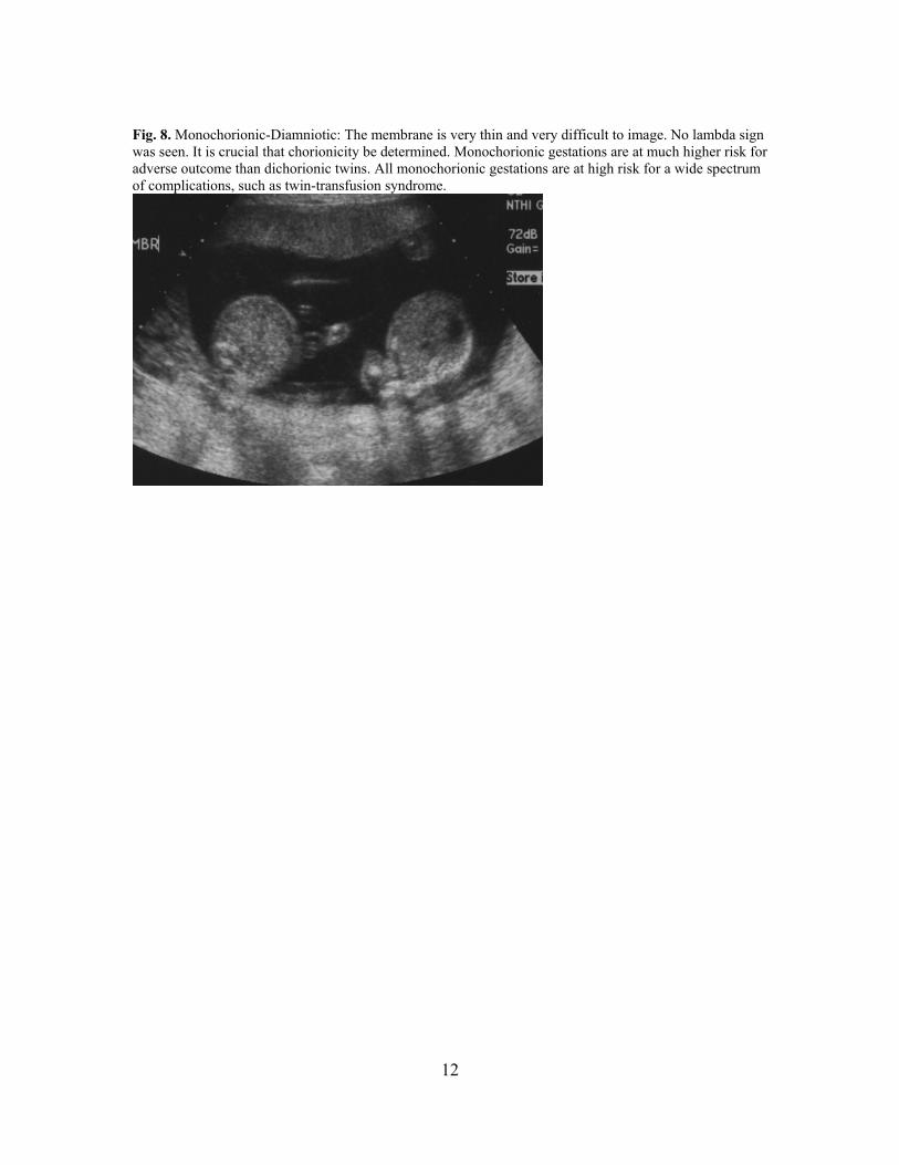
Fig. 8. Monochorionic-Diamniotic: The membrane is very thin and very difficult to image. No lambda sign was seen. It is crucial that chorionicity be determined. Monochorionic gestations are at much higher risk for adverse outcome than dichorionic twins. All monochorionic gestations are at high risk for a wide spectrum of complications, such as twin-transfusion syndrome.
13
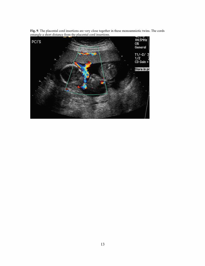
Fig. 9. The placental cord insertions are very close together in these monoamniotic twins. The cords entangle a short distance from the placental cord insertions.
14
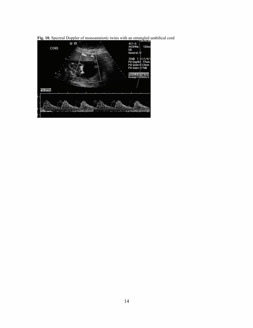
Fig. 10. Spectral Doppler of monoamniotic twins with an entangled umbilical cord

15
Fig. 11. Flowchart – how to tell chorionicity and amnionicity