multiple docking sites on substrate proteins form a ... · multiple docking sites on substrate...
TRANSCRIPT

Multiple docking sites on substrateproteins form a modular systemthat mediates recognition by ERKMAP kinaseDave Jacobs,1 Danielle Glossip,1 Heming Xing,2,3 Anthony J. Muslin,2,3 and Kerry Kornfeld1,4
1Department of Molecular Biology and Pharmacology, 2Department of Medicine, 3Department of Cell Biologyand Physiology, Washington University School of Medicine, St. Louis, Missouri 63110 USA
MAP kinases phosphorylate specific groups of substrate proteins. Here we show that the amino acid sequenceFXFP is an evolutionarily conserved docking site that mediates ERK MAP kinase binding to substrates inmultiple protein families. FXFP and the D box, a different docking site, form a modular recognition system, asthey can function independently or in combination. FXFP is specific for ERK, whereas the D box mediatesbinding to ERK and JNK MAP kinase, suggesting that the partially overlapping substrate specificities of ERKand JNK result from recognition of shared and unique docking sites. These findings enabled us to predict newERK substrates and design peptide inhibitors of ERK that functioned in vitro and in vivo.
[Key Words: MAP kinase; ERK; JNK; KSR; ETS transcription factor]
Received October 14, 1998; revised version accepted December 11, 1998.
Mitogen-activated protein (MAP) kinases are compo-nents of signaling cascades that regulate normal devel-opment and pathological processes such as oncogenesis.MAP kinases were identified during biochemicalsearches for serine/threonine-specific protein kinasesstimulated by growth factors in vertebrate cells (for re-view, see Sturgill and Wu 1991). MAP kinases were alsoidentified in screens for mutations that affect intercellu-lar signaling in yeast, worms, and flies (for review, seeFerrell 1996). Together, these investigations revealedthat MAP kinases function in many cell types, are regu-lated by a diverse group of extracellular stimuli, and me-diate a wide variety of cellular responses. MAP kinasescan be divided into subfamilies based on specific con-served residues, particularly a TXY motif in the activa-tion loop (Ferrell 1996). The three best-characterizedsubfamilies in vertebrates are named extracellular-regu-lated kinase (ERK), c-Jun amino-terminal kinase (JNK,also called stress-activated protein kinase), and p38.There are probably several additional vertebrate MAPkinase subfamilies, since Saccharomyces cerevisiae con-tains six different MAP kinases (Madhani and Fink1998). Here we use the name MAP kinase to refer to allmembers of the family, and the names ERK, JNK, andp38 to refer to members of those subfamilies.
MAP kinases function in modules composed of three
protein kinases (for review, see Marshall 1994). MAP ki-nase kinase kinases, such as Raf-1, phosphorylate andthereby activate MAP kinase kinases, such as MEK(MAP kinase kinase or ERK kinase). MAP kinase kinasesare serine/threonine and tyrosine-specific protein ki-nases that phosphorylate the TXY motif and thereby ac-tivate MAP kinases. In general, MAP kinases in differentsubfamilies are members of separate modules and areregulated by distinct extracellular stimuli (for review,see Whitmarsh and Davis 1996). For example, ERK isactivated strongly by receptor tyrosine kinases (RTK)such as the epidermal growth factor receptor, whereasJNK is activated strongly by stress stimuli such as ultra-violet light. Several of the signaling pathways leadingfrom extracellular stimuli to the activation of a MAPkinase module are well defined, whereas others have yetto be characterized in detail.
Whereas the upstream signaling events that regulateMAP kinases have been characterized extensively, con-siderably less is known about how MAP kinases regulatecell fates and contribute to the specificity of signalingpathways. Important questions that remain largely un-answered include: (1) How do MAP kinases recognizespecific proteins as substrates? (2) What proteins arephosphorylated by a particular MAP kinase in differentcell types and in different organisms? Answers to thesequestions will illuminate how the same MAP kinase me-diates different cell fates in different developmental con-texts and how MAP kinases from separate subfamiliesmediate different cellular responses.
4Corresponding author.E-MAIL [email protected]; FAX (314) 362-7058.
GENES & DEVELOPMENT 13:163–175 © 1999 by Cold Spring Harbor Laboratory Press ISSN 0890-9369/99 $5.00; www.genesdev.org 163

In the case of ERK, >50 different proteins have beenreported to be substrates (for reviews, see Davis 1993;Karin 1995; Treisman 1996; Whitmarsh and Davis 1996;Madhani and Fink 1998). These include signaling pro-teins likely to function upstream of ERK such as Son-of-sevenless (Sos) guanine nucleotide exchange factor andMEK; signaling proteins likely to function downstreamof ERK such the protein kinase pp90rsk; transcriptionfactors such as c-Fos, GATA-2, c-Myc, and ETS proteinsincluding Elk-1, LIN-1, and Aop/Yan; and proteins in-volved in a wide variety of other processes. These find-ings suggest that ERK plays a central role in signal propa-gation and feedback regulation. Furthermore, ERK is atransition point between signaling proteins and regula-tors of differentiation, suggesting it makes an importantcontribution to the specificity of RTK–Ras–ERK signal-ing pathways. Although a large number of ERK sub-strates have been identified, the understanding of ERKfunction remains fragmentary, as ERK probably phos-phorylates different substrates in different cell types andthe cellular context of most substrates has yet to be de-fined. In addition, many ERK substrates probably havenot been identified. Substrates of JNK have been charac-terized less extensively, but it is notable that they in-clude proteins that are also phosphorylated by ERK, suchas Elk-1, as well as unique substrates (for review, seeMinden and Karin 1997).
Little is known about how ERK recognizes such a di-verse group of substrates. Although the structure of ERKwas determined using X-ray crystallography (Zhang et al.1994; Canagarajah et al. 1997), this approach has not re-vealed how ERK interacts with substrate proteins, be-cause the structure of ERK bound to a substrate has yetto be determined. Studies of residues in substrate pro-teins that are phosphorylated by ERK and assays of pep-tide substrates identified a serine or threonine followedby a proline (S/TP) as the minimal consensus sequencefor phosphorylation by ERK (for review, see Davis 1993;Songyang et al. 1996). In addition, a proline at position −2is favorable, whereas a proline at position −1 is unfavor-able (the phosphoacceptor S/T is position 0). However,this information is not sufficient to explain how ERKrecognizes specific proteins as substrates, because manyproteins that contain S/TP sequences are not phosphory-lated by ERK. Studies of the interaction of JNK with itssubstrate c-Jun have identified a sequence positionedamino-terminal to the S/TP sites that is required for ef-ficient phosphorylation (the d domain) (Adler et al. 1992,1994; Hibi et al. 1993; Kallunki et al. 1996). This led tothe hypothesis that a docking site on the substrate pro-tein that is separate from the phosphorylation sites me-diates the interaction with JNK (Karin 1995). Other pro-tein kinases, such as cyclin–cdk2, also appear to interactwith a docking site on substrate proteins (Adams et al.1996). Although >50 proteins have been reported to beERK substrates, only recently has one such docking sitefor ERK been identified (Yang et al. 1998a,b). This dock-ing site, a domain of Elk-1 called the D box, is similar insequence to the d domain of c-Jun, and these two do-mains are interchangeable functionally, suggesting that
the d domain/D box is a docking site for both ERK andJNK.
Here we describe the identification and characteriza-tion of a different docking site, the amino acid sequenceFXFP, which mediates interactions with ERK but notJNK. These two docking sites define three classes of sub-strates: Proteins that contain only FXFP, only the d do-main/D box, or both. These findings suggest that amodular system of docking sites regulates interactions ofthe different MAP kinases with various substrates. Insubstrates that contain both docking sites, the sites func-tion additively to create a high-affinity interaction withERK. Thus, this system also modulates the affinity ofsubstrates for ERK and may determine which residuesare phosphorylated. We used this information to developpeptide inhibitors of ERK and identify new ERK sub-strates, including the kinase suppressor of ras (KSR) fam-ily of protein kinases.
Results
FQFP is necessary for high-affinity interactionsbetween ERK and ETS proteins in the Elk subfamily
The Caenorhabidtis elegans LIN-1 protein contains anETS DNA-binding domain and presumably regulatestranscription (Beitel et al. 1995). LIN-1 appears to beregulated directly by ERK, as LIN-1 is efficiently phos-phorylated by Erk2 in vitro and lin-1 is regulated nega-tively by RTK–Ras–ERK pathways in vivo (Jacobs et al.1998; Tan et al. 1998). We identified and characterizedsix gain-of-function (gf) mutations that impair the abilityof lin-1 to be regulated negatively by RTK–Ras–ERKpathways and disrupt vulval development (Jacobs et al.1998). Each mutation alters or eliminates FQFP, a se-quence located in the carboxy-terminal region of LIN-1,suggesting this motif is important for LIN-1 regulation(Fig. 1a). We analyzed the sequences of other ETS pro-teins and found FQFP in vertebrate Elk-1, SAP-1a, andNet/ERP/SAP-2, highly related proteins that comprisethe Elk subfamily of ETS proteins (Treisman 1994).FQFP is positioned near the carboxyl terminus of a con-served region named the C box that contains multipleS/TP motifs that are phosphorylated by ERK (Fig. 1a;Marais et al. 1993; Price et al. 1995). In addition, wefound FQFHP in a comparable position of DrosophilaAop/Yan (Fig. 1a). Aop/Yan also appears to be regulateddirectly by ERK (O’Neill et al. 1994). This combinationof sequence and functional similarities led us to proposethat LIN-1 and Aop/Yan are members of the Elk subfam-ily of ETS proteins (Jacobs et al. 1998). Based on theseobservations, we hypothesized that FQFP is an evolu-tionarily conserved docking site that mediates ERK bind-ing to these ETS proteins. According to this model, thelin-1(gf) mutations diminish phosphorylation of LIN-1by ERK because they alter or eliminate FQFP, resultingin constitutively active LIN-1.
To test this hypothesis, we produced wild-type LIN-1(residues 281–441), Elk-1 (residues 307–428), Aop/Yan(residues 480–732), or mutant proteins containing AAAP
Jacobs et al.
164 GENES & DEVELOPMENT

instead of FQFP in Escherichia coli. To minimize alter-ations of the overall protein structure, we did not mutatethe proline. Proteins contained an amino-terminal glu-tathione S-transferase (GST) moiety to facilitate purifi-cation by affinity chromatography (Fig. 2). Each protein
was assayed as a substrate for purified, recombinant, mu-rine Erk2. Increasing the concentration of substrate pro-tein resulted in saturation of phosphorylation. Thesedata were used to determine Km, a measure of the bind-ing affinity of a substrate and an enzyme (Fig. 2d).
Figure 1. Multiple protein families contain docking sites forMAP kinases. Numbers indicate the first and last residue in eachprotein and domain. DEFs are red, and adjacent amino-terminalregions containing multiple S/TP sites are yellow. In alignmentsof these regions, residues conserved in two or more proteins aregray, and S/TP and FXFP sequences are black. DEJLs are blue.Alternate names for conserved regions are shown in parentheses.DNA-binding domains are hatched. Protein kinase domains areleft diagonals. Phosphatase domains are right diagonals. (a,g)The Elk subfamily of ETS transcription factors: C. elegans LIN-1(GenBank accession no. (g) 3158478), human Elk-1 (g119291),human SAP-1a (DEF, residues 353–402; DEJL, residues 316–329;g730711), murine Net (DEF, residues 328–380; DEJL, residues290–303; g3041683), and D. melanogaster Aop/Yan (g418341).The positions and types of defect caused by the six lin-1(gf) mu-tations are shown above (Jacobs et al. 1998). n1790 and ky54encode truncated proteins that terminate at residue 351. n1761alters a splice site and probably results in ∼50 new amino acidsfollowing residue 379. n2515, n2525, and n1855 are missensemutations that change FQFP to FQFL or FQFS. (b) KSR proteinkinases and A-raf: C. elegans KSR-1 (g1245976), murine Ksr-1(g1171250), D. melanogaster Ksr (g1171240), and rat A-raf(g92443). (c) GATA transcription factors: murine GATA-2
(g2494682), human GATA-3 (g120962), and human GATA-4 (g1169845). (d) Dual-specificity protein phosphatases: human MAP kinasephosphatase-1 (MKP-1) (g1346900), Xenopus MKP (residues 298–345, g1050849), and human dual-specificity protein phosphatase-4(DUS4, g2499745). (e,g) ERK-specific MAP kinase kinases include human MEK1 (g400274), C. elegans MEK-2 (residues 3–16;g2133469), D. melanogaster MEK (residues 1–12; g2499636), S. cerevisiae Ste7 (g134968), and Schizosaccharomyces pombe BYR1(residues 1–15, g115194). JNK-specific MAP kinase kinases include human c-Jun amino-terminal kinase kinase 1 (JNKK1) (g1170596)and human JNKK2 (residues 23–34; g2558889). (f,g) c-Jun transcription factors: human c-Jun (g135298), chicken c-Jun (residues 26–40,g135295), and D. melanogaster Jun (residues 68–82; g135297). (g) Alignments of DEJLs, highly conserved positions are black. (h) TheDEF consensus sequence based on 15 proteins (a–d). Two slightly different DEJL consensus sequences; the upper is based on eightDNA-binding proteins (g, left columns) and the lower is based on seven MAP kinase kinases (g, right column). Similar motifs inotherwise unrelated proteins might be descendants of a common ancestral motif that was dispersed during evolution by a mechanismsuch as exon shuffling, or they might be descendants of separate ancestral sequences and represent convergent evolution.
A modular system of docking sites mediates ERK recognition
GENES & DEVELOPMENT 165
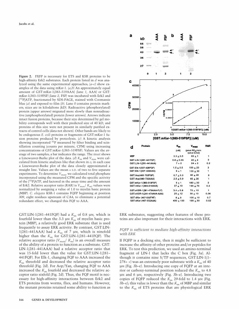
GST:LIN-1(281–441FQF) had a Km of 0.8 µM, which isfourfold lower than the 3.3 µM Km of myelin basic pro-tein (MBP), a relatively good ERK substrate that is usedfrequently to assay ERK activity. By contrast, GST:LIN-1(281–441AAA) had a Km of 7 µM, which is ninefoldhigher than the Km for GST:LIN-1(281–441FQF). Therelative acceptor ratio (Vmax/ Km) is an overall measureof the ability of a protein to function as a substrate. GST:LIN-1(281–441AAA) had a relative acceptor ratio thatwas 15-fold lower than the value for GST:LIN-1(281–441FQF). For Elk-1, changing FQF to AAA increased theKm threefold and decreased the relative acceptor ratiothreefold (Fig. 2d). For Aop/Yan, changing FQF to AAAincreased the Km fourfold and decreased the relative ac-ceptor ratio sixfold (Fig. 2d). Thus, the FQF motif is nec-essary for high-affinity interactions between Erk2 andETS proteins from worms, flies, and humans. However,the mutant proteins retained some ability to function as
ERK substrates, suggesting other features of these pro-teins are also important for their interactions with ERK.
FQFP is sufficient to mediate high-affinity interactionswith ERK
If FQFP is a docking site, then it might be sufficient toincrease the affinity of other proteins and/or peptides forERK. To test this prediction, we used an amino-terminalfragment of LIN-1 that lacks the C box (Fig. 3a). Al-though it contains nine S/TP sequences, GST:LIN-1(1–278+ +) was an extremely poor substrate with a Km of 40µM (Fig. 3b–e). Introducing one copy of FQFP at an inte-rior or carboxy-terminal position reduced the Km to 4.8µM and 6 µM, respectively (Fig. 3b–e). Introducing twocopies of FQFP reduced the Km 29-fold to 1.4 µM (Fig.3b–e); this value is lower than the Km of MBP and similarto the Km of ETS proteins that are physiological ERK
Figure 2. FXFP is necessary for ETS and KSR proteins to behigh-affinity Erk2 substrates. Each protein listed in d was ana-lyzed using the same experimental approaches; (a–c) show ex-amples of the data using mKsr-1. (a,b) An approximately equalamount of GST:mKsr-1(383–519AAA) (lane 1, AAA) or GST:mKsr-1(383–519FSF) (lane 2, FSF) was incubated with Erk2 and[32P]ATP, fractionated by SDS-PAGE, stained with Coomassieblue (a) and exposed to film (b). Lane 0 contains protein mark-ers; sizes are in kilodaltons (kD). Radioactive (phosphorylated)protein (upper arrows) migrated more slowly than nonradioac-tive (unphosphorylated) protein (lower arrows). Arrows indicateintact fusion proteins, because their size determined by gel mo-bility corresponds well with their predicted size of 40 kD, andproteins of this size were not present in similarly purified ex-tracts of control cells (data not shown). Other bands are likely tobe endogenous E. coli proteins or fragments of GST:mKsr-1 fu-sion proteins produced by proteolysis. (c) A kinetic analysisshowing incorporated 32P measured by filter binding and scin-tillation counting (counts per minute, CPM) using increasingconcentrations of GST:mKsr-1(383–519FSF). Values are the av-erage of two samples; a bar indicates the range. The inset showsa Lineweaver-Burke plot of the data. (d) Km and Vmax were cal-culated from kinetic analyses like that shown in c; in each casea Lineweaver-Burke plot of the data closely approximated astraight line. Values are the mean ± S.D. of two to five separateexperiments. To determine Vmax, we calculated total phosphateincorporated using the measured CPM and the specific activityof the [32P]ATP, and factored in the assay time and the amountof Erk2. Relative acceptor ratio (RAR) is Vmax/ Km; values werenormalized by assigning a value of 1.0 to myelin basic protein(MBP). C. elegans KSR-1 contains FQFP beginning at position309, eight residues upstream of CA4; to eliminate a potentialredundant effect, we changed this FQF to AAA.
Jacobs et al.
166 GENES & DEVELOPMENT
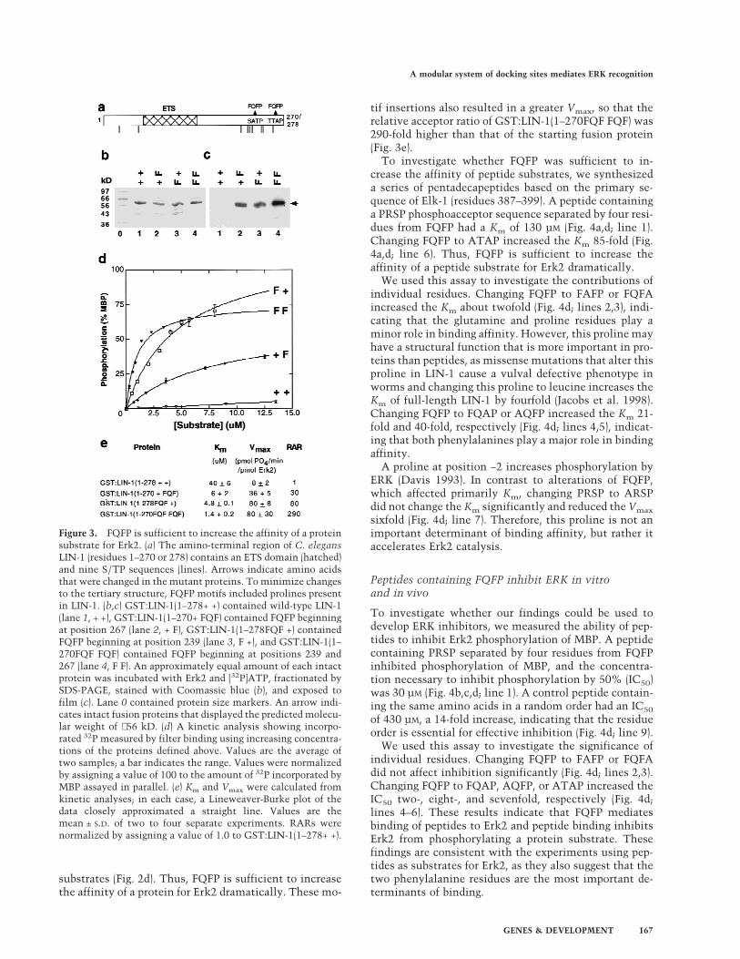
substrates (Fig. 2d). Thus, FQFP is sufficient to increasethe affinity of a protein for Erk2 dramatically. These mo-
tif insertions also resulted in a greater Vmax, so that therelative acceptor ratio of GST:LIN-1(1–270FQF FQF) was290-fold higher than that of the starting fusion protein(Fig. 3e).
To investigate whether FQFP was sufficient to in-crease the affinity of peptide substrates, we synthesizeda series of pentadecapeptides based on the primary se-quence of Elk-1 (residues 387–399). A peptide containinga PRSP phosphoacceptor sequence separated by four resi-dues from FQFP had a Km of 130 µM (Fig. 4a,d; line 1).Changing FQFP to ATAP increased the Km 85-fold (Fig.4a,d; line 6). Thus, FQFP is sufficient to increase theaffinity of a peptide substrate for Erk2 dramatically.
We used this assay to investigate the contributions ofindividual residues. Changing FQFP to FAFP or FQFAincreased the Km about twofold (Fig. 4d; lines 2,3), indi-cating that the glutamine and proline residues play aminor role in binding affinity. However, this proline mayhave a structural function that is more important in pro-teins than peptides, as missense mutations that alter thisproline in LIN-1 cause a vulval defective phenotype inworms and changing this proline to leucine increases theKm of full-length LIN-1 by fourfold (Jacobs et al. 1998).Changing FQFP to FQAP or AQFP increased the Km 21-fold and 40-fold, respectively (Fig. 4d; lines 4,5), indicat-ing that both phenylalanines play a major role in bindingaffinity.
A proline at position −2 increases phosphorylation byERK (Davis 1993). In contrast to alterations of FQFP,which affected primarily Km, changing PRSP to ARSPdid not change the Km significantly and reduced the Vmax
sixfold (Fig. 4d; line 7). Therefore, this proline is not animportant determinant of binding affinity, but rather itaccelerates Erk2 catalysis.
Peptides containing FQFP inhibit ERK in vitroand in vivo
To investigate whether our findings could be used todevelop ERK inhibitors, we measured the ability of pep-tides to inhibit Erk2 phosphorylation of MBP. A peptidecontaining PRSP separated by four residues from FQFPinhibited phosphorylation of MBP, and the concentra-tion necessary to inhibit phosphorylation by 50% (IC50)was 30 µM (Fig. 4b,c,d; line 1). A control peptide contain-ing the same amino acids in a random order had an IC50
of 430 µM, a 14-fold increase, indicating that the residueorder is essential for effective inhibition (Fig. 4d; line 9).
We used this assay to investigate the significance ofindividual residues. Changing FQFP to FAFP or FQFAdid not affect inhibition significantly (Fig. 4d; lines 2,3).Changing FQFP to FQAP, AQFP, or ATAP increased theIC50 two-, eight-, and sevenfold, respectively (Fig. 4d;lines 4–6). These results indicate that FQFP mediatesbinding of peptides to Erk2 and peptide binding inhibitsErk2 from phosphorylating a protein substrate. Thesefindings are consistent with the experiments using pep-tides as substrates for Erk2, as they also suggest that thetwo phenylalanine residues are the most important de-terminants of binding.
Figure 3. FQFP is sufficient to increase the affinity of a proteinsubstrate for Erk2. (a) The amino-terminal region of C. elegansLIN-1 (residues 1–270 or 278) contains an ETS domain (hatched)and nine S/TP sequences (lines). Arrows indicate amino acidsthat were changed in the mutant proteins. To minimize changesto the tertiary structure, FQFP motifs included prolines presentin LIN-1. (b,c) GST:LIN-1(1–278+ +) contained wild-type LIN-1(lane 1, + +), GST:LIN-1(1–270+ FQF) contained FQFP beginningat position 267 (lane 2, + F), GST:LIN-1(1–278FQF +) containedFQFP beginning at position 239 (lane 3, F +), and GST:LIN-1(1–270FQF FQF) contained FQFP beginning at positions 239 and267 (lane 4, F F). An approximately equal amount of each intactprotein was incubated with Erk2 and [32P]ATP, fractionated bySDS-PAGE, stained with Coomassie blue (b), and exposed tofilm (c). Lane 0 contained protein size markers. An arrow indi-cates intact fusion proteins that displayed the predicted molecu-lar weight of ∼56 kD. (d) A kinetic analysis showing incorpo-rated 32P measured by filter binding using increasing concentra-tions of the proteins defined above. Values are the average oftwo samples; a bar indicates the range. Values were normalizedby assigning a value of 100 to the amount of 32P incorporated byMBP assayed in parallel. (e) Km and Vmax were calculated fromkinetic analyses; in each case, a Lineweaver-Burke plot of thedata closely approximated a straight line. Values are themean ± S.D. of two to four separate experiments. RARs werenormalized by assigning a value of 1.0 to GST:LIN-1(1–278+ +).
A modular system of docking sites mediates ERK recognition
GENES & DEVELOPMENT 167

The SP acceptor site did not cause significant inhibi-tion, because changing PRSP to PRAP or ARSP did notaffect IC50 significantly (Fig. 4d; lines 7–8). These datademonstrate that FQFP-mediated binding to ERK doesnot require a phosphoacceptor site and support themodel that FQFP is a docking site that interacts with anERK binding pocket that is functionally distinguishablefrom the ERK active site.
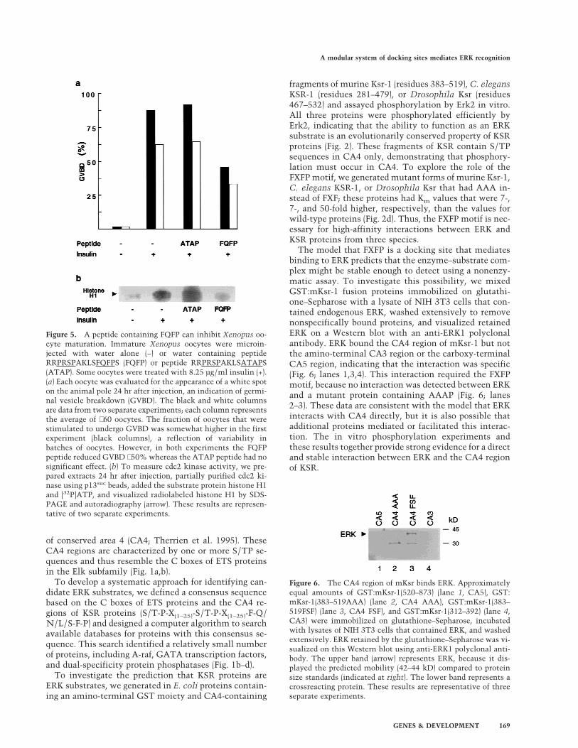
To determine if pentadecapeptides containing FQFPcan inhibit ERK in vivo, we assayed meiotic maturationof Xenopus oocytes. Treating immature oocytes with in-sulin activates the Ras–ERK pathway (Deshpande andKung 1987; Korn et al. 1987; Nebreda et al. 1993), whichresults in morphological changes such as germinalvesicle breakdown (GVBD) and biochemical changessuch as the activation of cdc2 kinase (Fig. 5; columns1–2). We injected insulin-stimulated oocytes with a pep-tide containing FQFP (RRPRSPAKLSFQFPS) or a controlpeptide lacking FQFP (RRPRSPAKLSATAPS). The pep-tide containing FQFP reduced cdc2 kinase activity andthe fraction of oocytes that displayed GVBD signifi-cantly, whereas the peptide lacking FQFP had no signifi-cant affect (Fig. 5; columns 3–4). To investigate furtherwhich step in this signaling pathway was inhibited bythe peptide containing FQFP, we analyzed the degree ofphosphorylation of ERK, an indirect measure of MEK
activity. Extracts of oocytes injected with a peptide con-taining FQFP or a control peptide were analyzed byWestern blotting using an antibody that recognizes phos-phorylated ERK specifically. A similar amount of phos-phorylated ERK was observed in both extracts, indicat-ing that the peptide containing FQFP did not affect theactivity of MEK or more upstream signaling events (datanot shown). These results demonstrate that FQFP is suf-ficient to cause a peptide to function as an inhibitor ofoocyte maturation. Furthermore, the peptide appeared tofunction at a step downstream of MEK and upstream ofcdc2 kinase activation, consistent with the model thatthe peptide inhibited ERK activity.
FXFP can be used to identify candidateERK substrates, including KSR proteinsthat are high-affinity ERK substrates
To investigate whether FQFP motifs mediate interac-tions between ERK and substrates besides ETS proteinsin the Elk subfamily and whether FQFP can be used topredict ERK substrates, we analyzed the sequences ofproteins involved in vulval induction and found that C.elegans KSR-1 contains FLFP. This motif is conserved inmurine Ksr-1 (FSFP) and Drosophila Ksr (FNFP) (Fig. 1b).These motifs are positioned at the carboxy-terminal end
Figure 4. FQFP is sufficient to increase the ability of peptidesto function as substrates and inhibitors of Erk2. (a) A kineticanalysis showing incorporated 32P measured by filter bindingusing increasing concentrations of RRPRSPAKLSFQFPS (closedtriangles, FQFP) or RRPRSPAKLSATAPS (open triangles,ATAP). Values are the average of two samples; a bar indicatesthe range. Values were normalized by assigning a value of 100 tothe amount of 32P incorporated by MBP assayed in parallel. TheATAP peptide displayed saturable phosphorylation at muchhigher concentrations than illustrated (data not shown). (b) In-hibition of Erk2 phosphorylation of MBP was measured by add-ing increasing concentrations of the FQFP peptide (top) or theATAP peptide (bottom) to reactions containing [32P]ATP and 18µM MBP. Reactions were fractionated by SDS-PAGE, and phos-phorylated MBP was visualized by autoradiography (arrow). (c)Inhibition assays like those shown in b were quantified using aPhosphorImager (Molecular Dynamics). Signals for the FQFPpeptide (closed triangles) or the ATAP peptide (open triangles)were normalized by assigning a value of 100% to assays con-taining no peptide and plotted on a logarithmic scale. Values arethe averages of two samples; a bar indicates the range. (d) High-lighted residues in peptide sequences are differences comparedto line 1. The peptide in line 9 contains the same residues as line1, but the order was scrambled. Km and Vmax were calculatedfrom kinetic analyses like those shown in a; in each case, aLineweaver-Burke plot of the data closely approximated astraight line. Values are the mean ± S.D. of two independent ex-periments. RARs were normalized by assigning a value of 1.0 tothe peptide in line 1. Peptides in lines 8 and 9 lack SP and werenot phosphorylated significantly. Inhibition curves like thoseshown in c were used to determine IC50—the concentration ofpeptide that reduced Erk2 phosphorylation of MBP by 50%. Val-ues are the mean ± S.D. of two independent experiments.
Jacobs et al.
168 GENES & DEVELOPMENT
of conserved area 4 (CA4; Therrien et al. 1995). TheseCA4 regions are characterized by one or more S/TP se-quences and thus resemble the C boxes of ETS proteinsin the Elk subfamily (Fig. 1a,b).
To develop a systematic approach for identifying can-didate ERK substrates, we defined a consensus sequencebased on the C boxes of ETS proteins and the CA4 re-gions of KSR proteins (S/T-P-X(1–25)-S/T-P-X(1–25)-F-Q/N/L/S-F-P) and designed a computer algorithm to searchavailable databases for proteins with this consensus se-quence. This search identified a relatively small numberof proteins, including A-raf, GATA transcription factors,and dual-specificity protein phosphatases (Fig. 1b–d).
To investigate the prediction that KSR proteins areERK substrates, we generated in E. coli proteins contain-ing an amino-terminal GST moiety and CA4-containing
fragments of murine Ksr-1 (residues 383–519), C. elegansKSR-1 (residues 281–479), or Drosophila Ksr (residues467–532) and assayed phosphorylation by Erk2 in vitro.All three proteins were phosphorylated efficiently byErk2, indicating that the ability to function as an ERKsubstrate is an evolutionarily conserved property of KSRproteins (Fig. 2). These fragments of KSR contain S/TPsequences in CA4 only, demonstrating that phosphory-lation must occur in CA4. To explore the role of theFXFP motif, we generated mutant forms of murine Ksr-1,C. elegans KSR-1, or Drosophila Ksr that had AAA in-stead of FXF; these proteins had Km values that were 7-,7-, and 50-fold higher, respectively, than the values forwild-type proteins (Fig. 2d). Thus, the FXFP motif is nec-essary for high-affinity interactions between ERK andKSR proteins from three species.
The model that FXFP is a docking site that mediatesbinding to ERK predicts that the enzyme–substrate com-plex might be stable enough to detect using a nonenzy-matic assay. To investigate this possibility, we mixedGST:mKsr-1 fusion proteins immobilized on glutathi-one–Sepharose with a lysate of NIH 3T3 cells that con-tained endogenous ERK, washed extensively to removenonspecifically bound proteins, and visualized retainedERK on a Western blot with an anti-ERK1 polyclonalantibody. ERK bound the CA4 region of mKsr-1 but notthe amino-terminal CA3 region or the carboxy-terminalCA5 region, indicating that the interaction was specific(Fig. 6; lanes 1,3,4). This interaction required the FXFPmotif, because no interaction was detected between ERKand a mutant protein containing AAAP (Fig. 6; lanes2–3). These data are consistent with the model that ERKinteracts with CA4 directly, but it is also possible thatadditional proteins mediated or facilitated this interac-tion. The in vitro phosphorylation experiments andthese results together provide strong evidence for a directand stable interaction between ERK and the CA4 regionof KSR.
Figure 5. A peptide containing FQFP can inhibit Xenopus oo-cyte maturation. Immature Xenopus oocytes were microin-jected with water alone (−) or water containing peptideRRPRSPAKLSFQFPS (FQFP) or peptide RRPRSPAKLSATAPS(ATAP). Some oocytes were treated with 8.25 µg/ml insulin (+).(a) Each oocyte was evaluated for the appearance of a white spoton the animal pole 24 hr after injection, an indication of germi-nal vesicle breakdown (GVBD). The black and white columnsare data from two separate experiments; each column representsthe average of ∼60 oocytes. The fraction of oocytes that werestimulated to undergo GVBD was somewhat higher in the firstexperiment (black columns), a reflection of variability inbatches of oocytes. However, in both experiments the FQFPpeptide reduced GVBD ∼50% whereas the ATAP peptide had nosignificant effect. (b) To measure cdc2 kinase activity, we pre-pared extracts 24 hr after injection, partially purified cdc2 ki-nase using p13suc beads, added the substrate protein histone H1and [32P]ATP, and visualized radiolabeled histone H1 by SDS-PAGE and autoradiography (arrow). These results are represen-tative of two separate experiments.
Figure 6. The CA4 region of mKsr binds ERK. Approximatelyequal amounts of GST:mKsr-1(520–873) (lane 1, CA5), GST:mKsr-1(383–519AAA) (lane 2, CA4 AAA), GST:mKsr-1(383–519FSF) (lane 3, CA4 FSF), and GST:mKsr-1(312–392) (lane 4,CA3) were immobilized on glutathione–Sepharose, incubatedwith lysates of NIH 3T3 cells that contained ERK, and washedextensively. ERK retained by the glutathione–Sepharose was vi-sualized on this Western blot using anti-ERK1 polyclonal anti-body. The upper band (arrow) represents ERK, because it dis-played the predicted mobility (42–44 kD) compared to proteinsize standards (indicated at right). The lower band represents acrossreacting protein. These results are representative of threeseparate experiments.
A modular system of docking sites mediates ERK recognition
GENES & DEVELOPMENT 169

FQFP and the D box function independentlyand additively as docking sites for ERK
The ETS proteins Elk-1, SAP-1a, and Net contain a con-served region called the D box that is characterized bythe amino acid sequence LXL carboxy-terminal to a clus-ter of basic residues (Lopez et al. 1994). We identifiedsequences similar to the D box in comparable positionsof C. elegans LIN-1 and Drosophila Aop/Yan (Fig. 1a,g).These observations support our hypothesis that LIN-1and Aop are members of the Elk subfamily and indicatethat the ancestral gene encoded a protein with an ETSdomain, a D box, and a C box. The D box of Elk-1 wasreported recently to function as a docking site for ERKand JNK (Yang et al. 1998a,b). To investigate the rela-tionship between the D box and the FQFP motif, weanalyzed the ability of Erk2 to phosphorylate Elk-1 pro-teins with mutations that disrupt the D box, the FQFPmotif, or both.
To disrupt the D box, we mutated the LEL sequence ofElk-1 to AEA; this change reduces the function of the Dbox as a docking site for ERK significantly (Yang et al.1998b). Wild-type GST:Elk-1(307–428) had a Km of 1.5µM (Fig. 7a,b,e). Changing FQFP to AAAP increased by3-fold the Km of Elk-1 with a wild-type D box (Fig. 7e;lines 1,2) and by 12-fold the Km of Elk-1 with a mutant Dbox (Fig. 7e, lines 3,4). Thus, FQFP mediated an interac-tion with ERK in the presence or absence of a D box.Changing LEL to AEA increased by 14-fold the Km ofElk-1 with wild-type FQFP (Fig. 7e; lines 1,3) and by 50-fold the Km of Elk-1 with a mutant FQFP motif (Fig. 7e;lines 2,4). Thus, the D box mediated an interaction withERK in the presence or absence of FQFP. The doublemutant protein had a Km of 260 µM, showing that thetwo sites in combination reduced the Km 170-fold. To-gether, these results indicate that the two docking sitesfunction independently and additively.
FQFP does not mediate interactions with JNKMAP kinase
Elk-1 is a substrate for JNK (Minden and Karin 1997). Toinvestigate whether FQFP mediates interactions withJNK, we used purified, recombinant, rat JNKb and thewild-type and mutant forms of Elk-1 described above.Wild-type GST:Elk-1(307–428) had a Km of 5 µM (Fig.7c-e). Disrupting the D box of wild-type Elk-1 or Elk-1with a mutation of FQFP increased the Km 22-fold and32-fold, respectively (Fig. 7e; lines 1,3 and 2,4). Thesedata are consistent with previous findings that the D boxmediates interactions with JNK (Yang et al. 1998b) andshow that the D box functions independently of FQFP.By contrast, disrupting the FQFP motif in either wild-type Elk-1 or Elk-1 with a mutation in the D box did notaffect Km significantly (Fig. 7e; lines 1,2 and 3,4). Thesedata indicate that FQFP does not mediate interactionswith JNK.
These results suggest that a protein that containsFXFP but lacks a D box will not function as a high-affinity substrate for JNK. To test this prediction, we
analyzed JNKb phosphorylation of Drosophila Ksr. GST:dKsr(467–532FNF) was a very poor substrate with a Km
of 180 ± 20 µM (mean ± S.D. of two separate trials). GST:dKsr (467–532AAA) had a Km of 110 ± 10 µM. Thus, FXFPdid not mediate an interaction between JNK and KSR.
Discussion
The results presented here lead to five major conclu-sions. First, FXFP is an evolutionarily conserved dockingsite that mediates high-affinity interactions betweenERK and substrate proteins in at least two different pro-tein families. Second, FXFP and the d domain/D box, adifferent docking site, can function separately or to-gether in substrate proteins to mediate binding to ERK.We refer to this as a modular system of docking sites.Third, the partially overlapping substrate specificities ofERK and JNK result from the ability of these enzymes torecognize shared and unique docking sites. Fourth, FXFP
Figure 7. FQFP and the D box mediate additive interactionswith ERK; FQFP does not mediate interactions with JNK.GST:Elk-1(307–428LEL-FQF) contained wild-type Elk-1 (lanes 1and 6, LEL-FQF), GST:Elk-1(307–428LEL-AAA) contained a sub-stitution of AAA for FQF (lanes 2 and 7, LEL-AAA), GST:Elk-1(307–428AEA-FQF) contained a substitution of AEA for LEL(lanes 3 and 8, AEA-FQF), and GST:Elk-1(307–428AEA-AAA)contained both substitutions (lanes 4 and 9, AEA-AAA). Pro-teins were incubated with Erk2 (a,b) or JNKb (c,d) and [32P]ATP,and approximately equal amounts were fractionated by SDS-PAGE, stained with Coomassie blue (a,c), and exposed to film(b,d). Lanes 0 and 5 contain protein size markers. Arrows indi-cate intact fusion proteins that migrated with the predicted mo-lecular weight of ∼39 kD. (e) Km was calculated from kineticanalyses; in each case a Lineweaver-Burke plot of the dataclosely approximated a straight line. Values are the mean ± S.D.of two separate experiments.
Jacobs et al.
170 GENES & DEVELOPMENT

can be used to identify ERK substrates, such as KSR.Fifth, peptides containing FXFP can be used to inhibitERK.
FXFP is a docking site for ERK
The evidence in support of this conclusion can be sum-marized as follows: (1) The Elk subfamily of ETS tran-scription factors includes some of the best documentedphysiological ERK substrates, since LIN-1, Aop/Yan, andElk-1 were independently shown to be regulated directlyby ERK in worms, flies, and vertebrates, respectively(O’Neill et al. 1994; Treisman 1994; Jacobs et al. 1998;Tan et al. 1998). The results presented here suggest thatthe KSR family of protein kinases are also physiologicalERK substrates (described below). FXFP is evolutionarilyconserved in both of these protein families, indicatingthat FXFP plays an important and perhaps similar role inthese proteins. FXFP is positioned just carboxy-terminalto ERK phosphorylation sites in both of these proteinfamilies, consistent with it being involved in ERK phos-phorylation. (2) FXFP was necessary for high-affinity in-teractions in vitro between ERK and C. elegans LIN-1and KSR-1, Drosophila Aop/Yan and Ksr, and vertebrateElk-1 and mKsr-1. The effects of mutating FXFP are notlikely to be the result of nonspecific alterations of pro-tein structure, because we observed a similar effect in allsix proteins and mutating FXFP did not diminish phos-phorylation of Elk-1 by JNK. (3) The addition of FXFP issufficient to increase the affinity of protein and peptidesubstrates for ERK dramatically in vitro and to increasethe efficacy of peptide inhibitors of ERK in vitro. Impor-tantly, these peptide inhibitors do not require an S/TPsite, demonstrating that FQFP-mediated binding to ERKcan occur independently of S/TP-mediated binding tothe active site of ERK. (4) Two different experimentssuggest FXFP mediates interactions with ERK in vivo:Mutations that alter or eliminate FQFP cause C. eleganslin-1 to be unresponsive to RTK–Ras–ERK-mediated sig-naling in worms (Jacobs et al. 1998); peptides containingFXFP interfere with RTK–Ras–ERK-mediated signalingduring Xenopus oocyte maturation. (5) Based on the sixETS and KSR proteins that were demonstrated to have amotif that mediates binding to ERK, we defined a con-sensus sequence: F-X-F-P (Fig. 1h). This consensus se-quence is consistent with our findings that peptide sub-strates and inhibitors required both phenylalanine resi-dues for high-affinity binding, whereas the residue inbetween was less significant. We speculate that ERK hasa binding pocket that interacts with these bulky, hydro-phobic residues. Additional experiments are necessary todefine further the effect of particular residues at eachposition. We propose that this motif be named DEF(docking site for ERK, FXFP).
This conclusion is significant because it reveals a fun-damental mechanism that enables ERK to interact withsubstrate proteins. This appears to be an ancient mecha-nism of substrate recognition, as it has been conservedduring the evolution of worms, flies, and vertebrates.The demonstration that FXFP mediates interactions be-
tween ERK and two different protein families, and thefinding that FXFP is present in additional demonstratedand candidate ERK substrates (described below), suggestthat many different ERK substrates may share the sameFXFP docking site.
Multiple docking sites on ERK substrate proteins forma modular recognition system
The d domain of c-Jun is a docking site for JNK (Karin1995). Yang et al. (1998a,b) extended the analysis of thismotif by showing that the D box of Elk-1 and the d do-main have a similar sequence, are functionally inter-changeable, and function as docking sites for both ERKand JNK. ERK-specific MAP kinase kinases are sub-strates for ERK (Matsuda et al. 1993), and the amino-terminal region of the ERK-specific MAP kinase kinaseof S. cerevisiae, Ste7, mediates an interaction with ERK(Bardwell et al. 1996). Bardwell and Thorner (1996) notedthat the amino termini of many ERK-specific MAP ki-nase kinases contain an evolutionarily conserved se-quence that was proposed to mediate interactions withERK and named a docking site. We noted that the se-quence of the docking site is similar to the sequence of ddomain and the D box, as all three motifs are character-ized by a cluster of basic residues amino-terminal to anL/I-X-L/I motif (Fig. 1g). Furthermore, we found a simi-lar motif in a comparable position of JNK-specific MAPkinase kinases (Fig. 1e,g), consistent with the possibilitythat this motif interacts with JNK and ERK. We proposethat the d domain, D box, and docking site are versions ofthe same motif, and this motif functions as a dockingsite for ERK and JNK. Figure 1h shows two slightly dif-ferent consensus motifs defined by these sequences. Wepropose that this motif be named DEJL (docking site forERK and JNK, LXL). Our goal is to initiate a systematicand informative nomenclature for MAP kinase dockingsites that can be used to name additional motifs de-scribed in the future.
Two types of evidence support the model that the DEFand DEJL form a modular system that mediates recogni-tion by ERK (Fig. 8). First is our analysis of Elk-1. Amutant Elk-1 protein lacking both motifs did not inter-act with ERK significantly. Restoring the DEF or DEJLdecreased the Km 12- and 50-fold, respectively, showingthat these motifs can mediate interactions with ERK in-dependently. Restoring both sequences decreased the Km
170-fold, showing that in combination these motifsfunction additively rather than redundantly or synergis-tically. These findings, together with the observationthat the sequences of the DEF and DEJL are not similar,suggest that the DEF and DEJL may interact with sepa-rate binding pockets of ERK. If ERK has separate bindingpockets, then ERK may be capable of simultaneouslyinteracting with the DEF and DEJL. However, the resultspresented here do not test this possibility.
Second, we analyzed the sequences of many proteinsreported to be ERK substrates to determine if they con-tained a candidate DEF or DEJL. This analysis revealedfour classes of substrate proteins (Fig. 8). Class I sub-
A modular system of docking sites mediates ERK recognition
GENES & DEVELOPMENT 171
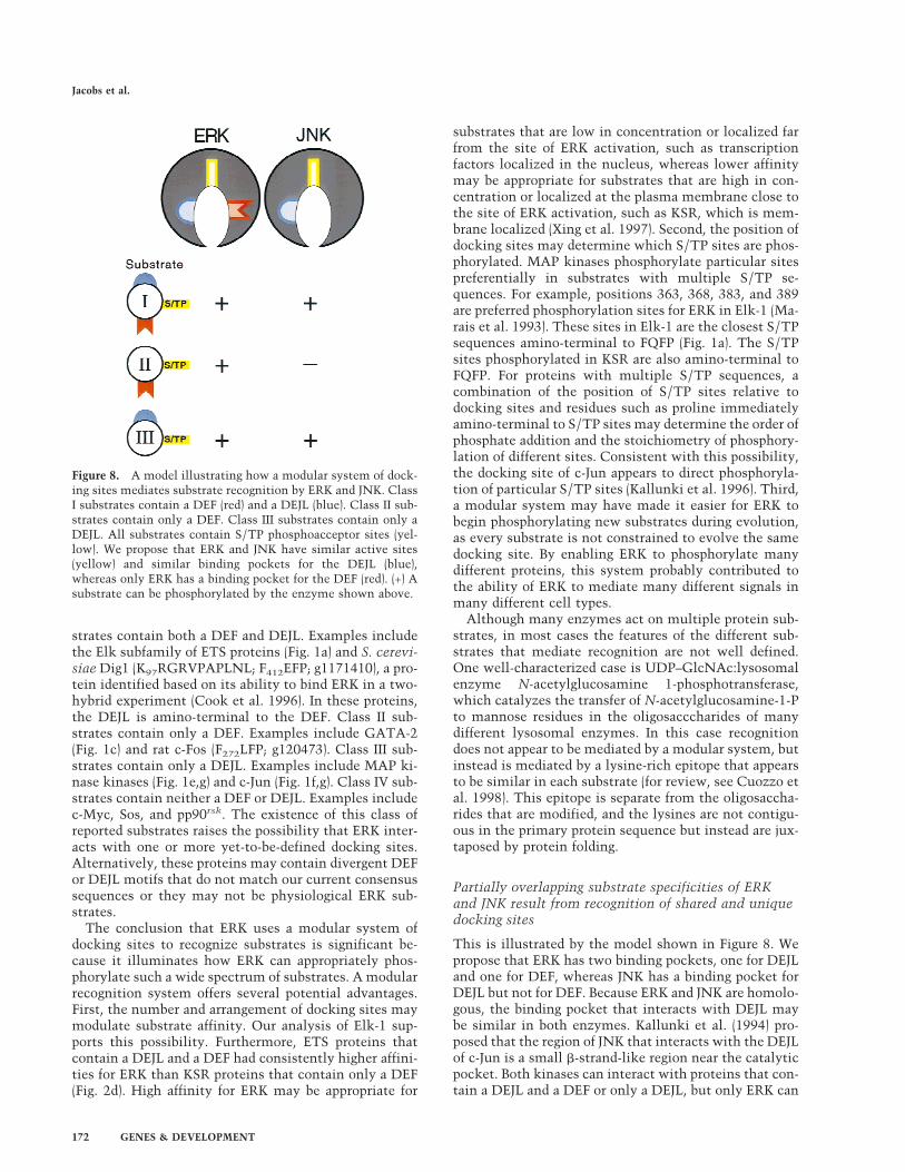
strates contain both a DEF and DEJL. Examples includethe Elk subfamily of ETS proteins (Fig. 1a) and S. cerevi-siae Dig1 (K97RGRVPAPLNL; F412EFP; g1171410), a pro-tein identified based on its ability to bind ERK in a two-hybrid experiment (Cook et al. 1996). In these proteins,the DEJL is amino-terminal to the DEF. Class II sub-strates contain only a DEF. Examples include GATA-2(Fig. 1c) and rat c-Fos (F272LFP; g120473). Class III sub-strates contain only a DEJL. Examples include MAP ki-nase kinases (Fig. 1e,g) and c-Jun (Fig. 1f,g). Class IV sub-strates contain neither a DEF or DEJL. Examples includec-Myc, Sos, and pp90rsk. The existence of this class ofreported substrates raises the possibility that ERK inter-acts with one or more yet-to-be-defined docking sites.Alternatively, these proteins may contain divergent DEFor DEJL motifs that do not match our current consensussequences or they may not be physiological ERK sub-strates.
The conclusion that ERK uses a modular system ofdocking sites to recognize substrates is significant be-cause it illuminates how ERK can appropriately phos-phorylate such a wide spectrum of substrates. A modularrecognition system offers several potential advantages.First, the number and arrangement of docking sites maymodulate substrate affinity. Our analysis of Elk-1 sup-ports this possibility. Furthermore, ETS proteins thatcontain a DEJL and a DEF had consistently higher affini-ties for ERK than KSR proteins that contain only a DEF(Fig. 2d). High affinity for ERK may be appropriate for
substrates that are low in concentration or localized farfrom the site of ERK activation, such as transcriptionfactors localized in the nucleus, whereas lower affinitymay be appropriate for substrates that are high in con-centration or localized at the plasma membrane close tothe site of ERK activation, such as KSR, which is mem-brane localized (Xing et al. 1997). Second, the position ofdocking sites may determine which S/TP sites are phos-phorylated. MAP kinases phosphorylate particular sitespreferentially in substrates with multiple S/TP se-quences. For example, positions 363, 368, 383, and 389are preferred phosphorylation sites for ERK in Elk-1 (Ma-rais et al. 1993). These sites in Elk-1 are the closest S/TPsequences amino-terminal to FQFP (Fig. 1a). The S/TPsites phosphorylated in KSR are also amino-terminal toFQFP. For proteins with multiple S/TP sequences, acombination of the position of S/TP sites relative todocking sites and residues such as proline immediatelyamino-terminal to S/TP sites may determine the order ofphosphate addition and the stoichiometry of phosphory-lation of different sites. Consistent with this possibility,the docking site of c-Jun appears to direct phosphoryla-tion of particular S/TP sites (Kallunki et al. 1996). Third,a modular system may have made it easier for ERK tobegin phosphorylating new substrates during evolution,as every substrate is not constrained to evolve the samedocking site. By enabling ERK to phosphorylate manydifferent proteins, this system probably contributed tothe ability of ERK to mediate many different signals inmany different cell types.
Although many enzymes act on multiple protein sub-strates, in most cases the features of the different sub-strates that mediate recognition are not well defined.One well-characterized case is UDP–GlcNAc:lysosomalenzyme N-acetylglucosamine 1-phosphotransferase,which catalyzes the transfer of N-acetylglucosamine-1-Pto mannose residues in the oligosacccharides of manydifferent lysosomal enzymes. In this case recognitiondoes not appear to be mediated by a modular system, butinstead is mediated by a lysine-rich epitope that appearsto be similar in each substrate (for review, see Cuozzo etal. 1998). This epitope is separate from the oligosaccha-rides that are modified, and the lysines are not contigu-ous in the primary protein sequence but instead are jux-taposed by protein folding.
Partially overlapping substrate specificities of ERKand JNK result from recognition of shared and uniquedocking sites
This is illustrated by the model shown in Figure 8. Wepropose that ERK has two binding pockets, one for DEJLand one for DEF, whereas JNK has a binding pocket forDEJL but not for DEF. Because ERK and JNK are homolo-gous, the binding pocket that interacts with DEJL maybe similar in both enzymes. Kallunki et al. (1994) pro-posed that the region of JNK that interacts with the DEJLof c-Jun is a small b-strand-like region near the catalyticpocket. Both kinases can interact with proteins that con-tain a DEJL and a DEF or only a DEJL, but only ERK can
Figure 8. A model illustrating how a modular system of dock-ing sites mediates substrate recognition by ERK and JNK. ClassI substrates contain a DEF (red) and a DEJL (blue). Class II sub-strates contain only a DEF. Class III substrates contain only aDEJL. All substrates contain S/TP phosphoacceptor sites (yel-low). We propose that ERK and JNK have similar active sites(yellow) and similar binding pockets for the DEJL (blue),whereas only ERK has a binding pocket for the DEF (red). (+) Asubstrate can be phosphorylated by the enzyme shown above.
Jacobs et al.
172 GENES & DEVELOPMENT
interact with proteins that contain only a DEF, such asKSR. We speculate that JNK phosphorylates unique sub-strates by recognizing one or more docking sites that donot bind ERK. Furthermore, other MAP kinases such asp38 may also employ a modular system that includesshared and unique docking sites to interact with sub-strate proteins.
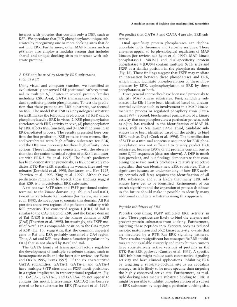
A DEF can be used to identify ERK substrates,such as KSR
Using visual and computer searches, we identified anevolutionarily conserved DEF positioned carboxy-termi-nal to multiple S/TP sites in several protein familiesincluding KSR, A-raf, GATA transcription factors, anddual-specificity protein phosphatases. To test the predic-tion that these proteins are ERK substrates, we focusedon KSR. The model that KSR is a physiological substratefor ERK makes the following predictions: (1) KSR can bephosphorylated by ERK in vitro; (2) KSR phosphorylationcorrelates with ERK activity in vivo; (3) phosphorylationby ERK affects KSR function; and (4) KSR functions in anERK-mediated process. The results presented here con-firm the first prediction; KSR proteins from worms, flies,and vertebrates were high-affinity substrates for ERK,and the DEF was necessary for these high-affinity inter-actions. These findings are consistent with the observa-tion that the amino-terminal region of mKsr-1 can inter-act with ERK-2 (Yu et al. 1997). The fourth predictionhas been demonstrated previously, as KSR positively me-diates RTK–Ras–ERK signaling in worms, flies and ver-tebrates (Kornfeld et al. 1995; Sundaram and Han 1995;Therrien et al. 1995; Xing et al. 1997). Although twopredictions remain to be tested, these findings suggestthat KSR is a physiological ERK substrate.
A-raf has two S/TP sites and FSFP positioned amino-terminal to the kinase domain (Fig. 1b). B-raf and Raf-1,two other vertebrate Raf proteins (for review, see Stormet al. 1990), do not appear to contain this domain. All Rafproteins share two regions of significant similarity withKSR proteins: The conserved region 1 (CR1) of Raf issimilar to the CA3 region of KSR, and the kinase domainof Raf (CR3) is similar to the kinase domain of KSR(CA5) (Therrien et al. 1995). Interestingly, the FXFP mo-tif of A-raf is in a comparable position to the CA4 regionof KSR (Fig. 1b), suggesting that the common ancestralgene of Raf and KSR probably contained a CA4 region.Thus, A-raf and KSR may share a function (regulation byERK) that is not shared by B-raf and Raf-1.
The GATA family of transcription factors regulatesthe development of multiple vertebrate tissues, such ashematopoeitic cells and the heart (for review, see Weissand Orkin 1995; Evans 1997). Of the six characterizedGATA subfamilies, GATA-2, GATA-3, and GATA-4have multiple S/TP sites and an FXFP motif positionedin a region implicated in transcriptional regulation (Fig.1c). GATA-1, GATA-5, and GATA-6 do not appear tocontain this motif. Interestingly, GATA-2 has been re-ported to be a substrate for ERK (Towatari et al. 1995).
We predict that GATA-3 and GATA-4 are also ERK sub-strates.
Dual specificity protein phosphatases can dephos-phorylate both threonine and tyrosine residues. Theseenzymes appear to be physiological regulators of MAPkinases (for review, see Byon et al. 1997). MAP kinasephosphatase-1 (MKP-1) and dual-specificity proteinphosphatase 4 (DUS4) contain multiple S/TP sites andFXFP at a similar position in the phosphatase domain(Fig. 1d). These findings suggest that FXFP may mediatean interaction between these phosphatases and ERK,which might facilitate phosphorylation of these phos-phatases by ERK, dephosphorylation of ERK by thesephosphatases, or both.
Three general approaches have been used previously toidentify MAP kinase substrates. First, candidate sub-strates like Elk-1 have been identified based on circum-stantial evidence such as involvement in a MAP kinase-mediated process or regulated phosphorylation (Treis-man 1994). Second, biochemical purification of a kinaseactivity that can phosphorylate a particular protein, suchas c-Jun, has resulted in the identification of MAP ki-nases, such as JNK (Karin 1995). Third, candidate sub-strates have been identified based on the ability to bindERK, such as Dig1 (Cook et al. 1996). The identificationof S/TP as a minimal consensus sequence for ERK phos-phorylation was not sufficient to reliably predict ERKsubstrates, because ∼90% of all proteins contain one ormore S/TP sequences (data not shown). A DEF is muchless prevalent, and our findings demonstrate that com-bining these two motifs produces a relatively selectivealgorithm that can identify new ERK substrates. This issignificant because an understanding of how ERK activ-ity controls cell fates requires the identification of allERK substrates, and it is likely that many ERK sub-strates have yet to be identified. Refinements of oursearch algorithm and the expansion of protein databasesin the future should make it possible to identify manyadditional candidate substrates using this approach.
Peptide inhibitors of ERK
Peptides containing FQFP inhibited ERK activity invitro. These peptides are likely to bind the enzyme andprevent protein substrates from docking. Furthermore,injecting these peptides into Xenopus oocytes reducedmeiotic maturation and cdc2 kinase activity, events thatare mediated by a RTK–Ras–ERK signaling pathway.These results are significant because specific ERK inhibi-tors are not available currently and many human tumorshave constitutively active versions of proteins in theRTK–Ras–ERK pathway (Cantley et al. 1991). A specificERK inhibitor might reduce such constitutive signalingactivity and have clinical applications. Inhibiting ERKby targeting a substrate-docking site is an appealingstrategy, as it is likely to be more specific than targetingthe highly conserved active site. Furthermore, as mul-tiple docking sites mediate substrate binding to ERK, itmight be possible to inhibit phosphorylation of a subsetof ERK substrates by targeting a particular docking site.
A modular system of docking sites mediates ERK recognition
GENES & DEVELOPMENT 173

Materials and methods
Protein production and MAP kinase assays
Expression plasmids were derived from cloned cDNAs andpGEX vectors (Pharmacia) and modified by in vitro mutagenesisusing standard techniques (Sambrook et al. 1989). Proteins wereexpressed in E. coli BL21, partially purified using glutathione–Sepharose (Pharmacia) essentially according to the manufactur-er’s instructions, and dialyzed into kinase assay buffer. Theamount of intact protein was estimated by comparison toknown amounts of purified bovine serum albumin present inadjacent lanes of Coomassie blue-stained SDS-PAGE gels. Puri-fied MBP was from GIBCO-BRL. Peptides were synthesized us-ing standard 9-fluorenylmethoxycarbonyl chemistry and puri-fied using HPLC by the Tufts Core Facility (Boston, MA).
Purified, recombinant, murine Erk2 (New England Biolabs) isfully active, because it is produced in E. coli containing consti-tutively active MEK. Assays were done according to Alessi et al.(1995). A 50-µl reaction contained 100 µM [32P]ATP (0.15 Ci/mmole) and 0.05 pmole Erk2 and was terminated after 15 minat 30°C, at which point 32P incorporation was linear with re-spect to time. Purified, recombinant, rat JNKb (Stratagene) ispartially active as a result of autophosphorylation. Assays weredone as described for Erk2 except that [32P]ATP was 0.6 Ci/mmole. For each protein, we used SDS-PAGE and autoradiog-raphy to establish that intact fusion protein contained most orall of the incorporated 32P. To quantify phosphorylation, wemeasured radioactive protein bound to phosphocellulose paper(P81, Whatman) using a scintillation counter.
Oocyte microinjection
Stage VI fully grown immature oocytes were obtained from ma-ture Xenopus laevis females and maintained in 1× modifiedBarth’s solution with HEPES, 1 mg/ml Ficoll 400, 1 mg/mlbovine serum albumin, and antibiotics (Muslin et al. 1993).Each oocyte was injected with 25–50 nl of water alone or 10 mM
peptide dissolved in water and incubated at 19°C for 24 hr in thepresence or absence of 8.25 µg/ml insulin. Using an estimate of10 µl for the volume of an average oocyte, we calculate that theinitial peptide concentration in the oocyte was ∼50 µM. GVBDwas defined as the presence of a broad white spot on the animalpole of the oocyte. Oocytes were pooled and lysed as describedby Muslin et al. (1993) and cdc2 kinase was assayed as describedby Xing et al. (1997).
Binding assay
NIH 3T3 cells were grown in DMEM + 10% calf serum to ∼80%confluence and lysed using NP-40 lysis buffer (Xing et al. 1997).Following low-speed centrifugation, cell extracts were incu-bated for 1 hr at 4°C with ∼2 µg of GST:mKsr-1 fusion proteinimmobilized on glutathione–Sepharose and washed three timeswith NP-40 lysis buffer for 10 sec at 4°C. Samples of beads wereboiled in SDS buffer, fractionated by SDS-PAGE, and transferredto a nitrocellulose membrane. Western blots were treated withrabbit, polyclonal, anti-ERK1, IgG antibody (C-16, Santa Cruz),anti-rabbit, secondary antibody conjugated to alkaline phospha-tase (Santa Cruz) and NBT/BCIP reagent (Promega).
Acknowledgments
We thank Andrew Turk for constructing plasmids, RichardMaurer, Marc Therrien, Ilaria Rebay, and Gerry Rubin for cD-
NAs, Sean Eddy for computer assistance, and Jeff Gordon, StuartKornfeld, Tim Schedl, and Dwight Towler for advice. This re-search was supported by the Edward Mallinckrodt, Jr. Founda-tion (K.K.), a Howard Hughes Medical Institute grant (K.K.), theBarnes-Jewish Hospital Foundation (A.J.M.), and the NationalInstitutes of Health (A.J.M.). K.K. is a recipient of the BurroughsWellcome Fund New Investigator Award in the Basic Pharma-cological Sciences.
The publication costs of this article were defrayed in part bypayment of page charges. This article must therefore be herebymarked ‘advertisement’ in accordance with 18 USC section1734 solely to indicate this fact.
References
Adams, P.D., W.R. Sellers, S.K. Sharma, A.D. Wu, C.M. Nalin,and W.G. Kaelin, Jr. 1996. Identification of a cyclin-cdk2recognition motif present in substrates and p21-like cyclin-dependent kinase inhibitors. Mol. Cell. Biol. 16: 6623–6633.
Adler, V., C.C. Franklin, and A.S. Kraft. 1992. Phorbol estersstimulate the phosphorylation of c-Jun but not v-Jun: Regu-lation by the N-terminal delta domain. Proc. Natl. Acad. Sci.89: 5341–5345.
Adler, V., T. Unlap, and A.S. Kraft. 1994. A peptide encoding thec-Jun d domain inhibits the activity of a c-Jun amino-termi-nal protein kinase. J. Biol. Chem. 269: 11186–11191.
Alessi, D.R., P. Cohen, A. Ashworth, S. Cowley, S.J. Leevers,and C.J. Marshall. 1995. Assay and expression of mitogen-activated protein kinase, MAP kinase kinase, and Raf. Meth-ods Enzymol. 255: 279–290.
Bardwell, L. and J. Thorner. 1996. A conserved motif at theamino termini of MEKs might mediate high-affinity inter-action with the cognate MAPKs. Trends Biol. Sci. 21: 373–374.
Bardwell, L., J.G. Cook, E.C. Chang, B.R. Cairns, and J. Thorner.1996. Signaling in the yeast pheromone response pathway:Specific and high-affinity interaction of the mitogen-acti-vated protein (MAP) kinases Kss1 and Fus3 with the up-stream MAP kinase kinase Ste7. Mol. Cell. Biol. 16: 3637–3650.
Beitel, G.J., S. Tuck, I. Greenwald, and H.R. Horvitz. 1995. TheCaenorhabditis elegans gene lin-1 encodes an ETS-domainprotein and defines a branch of the vulval induction path-way. Genes & Dev. 9: 3149–3162.
Byon, J.C.H., K.A. Kenner, A.B. Kusari, and J. Kusari. 1997.Regulation of growth factor-induced signaling by protein-tyrosine-phosphatases. Proc. Soc. Exp. Biol. Med. 216: 1–20.
Canagarajah, B.J., A. Khokhlatchev, M.H. Cobb, and E.J. Gold-smith. 1997. Activation mechanism of the MAP kinaseERK2 by dual phosphorylation. Cell 90: 859–869.
Cantley, L.C., K.R. Auger, C. Carpenter, B. Duckworth, A. Gra-ziani, R. Kapeller, and S. Soltoff. 1991. Oncogenes and signaltransduction. Cell 64: 281–302.
Cook, J.G., L. Bardwell, S.J. Kron, and J. Thorner. 1996. Twonovel targets of the MAP kinase Kss1 are negative regulatorsof invasive growth in the yeast Saccharomyces cerevisiae.Genes & Dev. 10: 2831–2848.
Cuozzo, J.W., K. Tao, M. Cygler, J.S. Mort, and G.G. Sahagian.1998. Lysine-based structure responsible for selective man-nose phosphorylation of cathepsin D and cathepsin L definesa common structural motif for lysosomal enzyme targeting.J. Biol. Chem. 273: 21067–21076.
Davis, R.J. 1993. The mitogen-activated protein kinase signaltransduction pathway. J. Biol. Chem. 268: 14553–14556.
Deshpande, A.K. and H.F. Kung. 1987. Insulin induction of
Jacobs et al.
174 GENES & DEVELOPMENT

Xenopus laevis oocyte maturation is inhibited by monoclo-nal antibody against p21 ras proteins. Mol. Cell. Biol.7: 1285–1288.
Evans, T. 1997. Regulation of cardiac gene expression by GATA-4/5/6. Trends Cardiovasc. Med. 7: 75–83.
Ferrell, J.E. 1996. MAP kinases in mitogenesis and develop-ment. Curr. Top. Dev. Biol. 33: 1–60.
Hibi, M., A. Lin, T. Smeal, A. Minden, and M. Karin. 1993.Identification of an oncoprotein- and UV-responsive proteinkinase that binds and potentiates the c-Jun activation do-main. Genes & Dev. 7: 2135–2148.
Jacobs, D., G.J. Beitel, S.G. Clark, H.R. Horvitz, and K. Kornfeld.1998. Gain-of-function mutations in the Caenorhabditis el-egans lin-1 ETS gene identify a C-terminal regulatory do-main phosphorylated by ERK MAP kinase. Genetics149: 1809–1822.
Kallunki, T., B. Su, I. Tsigelny, H.K. Sluss, B. Derijard, G.Moore, R. Davis, and M. Karin. 1994. JNK2 contains a speci-ficity-determining region responsible for efficient c-Jun bind-ing and phosphorylation. Genes & Dev. 8: 2996–3007.
Kallunki, T., T. Deng, M. Hibi, and M. Karin. 1996. c-Jun canrecruit JNK to phosphorylate dimerization partners via spe-cific docking interactions. Cell 87: 929–939.
Karin, M. 1995. The regulation of AP-1 activity by mitogen-activated protein kinases. J. Biol. Chem. 270: 16483–16486.
Korn, L.J., C.W. Siebel, F. McCormick, and R.A. Roth. 1987. Rasp21 as a potential mediator of insulin action in Xenopusoocytes. Science 236: 840–843.
Kornfeld, K., D.B. Hom, and H.R. Horvitz. 1995. The ksr-1 geneencodes a novel protein kinase involved in Ras-mediatedsignaling in C. elegans. Cell 83: 903–913.
Lopez, M., P. Oettgen, Y. Akbarali, U. Dendorfer, and T.A.Libermann. 1994. ERP, a new member of the ets transcrip-tion factor/oncoprotein family: Cloning, characterization,and differential expression during B-lymphocyte develop-ment. Mol. Cell. Biol. 14: 3292–3309.
Madhani, H.D. and G.R. Fink. 1998. The riddle of MAP kinasesignaling specificity. Trends Genet. 14: 151–155.
Marais, R., J. Wynne, and R. Treisman. 1993. The SRF accessoryprotein Elk-1 contains a growth factor-regulated transcrip-tional activation domain. Cell 73: 381–393.
Marshall, C.J. 1994. MAP kinase kinase kinase, MAP kinasekinase and MAP kinase. Curr. Opin. Gen. Dev. 4: 82–89.
Matsuda, S., Y. Gotoh, and E. Nishida. 1993. Phosphorylation ofXenopus mitogen-activated protein (MAP) kinase kinase byMAP kinase kinase kinase and MAP kinase. J. Biol. Chem.268: 3277–3281.
Minden, A. and M. Karin. 1997. Regulation and function of theJNK subgroup of MAP kinases. Biochim. Biophys. Acta1333: F85–F104.
Muslin, A.J., A.M. MacNicol, and L.T. Williams. 1993. Raf-1protein kinase is important for progesterone-induced Xeno-pus oocyte maturation and acts downstream of mos. Mol.Cell. Biol. 13: 4197–4202.
Nebreda, A.R., A. Porras, and E. Santos. 1993. p21ras-inducedmeiotic maturation of Xenopus oocytes in the absence ofprotein synthesis: MPF activation is preceded by activationof MAP and S6 kinases. Oncogene 8: 467–477.
O’Neill, E.M., I. Rebay, R. Tijan, and G.M. Rubin. 1994. Theactivities of two Ets-related transcription factors required forDrosophila eye development are modulated by the Ras/MAPK pathway. Cell 78: 137–147.
Price, M.A., A.E. Rogers, and R. Treisman. 1995. Comparativeanalysis of the ternary complex factors Elk-1, SAP-1a andSAP-2 (ERP/NET). EMBO J. 14: 2589–2601.
Sambrook, J., E.F. Fritsch, and T. Maniatis. 1989. Molecular
cloning: A laboratory manual, 2nd ed., Cold Spring HarborLaboratory Press, Cold Spring Harbor, NY.
Songyang, Z., K.P. Lu, Y.T. Kwon, L. Tsai, O. Filhol, C. Cochet,D.A. Brickey, T.R. Soderling, C. Bartleson, D.J. Graves, A.J.DeMaggio, M.F. Hoekstra, J. Blenis, T. Hunter, and L.C.Cantley. 1996. A structural basis for substrate specificities ofprotein Ser/Thr kinases: Primary sequence preference of ca-sein kinases I and II, NIMA, phosphorylase kinase, calmod-ulin-dependent kinase II, CDK5, and Erk1. Mol. Cell. Biol.16: 6486–6493.
Storm, S.M., U. Brennscheidt, G. Sithanandam, and U.R. Rapp.1990. raf oncogenes in carcinogenesis. Crit. Rev. Oncogen-esis 2: 1–8.
Sturgill, T.W. and J. Wu. 1991. Recent progress in characteriza-tion of protein kinase cascades for phosphorylation of ribo-somal protein S6. Biochim. Biophys. Acta 1092: 350–357.
Sundaram, M. and M. Han. 1995. The C. elegans ksr-1 geneencodes a novel Raf-related kinase involved in Ras-mediatedsignal transduction. Cell 83: 889–901.
Tan, P.B., M.R. Lackner, and S.K. Kim. 1998. MAP kinase sig-naling specificity mediated by the LIN-1 Ets/LIN-31 WHtranscription factor complex during C. elegans vulval induc-tion. Cell 93: 569–580.
Therrien, M., H.C. Chang, N.M. Solomon, F.D. Karim, D.A.Wassarman, and G.M. Rubin. 1995. KSR, a novel proteinkinase required for RAS signal transduction. Cell 83: 879–888.
Towatari, M., G.E. May, R. Marais, G.R. Perkins, C.J. Marshall,S. Cowley, and T. Enver. 1995. Regulation of GATA-2 phos-phorylation by mitogen-activated protein kinase and inter-leukin-3. J. Biol. Chem. 270: 4101–4107.
Treisman, R. 1994. Ternary complex factors: Growth factorregulated transcriptional activators. Curr. Opin. Gen. Dev.4: 96–101.
———. 1996. Regulation of transcription by MAP kinase cas-cades. Curr. Opin. Cell Biol. 8: 205–215.
Weiss, M.J. and S.H. Orkin. 1995. GATA transcription factors:Key regulators of hematopoiesis. Exp. Hematol. 23: 99–107.
Whitmarsh, A.J. and R.J. Davis. 1996. Transcription factor AP-1regulation by mitogen-activated protein kinase signal trans-duction pathways. J. Mol. Med. 74: 589–607.
Xing, H., K. Kornfeld, and A.J. Muslin. 1997. The protein kinaseKSR interacts with 14-3-3 protein and Raf. Curr. Biol.7: 294–300.
Yang, S.-H., P.R. Yates, A.J. Whitmarsh, R.J. Davis, and A.D.Sharrocks. 1998a. The Elk-1 ETS-domain transcription fac-tor contains a mitogen-activated protein kinase targetingmotif. Mol. Cell. Biol. 18: 710–720.
Yang, S.-H., A.J. Whitmarsh, R.J. Davis, and A.D. Sharrocks.1998b. Differential targeting of MAP kinases to the ETS-domain transcription factor Elk-1. EMBO J. 17: 1740–1749.
Yu, W., W.J. Fantl, G. Harrowe, and L.T. Williams. 1997. Regu-lation of the MAP kinase pathway by mammalian Ksrthrough direct interaction with MEK and ERK. Curr. Biol.8: 56–64.
Zhang, F., A. Strand, D. Robbins, M.H. Cobb, and E.J. Gold-smith. 1994. Atomic structure of the MAP kinase ERK2 at2.3A resolution. Nature 367: 704–711.
A modular system of docking sites mediates ERK recognition
GENES & DEVELOPMENT 175