multifaceted cytoprotection by synthetic polyacetylenes inspired by
TRANSCRIPT

Multifaceted cytoprotection by synthetic polyacetylenesinspired by the ginseng-derived naturalproduct, panaxytriolTing-Chao Choua, Huajin Donga, Xiuguo Zhanga, Xiaoguang Leib, John Hartungb, Yandong Zhangb, Jun Hee Leeb,Rebecca M. Wilsonc, and Samuel J. Danishefskyb,c,1
aPreclinical Pharmacology Core Facility and cBio-Organic Laboratory, Molecular Pharmacology and Chemistry Program, Memorial Sloan–Kettering CancerCenter, New York, NY 10065; and bDepartment of Chemistry, Columbia University, New York, NY 10027
Contributed by Samuel J. Danishefsky, July 13, 2011 (sent for review June 3, 2011)
We describe herein the discovery of a series of panaxytriol (PXT)-derived polyacetylene small molecules with promising cytoprotec-tive activity. In mouse xenograft models, we have demonstratedthe capacity of our synthetic analogs to mitigate a range of cancertherapeutic agent-induced toxicities, including body weight loss,lethality, neurotoxicity, and hematotoxicity. Our PXT analogs havealso been found to reduce radiation-induced body weight loss andlethality in mouse models. Moreover, several PXT analogs appearto exhibit moderate in vivo antiinflammatory activity as well asin vitro immunoenhancing capabilities. These compounds appearto derive their activity through induction of cancer preventivephase 2 enzymes. The studies described herein suggest thatcoadministration of a PXT-derived agent with cancer chemother-apeutics or radiation therapy may serve to mitigate a range oftherapy-associated toxicities.
chemoprotective | neutraceutical
The progression from structurally novel lead agent to viabledrug candidate is a high-risk enterprise of formidable pro-
portions.As the state of contemporary Pharma research can surelyattest, the path from discovery to the clinic and beyond is fraughtwith setbacks. Development candidates exhibiting considerablepromise in early laboratory screens fail, for reasons of lack ofefficiency or unacceptable toxicity, when introduced to complexliving systems. Although current Pharma drug discovery modelsfocus on the identification of lead agents through high-throughputscreening of multicompound libraries, the modest success ratesthat have been realized thus far, in terms of producing approvednew drugs, may well warrant reappraisal of this approach (1–3).By contrast, Nature has historically proven to be a rich source
of structurally diverse and biologically active compounds leadingto valuable drugs. Thus, even despite the practice of Pharma todeprioritize natural products-based research in recent years, a re-markable proportion of the new drug agents approved by the USFood and Drug Administration continue to be those derived fromnatural sources, either directly or through modification of a parentnatural product (4–6). It is difficult to detail the reasons for thedisproportionate success rates achieved by building on naturalproduct-based scaffolds. However, it can be safely surmised that, asa precondition of its existence, the natural product must realizecompatibility with a living system. Furthermore, the small-moleculenatural product is presumably biosynthesized by its host to performa specific role, which could well entail binding to the active site ofa protein, oligosaccharide, or oligonucleotide. This type of bindingcharacteristic is likely to be required for activity as a drug.In practice, valuable lead compounds have been gleaned from
all reaches of the vast natural product estate. Another productivesource of discovery has been realized from traditional medicine.Throughout history, certain plants have been held in particularesteem by their native cultures because of their purported me-dicinal properties. Scientists have long sought to identify theactive components of these medicinal plants in the hopes of
using them as lead agents for further drug development. Towardthis end, a significant amount of research has been devoted tothe investigation of the therapeutic properties of the ginsengplant. For thousands of years, ginseng root has been a staple ofthe medicinal traditions of its native regions of Asia, and it is stillwidely used today for the promotion of general well-being andthe treatment of a range of specific ailments. There are at least12 species of botanical ginseng, belonging the Araliaceae family.Among the most prominent is Panax ginseng C. A. Meyer, alsoreferred to as Chinese or Korean red ginseng.Among the many scientific studies on the ginseng root and its
individual components, there are indications of its ability to pro-mote general well-being in animal models through prolongationof life span (7), mitigation of stress response (8), and enhance-ment of serum HDL cholesterol levels (9). The ginseng saponinshave been reported to enhance production of IgM antibodies inmice (10), to stimulate serum protein biosynthesis (11), and topromote the expression of GM-CSF from human endothelial cellsand monocytes (12). Moreover, ginseng extracts have been shownto exhibit a range of chemoprotective effects that could be ofparticular value. Notably, P. ginseng reduces the rate of adriamy-cin-induced heart failure in rat models (13) and red ginseng ex-tract has been found to mitigate the side effects of nausea andvomiting associated with cisplatin treatment (14).Our laboratory has long been devoted to the total synthesis
and evaluation of biologically active natural products of potentialtherapeutic import (15). Toward this end, we took note of thedisclosure of a series of biologically active polyacetylene naturalproducts isolated from the P. ginseng root. We were originallydrawn, in particular, to (3R, 9R, 10S)-panaxytriol (PXT) (16) onthe basis of reports of its in vitro cytotoxic activity against a rangeof human tumor cells, including breast carcinoma (breast M25-SF) (17) and gastric carcinoma (MK-1) (18). In one in vivo study,PXT was reported to suppress the growth of B16 melanoma cellsin mouse models (19). On the basis of these reports, we set out tosynthesize PXT, anticipating that a successful program directedto this natural product could be adapted to reaching analogsworthy of further investigation.As described in detail below, our investigations served to
confirm the modest cytotoxic activity of both PXT and its syn-thetic analogs. However, as will be seen (vide infra), of particularinterest to our laboratory was the finding that even at sub-therapeutic dosages, the PXT-based compounds are markedlyable to alleviate the toxic and neuropathy-inducing side effects
Author contributions: T.-C.C. and S.J.D. designed research; H.D. and X.Z. performed re-search; X.L., J.H., Y.Z., and J.H.L. contributed new reagents/analytic tools; T.-C.C., H.D., andX.Z. analyzed data; and T.-C.C., J.H., R.M.W., and S.J.D. wrote the paper.
The authors declare no conflict of interest.1To whom correspondence should be addressed. E-mail: [email protected].
This article contains supporting information online at www.pnas.org/lookup/suppl/doi:10.1073/pnas.1111332108/-/DCSupplemental.
14336–14341 | PNAS | August 23, 2011 | vol. 108 | no. 34 www.pnas.org/cgi/doi/10.1073/pnas.1111332108
associated with chemotherapeutic and radiation-based anticancertreatments.
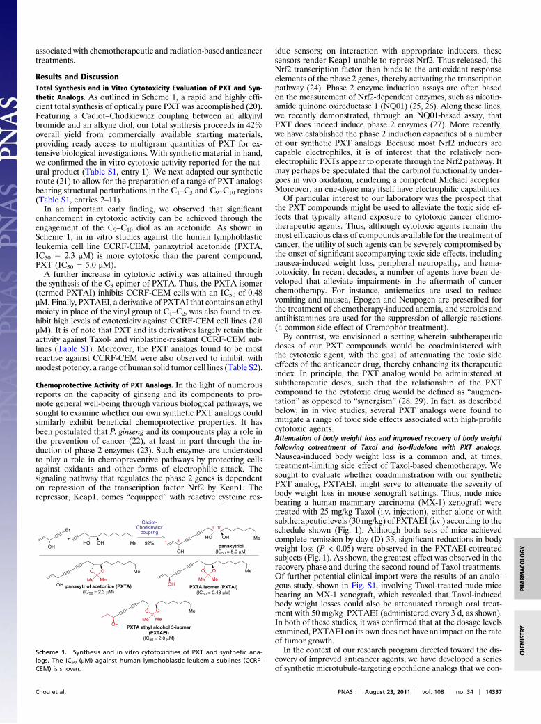
Results and DiscussionTotal Synthesis and in Vitro Cytotoxicity Evaluation of PXT and Syn-thetic Analogs. As outlined in Scheme 1, a rapid and highly effi-cient total synthesis of optically pure PXT was accomplished (20).Featuring a Cadiot–Chodkiewicz coupling between an alkynylbromide and an alkyne diol, our total synthesis proceeds in 42%overall yield from commercially available starting materials,providing ready access to multigram quantities of PXT for ex-tensive biological investigations. With synthetic material in hand,we confirmed the in vitro cytotoxic activity reported for the nat-ural product (Table S1, entry 1). We next adapted our syntheticroute (21) to allow for the preparation of a range of PXT analogsbearing structural perturbations in the C1–C3 and C9–C10 regions(Table S1, entries 2–11).In an important early finding, we observed that significant
enhancement in cytotoxic activity can be achieved through theengagement of the C9–C10 diol as an acetonide. As shown inScheme 1, in in vitro studies against the human lymphoblasticleukemia cell line CCRF-CEM, panaxytriol acetonide (PXTA,IC50 = 2.3 μM) is more cytotoxic than the parent compound,PXT (IC50 = 5.0 μM).A further increase in cytotoxic activity was attained through
the synthesis of the C3 epimer of PXTA. Thus, the PXTA isomer(termed PXTAI) inhibits CCRF-CEM cells with an IC50 of 0.48μM.Finally, PXTAEI, a derivative of PXTAI that contains an ethylmoiety in place of the vinyl group at C1–C2, was also found to ex-hibit high levels of cytotoxicity against CCRF-CEM cell lines (2.0μM). It is of note that PXT and its derivatives largely retain theiractivity against Taxol- and vinblastine-resistant CCRF-CEM sub-lines (Table S1). Moreover, the PXT analogs found to be mostreactive against CCRF-CEM were also observed to inhibit, withmodest potency, a range of human solid tumor cell lines (Table S2).
Chemoprotective Activity of PXT Analogs. In the light of numerousreports on the capacity of ginseng and its components to pro-mote general well-being through various biological pathways, wesought to examine whether our own synthetic PXT analogs couldsimilarly exhibit beneficial chemoprotective properties. It hasbeen postulated that P. ginseng and its components play a role inthe prevention of cancer (22), at least in part through the in-duction of phase 2 enzymes (23). Such enzymes are understoodto play a role in chemopreventive pathways by protecting cellsagainst oxidants and other forms of electrophilic attack. Thesignaling pathway that regulates the phase 2 genes is dependenton repression of the transcription factor Nrf2 by Keap1. Therepressor, Keap1, comes “equipped” with reactive cysteine res-
idue sensors; on interaction with appropriate inducers, thesesensors render Keap1 unable to repress Nrf2. Thus released, theNrf2 transcription factor then binds to the antioxidant responseelements of the phase 2 genes, thereby activating the transcriptionpathway (24). Phase 2 enzyme induction assays are often basedon the measurement of Nrf2-dependent enzymes, such as nicotin-amide quinone oxireductase 1 (NQ01) (25, 26). Along these lines,we recently demonstrated, through an NQ01-based assay, thatPXT does indeed induce phase 2 enzymes (27). More recently,we have established the phase 2 induction capacities of a numberof our synthetic PXT analogs. Because most Nrf2 inducers arecapable electrophiles, it is of interest that the relatively non-electrophilic PXTs appear to operate through the Nrf2 pathway. Itmay perhaps be speculated that the carbinol functionality under-goes in vivo oxidation, rendering a competent Michael acceptor.Moreover, an ene-diyne may itself have electrophilic capabilities.Of particular interest to our laboratory was the prospect that
the PXT compounds might be used to alleviate the toxic side ef-fects that typically attend exposure to cytotoxic cancer chemo-therapeutic agents. Thus, although cytotoxic agents remain themost efficacious class of compounds available for the treatment ofcancer, the utility of such agents can be severely compromised bythe onset of significant accompanying toxic side effects, includingnausea-induced weight loss, peripheral neuropathy, and hema-totoxicity. In recent decades, a number of agents have been de-veloped that alleviate impairments in the aftermath of cancerchemotherapy. For instance, antiemetics are used to reducevomiting and nausea, Epogen and Neupogen are prescribed forthe treatment of chemotherapy-induced anemia, and steroids andantihistamines are used for the suppression of allergic reactions(a common side effect of Cremophor treatment).By contrast, we envisioned a setting wherein subtherapeutic
doses of our PXT compounds would be coadministered withthe cytotoxic agent, with the goal of attenuating the toxic sideeffects of the anticancer drug, thereby enhancing its therapeuticindex. In principle, the PXT analog would be administered atsubtherapeutic doses, such that the relationship of the PXTcompound to the cytotoxic drug would be defined as “augmen-tation” as opposed to “synergism” (28, 29). In fact, as describedbelow, in in vivo studies, several PXT analogs were found tomitigate a range of toxic side effects associated with high-profilecytotoxic agents.Attenuation of body weight loss and improved recovery of body weightfollowing cotreatment of Taxol and iso-fludelone with PXT analogs.Nausea-induced body weight loss is a common and, at times,treatment-limiting side effect of Taxol-based chemotherapy. Wesought to evaluate whether coadministration with our syntheticPXT analog, PXTAEI, might serve to attenuate the severity ofbody weight loss in mouse xenograft settings. Thus, nude micebearing a human mammary carcinoma (MX-1) xenograft weretreated with 25 mg/kg Taxol (i.v. injection), either alone or withsubtherapeutic levels (30mg/kg) of PXTAEI (i.v.) according to theschedule shown (Fig. 1). Although both sets of mice achievedcomplete remission by day (D) 33, significant reductions in bodyweight loss (P < 0.05) were observed in the PXTAEI-cotreatedsubjects (Fig. 1). As shown, the greatest effect was observed in therecovery phase and during the second round of Taxol treatments.Of further potential clinical import were the results of an analo-gous study, shown in Fig. S1, involving Taxol-treated nude micebearing an MX-1 xenograft, which revealed that Taxol-inducedbody weight losses could also be attenuated through oral treat-ment with 50 mg/kg PXTAEI (administered every 3 d, as shown).In both of these studies, it was confirmed that at the dosage levelsexamined, PXTAEI on its own does not have an impact on the rateof tumor growth.In the context of our research program directed toward the dis-
covery of improved anticancer agents, we have developed a seriesof synthetic microtubule-targeting epothilone analogs that we con-
MeHO OH
OHpanaxytriol
(IC50 = 5.0 µM)
MeHO OH
Br
OH
Cadiot-Chodkiewicz
coupling
92%+ 3
9 10
1
Me
OH panaxytriol acetonide (PXTA)(IC50 = 2.3 µM)
O O
Me Me
Me
OH PXTA isomer (PXTAI)(IC50 = 0.48 µM)
O O
Me Me
Me
OH PXTA ethyl alcohol 3-isomer (PXTAEI)
(IC50 = 2.0 µM)
O O
Me Me
Scheme 1. Synthesis and in vitro cytotoxicities of PXT and synthetic ana-logs. The IC50 (μM) against human lymphoblastic leukemia sublines (CCRF-CEM) is shown.
Chou et al. PNAS | August 23, 2011 | vol. 108 | no. 34 | 14337
PHARM
ACO
LOGY
CHEM
ISTR
Y

sider to possess truly remarkable therapeutic potential at the clin-ical level (30–32). One such candidate, termed iso-fludelone (iso-flu), is currently being readied for phase I clinical trials. As outlinedin Fig. 2, sets of nude mice bearing a human colon carcinoma(HCT-116) xenograft were treated with iso-flu (25 mg/kg adminis-tered via i.v. infusion every 12 d fromD9 to D45), either on its ownor with the synthetic PXT analog, PXTAI (10 mg/kg administeredas an i.v. injection according to the schedule shown). The bodyweights of the mice were monitored. Once more, cotreatment witheven low levels of PXTAI resulted in alleviation of iso-flu–inducedbody weight losses over the four rounds of treatment.Mitigation of Taxol-induced neurotoxicity by PXTAEI. A limitation ofTaxol-based cancer chemotherapy has been the high incidenceof peripheral neuropathy reported as a significant side effect oftreatment. Along these lines, we took note of a number of pro-mising studies suggesting a neuroprotective role for various gin-seng components. Specifically, ginsenoside Rd has been shown toexhibit neuroprotective effects against oxygen-glucose deprivationin cultured hippocampal neurons (33). It has been postulated thatthese effects may be attributed to the antioxidative activity of thecompound, which serves to reduce intracellular reactive oxygenspecies, increase glutathione content, and enhance the antioxidantactivities of a range of enzymes (catalase, superoxide dismutase,and glutathione peroxidase). Given the reported neuroprotectiveactivity of these ginseng extracts, we sought to examine whetherour PXT-based compounds might possess valuable neuroprotec-tive activity, which would allow for the alleviation of Taxol-induced neurotoxicity.
Twenty nude mice bearing MX-1 xenografts were divided intotwo groups. The control group (n = 10) received repeated high-dose i.v. injections of Taxol (25 mg/kg) on D9, D11, D13, andD15 following tumor implantation. The treated group (n = 10)received the same Taxol treatment but were coadministeredPXTAEI [30 mg/kg administered as an i.v. injection every 4 dthroughout the course of the study, including pretreatment onD(−4)]). In the first round (cycle 1), peripheral neuropathy wasscored from D9 to D27 (Table S3). The following rating systemwas used for the evaluation of neuropathy: −, no detectable neu-rotoxicity; +, slight impairment of balance; ++, paralysis of twohind legs; and +++, paralysis of all four legs, which would lead todeath without intervention with food and water. On D27, thecontrol group, receiving treatment with Taxol alone, carried anaggregate neuropathy score of 53, whereas the group receivingcotreatment with PXTAEI received a score of only 27. Thus, incycle 1, a significant reduction of peripheral neuropathy wasachieved through cotreatment with PXTAEI. Moreover, the ob-served neuroprotective effects of PXTAEI were maintainedthroughout the course of a second round of Taxol treatment. Thus,on D29, when the mice had mainly recovered from the neurotoxiceffects induced by the first round of treatment, a second cycleanalogous to the first was commenced. As shown in Table S4, thissecond treatment cycle led to elevated levels of neuropathy in bothsets of mice, presumably attributable to carryover toxicity accu-mulated from the first round. Nonetheless, coadministration withPXTAEI did measurably serve to attenuate the severity of theperipheral neuropathy. The aggregate neurotoxicity score ofthe control group in cycle 2 was calculated to be 181, whereas the
Fig. 1. Alleviation of Taxol-induced body weight losses by PXTAEI in nude mice bearing a human mammary carcinoma MX-1 xenograft. The doses of Taxoland PXTAEI and their schedules of i.v. injection are shown (n = 10). Taxol was used as a Cremophor formulation (60 mg of Taxol in 2.5 mL of ethanol and2.5 mL of Cremophor). The saline-diluted Taxol solution was used within 2 h of its preparation; 0.2 mL of the solution was introduced through i.v. injection viathe tail vein. The PXTAEI stock solution was prepared in 10 mg/mL DMSO. The solution was diluted with saline containing 0.5% Tween 80 for i.v. injection intomice. This formulation caused no detectible toxicity in mice. The Taxol-induced body weight losses (green △) were reduced by coadministration of PXTAEI(pink▲) with a P value <0.05 (**) or <0.1 (*) as shown. The numbers indicate the average body weight of 10 mice (mean ± SE). Both treated groups achievedcomplete remission of xenograft tumors by D33. The PXTAEI and control groups were killed on D25 because of excessive tumor burden. All P values weredetermined by the Student t test for paired comparisons (Excel; Microsoft Corp.).
14338 | www.pnas.org/cgi/doi/10.1073/pnas.1111332108 Chou et al.

PXTAEI cotreated group scored 145. Over the course of bothcycles, the PXTAEI-treated group achieved an overall reductionin peripheral neuropathy of ∼26%.Mitigation of 5-fluorouracil–induced toxicity and lethality throughcotreatment with PXT analogs. Among the common side effects oftreatment with the anticancer drug 5-fluorouracil (5-FU) are lowRBC and WBC counts, which can lead to increased risk for in-fection as well as anemia. We set out to determine whether thehematotoxicity associated with 5-FU might be mitigated throughexposure to the PXT analog PXTAEI. Normal CD-1 mice weretreated with either 5-FU alone (50mg/kg administered i.v. QD for4 d) or 5-FU with PXTAEI (30 mg/kg administered i.v. on D(−2),D1, D4, and D7), and blood samples were taken on D8. Asexpected, following treatment with 5-FU alone, the levels ofWBCs, lymphocytes, RBCs, platelets, and hemoglobin were allfound to be markedly diminished (Fig. 3). Interestingly, cotreat-ment with PXTAEI led to alleviation of hematotoxicity, asdemonstrated by enhanced blood cell counts, in comparison withthe 5-FU–treated group.In contrast to Taxol and the epothilone-based cytotoxic
agents, cotreatment with 5-FU and PXT analogs did not resultin significant attenuation of body weight losses. However, co-administration with PXTAI did lead to a marked reduction in5-FU–induced lethality. Thus, nude mice bearing MX-1 xeno-graft tumors were treated with 5-FU (50 mg/kg administered asan i.v. injection QD for 5 d) either alone or coadministered withPXTAI (10 mg/kg administered via i.v. injection every 4 d). Inboth sets of mice, significant body weight losses were observed(Fig. S2). However, of the eight 5-FU–treated mice, five diedduring the course of treatment (2 on D18 and 1 each on D19–D21), although only two of the eight mice cotreated with PXTAIdied (1 each on D19 and D20). These findings raise the in-triguing possibility that PXTAI is perhaps operating through anas yet undefined chemoprotective pathway.Reduction of radiation-induced toxicity by PXTAEI. Several ginseng-derived natural products, including the polysaccharide ginsan(34) and several ginsenosides (35), have been reported to exhibitradioprotective effects in irradiated mice. Having established the
chemoprotective abilities of our PXT compounds in cotreatmentwith cytotoxic agents, we speculated that these agents mightdemonstrate the capacity to alleviate similar side effects associ-ated with radiation therapy, namely, body weight loss, lethality,and hematotoxicity.Thus, in a control group, CD-1 mice irradiated with 7 Gy
suffered significant body weight losses, peaking at 12.3% on D15following irradiation. A separate group of mice treated with 7 Gywere coadministered PXTAEI (30 mg/kg administered as ani.v. injection every 4 d, as shown). As outlined in Fig. 4, signif-icant alleviation of body weight loss was observed on D3–D8(P < 0.05). Over the course of this study, comparison of the body
Fig. 3. Reduction of 5-FU–induced hematotoxicity by PXTAEI in normalCD-1 mice. Heavy and light bars indicate with or without PXTAEI treatment,respectively. The PXTAEI dose was 30 mg/kg administered i.v. on D(−2), D1,D4, and D7, and the 5-FU dose was 50 mg/kg administered i.v. QD for 4 d. Allmeasurements were taken on D8 (n = 17). Blood was collected in 100 μL ofpotassium EDTA in a round-bottomed inner tube (Microvette 100). Blood cellcounts in samples (2.0 μL) were determined by a flow Multispecies Hema-tology System (Hemavet HV950FS). The lymphocyte reduction was signifi-cantly alleviated by PXTAEI (P < 0.05), whereas other parameters showeda trend toward significance (P < 0.2). PLT, platelet.
Fig. 2. Alleviation of iso-flu–induced body weight losses by PXTAI in nude mice bearing human colon carcinoma HCT-116 xenografts. The doses of iso-flu (i.v.infusion) and PXTAI (i.v. injection) and their schedules are shown (n = 6). The iso-flu–induced body weight losses were reduced, and recovery was improved bycoadministration of PXTAI with P < 0.05 (**) and P < 0.1 (*) as shown. The numbers indicate the average body weight of six mice (mean ± SE). The iso-flu–treated groups (green △) achieved complete remission of xenograft tumors between D36 and D57, and the cotreated group (pink ▲) achieved completeremission between D23 and D57. The PXTAI control groups (orange ○) were killed on D27 because of excessive tumor burden.
Chou et al. PNAS | August 23, 2011 | vol. 108 | no. 34 | 14339
PHARM
ACO
LOGY
CHEM
ISTR
Y

weight losses of PXTAEI-treated and untreated mice suggestsa trend toward significance.In a separate study, the X-irradiation dose was increased to 8
Gy, leading to the eventual death of all the CD-1 mice examined,regardless of whether they were cotreated with PXTAEI or not
(Fig. 5). However, the mean postirradiation survival times in-creased from 12.4 ± 1.5 d for the untreated mice to 14.0 ± 1.3 dfor mice cotreated with PXTAEI [30 mg/kg administered i.v. onD(−2), D1, D5, and D9]. Thus, cotreatment with PXTAEI led toa statistically meaningful (P < 0.03) increase in life span fol-lowing high levels of irradiation.Finally, we examined the ability of PXTAEI to alleviate X-
irradiation–induced hematotoxicity. Thus, three sets of CD-1mice were irradiated with 8 Gy. The control group received nofurther treatment, a second group was treated with the free rad-ical scavenger and well-established radioprotective agent Ami-fostine (100 mg/kg), and the third group received treatment withPXTAEI (30 mg/kg). Both Amifostine and PXTAEI were ad-ministered by i.v. injection on D(−2), D0, D4, and D8. On D10,blood samples were collected from each group and blood cellcounts were determined through hematocytometric analysis (Fig.6). As expected, the untreated control group exhibited signifi-cantly diminished WBC, lymphocyte, RBC, hemoglobin, andplatelet levels. Notably, Amifostine and PXTAEI were both ableto attenuate the hematotoxic effects of irradiation, with generallycomparable levels of efficacy.
ConclusionIn summary, we have described herein the discovery and evalu-ation of a promising class of biologically active compounds basedon the ginseng-derived natural product PXT. Of particular in-terest to our laboratory is the ability of this unique series ofpolyacetylene agents to alleviate a range of toxic side effectsassociated with standard cancer treatments. In particular, inmouse models, we observed mitigation of body weight loss, le-thality, peripheral neuropathy, and hematotoxicity, all of whichare side effects that may be associated with treatment withstandard chemotherapeutic agents, including Taxol, vincristine,epothilones, cyclophosphamide, and 5-FU, as well as radiation
Fig. 4. Reduction of radiation-induced body weight loss by PXTAEI in CD-1mice. Female CD-1 mice, 10 per group, were treated with 0 Gy of X-radiation(●); 7 Gy of X-radiation using anX-RAD320 irradiatorwith an IACUC-approvedprotocol and radiation user’s license (green ◇); and 7 GY of X-radiation plus30 mg/kg of PXTAEI administered i.v. on D(−4), D1, D5, D9, D13, and D17(orange△). Twomice in the radiation-treated group and the cotreated groupdied on D16 and D18. The cotreated group had significantly (P < 0.05) lessbody weight losses than the radiation-treated group.
Fig. 5. Extension of life span for lethal dose radiation-treated CD-1 miceby PXTAEI. Female CD-1 mice, 10 per group, were treated with (●) 0 Gy ofX-radiation; 8 Gy of X-radiation using an X-RAD320 irradiator with anIACUC-approved protocol and radiation user’s license (blue □); or 8 Gy of X-radiation plus 30 mg/kg of PXTAEI administered i.v. on D(−2), D1, D5, and D9(orange △). All 10 mice died in both the radiation-only group and thePXTAEI-cotreated group. However, the cotreated group had longer survivaltimes (14.0 ± 1.33 d) than the radiation-only group (12.4 ± 1.51 d) (P < 0.03).In addition, the body weight loss of the cotreated group was significantlyless than that of the radiation-only group (P < 0.05).
Fig. 6. Effects of PXTAEI on X-irradiation–induced hematological suppres-sion in CD-1 mice. CD-1 mice underwent X-irradiation with 8 Gy, and bloodsamples were collected on D10. PXTAEI at a dose of 30 mg/kg was admin-istered i.v. on D(−2), D0, D4, and D8 (green bars). The well-established ra-diation protection agent, Amifostine (Sigma–Aldrich) was administered i.v.at a dose of 100 mg/kg on D(−2), D0, D4, and D8 to serve as the positivecontrol (pink bars). The solvent-treated group (open bars) served as thenegative control. Each group comprised 20 CD-1 mice. Blood cell counts insamples (2.0 μL) were determined by a flow Multispecies Hematology System(Hemavet HV950FS). Although all parameters tested showed consistent al-leviation of X-irradiation–induced hematological suppression on D10, theP value calculated for n = 20 did not reach the level of significance.
14340 | www.pnas.org/cgi/doi/10.1073/pnas.1111332108 Chou et al.

therapy. The indications for these treatments cover a broadspectrum of cancer types. Through coadministration of a sub-therapeutic dose of PXT-based drug agent, it is conceivablypossible to increase the therapeutic indices of these commonlyused cytotoxic agents. Based on these findings, we are optimisticthat a PXT-derived lead agent may provide a clinically viablemeans by which to enhance chemotherapeutic and radiation-basedtreatments. We do not claim to have yet developed the optimalcandidate structure. Indeed, investigations along these lines areongoing in our laboratories. However, we believe that this “cyto-protection”-based approach may well represent a promising para-digm in the development of cancer treatment strategies. Accord-ingly, we have expanded our structure-activity relationship (SAR)study, and the results will be described in due course. Studiesaddressed to the possible mode of action of these compounds arein progress. Already, however, the results shown here serve to helpvalidate the idea that small molecule natural products providea menu of possibilities for drug discovery (15). Above, we havedemonstrated the possibility of combining chemistry and biology toadvance from anecdotally based neutraceuticals to the more chal-lenging domain of pharmaceuticals.
Materials and MethodsThe PXTs and iso-flu were synthesized in-house according to previouslyreported methods (17, 24). In vitro cell growth inhibition was measured bycell counting kit 8 (CCK-8) assay (Dojindo Molecular Technologies Inc.) fol-
lowing a 72-h incubation using a Powerwave XS microplate spectropho-tometer (BioTek Instruments, Inc.). IC50 values were determined in duplicateor triplicate from the dose–effect relationship at six or seven concentrationsof each drug using CompuSyn software (ComboSyn, Inc.) based on the me-dian-effect principle and plot and serial deletion analysis.
In in vivo studies with xenograft-bearing nude mice, Taxol was used asa Cremophor formulation (60 mg of Taxol in 2.5 mL of ethanol and 2.5 mL ofCremophor). The saline-diluted Taxol solution was used within 2 h of itspreparation; 0.2 mL of solution was introduced through i.v. injection via thetail vein. The PXT stock solutions were prepared in 10 mg/mL DMSO. Thesolutions were diluted with saline containing 0.5% Tween 80 for i.v. injectionintomice. Iso-fluwas introduced in the tail vein through a 6-h i.v. infusion. Forthe 5-FU– and radiation-induced hematotoxicity studies, blood was collectedin 100 μL of potassium EDTA in a round-bottomed inner tube (Microvette100; Sarstedt). Blood cell counts in the sample (2.0 μL) were determined bya flow Multispecies Hematology System (Hemavet HV950FS; Drew Scientific,Inc.). The radiation studies were performed with an X-RAD320 irradiator(Precision X-Ray) with an Institutional Animal Care and Use Committee(IACUC)-approved protocol and radiation user’s license. All animal studieswere conducted in accordance with the guidelines of the National Institutesof Health Guide for the Care and Use of Animals, and the protocol wasapproved by the Memorial Sloan–Kettering Cancer Center’s IACUC.
ACKNOWLEDGMENTS. We thank Grace Kang for assistance in the prepara-tion of this manuscript and Quen-Hui Tan and Luan-Ing Chen for technicalassistance. This work is supported, in part, by National Institutes of HealthGrant HL25848 (to S.J.D.) and by the Sloan-Kettering Institute General Fund(to T.-C.C.) from the Memorial Sloan–Kettering Cancer Center.
1. Strohl WR (2000) The role of natural products in a modern drug discovery program.Drug Discov Today 5:39–41.
2. Cordell GA (2002) Natural products in drug discovery—Creating a new vision. Phy-tochem Rev 1:261–273.
3. Rouhi AM (2003) Rediscovering natural products. Chem Eng News 81:77–91.4. Newman DJ, Cragg GM, Snader KM (2000) The influence of natural products upon
drug discovery. Nat Prod Rep 17:215–234.5. Newman DJ, Cragg GM, Snader KM (2003) Natural products as sources of new drugs
over the period 1981-2002. J Nat Prod 66:1022–1037.6. Butler MS (2004) The role of natural product chemistry in drug discovery. J Nat Prod
67:2141–2153.7. Bittles AH, Fulder SJ, Grant EC, Nicholls MR (1979) The effect of ginseng on lifespan
and stress responses in mice. Gerontology 25:125–131.8. Rai D, Bhatia G, Sen T, Palit G (2003) Anti-stress effects of Ginkgo biloba and Panax
ginseng: a comparative study. J Pharmacol Sci 93:458–464.9. Yamamoto M, Uemura T, Nakama S, Uemiya M, Kumagai A (1983) Serum HDL-cho-
lesterol-increasing and fatty liver-improving actions of Panax ginseng in high cho-lesterol diet-fed rats with clinical effect on hyperlipidemia in man. Am J Chin Med 11:96–101.
10. Mita A, Shida R, Kasai N, Shoji J (1979) Enhancement and suppression in production ofIgM-antibody in mice treated with purified saponins. Biomedicine 31:223–227.
11. Oura H, Hiai S, Odaka Y, Yokozawa T (1975) Studies on the biochemical action ofginseng saponin. I. Purification from ginseng extract of the active component stim-ulating serum protein biosynthesis. J Biochem 77:1057–1065.
12. Chen D, Wang SL, Wang YP (2003) [Experimental study on expression of GM-CSF fromhuman endothelial cells and monocytes induced by total saponins of panax ginseng].Zhongguo Zhong Xi Yi Jie He Za Zhi 23:845–847.
13. You JS, Huang HF, Chang YL (2005) Panax ginseng reduces adriamycin-induced heartfailure in rats. Phytother Res 19:1018–1022.
14. Kim JH, et al. (2005) Effects of Korean red ginseng extract on cisplatin-induced nauseaand vomiting. Arch Pharm Res 28:680–684.
15. Wilson RM, Danishefsky SJ (2006) Small molecule natural products in the discovery oftherapeutic agents: the synthesis connection. J Org Chem 71:8329–8351.
16. Kitagawa I, Yoshikawa M, Yoshihara M, Hayashi T, Taniyama T (1983) Chemicalstudies on crude drug procession. 1. On the constituents of Ginseng radix rubra.Yakugaku Zasshi 103:612–622.
17. Matsunaga H, Saita T, Nagumo F, Mori M, Katano M (1995) A possible mechanism forthe cytotoxicity of a polyacetylenic alcohol, panaxytriol: inhibition of mitochondrialrespiration. Cancer Chemother Pharmacol 35:291–296.
18. Saita T, et al. (1995) Screening of polyacetylenic alcohols in crude drugs using theELISA for panaxytriol. Biol Pharm Bull 18:933–937.
19. Katano M, et al. (1990) [Cell growth inhibitory substance isolated from Panax ginsengroot: panaxytriol]. Gan To Kagaku Ryoho 17:1045–1049.
20. Yun HD, Danishefsky SJ (2003) Straightforward synthesis of panaxytriol: an activecomponent of Red Ginseng. J Org Chem 68:4519–4522.
21. Yun H, et al. (2005) Total synthesis as a resource in drug discovery: the first in vivoevaluation of panaxytriol and its derivatives. J Org Chem 70:10375–10380.
22. Helms S (2004) Cancer prevention and therapeutics: Panax ginseng. Altern Med Rev 9:259–274.
23. Halim M, Yee DJ, Sames D (2008) Imaging induction of cytoprotective enzymes inintact human cells: coumberone, a metabolic reporter for human AKR1C enzymesreveals activation by panaxytriol, an active component of red ginseng. J Am Chem Soc130:14123–14128.
24. Kensler TW, Wakabayashi N, Biswal S (2007) Cell survival responses to environmentalstresses via the Keap1-Nrf2-ARE pathway. Annu Rev Pharmacol Toxicol 47:89–116.
25. Talalay P, et al. (2007) Sulforaphane mobilizes cellular defenses that protect skinagainst damage by UV radiation. Proc Natl Acad Sci USA 104:17500–17505.
26. Liu H, Dinkova-Kostova AT, Talalay P (2008) Coordinate regulation of enzymemarkers for inflammation and for protection against oxidants and electrophiles. ProcNatl Acad Sci USA 105:15926–15931.
27. Ng F, et al. (2008) (3R, 9R, 10R)-Panaxytriol: A molecular-based nutraceutical withpossible application to cancer prevention and treatment. Tetrahedron Lett 49:7178–7179.
28. Chou TC, Talalay P (1984) Quantitative analysis of dose-effect relationships: thecombined effects of multiple drugs or enzyme inhibitors. Adv Enzyme Regul 22:27–55.
29. Chou TC (2006) Theoretical basis, experimental design, and computerized simulationof synergism and antagonism in drug combination studies. Pharmacol Rev 58:621–681.
30. Rivkin A, Chou TC, Danishefsky SJ (2005) On the remarkable antitumor properties offludelone: how we got there. Angew Chem Int Ed Engl 44:2838–2850.
31. Chou TC, Dong H, Zhang X, Tong WP, Danishefsky SJ (2005) Therapeutic cure againsthuman tumor xenografts in nude mice by a microtubule stabilization agent, flude-lone, via parenteral or oral route. Cancer Res 65:9445–9454.
32. Chou TC, et al. (2008) Therapeutic effect against human xenograft tumors in nudemice by the third generation microtubule stabilizing epothilones. Proc Natl Acad SciUSA 105:13157–13162.
33. Ye R, et al. (2009) Neuroprotective effects of ginsenoside Rd against oxygen-glucosedeprivation in cultured hippocampal neurons. Neurosci Res 64:306–310.
34. Song JY, et al. (2003) Radioprotective effects of ginsan, an immunomodulator. RadiatRes 159:768–774.
35. Lee JH, et al. (2006) Effects of ginsenosides and their metabolites on voltage-dependent Ca(2+) channel subtypes. Mol Cells 21:52–62.
Chou et al. PNAS | August 23, 2011 | vol. 108 | no. 34 | 14341
PHARM
ACO
LOGY
CHEM
ISTR
Y