mtdna-maintenance defects: syndromes and genes
TRANSCRIPT

SSIEM 2016
MtDNA-maintenance defects: syndromes and genes
Carlo Viscomi1 & Massimo Zeviani1
Received: 24 January 2017 /Revised: 10 February 2017 /Accepted: 13 February 2017# The Author(s) 2017. This article is published with open access at Springerlink.com
Abstract A large group of mitochondrial disorders, rangingfrom early-onset pediatric encephalopathic syndromes to late-onset myopathy with chronic progressive externalophthalmoplegia (CPEOs), are inherited as Mendelian disor-ders characterized by disturbed mitochondrial DNA (mtDNA)maintenance. These errors of nuclear-mitochondrialintergenomic signaling may lead to mtDNA depletion, accu-mulation of mtDNA multiple deletions, or both, in criticaltissues. The genes involved encode proteins belonging to atleast three pathways: mtDNA replication and maintenance,nucleotide supply and balance, and mitochondrial dynamicsand quality control. In most cases, allelic mutations in thesegenes may lead to profoundly different phenotypes associatedwith either mtDNA depletion or multiple deletions.
Introduction
Mitochondria are essential organelles found in nearly all eukary-otic cells (Vafai and Mootha 2012), where they are mainly re-lated to energy metabolism. The most prominent role for mito-chondria is to generate ATP through the respiratory chain (RC)by means of oxidative phosphorylation (OXPHOS). However,mitochondria also take part in a vast array of important bio-chemical pathways including, among others, heat production,
apoptosis, generation and detoxification of reactive oxygen spe-cies (ROS), intracellular Ca2+ regulation, steroid hormone andheme synthesis and lipid metabolism (Wallace 2005).
The RC is composed of four multiheteromeric complexes(CI-IV), which transfer the electrons extracted from nutrientsbymeans of sequential redox reactions to molecular oxygen toform water. This process is coupled to the translocation ofproton across the IM by three of the four canonical respiratorychain complexes (CI, CIII, and CIV), generating a protongradient, which is then exploited by ATP synthase (or com-plex V, CV) to convert ADP into ATP.
Mitochondria are semi-autonomous organelles because theyhave their own DNA (mitochondrial DNA, mtDNA), whichencodes for 13 essential subunits of CI, CIII, CIV, and CV(Schon et al 2012), whereas all the other components of theRC, and indeed all of the other ≈1500 polypeptides constitutingthe mitochondrial proteome, are encoded by the nuclear genome.Therefore, mitochondrial bioenergetics and related homeostaticand execution pathways are under the double genetic control ofboth nuclear and mtDNA. The genetic duality of mitochondrialmetabolism has relevant consequences for human pathology, asmutations in either nuclear or mtDNA can lead to mitochondrialdysfunction and disease. These genetic features explain why mi-tochondrial disorders can be transmitted as dominant, recessive,X-linked or maternally-inherited traits (Zeviani and Di Donato2004). Hundreds of pathogenic mtDNA mutations have beenreported (MITO-MAP 2012). Whilst mtDNA in normal condi-tions is essentially uniform, a condition called homoplasmy,mostof the pathogenic mutations of mtDNA co-exist with a variablepercentage of wild-type mtDNA, in a condition termedheteroplasmy. Mutations of mtDNA or of OXPHOS-related nu-clear genes can affect virtually every tissue in the body, and leadto different phenotypes depending on the organ involved, intrin-sic severity of the mutation, targeted gene, and, in case ofmtDNA mutations, heteroplasmy levels. Tissue and organ
Communicated by: Shamima Rahman
Presented at the Annual Symposium of the Society for the Study ofInborn Errors of Metabolism, Rome, Italy, September 6–9, 2016
* Massimo [email protected]
1 MRC-Mitochondrial Biology Unit, MRC MBU, Wellcome Trust/MRC Building, Hills Road, Cambridge CB2 0XY, UK
J Inherit Metab DisDOI 10.1007/s10545-017-0027-5

functions critically depend on adequate ATP production, espe-cially when energy demand is high, like in neurons and musclefibers (Koopman et al 2013). This explains why primary disor-ders of mitochondrial bioenergetics usually cause neurodegener-ation and/or myopathy, often in combination, to determineencephalomyopathies affecting children or adults. However,many mitochondrial disorders also involve additional organs,e.g., causing heart impairment (for instance cardiac failure dueto cardiomyopathy, or heart conduction defects), liver dysfunc-tion, diabetes mellitus, or alterations in special senses or specificdistricts, e.g. sensory-neural deafness, ophthalmoparesis (due toweakness of the extrinsic eyemuscles), ptosis (drooping eyelids),retinitis pigmentosa, and optic neuropathy causing blindness dueto degeneration of the optic nerve. Mutations in mtDNA canaffect specific proteins of the respiratory chain or the in situ,autochthonous synthesis of mitochondrial proteins as a whole(when mutations or deletions involve tRNA or rRNA genes)and can be in turn classified into large-scale rearrangements(i.e., partial deletions or duplications) and point mutations. Bothgroups have been associated with well-defined clinical syn-dromes. Whilst single large-scale rearrangements are usuallysporadic, point mutations are tipically maternally inherited.Large-scale rearrangements include several genes and are invari-ably heteroplasmic. In contrast, point mutations may beheteroplasmic or homoplasmic, the latter being characterizedby incomplete penetrance. (e.g., Leber’s hereditary opticneuropathy).
In addition to sporadic or maternally inherited disorders dueto mutations of the mitochondrial genome, mitochondrial dis-eases can also be transmitted asMendelian traits. Here, we shallfocus on those Mendelian disorders that alter the stability andthe integrity of mtDNA. In 1989, Zeviani and colleagues de-scribed an Italian family with adult-onset mitochondrial myop-athy characterized by chronic progressive externalophthalmoplegia, CPEO, and inherited in an autosomal domi-nant fashion (Zeviani et al 1989). Maternal inheritance wasexcluded because the male patients also transmitted the diseaseto their offspring. Since then, many additional autosomal dom-inant CPEO families have been described. A second group ofsyndromes, characterized by infantile myopathy orhepatopathy, was then associated with depletion of mitochon-drial DNA in affected tissues (Moraes et al 1991). Multipledeletions and depletion of mitochondrial DNAwere also foundin skeletal muscle in a complex, multisystem syndrome com-biningmuscle, brain, and gastrointestinal symptoms (mitochon-drial neurogastrointestinal encephalomyopathy or MNGIE)(Hirano et al 1998). Finally, mutations in the gene encodingthe DNA polymerase gamma (POLG), the master enzyme ofmitochondrial DNA replication, were found in severe, early-onset neurologic disorders, namely Alpers-Huttenlocherhepatopathic poliodystrophy, sensory-ataxia neuropathy withdysarthria and ophthalmoplegia, and spinocerebellar ataxia-epilepsy syndrome (Naviaux and Nguyen 2005).
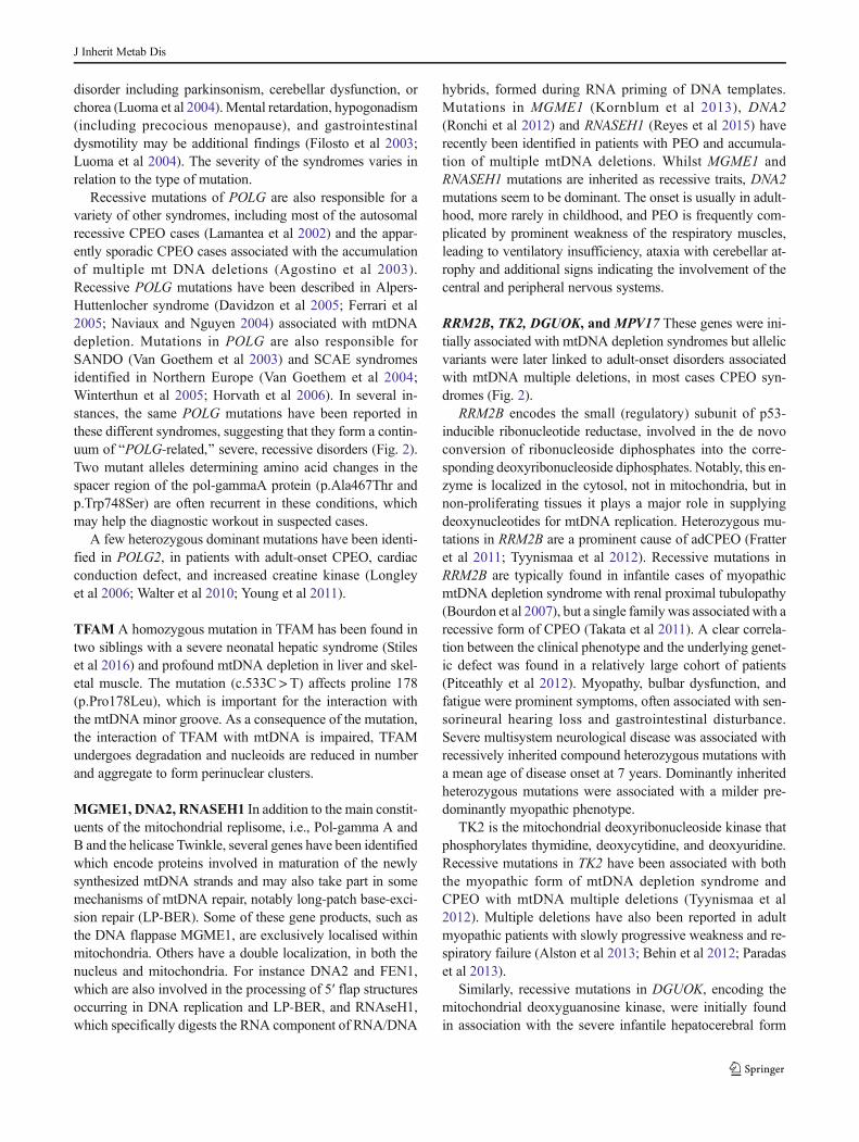
Over the last decade, an increasing number of genes havebeen identified in association with mtDNA multiple deletionsor depletion with variable phenotypes hallmarked bysyndromic CPEO, encephalomyopathy, and cardiomyopathy.The vast majority of them involve proteins with a role in themtDNA replisome (POLG and 2, TWNK, DNA2, MGME1)or dNTP supply for mtDNA synthesis (TP, TK2, DGUOK,RRM2B, SUCLA2, SUCLG1). A novel category of proteinsinvolved in accumulation of mtDNAmultiple deletions is rep-resented by OPA1 and MFN2, which are part of the complexmachinery regulating mitochondrial dynamics, specificallymitochondrial fusion; and paraplegin and AFG3L2, whichplay an important role in the protein quality control ofmitochondria.
Clinical features
Autosomal disorders classified as defects of mtDNA mainte-nance due to disturbed nuclear-mitochondrial intergenomiccommunication can be associated with the accumulation ofmtDNA large-scale rearrangements (mtDNA multiple dele-tions) or by severe reduction of the mtDNA copy number(mtDNA depletion syndromes) (Spinazzola and Zeviani2005). The most frequent clinical presentations (Table 1) are:
(1) adult-onset encephalomyopathy, defined clinically byCPEO, genetically by autosomal dominant or recessivetransmission, and molecularly by the presence of multi-ple deletions of mtDNA.
(2) an autosomal recessive multisystem disorder known asmitochondrial neurogastrointestinal encephalomyopathy(MNGIE), characterized by combined accumulation ofmultiple deletions and partial depletion of mtDNA.
(3) a spectrum of recessive neurologic syndromes rangingfrom typical infantile hepatopathic poliodystrophy(Alpers-Huttenlocher syndrome) to juvenile-onset senso-ry-ataxia neuropathy, dysarthria, and ophthalmoplegia(SANDO) to a combination of spinocerebellar ataxiaand epilepsy (SCAE) with or without externalophthalmoplegia.
(4) early-onset, organ-specific autosomal recessive syn-dromes associated with profound mtDNA depletion.
Autosomal dominant chronic progressive externalophthalmoplegia (adCPEO) CPEO is frequent in mitochon-drial disorders. The clinical hallmark is extraocular muscleinvolvement; all patients have ptosis with limitation of eyemovements. The first symptoms typically appear when pa-tients are 20 to 40 years old. Generalized muscle weaknessis frequently present. Additional features vary among families;
J Inherit Metab Dis

they may include ataxia, sensorineural hearing loss, cataracts,hypogonadism, parkinsonism, and psychiatric abnormalitiesconsisting of severe depression and avoidant personality.Dysphagia, dysphonia, weakness of facial muscles, and pe-ripheral neuropathy may be prominent symptoms in somefamilies. At rest, elevated levels of plasma lactate are detectedonly in severely affected patients. Symptoms seem to progresswith age. Muscle biopsies show ragged-red fibers due to thesubsarcolemmal accumulation of abnormal mitochondria. Inaddition, the histochemical reaction for cytochrome c oxidase(COX, complex IV) is decreased or absent in scattered fibers,and neurogenic changes may also be observed .
Biochemically, the activities of mtDNA-related respiratorycomplexes (complex I, III, IV, and V) in muscle homogenatecan range from normal to about 50% of the controls' mean(Servidei et al 1991). Presymptomatic patients appear normalbut often have laboratory, electrophysiological, morphologi-cal, and biochemical features of a subclinical mitochondrialencephalomyopathy. Autosomal dominant optic atrophy hasbeen reported in association with CPEO and multiple mtDNAdeletions. Additional signs included deafness, ataxia, axonalsensory-motor polyneuropathy, and mitochondrial myopathywith cytochrome c oxidase negative and ragged red fibers(Yu-Wai-Man et al 2010).
Table 1 Genes and phenotypesaffecting mtDNA maintenance Gene mtDNA alteration Inheritance Main clinical phenotype OMIM
SLC25A4 Multiple deletions AD ad/arCPEO 157640/258450
Multiple deletions AR myopathy and cardiomyopathy 615418
Depletion AD myopathy and cardiomyopathy
TWNK Multiple deletions AD adCPEO 609286
Multiple deletions AR IOSCA 271245
Depletion AR Alpers-like
POLG Multiple deletions AD adCPEO 157640
Multiple deletions AR arCPEO 258450
Depletion AR Alpers-Huntenlocher 203700
Multiple deletions AR SANDO/SCAE 607459
POLG2 Multiple deletions AD adCPEO 610131
TFAM Depletion AR Hepatocerebral syndrome 617156
MGME1 Multiple deletions AR arCPEO 615076
DNA2 Multiple deletions AD adCPEO 615156
RNASEH1 Multiple deletions AR arCPEO 616479
RRM2B Multiple deletions AD adCPEO 613077
Depletion AR myopathy and tubulopathy 612075
TK2 Depletion AR myopathy 609560
Multiple deletions AR arCPEO 617069
DGUOK Depletion AR Hepatocerebral syndrome 251880
Multiple deletions AR Myopathy with or w/o CPEO 617070
Multiple deletions AR lower motor neuron syndrome
MPV17 Depletion AR Hepatocerebral syndrome 256810
Multiple deletions AR arCPEO, leukoencephalopathy andparkinsonism
OPA1 Multiple deletions AD DOA 165500
Multiple deletions AD DOA plus 125250
MFN2 Multiple deletions AD DOA plus 608507
SPG7 Multiple deletions AR arCPEO and ataxia 602783
AFG3L2 Multiple deletions AD arCPEO and ataxia 604581
TYMP Multiple deletions anddepletion
AR MNGIE 603041
SUCLA2 Depletion AR Hepatocerebral syndrome 612073
SUCLG1 Depletion AR Hepatocerebral syndrome 245400
ABAT Multiple deletions AR Encephalomyopathy 613163
FBXL4 Depletion AR Encephalomyopathy 615471
GFER Multiple deletions AR myopathy 613076
J Inherit Metab Dis

Autosomal recessive chronic progressive externalophthalmoplegia (arCPEO)Multiple deletions of mitochon-drial DNA have been reported in numerous sporadic CPEOcases or in families in which CPEO was clearly transmitted asa recessive trait (Lamantea et al 2002).
Mitochondrial neurogastrointestinal encephalomyopathy(MNGIE) This is an autosomal recessive disease character-ized by the unusual combination of six features: (1) progres-sive external ophthalmoplegia, (2) severe gastrointestinaldysmotility, (3) cachexia, (4) peripheral neuropathy, (5) dif-fuse leukoencephalopathy on brain MRI, due to alteredblood–brain barrier, and (6) evidence of mitochondrial dys-function (histological, biochemical, or genetic abnormalitiesof the mitochondria) (Hirano et al 1994). The mean age atonset is 19 years, and the mean age at death is 37.6 years.Gastrointestinal manifestations comprise the most prominentand debilitating feature; painful gastrointestinal dysmotilitymay cause gastroparesis, frequent diarrhea, and intestinalpseudo-obstruction. Skeletal muscle biopsies reveal neuro-genic changes and occasional ragged-red and cytochrome coxidase-deficient fibers, reflecting the neuropathy and mito-chondrial myopathy. The peripheral neuropathy is predomi-nantly demyelinating, but electrophysiological data have alsoshown evidence of axonopathy in about one half of the pa-tients. In the gastrointestinal system, histological studies haverevealed abnormalities of both the intestinal smooth muscleand the enteric nervous system, thus accounting for the severegastrointestinal problems. In particular, studies of postmortemtissues from MNGIE patients revealed a selective, profoundmtDNA depletion, and marked atrophy of the external layer ofthe muscularis propria of the small intestine, consistent with avisceral myopathy leading to the prominent gastrointestinalinvolvement typical of this disorder (Giordano et al 2006,2008). Later-onset and longer survival MNGIE patients havebeen reported (Marti et al 2005).
Alpers-Huttenlocher hepatopathic poliodystrophy;s en so ry - a t ax i a neuropa thy, dy sa r th r i a andophthalmoplegia (SANDO); and spinocerebellar ataxia-epilepsy (SCAE)Anumber of recessive syndromes have beenassociated with qualitative or quantitative mtDNA abnormal-ities. These syndromes are all characterized by an associationwith recessive mutations of POLG, the master gene ofmtDNA replication. Alpers-Huttenlocher hepatopathicpoliodystrophy is an early onset, fatal disease, characterizedby hepatic failure and intractable seizures, evolving intoepilepsia partialis continua and global neurologic deteriora-tion. The liver dysfunction is usually progressive as well,evolving from microvesicular steatosis with bile duct prolifer-ation into cirrhosis. Childhood-onset or juvenile-onset autoso-mal recessive, progressive sensory-ataxic syndromes, with orwithout epilepsy, have been reported, more frequently in
families from Northern European countries. The associationof sensory ataxic neuropathy with dysarthria andophthalmoplegia (SANDO) defines some of these families,but in other families, cerebellar signs, myoclonus, and seizuresare additional prominent findings. The use of valproate tocontrol epilepsy is contraindicated in these patients becauseof their exquisite sensitivity to valproate hepatotoxicity, whichmay lead to fatal acute hepatic failure (Davidzon et al 2005;Gauthier-Villars et al 2001; Horvath et al 2006; Naviaux andNguyen 2005; Naviaux et al 1999; Tzoulis et al 2006).
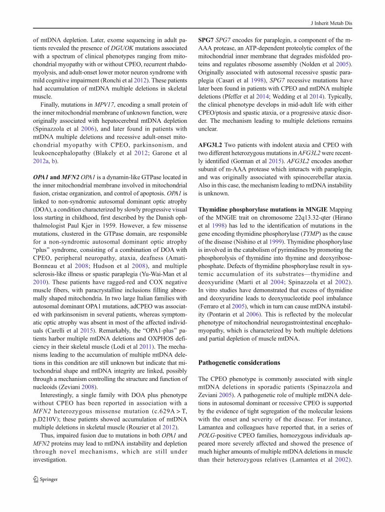
A tentative classification of these entities has been pro-posed (Copeland and Longley 2014), according to the follow-ing nomenclature: Alpers-Huttenlocher hepatopathicpoliodystrophy (AHS), childhood myocerebrohepatopathyspectrum (MCHS), myoclonic epilepsy myopathy sensoryataxia (MEMSA), and the ataxia neuropathy spectrum(ANS). It is however important to stress that these entitiesconstitute a continuous spectrum of syndromes with overlap-ping features (Fig. 2), with the exception perhaps of AHSwhich has rather specific clinical and neuroimaginghallmarks.
Mitochondrial DNA depletion syndromes In contrast toother types of mtDNA defects, mtDNA depletion syndromeis a quantitative abnormality: there is paucity of mtDNA , butthe remaining mtDNA does not harbor any mutations or rear-rangements. Mitochondrial DNA depletion syndrome is trans-mitted as an autosomal recessive trait and is clinically andgenetically heterogeneous.
Some children present with myopathy, others with liverfailure in infancy, and some with multisystem involvement.Consistent with the different phenotypes, mtDNA depletionmay affect either a specific tissue (most commonly muscle orliver and brain) or multiple organs, including heart, brain, andkidney.
Myopathic form Typically, affected children are born after anuncomplicated pregnancy, although decreased fetal move-ments are noted in some cases. A few patients hadarthrogryposis and clubfeet, but facial dysmorphic featuresare rare. The patient usually presents in the first year of lifewith feeding difficulty, failure to thrive, hypotonia, weakness,and occasionally progressive external ophthalmoplegia. Deathis usually due to pulmonary insufficiency and infections, butsome patients survive into their teens (Moraes et al 1991;Tritschler et al 1992). The clinical spectrum has now expand-ed to include spinal muscular atrophy type 3-like presentation,rigid spine syndrome, severe muscle weakness with markeddystrophic alterations, encephalopathy, and seizures (Galbiatiet al 2006), and a milder myopathic phenotype without motorregression and with longer survival (Oskoui et al 2006).mtDNA depletion in muscle has been reported in patients withearly-onset myopathy, lactic acidosis, and renal proximal
J Inherit Metab Dis

tubulopathy with nephrocalcinosis (Bourdon et al 2007). Inpatients with earlier onset and rapid courses, all muscle fibershave little or no immunoreactivity to DNA antibodies in mito-chondria and are cytochrome c oxidase-deficient, whereas inlater-onset patients the pattern of involvement is mosaic; somefibers appear normal, whereas others lack both COX activityand mtDNA. Proliferation of mitochondria (ragged-red fibers)is not a consistent feature, but their number can increase withage. Biochemical defects of all complexes containing mtDNA-encoded subunits are always present in muscle mitochondria.Patients with mtDNA depletion in muscle tend to have elevat-ed serum creatine kinase levels, ranging from 2 to 30 times theupper limit of normal. This is an important clue for the diag-nosis because increased serum creatine kinase is relatively un-common in patients with other mitochondrial myopathies.
Encephalomyopathic forms Two forms have been reported,both caused by a block of succinyl-CoA lyase activity in theKrebs cycle. The first is characterized by high lactate in blood,severe psychomotor retardation with muscle hypotonia, im-paired hearing, and generalized seizures followed by kneeand hip contractures, finger and limb dystonia, and mild pto-sis. Brain MRI is suggestive of Leigh syndrome. ModeratemtDNA depletion (about 32%) has been documented in skel-etal muscle (Carrozzo et al 2007; Elpeleg et al 2005;Ostergaard et al 2007a). Mutations in the gene encoding theATP-dependent succinyl-CoA synthase (SCS) activity,SUCLA2, are responsible for this form. The second, extreme-ly severe form is due to mutations in SUCLG1, the geneencoding the GTP-dependent isoform SUCLG1 (see below)and is associated with combined muscle and liver mtDNAdepletion, dysmorphic features, connatal lactic acidosis, anddeath in the first days of life (Ostergaard et al 2007b). Bothsyndromes are hallmarked by methylmalonic aciduria, whichaccumulates because of impaired conversion into succinyl-CoA of propionyl-CoA, derived from the beta oxidation ofodd-number fatty acids. A large cohort of patients with muta-tions in SUCLA2 and SUCLG1 has been recently described(Carrozzo et al 2016), showing significantly longer survival inSUCLA2 patients compared to SUCLG1 patients.Hypertrophic cardiomyopathy and liver involvement were ex-clusively found in patients with SUCLG1 mutations, whereasepilepsy was much more frequent in patients with SUCLA2mutations compared to patients with SUCLG1 mutations.
Finally, patients with mutations in the ABAT gene,encoding 4-aminobutyrate aminotransferase have elevatedlevels of GABA along with severe psychomotor retardation,intractable seizures, hypotonia, and hyperreflexia, associatedwith profound mtDNA depletion (Besse et al 2015). ABAT,besides its role in GABA biosynthesis, encodes another com-ponent of the SCS.
The mechanism of mtDNA depletion in mutations of theSCS components is unclear but could be related to the
physical interaction of SCS with nucleoside diphosphate ki-nase, NDPK, an enzyme involved in the salvage pathway ofmitochondrial nucleotides.
Hepatocerebral form In addition to patients with Alpers-Huttenlocher syndrome, hepatic mtDNA depletion is seen insome infants with liver failure (Mazziotta et al 1992). Onset ofsymptoms is between birth and 6months; death usually occurswithin 1 year of age. The most common symptoms and signsinclude persistent vomiting, failure to thrive, hypotonia, andhypoglycemia associated with progressive neurologic symp-toms. Histological changes on liver biopsy include fatty de-generation, bile duct proliferation, fibrosis, and collapse oflobular architecture. Generalized reduction in COX histo-chemistry and decreased mitochondrial respiratory chain en-zyme activities were found in the liver of a few patients. Avariant form of hepatocerebral mitochondrial DNA depletionsyndrome affects the Navajo people with a prevalence of 1 in1600 live births, hence the termNavajo neurohepatopathy (Vuet al 2001).
Molecular genetics
Numerous genes have been associated with syndromes causedby mtDNA instability (Fig. 1) but a substantial fraction ofthese conditions remain genetically undefined.
ANT1 Encoded by SLC25A4, ANT1 is the muscle-specificisoform of the mitochondrial adenine nucleotide translocator,and it is also expressed in heart and brain. Several mutations inSLC25A4 have been linked to mitochondrial disorders and fallinto two distinct clinical phenotypes. The first is caused byseveral single heterozygous mutations, and manifests asadCPEO (Deschauer et al 2005; Kaukonen et al 2000;Komaki et al 2002; Napoli et al 2001; Siciliano et al 2003).The second consists of a relatively benign mitochondrial my-opathy and cardiomyopathy phenotype that presents in child-hood or early adulthood and is characterized by fatigue andexercise intolerance, associated with recessive null mutations(Echaniz-Laguna et al 2012; Korver-Keularts et al 2015;Palmieri et al 2005; Strauss et al 2013). From the molecularpoint of view, both dominant and recessive mutations lead tothe accumulation of multiple mtDNA deletions, and their clin-ical course is relatively benign. However, de novo dominantmutations in SLC25A4 have recently been identified by wholeexome sequencing in seven patients presenting with profoundcongenital hypotonia and muscle weakness, leading to deathin the neonatal period. Profound mtDNA depletion and im-pairment of ATP transport capacity is the hallmark of thisearly onset form of ANT1-related disease (Thompson et al2016). However, the reason why either recessive or dominantmutations in a monomeric protein (Bamber et al 2007) give
J Inherit Metab Dis

rise to such a wide spectrum of clinical and molecular pheno-types is still unknown.
TWNK This gene (previously designated as C10orf2) en-codes Twinkle, the mitochondrial DNA and RNA helicase in-volved in replication of the mitochondrial genome.Heterozygous mutations in this gene are responsible foradCPEO (Spelbrink et al 2001) with accumulation of multiplemtDNA deletions. Clinical presentations include CPEO, oftenassociated with proximal muscle and facial weakness, dyspha-gia and dysphonia, mild ataxia, and peripheral neuropathy.Symptoms are much more severe in a few homozygous mutantpatients described in consanguineous families. A specific, re-cessive TWNK mutation is responsible for infantile onsetspinocerebellar ataxia, IOSCA (Nikali et al 2005), which is partof the Finnish disease heritage. Onset usually is between 1 and2 years of age. Patients suffer from a combination of ataxia,athetosis, areflexia, muscle hypotonia, and severe epilepsy.Other features such as ophthalmoplegia, hearing loss, and opticatrophy appear later in the disease course. Some patients showreduced mental capacity, and hypergonadotropichypogonadism may occur in girls. Morphologic studies revealsensory axonal neuropathy and progressive atrophy of the cer-ebellum, brainstem and spinal cord (Koskinen et al 1994).Besides infantile onset spinocerebellar ataxia, an Alpers-likeclinical phenotype has also been associated with recessive mu-tations in TWNK (Fig. 2) (Hakonen et al 2007; Sarzi et al 2007).
Pol-gamma mutations The mitochondrial DNA polymerase(pol-gamma) is essential for mitochondrial DNA replicationand proofreading-based repair. It is composed of a 140-kDa
catalytic subunit (pol-[gamma]A) and a 55-kDa accessorysubunit (pol-[gamma]B), which functions as a DNA bindingfactor, increasing the processivity of the polymerase holoen-zyme. The holoenzyme works as an A1B2 heterotrimer. Thecatalytic subunit is encoded by POLG on chromosome 15q25,whereas the accessory subunits are coded by POLG2 on chro-mosome 17q.Mutations of POLG are a major cause of humanmitochondrial disease. So far more than 100 mutations in pol-gamma have been reported (http://tools.niehs.nih.gov/polg).This gene is the most frequent cause of adCPEO (50% ofthe cases in our series) (Lamantea et al 2002; Van Goethemet al 2001). In addition to CPEO, prominent features are se-vere dysphagia and dysphonia and, occasionally, a movement
Fig. 1 Pathways involved inmtDNA maintenance
Fig. 2 Scheme representing the continuous spectrum of clinicalphenotypes associated with mutations in mtDNA-maintenance-relatedgenes
J Inherit Metab Dis
disorder including parkinsonism, cerebellar dysfunction, orchorea (Luoma et al 2004). Mental retardation, hypogonadism(including precocious menopause), and gastrointestinaldysmotility may be additional findings (Filosto et al 2003;Luoma et al 2004). The severity of the syndromes varies inrelation to the type of mutation.
Recessive mutations of POLG are also responsible for avariety of other syndromes, including most of the autosomalrecessive CPEO cases (Lamantea et al 2002) and the appar-ently sporadic CPEO cases associated with the accumulationof multiple mt DNA deletions (Agostino et al 2003).Recessive POLG mutations have been described in Alpers-Huttenlocher syndrome (Davidzon et al 2005; Ferrari et al2005; Naviaux and Nguyen 2004) associated with mtDNAdepletion. Mutations in POLG are also responsible forSANDO (Van Goethem et al 2003) and SCAE syndromesidentified in Northern Europe (Van Goethem et al 2004;Winterthun et al 2005; Horvath et al 2006). In several in-stances, the same POLG mutations have been reported inthese different syndromes, suggesting that they form a contin-uum of BPOLG-related,^ severe, recessive disorders (Fig. 2).Two mutant alleles determining amino acid changes in thespacer region of the pol-gammaA protein (p.Ala467Thr andp.Trp748Ser) are often recurrent in these conditions, whichmay help the diagnostic workout in suspected cases.
A few heterozygous dominant mutations have been identi-fied in POLG2, in patients with adult-onset CPEO, cardiacconduction defect, and increased creatine kinase (Longleyet al 2006; Walter et al 2010; Young et al 2011).
TFAM A homozygous mutation in TFAM has been found intwo siblings with a severe neonatal hepatic syndrome (Stileset al 2016) and profound mtDNA depletion in liver and skel-etal muscle. The mutation (c.533C > T) affects proline 178(p.Pro178Leu), which is important for the interaction withthe mtDNA minor groove. As a consequence of the mutation,the interaction of TFAM with mtDNA is impaired, TFAMundergoes degradation and nucleoids are reduced in numberand aggregate to form perinuclear clusters.
MGME1, DNA2, RNASEH1 In addition to the main constit-uents of the mitochondrial replisome, i.e., Pol-gamma A andB and the helicase Twinkle, several genes have been identifiedwhich encode proteins involved in maturation of the newlysynthesized mtDNA strands and may also take part in somemechanisms of mtDNA repair, notably long-patch base-exci-sion repair (LP-BER). Some of these gene products, such asthe DNA flappase MGME1, are exclusively localised withinmitochondria. Others have a double localization, in both thenucleus and mitochondria. For instance DNA2 and FEN1,which are also involved in the processing of 5′ flap structuresoccurring in DNA replication and LP-BER, and RNAseH1,which specifically digests the RNA component of RNA/DNA
hybrids, formed during RNA priming of DNA templates.Mutations in MGME1 (Kornblum et al 2013), DNA2(Ronchi et al 2012) and RNASEH1 (Reyes et al 2015) haverecently been identified in patients with PEO and accumula-tion of multiple mtDNA deletions. Whilst MGME1 andRNASEH1 mutations are inherited as recessive traits, DNA2mutations seem to be dominant. The onset is usually in adult-hood, more rarely in childhood, and PEO is frequently com-plicated by prominent weakness of the respiratory muscles,leading to ventilatory insufficiency, ataxia with cerebellar at-rophy and additional signs indicating the involvement of thecentral and peripheral nervous systems.
RRM2B, TK2, DGUOK, and MPV17 These genes were ini-tially associated with mtDNA depletion syndromes but allelicvariants were later linked to adult-onset disorders associatedwith mtDNA multiple deletions, in most cases CPEO syn-dromes (Fig. 2).
RRM2B encodes the small (regulatory) subunit of p53-inducible ribonucleotide reductase, involved in the de novoconversion of ribonucleoside diphosphates into the corre-sponding deoxyribonucleoside diphosphates. Notably, this en-zyme is localized in the cytosol, not in mitochondria, but innon-proliferating tissues it plays a major role in supplyingdeoxynucleotides for mtDNA replication. Heterozygous mu-tations in RRM2B are a prominent cause of adCPEO (Fratteret al 2011; Tyynismaa et al 2012). Recessive mutations inRRM2B are typically found in infantile cases of myopathicmtDNA depletion syndrome with renal proximal tubulopathy(Bourdon et al 2007), but a single family was associatedwith arecessive form of CPEO (Takata et al 2011). A clear correla-tion between the clinical phenotype and the underlying genet-ic defect was found in a relatively large cohort of patients(Pitceathly et al 2012). Myopathy, bulbar dysfunction, andfatigue were prominent symptoms, often associated with sen-sorineural hearing loss and gastrointestinal disturbance.Severe multisystem neurological disease was associated withrecessively inherited compound heterozygous mutations witha mean age of disease onset at 7 years. Dominantly inheritedheterozygous mutations were associated with a milder pre-dominantly myopathic phenotype.
TK2 is the mitochondrial deoxyribonucleoside kinase thatphosphorylates thymidine, deoxycytidine, and deoxyuridine.Recessive mutations in TK2 have been associated with boththe myopathic form of mtDNA depletion syndrome andCPEO with mtDNA multiple deletions (Tyynismaa et al2012). Multiple deletions have also been reported in adultmyopathic patients with slowly progressive weakness and re-spiratory failure (Alston et al 2013; Behin et al 2012; Paradaset al 2013).
Similarly, recessive mutations in DGUOK, encoding themitochondrial deoxyguanosine kinase, were initially foundin association with the severe infantile hepatocerebral form
J Inherit Metab Dis
of mtDNA depletion. Later, exome sequencing in adult pa-tients revealed the presence of DGUOK mutations associatedwith a spectrum of clinical phenotypes ranging from mito-chondrial myopathy with or without CPEO, recurrent rhabdo-myolysis, and adult-onset lower motor neuron syndrome withmild cognitive impairment (Ronchi et al 2012). These patientshad accumulation of mtDNA multiple deletions in skeletalmuscle.
Finally, mutations in MPV17, encoding a small protein ofthe inner mitochondrial membrane of unknown function, wereoriginally associated with hepatocerebral mtDNA depletion(Spinazzola et al 2006), and later found in patients withmtDNA multiple deletions and recessive adult-onset mito-chondrial myopathy with CPEO, parkinsonism, andleukoencephalopathy (Blakely et al 2012; Garone et al2012a, b).
OPA1 andMFN2 OPA1 is a dynamin-like GTPase located inthe inner mitochondrial membrane involved in mitochondrialfusion, cristae organization, and control of apoptosis. OPA1 islinked to non-syndromic autosomal dominant optic atrophy(DOA), a condition characterized by slowly progressive visualloss starting in childhood, first described by the Danish oph-thalmologist Paul Kjer in 1959. However, a few missensemutations, clustered in the GTPase domain, are responsiblefor a non-syndromic autosomal dominant optic atrophyBplus^ syndrome, consisting of a combination of DOA withCPEO, peripheral neuropathy, ataxia, deafness (Amati-Bonneau et al 2008; Hudson et al 2008), and multiplesclerosis-like illness or spastic paraplegia (Yu-Wai-Man et al2010). These patients have ragged-red and COX negativemuscle fibers, with paracrystalline inclusions filling abnor-mally shaped mitochondria. In two large Italian families withautosomal dominant OPA1 mutations, adCPEO was associat-ed with parkinsonism in several patients, whereas symptom-atic optic atrophy was absent in most of the affected individ-uals (Carelli et al 2015). Remarkably, the BOPA1-plus^ pa-tients harbor multiple mtDNA deletions and OXPHOS defi-ciency in their skeletal muscle (Lodi et al 2011). The mecha-nisms leading to the accumulation of multiple mtDNA dele-tions in this condition are still unknown but indicate that mi-tochondrial shape and mtDNA integrity are linked, possiblythrough a mechanism controlling the structure and function ofnucleoids (Zeviani 2008).
Interestingly, a single family with DOA plus phenotypewithout CPEO has been reported in association with aMFN2 heterozygous missense mutation (c.629A > T,p.D210V); these patients showed accumulation of mtDNAmultiple deletions in skeletal muscle (Rouzier et al 2012).
Thus, impaired fusion due to mutations in both OPA1 andMFN2 proteins may lead to mtDNA instability and depletionthrough novel mechanisms, which are sti ll underinvestigation.
SPG7 SPG7 encodes for paraplegin, a component of the m-AAA protease, an ATP-dependent proteolytic complex of themitochondrial inner membrane that degrades misfolded pro-teins and regulates ribosome assembly (Nolden et al 2005).Originally associated with autosomal recessive spastic para-plegia (Casari et al 1998), SPG7 recessive mutations havelater been found in patients with CPEO and mtDNA multipledeletions (Pfeffer et al 2014; Wedding et al 2014). Typically,the clinical phenotype develops in mid-adult life with eitherCPEO/ptosis and spastic ataxia, or a progressive ataxic disor-der. The mechanism leading to multiple deletions remainsunclear.
AFG3L2 Two patients with indolent ataxia and CPEO withtwo different heterozygous mutations in AFG3L2were recent-ly identified (Gorman et al 2015). AFG3L2 encodes anothersubunit of m-AAA protease which interacts with paraplegin,and was originally associated with spinocerebellar ataxia.Also in this case, the mechanism leading tomtDNA instabilityis unknown.
Thymidine phosphorylase mutations in MNGIE Mappingof the MNGIE trait on chromosome 22q13.32-qter (Hiranoet al 1998) has led to the identification of mutations in thegene encoding thymidine phosphorylase (TYMP) as the causeof the disease (Nishino et al 1999). Thymidine phosphorylaseis involved in the catabolism of pyrimidines by promoting thephosphorolysis of thymidine into thymine and deoxyribose-phosphate. Defects of thymidine phosphorylase result in sys-temic accumulation of its substrates—thymidine anddeoxyuridine (Marti et al 2004; Spinazzola et al 2002).In vitro studies have demonstrated that excess of thymidineand deoxyuridine leads to deoxynucleotide pool imbalance(Ferraro et al 2005), which in turn can cause mtDNA instabil-ity (Pontarin et al 2006). This is reflected by the molecularphenotype of mitochondrial neurogastrointestinal encephalo-myopathy, which is characterized by both multiple deletionsand partial depletion of muscle mtDNA.
Pathogenetic considerations
The CPEO phenotype is commonly associated with singlemtDNA deletions in sporadic patients (Spinazzola andZeviani 2005). A pathogenetic role of multiple mtDNA dele-tions in autosomal dominant or recessive CPEO is supportedby the evidence of tight segregation of the molecular lesionswith the onset and severity of the disease. For instance,Lamantea and colleagues have reported that, in a series ofPOLG-positive CPEO families, homozygous individuals ap-peared more severely affected and showed the presence ofmuch higher amounts of multiple mtDNA deletions in musclethan their heterozygous relatives (Lamantea et al 2002).
J Inherit Metab Dis

Moslemi and colleagues have demonstrated close correlationbetween the accumulation of deletions and the segmentalragged-red cytochrome c oxidase-negative transformation ofmuscle fibers (Moslemi et al 1996). These authors showed thatwithin a single cytochrome c oxidase-deficient muscle fibersegment, only one single deletion could be detected.However, different deletions were identified in different seg-ments. These results indicate clonal expansion of a single de-leted mtDNA in each cytochrome c oxidase-deficient musclefiber segment. A two-hit mechanism can, therefore, be hypoth-esized, consisting of the combination of a nuclear factor thatsomehow predisposes to mtDNA deletions, followed by clonalexpansion of each deleted mtDNA molecule in muscle andother stable tissues (Moslemi et al 1996). Deletions are absentin cultured fibroblasts, peripheral blood cells, and culturedmyoblasts but can be detected in stable tissues, including (be-sides skeletal muscle) brain, heart, and in lesser amount, kidney,and liver. Disturbances of the nucleotide pool available formtDNA replication, as well as abnormalities in either the mito-chondrial helicase or mtDNA polymerase, are likely to affectthe rate or process of DNA replication, which could ultimatelylead to the exaggerated production of rearranged mtDNA mol-ecules (Graziewicz et al 2004). In addition to large-scale rear-rangements, increased frequency of mtDNA point mutationshave been reported in MNGIE (Nishigaki et al 2003). Severalsomatic mutations, mostly T > C transitions preceded by 5′-Ansequences, were scattered throughout the mtDNA molecule oftissues and cultured cells from MNGIE patients. Some muta-tions were clearly pathogenic, as they predict loss or abnormalfunction of mtDNA-encoded proteins or tRNA. The accumu-lation of these mutations is likely to be due to next-nucleotideeffects and dislocation mutagenesis, as a consequence of in-creased levels of mitochondrial deoxy-thymidine and deoxy-uridine pools (Nishigaki et al 2003).
Finally, the mtDNA damage caused by POLGmutations inAlpers-Huttenlocher syndrome, SANDO, and SCAE syn-drome is unclear. Depletion of mtDNA in critical organs (liverand brain) has been demonstrated in a few cases (Ferrari et al2005; Tzoulis et al 2014), which may explain the early onsetand severity of these conditions.
Balance and control of the mitochondrial deoxynucleotidepools are essential for the maintenance of mtDNA integrityand copy number. Perturbation of this homeostatic control, asdetermined by defects of dGK and TK2, and possibly of thy-midine phosphorylase, RRM2B, and ANT1 as well, can leadto mtDNA depletion or multiple deletions. In particular, dGKand TK2 are involved in the salvage pathways of mitochon-drial deoxynucleotides, which constitute the major source ofmtDNA precursors in stable tissues such as liver, brain, andmuscle. Although a role for MPV17 has been proposed in thecellular response to metabolic stress and maintenance of nu-cleotide pools (Dalla Rosa et al 2016; Spinazzola et al 2006),its function remains largely unknown.
The identification ofOPA1mutations as a cause of mtDNAmultiple deletions in skeletal muscle, which is now extendedto the cognate mitochondrial fusion protein MFN2 (Rouzieret al 2012), points to the role played bymitochondrial networkdynamics in mtDNA maintenance (Yu-Wai-Man andChinnery 2012; Zeviani 2008). Impaired mitochondrial fusionin cells and in recombinant mouse models is indeed associatedwith mtDNA instability and worsening of the phenotype ef-fects of mtDNA mutations (Chen et al 2010). Likewise, mul-tiple deletions associated with mutations in SPG7 (paraplegin)and AFG3L2 indicate a causative association betweenmtDNA instability and impaired protein quality control inmitochondria.
Additional genes
The mechanisms leading to mtDNA instability are rather con-troversial for several of the genes associated with the disease.
For instance, SUCLA1 and SUCLG1 encode for compo-nents of the Krebs cycle and their role in the last step of themitochondria dNTPs salvage pathway, because their associa-tion with NDPK, has been postulated but not conclusivelyproven. Similarly, ABAT is involved in GABA biosynthesisbut also interacts with SCS and has been proposed to have arole in dNTP salvage pathway.
Another gene with an unclear role in mtDNA maintenanceis FBXL4. This gene has been recently associated with earlyonset encephalopathy with mtDNA depletion (Bonnen et al2013; Gai et al 2013). FBXL4 encodes for F-box and leucine-rich repeat 4 protein targeted to the intermembrane space ofmitochondria. Although pathogenic mutations are associatedwith substantially decreased mtDNA content and mitochon-drial respiratory chain deficiency in muscle and fibroblasts, itsrole in mtDNA maintenance is still unclear.
Finally, a homozygous mutation in the humanGFER gene,coding for a sulfhydryl oxidase (DRS) of the mitochondrialintermembrane space, has been reported in an inbredMoroccan family (Di Fonzo et al 2009). Three siblings wereaffected by congenital cataract, progressive muscular hypoto-nia, sensorineural hearing loss, and developmental delay.Muscle biopsy showed scattered COX deficiency andmtDNA multiple deletions. DRS is a protein involved in oneof the mitochondrial protein import pathways. The pathogeniclink between DRS mutation and mtDNA multiple deletions ispresently unknown.
Concluding remarks
The development of next-generation sequencing (NGS) technol-ogies (whole exome sequencing andwhole genome sequencing)and their use in the diagnostic workout formitochondrial disease
J Inherit Metab Dis

has had a tremendous impact on the pace of gene discovery inrecent years, and a huge set of new genes have indeed beenassociated with either mtDNA multiple deletions or mtDNAdepletion. Nevertheless, a substantial fraction of the cases asso-ciated with mtDNA instability remains unsolved. This may bedue to a number of reasons. First, we now know that genesencoding proteins whose function is not directly related tomtDNA replication or repair, such as OPA1, MFN2, FBXL4,can affect mtDNA metabolism. This consideration requires asubstantial expansion of the territory of gene hunting, well be-yond that of the Busual suspects^. Second, mtDNA defects canbe due to sporadic, heterozygous mutations, affecting singletoncases, as in the case of the recent identification of lethal de novomutations in ANT1 (Thompson et al 2016). These cases canescape detection, particularly if the screening does not includeparents and direct relatives of the proband. Third, the mutationcan affect deep intronic regions, and may escape detection if adhoc gene panels, or whole exome sequencing, WES, are carriedout or are considered a benign variant. Fourth, in case of spo-radic patients, oligo- or multi-genic inheritance cannot be ex-cluded, which complicates the diagnostic work-up. Fifth, theexpression of a pathological phenotype may depend on dual ormultiple mechanisms, for instance the mtDNA haplogroup, thegender, or behavioral habits. Finally, in several cases the func-tion of the culprit gene is unknown, and the attribution of apathogenic role to these Bunknown factors^may not be obvious.The accumulation ofmoreWES andwhole genome sequencing,WGS, data in the human population and the systematic use oftrio sequencing will possibly reduce the gap between the bio-chemical and genetic diagnosis, not only in the syndromes as-sociated with mtDNA instability but in mitochondrial medicineas a whole.
A tissue biopsy, typically muscle, remains the gold stan-dard for the diagnosis of mtDNA instability syndromes.However, in an increasing number of cases the syndromiccharacterization is specific enough to consent the screeningof suspected genes, by ad hoc gene panels or WES, beforeor alternative to the investigation of muscle biopsy. Muscleand, in specific cases, liver biopsies should be performed inthe routine analysis for mitochondrial disease when the diag-nosis cannot be confirmed by direct targeted DNA testing.
Functional in vitro assays in tissue (typically muscle) havebeen the mainstay of diagnosis of mitochondrial disorders,especially prior to the recent advances in genomics.Functional assays remain important measures of mitochondri-al function. All of the mitochondrial disease guidelines anddiagnostic criteria developed prior to the recent advances ingenetic techniques and understanding include results of suchbiochemical studies to help establish a mitochondrial diseasediagnostic workup (Nishigaki et al 2003; Nishino et al 1999;Nolden et al 2005; Oskoui et al 2006; Ostergaard et al 2007a).
These tests evaluate the various functions of the mitochon-drial ETC or respiratory chain. Functional assays include
enzyme activities of the individual components of the ETC,measurements of the activity of respiratory chain complexesand ATP synthase, blue-native gel electrophoresis, measure-ment of the presence of various protein components withincomplexes and supercomplexes (achieved via western blotsand gel electrophoresis), and consumption of oxygen usingvarious substrates and inhibitors. In any case we recommendthat these assays should be carried out in specialized centers,as they require ad hoc expertise and standardized, verified andcomplex technologies and diagnostic protocols.
As for the majority of mitochondrial disorders, no treat-ment is currently available for disease associated withmtDNA instability. However, it has been recently demonstrat-ed in mouse models knockout for Tymp and knockin for theTk2 H126N mutation that the supplementation of deoxyribo-nucleotides can effectively correct mtDNA depletion (Camaraet al 2014; Garone et al 2014). Albeit preliminary, these newexperimental approaches open the possibility for ad hoc ther-apies for diseases due to mtDNA instability.
Acknowledgments This work was supported by: ERC FP7-322424grant (to MZ), CoEN grant 3038 (to MZ and CV) and the MRC coregrant to the Mitochondrial Biology Unit.
Compliance with ethical standards
Conflict of interest None.
Open Access This article is distributed under the terms of the CreativeCommons At t r ibut ion 4 .0 In te rna t ional License (h t tp : / /creativecommons.org/licenses/by/4.0/), which permits unrestricted use,distribution, and reproduction in any medium, provided you give appro-priate credit to the original author(s) and the source, provide a link to theCreative Commons license, and indicate if changes were made.
References
Agostino A, Valletta L, Chinnery PF, Ferrari G, Carrara F, Taylor RW,Schaefer AM, Turnbull DM, Tiranti V, Zeviani M (2003) Mutationsof ANT1, Twinkle, and POLG1 in sporadic progressive externalophthalmoplegia (PEO). Neurology 60:1354–1356
Alston CL, Schaefer AM, Raman P, Solaroli N, Krishnan KJ, Blakely EL,He LP, Craig K, Roberts M, Vyas A et al (2013) Late-onset respira-tory failure due to tk2 mutations causing multiple mtDNA deletions.Neurology 81:2051–2053
Amati-Bonneau P, Valentino ML, Reynier P, Gallardo ME, Bornstein B,Boissiere A, Campos Y, Rivera H, de la Aleja JG, Carroccia R et al(2008) OPA1 mutations induce mitochondrial DNA instability andoptic atrophy ‘plus’ phenotypes. Brain 131:338–351
Bamber L, Harding M, Monne M, Slotboom DJ, Kunji ERS (2007) Theyeast mitochondrial ADP/ATP carrier functions as a monomer inmitochondrial membranes. Proc Natl Acad Sci U S A 104:10830–10834
Behin A, Jardel C, Claeys KG, Fagart J, LouhaM, RomeroNB, Laforet P,Eymard B, Lombes A (2012) Adult cases of mitochondrial DNA
J Inherit Metab Dis

depletion due to TK2 defect—an expanding spectrum. Neurology78:644–648
Besse A,Wu P, Bruni F, Donti T, GrahamBH, CraigenWJ,McFarland R,Moretti P, Lalani S, Scott KL et al (2015) The GABA transaminase,ABAT, is essential for mitochondrial nucleoside metabolism. CellMetab 21:417–427
Blakely EL, Butterworth A, Hadden RDM, Bodi I, He LP, McFarland R,Taylor RW (2012) MPV17 mutation causes neuropathy andleukoencephalopathy with multiple mtDNA deletions in muscle.Neuromuscul Disord 22:587–591
Bonnen PE, Yarham JW, Besse A, Wu P, Faqeih EA, Al-Asmari AM,Saleh MAM, Eyaid W, Hadeel A, He LP et al (2013) Mutations inFBXL4 cause mitochondrial encephalopathy and a disorder of mi-tochondrial DNA maintenance. Am J Hum Genet 93:773–773
Bourdon A, Minai L, Serre V, Jais JP, Sarzi E, Aubert S, Chretien D, deLonlay P, Paquis-Flucklinger V, Arakawa H et al (2007)Mutation ofRRM2B, encoding p53-controlled ribonucleotide reductase(p53R2), causes severe mitochondrial DNA depletion. Nat Genet39:776–780
Camara Y, Gonzalez-Vioque E, Scarpelli M, Torres-Torronteras J,Caballero A, Hirano M, Marti R (2014) Administration ofdeoxyribonucleosides or inhibition of their catabolism as a pharma-cological approach for mitochondrial DNA depletion syndrome.Hum Mol Genet 23:2459–2467
Carelli V, Musumeci O, Caporali L, Zanna C, La Morgia C, Del Dotto V,Porcelli AM, Rugolo M, Valentino ML, Iommarini L et al (2015)Syndromic parkinsonism and dementia associated with OPA1 mis-sense mutations. Ann Neurol 78:21–38
Carrozzo R, Dionisi-Vici C, Steuerwald U, Lucioli S, Deodato F, DiGiandomenico S, Bertini E, Franke B, Kluijtmans LAJ, MeschiniMC et al (2007) SUCLA2 mutations are associated with mildmethylmalonic aciduria, Leigh-like encephalomyopathy, dystoniaand deafness. Brain 130:862–874
Carrozzo R, Verrigni D, Rasmussen M, de Coo R, Amartino H, BianchiM, Buhas D,Mesli S, Naess K, Born AP et al (2016) Succinate-CoAligase deficiency due to mutations in SUCLA2 and SUCLG1: phe-notype and genotype correlations in 71 patients. J Inherit Metab Dis39:243–252
Casari G, De Fusco M, Ciarmatori S, Zeviani M, Mora M, Fernandez P,De Michele G, Filla A, Cocozza S, Marconi R et al (1998) Spasticparaplegia and OXPHOS impairment caused by mutations inparaplegin, a nuclear-encoded mitochondrial metalloprotease. Cell93:973–983
Chen H, Vermulst M, Wang YE, Chomyn A, Prolla TA, McCaffery JM,Chan DC (2010) Mitochondrial fusion is required for mtDNA sta-bility in skeletal muscle and tolerance of mtDNA mutations. Cell141:280–289
Copeland WC, Longley MJ (2014) Mitochondrial genome maintenancein health and disease. DNA Repair (Amst) 19:190–198
Dalla Rosa I, Camara Y, Durigon R, Moss CF, Vidoni S, Akman G, HuntL, Johnson MA, Grocott S, Wang L et al (2016) MPV17 loss causesdeoxynucleotide insufficiency and slow DNA replication in mito-chondria. PLoS Genet 12:e1005779
Davidzon G, Mancuso M, Ferraris S, Quinzii C, Hirano M, Peters HL,Kirby D, Thorburn DR, DiMauro S (2005) POLG mutations andAlpers syndrome. Ann Neurol 57:921–923
Deschauer M, Hudson G, Muller T, Taylor RW, Chinnery PF, Zierz S(2005) A novel ANT1 gene mutation with probable germline mo-sa ic ism in autosomal dominant progress ive externalophthalmoplegia. Neuromuscul Disord 15:311–315
Di Fonzo A, Ronchi D, Lodi T et al (2009) The mitochondrial disulfiderelay system protein GFER is mutated in autosomal-recessive my-opathy with cataract and combined respiratory-chain deficiency. AmJ Hum Genet 84(5):594–604
Echaniz-Laguna A, Chassagne M, Ceresuela J, Rouvet I, Padet S,Acquaviva C, Nataf S, Vinzio S, Bozon D, Mousson de Camaret
B (2012) Complete loss of expression of the ANT1 gene causingcardiomyopathy and myopathy. J Med Genet 49:146–150
Elpeleg O, Miller C, Hershkovitz E, Bitner-Glindzicz M, Bondi-Rubinstein G, Rahman S, Pagnamenta A, Eshhar S, Saada A(2005) Deficiency of the ADP-forming succinyl-CoA synthase ac-tivity is associated with encephalomyopathy and mitochondrialDNA depletion. Am J Hum Genet 76:1081–1086
Ferrari G, Lamantea E, Donati A, FilostoM, Briem E, Carrara F, Parini R,Simonati A, Santer R, Zeviani M (2005) Infantile hepatocerebralsyndromes associated with mutations in the mitochondrial DNApolymerase-gammaA. Brain 128:723–731
Ferraro P, Pontarin G, Crocco L, Fabris S, Reichard P, Bianchi V (2005)Mitochondrial deoxynucleotide pools in quiescent fibroblasts — apossible model for mitochondrial neurogastrointestinal encephalo-myopathy (MNGIE). J Biol Chem 280:24472–24480
Filosto M, Mancuso M, Nishigaki Y, Pancrudo J, Harati Y, Gooch C,Mankodi A, Bayne L, Bonilla E, Shanske S et al (2003) Clinicaland genetic heterogeneity in progressive external ophthalmoplegiadue to mutations in polymerase gamma. Arch Neurol 60:1279–1284
Fratter C, Raman P, Alston CL, Blakely EL, Craig K, Smith C, Evans J,Seller A, Czermin B, Hanna MG et al (2011) RRM2Bmutations arefrequent in familial PEO with multiple mtDNA deletions.Neurology 76:2032–2034
Gai XW, Ghezzi D, Johnson MA, Biagosch CA, Shamseldin HE, HaackTB, Reyes A, Tsukikawa M, Sheldon CA, Srinivasan S et al (2013)Mutations in FBXL4, Encoding a Mitochondrial Protein, CauseEarly-Onset Mitochondrial Encephalomyopathy. Am J Hum Genet93:482–495
Galbiati S, Bordoni A, Papadimitriou D, Toscano A, Rodolico C,Katsarou E, Sciacco M, Garufi A, Prelle A, Aguennouz M et al(2006) New mutations in TK2 gene associated with mitochondrialDNA depletion. Pediatr Neurol 34:177–185
Garone C, Rubio JC, Calvo S, Naini A, Tanji K, DiMauro S, Mootha V,Hirano M (2012) New MPV17Mutations Associated with MultipleDeletions in Skeletal Muscle. Neurology 78
Garone C, Rubio JC, Calvo SE, Naini A, Tanji K, DiMauro S, MoothaVK, Hirano M (2012b) MPV17 mutations causing adult-onsetmultisystemic disorder with multiple mitochondrial DNA deletions.Arch Neurol 69:1648–1651
Garone C, Garcia-Diaz B, Emmanuele V, Lopez LC, Tadesse S, AkmanHO, Tanji K, Quinzii CM, Hirano M (2014) Deoxypyrimidinemonophosphate bypass therapy for thymidine kinase 2 deficiency.EMBO Mol Med 6:1016–1027
Gauthier-Villars M, Landrieu P, Cormier-Daire V, Jacquemin E, ChretienD, Rotig A, Rustin P, Munnich A, de Lonlay P (2001) Respiratorychain deficiency in Alpers syndrome. Neuropediatrics 32:150–152
Giordano C, SebastianiM, Plazzi G, Travaglini C, Sale P, Pinti M, TancrediA, Liguori R, Montagna P, Bellan M et al (2006) Mitochondrialneurogastrointestinal encephalomyopathy: evidence of mitochondrialDNA depletion in the small intestine. Gastroenterology 130:893–901
Giordano C, Sebastiani M, De Giorgio R, Travaglini C, Tancredi A,Valentino ML, Bellan M, Cossarizza A, Hirano M, d’Amati Get al (2008) Gastrointestinal dysmotility in mitochondrialneurogastrointestinal encephalomyopathy is caused bymitochondri-al DNA depletion. Am J Pathol 173:1120–1128
Gorman GS, Pfeffer G, Griffin H, Blakely EL, Kurzawa-Akanbi M,Gabriel J, Sitarz K, Roberts M, Schoser B, Pyle A et al (2015)Clonal expansion of secondary mitochondrial DNA deletions associ-ated with spinocerebellar ataxia type 28. JAMA Neurol 72:106–111
Graziewicz MA, Longley MJ, Bienstock RJ, Zeviani M, Copeland WC(2004) Structure-function defects of human mitochondrial DNA po-lymerase in autosomal dominant progressive externalophthalmoplegia. Nat Struct Mol Biol 11:770–776
Hakonen AH, Isohanni P, Paetau A, Herva R, Suomalainen A, LonnqvistT (2007) Recessive Twinkle mutations in early onset encephalopa-thy with mtDNA depletion. Brain 130:3032–3040
J Inherit Metab Dis

Hirano M, Silvestri G, Blake DM, Lombes A, Minetti C, Bonilla E, HaysAP, Lovelace RE, Butler I, Bertorini TE et al (1994) Mitochondrialneurogastrointestinal encephalomyopathy (MNGIE): clinical, bio-chemical, and genetic features of an autosomal recessive mitochon-drial disorder. Neurology 44:721–727
Hirano M, Garcia-de-Yebenes J, Jones AC, Nishino I, DiMauro S, CarloJR, Bender AN, Hahn AF, Salberg LM, Weeks DE et al (1998)Mitochondrial neurogastrointestinal encephalomyopathy syndromemaps to chromosome 22q13.32-qter. Am J Hum Genet 63:526–533
Horvath R, Hudson G, Ferrari G, Futterer N, Ahola S, Lamantea E,Prokisch H, Lochmuller H, McFarland R, Ramesh V et al (2006)Phenotypic spectrum associated with mutations of the mitochondrialpolymerase gamma gene. Brain 129:1674–1684
Hudson G, Amati-Bonneau P, Blakely EL, Stewart JD, He L, Schaefer AM,Griffiths PG, Ahlqvist K, Suomalainen A, Reynier P et al (2008)Mutation of OPA1 causes dominant optic atrophy with externalophthalmoplegia, ataxia, deafness and multiple mitochondrial DNAdeletions: a novel disorder ofmtDNAmaintenance. Brain 131:329–337
Kaukonen J, Juselius JK, Tiranti V, Kyttala A, Zeviani M, Comi GP,Keranen S, Peltonen L, Suomalainen A (2000) Role of adenine nu-cleotide translocator 1 in mtDNAmaintenance. Science 289:782–785
Komaki H, Fukazawa T, Houzen H, Yoshida K, Nonaka I, Goto Y (2002)A novel D104G mutation in the adenine nucleotide translocator 1gene in autosomal dominant progressive external ophthalmoplegiapatients with mitochondrial DNA with multiple deletions. AnnNeurol 51:645–648
KoopmanWJ, Distelmaier F, Smeitink JA,Willems PH (2013) OXPHOSmutations and neurodegeneration. EMBO J 32:9–29
Kornblum C, Nicholls TJ, Haack TB, Scholer S, Peeva V, Danhauser K,Hallmann K, Zsurka G, Rorbach J, Iuso A et al (2013) Loss-of-functionmutations inMGME1 impair mtDNA replication and causemultisystemic mitochondrial disease. Nat Genet 45:214–219
Korver-Keularts IM, de Visser M, Bakker HD, Wanders RJ, Vansenne F,Scholte HR, Dorland L, Nicolaes GA, Spaapen LM, Smeets HJ et al(2015) Two novel mutations in the SLC25A4 gene in a patient withmitochondrial myopathy. JIMD Rep 22:39–45
Koskinen T, Sainio K, Rapola J, Pihko H, Paetau A (1994) Sensoryneuropathy in infantile onset spinocerebellar ataxia (Iosca). MuscleNerve 17:509–515
Lamantea E, Tiranti V, Bordoni A, Toscano A, Bono F, Servidei S,Papadimitriou A, Spelbrink H, Silvestri L, Casari G et al (2002)Mutations of mitochondrial DNA polymerase gammaA are a fre-quent cause of autosomal dominant or recessive progressive externalophthalmoplegia. Ann Neurol 52:211–219
Lodi R, Tonon C, Valentino ML, Manners D, Testa C, Malucelli E, LaMorgia C, Barboni P, Carbonelli M, Schimpf S et al (2011)Defective mitochondrial adenosine triphosphate production in skel-etal muscle from patients with dominant optic atrophy due to OPA1mutations. Arch Neurol 68:67–73
Longley MJ, Clark S, Man CYW, Hudson G, Durham SE, Taylor RW,Nightingale S, Turnbull DM, Copeland WC, Chinnery PF (2006)Mutant POLG2 disrupts DNA polymerase gamma subunits andcauses progressive external ophthalmoplegia. Am J Hum Genet78:1026–1034
Luoma P, Melberg A, Rinne JO, Kaukonen JA, Nupponen NN, ChalmersRM, Oldfors A, Rautakorpi I, Peltonen L, Majamaa K et al (2004)Parkinsonism, premature menopause, and mitochondrial DNA po-lymerase gamma mutations: clinical and molecular genetic study.Lancet 364:875–882
Marti R, Spinazzola A, Tadesse S, Nishino I, Nishigaki Y, Hirano M(2004) Definitive diagnosis of mitochondrial neurogastrointestinalencephalomyopathy by biochemical assays. Clin Chem 50:120–124
Marti R, Verschuuren JJGM, Buchman A, Hirano I, Tadesse S, vanKuilenburg ABP, van Gennip AH, Poorthuis BJHM, Hirano M(2005) Late-onset MNGIE due to partial loss of thymidine phos-phorylase activity. Ann Neurol 58:649–652
Mazziotta MRM, Ricci E, Bertini E, Vici CD, Servidei S, Burlina AB,Sabetta G, Bartuli A, Manfredi G, Silvestri G et al (1992) Fatalinfantile liver-failure associated with mitochondrial-DNA depletion.J Pediatr 121:896–901
Moraes CT, Shanske S, Tritschler HJ, Aprille JR, Andreetta F, Bonilla E,Schon EA, DiMauro S (1991) mtDNAdepletion with variable tissueexpression: a novel genetic abnormality in mitochondrial diseases.Am J Hum Genet 48:492–501
Moslemi AR, Melberg A, Holme E, Oldfors A (1996) Clonal expansionof mitochondrial DNA with multiple deletions in autosomal domi-nant progressive external ophthalmoplegia. AnnNeurol 40:707–713
Napoli L, Bordoni A, Zeviani M, Hadjigeorgiou GM, Sciacco M, TirantiV, Terentiou A,MoggioM, Papadimitriou A, Scarlato G et al (2001)A novel missense adenine nucleotide translocator-1 gene mutationin a Greek adPEO family. Neurology 57:2295–2298
Naviaux RK, Nguyen KV (2004) POLG mutations associated withAlpers’ syndrome and mitochondrial DNA depletion. Ann Neurol55:706–712
Naviaux RK, Nguyen KV (2005) POLG mutations associated withAlpers syndrome and mitochondrial DNA depletion. Ann Neurol58:491–491
Naviaux RK, NyhanWL, Barshop BA, Poulton J,Markusic D, KarpinskiNC, Haas RH (1999) Mitochondrial DNA polymerase gamma defi-ciency and mtDNA depletion in a child with Alpers’ syndrome. AnnNeurol 45:54–58
Nikali K, Suomalainen A, Saharinen J, Kuokkanen M, Spelbrink JN,Lonnqvist T, Peltonen L (2005) Infantile onset spinocerebellar atax-ia is caused by recessive mutations in mitochondrial proteinsTwinkle and Twinky. Hum Mol Genet 14:2981–2990
Nishigaki Y, Marti R, Copeland WC, Hirano M (2003) Site-specific so-matic mitochondrial DNA point mutations in patients with thymi-dine phosphorylase deficiency. J Clin Invest 111:1913–1921
Nishino I, Spinazzola A, Hirano M (1999) Thymidine phosphorylasegene mutations in MNGIE, a human mitochondrial disorder.Science 283:689–692
Nolden M, Ehses S, Koppen M, Bernacchia A, Rugarli EI, Langer T(2005) The m-AAA protease defective in hereditary spastic paraple-gia controls ribosome assembly in mitochondria. Cell 123:277–289
Oskoui M, Davidzon G, Pascual J, Erazo R, Gurgel-Giannetti J, KrishnaS, Bonilla E, De Vivo DC, Shanske S, DiMauro S (2006) Clinicalspectrum of mitochondrial DNA depletion due to mutations in thethymidine kinase 2 gene. Arch Neurol 63:1122–1126
Ostergaard E, Christensen E, Kristensen E, Mogensen B, Duno M,Shoubridge EA, Wibrand F (2007a) Deficiency of the alpha subunitof succinate-coenzyme A ligase causes fatal infantile lactic acidosiswith mitochondrial DNA depletion. Am J Hum Genet 81:383–387
Ostergaard E, Hansen FJ, Sorensen N, Duno M, Vissing J, Larsen PL,Faeroe O, Thorgrimsson S, Wibrand F, Christensen E et al (2007b)Mitochondrial encephalomyopathy with elevated methylmalonicacid is caused by SUCLA2 mutations. Brain 130:853–861
Palmieri L, Alberio S, Pisano I, Lodi T, Meznaric-Petrusa M, Zidar J,Santoro A, Scarcia P, Fontanesi F, Lamantea E et al (2005) Completeloss-of-function of the heart/muscle-specific adenine nucleotidetranslocator is associated with mitochondrial myopathy and cardio-myopathy. Hum Mol Genet 14:3079–3088
Paradas C, Rios PG, Rivas E, Carbonell P, HiranoM, DiMauro S (2013) Tk2Mutation Presenting as Indolent Myopathy. Neurology 80:504–506
Pfeffer G, Gorman GS, Griffin H, Kurzawa-Akanbi M, Blakely EL,Wilson I, Sitarz K, Moore D, Murphy JL, Alston CL et al (2014)Mutations in the SPG7 gene cause chronic progressive externalophthalmoplegia through disordered mitochondrial DNA mainte-nance. Brain 137:1323–1336
Pitceathly RDS, Smith C, Fratter C, Alston CL, He LP, Craig K, BlakelyEL, Evans JC, Taylor J, Shabbir Z et al (2012) Adults with RRM2B-related mitochondrial disease have distinct clinical and molecularcharacteristics. Brain 135:3392–3403
J Inherit Metab Dis

Pontarin G, Ferraro P, Valentin ML, Hirano M, Reichard P, Bianchi V(2006)Mitochondrial DNAdepletion and thymidine phosphate pooldynamics in a cellular model of mitochondrial neurogastrointestinalencephalomyopathy. J Biol Chem 281:22720–22728
Reyes A, Melchionda L, Nasca A, Carrara F, Lamantea E, Zanolini A,Lamperti C, Fang M, Zhang J, Ronchi D et al (2015) RNASEH1mutations impair mtDNA replication and cause adult-onset mito-chondrial encephalomyopathy. Am J Hum Genet 97:186–193
Ronchi D, Garone C, Bordoni A, Gutierrez Rios P, Calvo SE, RipoloneM, Ranieri M, Rizzuti M, Villa L, Magri F et al (2012) Next-generation sequencing reveals DGUOK mutations in adult patientswith mitochondrial DNA multiple deletions. Brain 135:3404–3415
Rouzier C, Bannwarth S, Chaussenot A, Chevrollier A, Verschueren A,Bonello-Palot N, Fragaki K, Cano A, Pouget J, Pellissier JF et al(2012) The MFN2 gene is responsible for mitochondrial DNA in-stability and optic atrophy ‘plus’ phenotype. Brain 135:23–34
Sarzi E, Goffart S, Serre V, Chretien D, Slama A, Munnich A, SpelbrinkJN, Rotig A (2007) Twinkle helicase (PEO1) gene mutation causesmitochondrial DNA depletion. Ann Neurol 62:579–587
Schon EA, DiMauro S, Hirano M (2012) Human mitochondrial DNA:roles of inherited and somatic mutations. Nature reviews. Genetics13:878–890
Servidei S, Zeviani M, Manfredi G, Ricci E, Silvestri G, Bertini E, Gellera C,Di Mauro S, Di Donato S, Tonali P (1991) Dominantly inherited mito-chondrialmyopathywithmultiple deletions ofmitochondrial DNA: clin-ical, morphologic, and biochemical studies. Neurology 41:1053–1059
Siciliano G, Tessa A, Petrini S, Mancuso M, Bruno C, Grieco GS,Malandrini A, DeFlorio L, Martini B, Federico A et al (2003)Autosomal dominant external ophthalmoplegia and bipolar affectivedisorder associated with a mutation in the ANT1 gene. NeuromusculDisord 13:162–165
Spelbrink JN, Li FY, Tiranti V, Nikali K, Yuan QP, Tariq M, Wanrooij S,Garrido N, Comi G, Morandi L et al (2001) Human mitochondrialDNA deletions associated with mutations in the gene encodingTwinkle, a phage T7 gene 4-like protein localized in mitochondria(vol 28, pg 223, 2001). Nat Genet 29:100–100
Spinazzola A, Zeviani M (2005) Disorders of nuclear-mitochondrialintergenomic signaling. Gene 354:162–168
Spinazzola A,Marti R, Nishino I, Andreu AL, Naini A, Tadesse S, Pela I,Zammarchi E, Donati MA, Oliver JA et al (2002) Altered thymidinemetabolism due to defects of thymidine phosphorylase. J Biol Chem277:4128–4133
Spinazzola A, Viscomi C, Fernandez-Vizarra E, Carrara F, D’Adamo P,Calvo S, Marsano RM, Donnini C, Weiher H, Strisciuglio P et al(2006) MPV17 encodes an inner mitochondrial membrane proteinand is mutated in infantile hepatic mitochondrial DNA depletion.Nat Genet 38:570–575
Stiles AR, Simon MT, Stover A, Eftekharian S, Khanlou N, Wang HL,Magaki S, Lee H, Partynski K, Dorrani N et al (2016) Mutations inTFAM, encoding mitochondrial transcription factor A, cause neona-tal liver failure associated with mtDNA depletion. Mol Genet Metab119:91–99
Strauss KA, DuBiner L, Simon M, Zaragoza M, Sengupta PP, Li P,Narula N, Dreike S, Platt J, Procaccio V et al (2013) Severity ofcardiomyopathy associated with adenine nucleotide translocator-1deficiency correlates with mtDNA haplogroup. Proc Natl Acad SciU S A 110:3453–3458
Takata A, Kato M, Nakamura M, Yoshikawa T, Kanba S, Sano A, Kato T(2011) Exome sequencing identifies a novel missense variant inRRM2B associated with autosomal recessive progressive externalophthalmoplegia. Genome Biol 12:R92
Thompson K, Majd H, Dallabona C, Reinson K, King MS, Alston CL,He L, Lodi T, Jones SA, Fattal-Valevski A et al (2016) Recurrent denovo dominant mutations in SLC25A4 cause severe early-onsetmitochondrial disease and loss of mitochondrial DNA copy number.Am J Hum Genet 99:860–876
Tritschler HJ, Andreetta F, Moraes CT, Bonilla E, Arnaudo E, DanonMJ,Glass S, Zelaya BM, Vamos E, Telerman-Toppet N et al (1992)Mitochondrial myopathy of childhood associated with depletion ofmitochondrial DNA. Neurology 42:209–217
Tyynismaa H, Sun R, Ahola-Erkkila S, Almusa H, Poyhonen R, KorpelaM, Honkaniemi J, Isohanni P, Paetau A, Wang L et al (2012)Thymidine kinase 2 mutations in autosomal recessive progressiveexternal ophthalmoplegia with multiple mitochondrial DNA dele-tions. Hum Mol Genet 21:66–75
Tzoulis C, Engelsen BA, Telstad W, Aasly J, Zeviani M, Winterthun S,Ferrari G, Aarseth JH, Bindoff LA (2006) The spectrum of clinicaldisease caused by the A467T and W748S POLG mutations: a studyof 26 cases. Brain 129:1685–1692
Tzoulis C, Tran GT, Coxhead J, Bertelsen B, Lilleng PK, Balafkan N,Payne B, Miletic H, Chinnery PF, Bindoff LA (2014) Molecularpathogenesis of polymerase gamma-related neurodegeneration.Ann Neurol 76:66–81
Vafai SB, Mootha VK (2012) Mitochondrial disorders as windows intoan ancient organelle. Nature 491:374–383
Van Goethem G, Dermaut B, Lofgren A, Martin JJ, Van Broeckhoven C(2001) Mutation of POLG is associated with progressive externalophthalmoplegia characterized by mtDNA deletions. Nat Genet 28:211–212
Van Goethem G, Martin JJ, Dermaut B et al (2003) POLG mutationspresenting with sensory and ataxic neuropathy in compound hetero-zygote patients with progressive external ophthalmoplegia.Neuromuscul Disord 13:133–142
Van Goethem G, Luoma P, Rantamaki M et al (2004) POLGmutations inneurodegenerative disorders with ataxia but nomuscle involvement.Neurology 63:1251–1257
Vu TH, Tanji K, Holve SA, Bonilla E, Sokol RJ, Snyder RD, Fiore S,Deutsch GH, DiMauro S, DeVivo D (2001) Navajo neurohepatopathy:a mitochondrial DNA depletion syndrome? Hepatology 34:116–120
Wallace DC (2005) A mitochondrial paradigm of metabolic and degen-erative diseases, aging, and cancer: a dawn for evolutionary medi-cine. Annu Rev Genet 39:359–407
WalterMC, Czermin B, Muller-Ziermann S, Bulst S, Stewart JD, HudsonG, Schneiderat P, Abicht A, Holinski-Feder E, Lochmuller H et al(2010) Late-onset ptosis and myopathy in a patient with a heterozy-gous insertion in POLG2. J Neurol 257:1517–1523
Wedding IM, Koht J, Tran GT, Misceo D, Selmer KK, Holmgren A,Frengen E, Bindoff L, Tallaksen CME, Tzoulis C (2014) Spasticparaplegia type 7 is associated with multiple mitochondrial DNAdeletions. PloS One 9
Winterthun S, Ferrari G, He L, Taylor RW, Zeviani M, Turnbull DM,Engelsen BA, Moen G, Bindoff LA (2005) Autosomal recessivemitochondrial ataxic syndrome due to mitochondrial polymerasegamma mutations. Neurology 64:1204–1208
Young MJ, Longley MJ, Li FY, Kasiviswanathan R, Wong LJ, CopelandWC (2011) Biochemical analysis of human POLG2 variants asso-ciated with mitochondrial disease. Hum Mol Genet 20:3052–3066
Yu-Wai-Man P, Chinnery PF (2012) Dysfunctional mitochondrial main-tenance: what breaks the circle of life? Brain 135:9–11
Yu-Wai-Man P, Griffiths PG, Gorman GS, Lourenco CM, Wright AF,Auer-Grumbach M, Toscano A, Musumeci O, Valentino ML,Caporali L et al (2010) Multi-system neurological disease is com-mon in patients with OPA1 mutations. Brain 133:771–786
Zeviani M (2008) OPA1 mutations and mitochondrial DNA damage:keeping the magic circle in shape. Brain 131:314–317
Zeviani M, Di Donato S (2004) Mitochondrial disorders. Brain 127:2153–2172
Zeviani M, Servidei S, Gellera C, Bertini E, DiMauro S, DiDonato S(1989) An autosomal dominant disorder with multiple deletions ofmitochondrial DNA starting at the D-loop region. Nature 339:309–311
J Inherit Metab Dis