mst 3 - jges.net · 1 序(introduction) 1.1 開発(development) since computers became more...
TRANSCRIPT

MINIMAL STANDARD TERMINOLOGY FOR
GASTROINTESTINAL ENDOSCOPY
Copyright 2008 Organization Mondiale Endoscopia Digestive(OMED). All rights reserved.
Permission is hereby granted without written agreement and without license or royalty fees to use copy or distribute the Minimal Standard Terminology(MST)for any purpose so long as this copyright notice appears on any copies of the MST and that the following conditions are met.
・ The notice of OMED copyright(above)should be displayed on every copy of the MST on all manuals and other materials used in connection with the MST including electronic media(disks CD ROMs etc.)and should be apparent in text files loaded on these disks or onto the Internet.
・ The content of the defined core MST fields must not be changed. Users may add list items and sub-classification of items as needed as long as the diversion from the core MST structure is documented and traceable.
OMED and the members of the OMED Committee for Standardization and Terminology do not accept liability for any omissions or errors in the MST and all EXPRESS AND IMPLIED WARRANTIES INCLUDING THOSE RELATING TO MERCHANTABILITY OR FITNESS FOR A PARTICULAR PURPOSE ARE DISCLAIMED.
MST 3.0日本語版

1 序(introduction) —————————————————————————————236
1.1 開発(development) ……………………………………………………………………… 2361.2 MST第 3版(MST 3.0) ………………………………………………………………… 2371.3 MSTの修正について(modifications to the MST) ……………………………………… 237
2 解剖学的構造(anatomical structures) ————————————————————237
2.1 管腔の解剖用語(luminal anatomy) ……………………………………………………… 2382.2 肝・胆道の解剖用語(hepatobiliary anatomy) ………………………………………… 2402.3 手技/臓器別一覧(procedure/organ diagram) ………………………………………… 2412.4 超音波内視鏡の解剖用語(EUS anatomy) ……………………………………………… 242
3 内視鏡所見とその属性(endoscopic findings and their attributes) ———————245
3.1 内腔の所見(luminal findings) …………………………………………………………… 2463.2 臓器別管腔所見(luminal findings per organ) …………………………………………… 2513.3 ERCP所見(ERCP findings) ……………………………………………………………… 2553.4 臓器別 ERCP所見(ERCP findings per organ) ………………………………………… 2583.5 EUS所見(EUS findings) ………………………………………………………………… 2603.6 EUS病変─個別属性のリスト(EUS lesion─specific attributes lists) ………………… 2693.7 臓器別 EUS所見(EUS findings per organ) …………………………………………… 273
4 内視鏡検査の理由(reasons for endoscopy) —————————————————274
4.1 上部消化管内視鏡(upper endoscopy) ………………………………………………… 2754.2 大腸内視鏡(colonoscopy) ……………………………………………………………… 2764.3 小腸内視鏡(enteroscopy) ……………………………………………………………… 2774.4 ERCP …………………………………………………………………………………… 2784.5 EUS ……………………………………………………………………………………… 279
5 内視鏡診断(endoscopic diagnosis) ————————————————————280
5.1 上部消化管内視鏡(upper endoscopy) ………………………………………………… 2805.2 大腸内視鏡(colonoscopy) ……………………………………………………………… 2815.3 小腸内視鏡(enteroscopy) ……………………………………………………………… 2825.4 ERCP ……………………………………………………………………………………… 2825.5 EUS ……………………………………………………………………………………… 283
6 手技(procedures) —————————————————————————————284
6.1 管腔内視鏡手技(luminal procedures) …………………………………………………… 2846.2 ERCP手技(ERCP procedures) …………………………………………………………… 2876.3 EUS手技(EUS procedures) ……………………………………………………………… 289
7 偶発症(adverse events) ——————————————————————————290
7.1 術中の偶発症(intraprocedure events) …………………………………………………… 2907.2 術後の偶発症(postprocedure events) …………………………………………………… 2917.3 行為とその結果(actions and outcomes) ………………………………………………… 292
8 付録(appendices) ————————————————————————————292
8.1 分類(classifications) ………………………………………………………………… 292

1 序(introduction)
1.1 開発(development)Since computers became more readily available and relatively inexpensive, there has been
increasing interest in their use for recording the findings at endoscopy. The advantages are that it is possible to search any database created, perform statistical analysis, and avoid the need for hand-written or typed reports. Around the world, a considerable number of endoscopy record systems have been developed but there has been no standardization of the terminology used. As a result, a golden opportunity has been lost for sharing and comparing data collected from different centers.
Following a meeting on Computers in Endoscopy organized by Pr. M. Classen in Munich in 1991, it became apparent that this important problem needed resolution. ESGE organized a committee under the chairmanship of Pr. M. Crespi and included a number of experts from Belgium, France, Germany, Hungary, Italy, Spain and the United Kingdom . Dr. Maratka from Czech Republic was invited to join the Committee because of his hallmark on endoscopic terminology for the Organisation Mondiale d'Endoscopie Digestive(OMED). At an early stage, it was felt important that the other World Zones be represented and representatives from the USA and Japan were added to the Committee. Additionally, the three major endoscope manufacturers(Fujinon, Olympus and Pentax)and the publisher Normed-Verlag were invited to join the committee as it was imperative that industry should be involved in this work as they were developing their own systems and compatibility between these was regarded as vital if the opportunities for sharing data were to be optimized. It was also important that these companies be involved in discussing other aspects, such as image capture, storage and transfer.
Between 1992 and 1993, a series of meetings of this Committee were held, concluding with a joint meeting of the ESGE group and the Computer Committee of the American Society for Gastrointestinal Endoscopy(ASGE). At this time, the work was reviewed and modified and the Committee was constituted as the Working Party for this report for the World Congresses of Gastroenterology and Digestive Endoscopy.
The major aim of the project was to devise a minimal list of terms that could be included within any computer system used to record the results of a gastrointestinal endoscopic examination. The lists should not be exhaustive, and the work should not result in complete software. Rather, the MST should for the basis for various software vendors to facilitate common structure and language. In addition, the MST should provide assistance in the standardization of endoscopic image storage and transfer between individual systems and in the structure of reports.
The list of terms proposed relied heavily upon the original and detailed work performed by the OMED committee under the chairmanship and guidance of Pr. Z. Maratka. His book provides the framework, as well as the definitions for most of the MST terminology. This will provide a reference for users unfamiliar with the words employed.
MST 1.0 formed the basis for prospective testing of the Terminology in Europe and the United States. This testing was funded by the European Commission through the Gaster Project and the American Digestive Health Foundation . This work resulted in a number of modifications implemented in the MST 2.0. in 2000. Since then, this version of the MST has been implemented in a number of software solutions, mostly with various modifications.

1.2 MST第 3版(MST 3.0)The MST copyright and responsibility was transferred to the OMED society for further
development. The committee of terminology and standardization has been in charge of this task, which has resulted in the present MST 3.0 version. While the original ideas of Prof. Maratka, ESGE, ASGE and the Gaster project have been retained, some modifications have still been put in place in this revision. ・EUS and enteroscopy(including capsule endoscopy)have been included・ The lists of findings have been reorganized, with one generic list for each main category(luminal,
ERCP, EUS). This is coupled with a table to indicate which findings are relevant for which organ. ・ The ERCP terminology has been revised to allow more precise description of maneuvers, as well
as findings・The lists for indications and diagnoses have been extended and somewhat revised. ・New sections on therapy and adverse events have been included. ・Updated classifications have been included as attributes wherever relevant.
1.3 MSTの修正について(modifying the MST)The discipline of endoscopy is constantly evolving, and it is performed quite differently between
centers, countries and cultures. Thus, although there are some items of the endoscopic language and structure that remain, there is a continuous need for flexibility and customization. This has implications for the MST document. While offering standards for core items, it should offer, even encourage, enough flexibility for users to accept the inherent structure and limitation of any standard. Thus, making modifications within the recommendations in the copyright statement is probably vital to an acceptable role of the MST.
Also, by presenting the MST 3.0 in an interactive context on the OMED website, we hope to collect feedback from users that will help improving the document even more.
The MST offers a selection of terms and attributes for appropriate description of findings, procedures and complications. It does not offer a complete reference for the endoscopic report. However, when developing software for endoscopic reporting the MST should be considered as structuring guidance and for initial selection of list terms available.
The relevant local modifications would be・Adding items to a list・Removing irrelevant items from a list・Adding sub-classifications within the main MST items・Adding attributes with corresponding values
2 解剖学的構造(anatomical structures)All findings should be have a location attribute. The lists below show the appropriate locations
within each organ. In addition, certain modifying terms, e.g. cm from incisors may be applied.

2.1 管腔の解剖用語(luminal anatomy)
臓器(organ) 部位(site) 修飾語(modifier)
食道(esophagus) 食道(esophagus) 切歯列より xx cm(xx cm from incisors/nares)
輪状咽頭(cricopharynx)上 1/3(upper third)中 1/3(middle third) 下 1/3(Lower third)Z-ライン(Z-line)噴門(cardia)全食道(whole esophagus)吻合部(anastomosis)
胃(stomach) 胃(stomach) 大彎(greater curve)噴門(cardia) 小彎(lesser curve)
胃底部(fundus) 前壁(anterior wall)胃体部(body) 後壁(posterior wall)胃角部(incisura/angulus) 切歯列より xx cm
(xx cm from incisors) 前庭部(antrum) GE接合部より xx cm(xx cm
from the GE junction)前幽門部(prepyloric region) 幽門より xx cm(xx cm from
pylorus)幽門(pylorus)全胃(whole stomach)吻合部(anastomosis)裂孔ヘルニア(hiatal hernia)横隔膜孔(diaphragma orifice)
十二指腸(duodenum) 十二指腸(duodenum) 近位(proximal)十二指腸球部(duodenal bulb) 遠位(distal)
上壁下壁
十二指腸下行部(第 2部)(D2 - 2nd part of duodenum)
全(whole)
十二指腸水平部(第 3部)(D3 - 3rd part of duodenum)
すべて検査した(whole exam-ined)
十二指腸第 4部(D4 - 4th part of duodenum)上十二指腸角(upper duodenal knee/superior duodenal angulus)
(つづく)

臓器(organ) 部位(site) 修飾語(modifier)
乳頭部(ampullary region)主乳頭(major papilla)副乳頭(minor papilla)下十二指腸角(lower duodenal knee/ inferior duodenal angulus)
吻合部(anastomosis)
空腸(jejunum) 空腸(jejunum) トライツ靱帯より xx cm(xx cm from lig of Treitz)
トライツ靱帯(ligament of Treitz) 幽門より xx cm(xx cm from pylorus)
輸入脚(afferent loop) 近位(proximal)輸出脚(efferent loop) 遠位(distal)鞍部(jejunal crest) 全(whole)Roux-y脚(roux-y-limb) すべて検査した(whole examined)吻合部(anastomosis) 型(記載)[type(specify)]人工肛門(stoma)
回腸(ileum)1 回腸(ileum) 回盲弁より xx cm(xx cm from ileocecal valve)
人工肛門(stoma) 人工肛門より xx cm(xx cm from stoma)盲腸より xx cm(xx cm from cecum) 近位(proximal)遠位(distal)全(whole)すべて検査した(whole exam-ined)
大腸(colon) 大腸(colon) 肛門から xx cm(xx cm from anus)肛門(anus) 吻合部から xx cm(xx cm from
stoma)直腸(rectum) 近位(proximal)S状結腸(sigmoid colon) 中間(mid)下行結腸(descending colon) 遠位(distal)脾彎曲(splenic flexure) 全(whole)
2.1 管腔の解剖用語(つづき)
(つづく)1 Jejunum-ileal transition : Where the typical jejunal mucosal pattern disappears and allows to guess where the endoscope reaches the upper ileal segment.

臓器(organ) 部位(site) 修飾語(modifier)
横行結腸(transverse colon) 全部検査した(whole examined)肝彎曲(hepatic flexure)上行結腸(ascending colon)盲腸(cecum)回盲弁(ileocecal valve)吻合部(anastomosis) 様式(指定して記載)
[type(specify)]パウチ(pouch)人工肛門(stoma)
2.2 肝・胆道の解剖用語(hepatobiliary anatomy)
臓器(organ) 部位(site) 修飾語(modifier)
胆道(biliary tract) 主乳頭(major papilla)全胆道(whole biliary tree)総胆管(common bile duct)2 全(whole)
上(upper)中(mid)下(lower)終末(terminal)乳頭より xx cm(xx cm from papilla)の上(above)の下(below)
胆囊管(cystic duct) 胆囊管分岐部(cystic duct take off)
胆囊(gallbladder) 全体,頸部,体部,底部分岐部 /肝門部(bifurcation/hilum)吻合部(anastomosis)左胆管(left hepatic duct) 中枢側(central)3
右胆管(right hepatic duct) 末梢側(peripheral)左肝内胆管枝(left intrahepatic branches)
2.1 管腔の解剖用語(つづき)
2 The common bile duct denotes the entire extrahepatic bile duct, excluding the cystic duct.3 Central and peripheral relate to all intrahepatic structures.
(つづく)

臓器(organ) 部位(site) 修飾語(modifier)
右肝内胆管枝(right intrahepatic branches)xx区域枝(segment xx branch)
膵管(pancreatic duct)主乳頭(major papilla) 乳頭より xx cm(xx cm from the papilla)
副乳頭(minor papilla) 全(whole)全膵管(whole pancreatic duct) 上流(upstream)頭部(head) 下流(downstream)頸部(neck) 中枢側(central)体部(body) 末梢側(peripheral)尾部(tail)膵鉤状部(鉤状突起)(uncinate process)腹側膵管(ventral duct)背側膵管(dorsal duct)サントリニ管(副膵管)(duct of Santorini)ウィルスンク管(主膵管)(duct of Wirsung)
全体
分枝管(branches) 全体
2.3 手技 /臓器別一覧(procedure/organ diagram)This diagram shows what organs would be relevant for individual endoscopic procedures.
上部消化管(EGD)
小腸鏡(enteroscopy)
カプセル内視鏡(VCE)
大腸鏡(colonoscopy)
ERCP
食道(esophagus) × × × ×胃(stomach) × × × ×十二指腸(duodenum) × × × ×空腸(jejunum) × × ×回腸(ileum) × × ×大腸(colon) × × ×胆道(biliary tract) ×膵管(pancreatic duct) ×
2.2 肝・胆道の解剖用語(つづき)

2.4 超音波内視鏡の解剖用語(EUS anatomy)
臓器(organ) 部位(site) 修飾語(modifier)
食道(esophagus) 食道壁(esophageal wall) 右(right)上部食道(upper esophagus) 左(left)中部食道(mid esophagus)下部食道(lower esophagus)
切歯列より xx cm(xx cm from incisors)
噴門(cardia)縦隔(mediastinum)食道周囲(periesophageal)傍食道(paraesophageal)分岐下(subcarina)肋膜(pleura)肺(lung)気管(trachea)主気管支(main bronchus)左心房(left atrium)心周囲(pericardium)大動脈-肺間隙(aortopulmonary window)横隔膜(diaphragm)脚(crus)脊柱(spine)頸動脈(carotid artery)鎖骨下動脈(subclavian artery)大動脈弓(aortic arch)胸部大動脈(thoracic aorta)肺動脈(pulmonary artery)上大静脈(superior vena cava)下大静脈(inferior vena cava)奇静脈(azygos vein)xxのリンパ節(lymph node station xx)
部位名(station)
胃(stomach) 胃(stomach) 大彎(greater curve)噴門(cardia) 小彎(lesser curve)胃底部(fundus) 前壁(anterior wall)体部(body) 後壁(posterior wall)胃角部(incisura) 切歯列より xx cm(xx cm from
incisors)前庭部(antrum) GE接合部より xx cm(xx cm
from the GE junction)(つづく)

臓器(organ) 部位(site) 修飾語(modifier)
幽門前部(prepyloric region) 幽門より xx cm(xx cm from pylorus)
幽門(Pylorus) 胃腸壁より xx mm(xx mm from GI wall)
全胃(whole stomach)吻合部(anastomosis)裂孔ヘルニア(hiatal hernia)胃壁(gastric wall)腹腔動脈幹(celiac axis)胃周囲の(perigastric)左腎(left kidney)脾(spleen)脾門部(splenic hilum)左副腎(left adrenal)門脈(portal vein)脾静脈(splenic vein)門脈合流部(portal confluence)脾動脈(splenic artery)左腎動脈(left renal artery)左腎静脈(left renal vein)肝動脈(hepatic artery)上腸間膜動脈(superior mesentric artery)上腸間膜静脈(superior mesentric vein)左胃動脈(left gastric artery)
十二指腸(duodenum) 十二指腸(duodenum) 近位(proximal)十二指腸球部(duodenal bulb) 遠位(distal)十二指腸下行部(第 2部)(D2 - 2nd part of duodenum)十二指腸水平部(第 3部)(D3 - 3rd part of duodenum)十二指腸第 4部(D4 - 4th part of duodenum)上十二指腸角(upper duodenal knee/superior duodenal angulus)乳頭部(ampullary region)主乳頭(major papilla)副乳頭(minor papilla)
2.4 超音波内視鏡の解剖用語(つづき)
(つづく)

臓器(organ) 部位(site) 修飾語(modifier)
吻合部(anastomosis)十二指腸壁(duodenal wall)十二指腸周囲(periduodenal)右腎(right kidney)右副腎(right adrenal)下大静脈(inferior vena cava)腹部大動脈[aorta(abdominal)]
胆管(biliary tract) 主乳頭(major papilla)全胆道(whole biliary tract)総胆管(common bile duct) 全(whole)
上部(upper)中部(mid)下部(lower)末梢側(terminal)乳頭より xx cm(xx cm from papilla)の上(above)の下(below)
胆囊管(cystic duct)胆囊管分岐部(cystic duct takeoff gallbladder)分岐部(bifurcation)全般的な(generalized)胆管周辺(peribiliary)
膵(pancreas) 主乳頭(major papilla) 乳頭より xx cm(xx cm from the papilla)
副乳頭(minor papilla) 全(whole)全膵管(whole pancreatic duct) 上方へ(upstream) 頭部(head) 下方へ(downstream)頸部(neck) 中央(central)体部(body) 末端(peripheral)尾部(tail)膵鉤状部(鉤状突起)(uncinate process)腹側膵管(ventral duct)背側膵管(dorsal duct)サントリニ管(副膵管)(duct of Santorini)
2.4 超音波内視鏡の解剖用語(つづき)
(つづく)

臓器(organ) 部位(site) 修飾語(modifier)
ウィルスンク管(主膵管)(duct of Wirsung)分枝管(side branches)全般的な(generalized)膵周辺の(peripancreatic)
大腸(colorectum) 大腸壁(colorectal wall) 近位(proximal)肛門管(anal canal) 中央(mid)下部直腸(lower rectum) 遠位(distal)中部直腸(mid rectum) 肛門より xx cm(xx cm from anus)上部直腸(upper rectum) 吻合口より xx cm(xx cm from
stoma)直腸 S状部(rectosigmoid junc-tion) S状結腸(sigmoid)下行結腸(descending colon)横行結腸(transverse colon)上行結腸(ascending colon)盲腸(cecum)回腸終末部(terminal ileum)肛門周囲(perianal)直腸周囲(perirectal)大腸周囲(pericolonic)前立腺(prostate gland)精囊(seminal vesicles)膀胱(urinary bladder)子宮(uterus)腟(vagina)仙骨(sacrum)直腸恥骨筋(puborectalis muscle)内括約筋(internal sphincter)外括約筋(external sphincter)吻合部(anastomosis)
3 内視鏡所見とその属性(endoscopic findings and their attributes)Most of these findings are general and relate to all or most of the organs and structures available to
GI endoscopy. the findings share the same attributes regardless of location. Thus, they are described together, with an additional table to indicate which findings are relevant for which organs.
For each finding, the recommended attributes that should be described are listed. In addition, the location attribute(chapter 3)applies to all findings.
2.4 超音波内視鏡の解剖用語(つづき)

3.1 内腔の所見(luminal findings)
項目(heading) 用語(term) 属性(attribute) 属性値(attr values)
内腔(lumen) 正常(normal)拡張(dilation)狭窄(stenosis) 所見(appearance) 良性(benign)
悪性(malignant)長さ(length) xx cm通過できる(traversable)
はい(yes)
拡張術後(after dilation)いいえ(no)
圧迫(compression) サイズ(size) 小(small)大(large)
憩室(diverticulum) 個数(number) 単発(single)多発(multiple)
大きさ(size) 小(small)大(large)
入口部(neck) 狭い(narrow)広い(wide)
内容物(content) なし(none)食物(food)血液(blood)凝血塊(clot)
過去の手術(previous surgery)
タイプ(type) 記述する(specify)
変形(deformity) タイプ(type) 記述する(specify)輪(ring/web) 内腔(lumen) xx cm(xxcm)裂孔ヘルニア(hiatal her-nia)
上縁(upper border) 切歯列より xx cm(xx cm from incisors)
下縁(lower border) 切歯列より xx cm(xx cm from incisors)
Z-ライン(Z-line) 位置(position) 切歯列より xx cm(xx cm from incisors)
内容物(contents)
異物(foreign body) 形状(type) 記述する(specify)血液(blood) 形態(type) 新鮮血(fresh blood)
凝結(clot)ヘマチン(hematin)
食物(food)胃石(bezoar) 形状(type) 記述する(specify)液体(fluid) 形状(type) 記述する(specify)寄生虫(parasites) 形状(type) 記述する(specify)
(つづく)

項目(heading) 用語(term) 属性(attribute) 属性値(attr values)
ステント(stent) 形状(type) 記述する(specify)胃瘻(gastrostomy) 形状(type) 記述する(specify)糞便(feces)滲出液(exudate)
粘膜(mucosa) 発赤(erythematous) 広がりまたは範囲(distribution)
限局性(localized)
褪色(pale)浮腫状(edematous) 斑状(patchy)顆粒状(granular) 全般的な(generalized)結節状(nodular) チェック(✓)脆弱な(friable) チェック(✓)出血性(hemorrhagic) チェック(✓)点状出血(petechial) チェック(✓)萎縮性(atrophic) チェック(✓)硬化 /瘢痕化(sclerosis/scarring)バレット食道(Barrett's esophagus)
範囲(extent) CM分類(CM-classifi-cation)
食道炎(esophagitis) グレード(grade) LA分類(LA-classifi-cation)
出血(bleeding) あり(yes)出血の痕跡(bleeding stigmata)なし(no)
カンジダ症(candidosis/candidiasis)
範囲(distribution) 限局性(localized)
波状変形(scalloping) 斑状(patchy)異常血管像(pathological vascular pattern)
全般的な(generalized)
潰瘍性粘膜(ulcerated mucosa)
チェック(✓)
偽膜(pseudomembranes) チェック(✓)メラノーシス(melanosis) チェック(✓)
ヨード染色性
平坦病変(flat lesions)
血管拡張症(angioectasia) 数(number) 単発(single)多発(multiple)
出血(bleeding) あり(yes)なし(no)
3.1 内腔の所見(つづき)
(つづく)

項目(heading) 用語(term) 属性(attribute) 属性値(attr values)
デュラフォア病変(Dieulafoy lesion)
数(number) 単発(single)
多発(multiple)出血(bleeding) 噴出性(spurting)
滲み出る(oozing)凝血(clot)いいえ(no)
異所性胃粘膜(ectopic gastricmucosa)
大きさ(size) 最大 xx mm[xx mm(max)]
平坦隆起表在性病変(flat/elevated superficial lesion)
数(number) 単発(single)
多発(multiple)大きさ(size) 最大 xx mm[xx mm
(max)]辺縁不鮮明(free margins)
連続構造[to relevant structure(s)]
形状(type) 日本の分類(Japan classi-fication)
ピットパターン(pit pattern)
工藤分類(Kudo classifica-tion)
出血(bleeding) あり(yes)出血斑(stigmata)なし(no)
隆起病変(protruding lesions)
小結節(nodule) 数(number) 単発(single)
多発(multiple)ボリープ(polyp) 数(number) 単発(single)
多発(multiple)大きさ(size) 最大 xx mm[xx mm
(max)]形(shape) 有茎性(pedunculated)
亜有茎性(semipedunculated)無茎性(sessile)平坦隆起(flat elevated)中心陥凹のある無茎性
ピットパターン 工藤分類(Kudo classi-
3.1 内腔の所見(つづき)
(つづく)

項目(heading) 用語(term) 属性(attribute) 属性値(attr values)
(pit pattern) fication)肉眼所見(appearance)
悪性(malignant)
腺腫性(adenomatous)過形成性(hyperplastic)炎症性(inflammatory)偽ポリープ(pseudopolyp)
出血(bleeding) あり(yes)出血斑(stigmata)なし(no)
腫瘤(tumor/mass) 数(number) 単発(single)多発(multiple)
大きさ(size) 最大 xx mm[xx mm(max)]
辺縁不鮮明(free margins)
連続構造[to relevant struc-ture(s)]
形状(type) パリ分類 0~4型(Paris type 0~4)日本の分類(0~xで)(Japan class for type 0)腫瘤形成性,潰瘍形成性
ピットパターン(pit pattern)
工藤分類(Kudo classi-fication)
出血(bleeding) あり(yes)出血斑(stigmata)なし(no)
静脈瘤(varices) 数(number) #グレード(grade) 1-3出血(bleeding) 噴出性(spurting)
滲み出る(oozing)出血斑(stigmata)なし(no)
発赤所見(red signs)
あり(yes)
なし(no)形態 F0, F1, F2, F3
肥大した皺襞(enlarged folds)
広がりまたは範囲(distribution)
限局(localized)
全般的な(generalized)
3.1 内腔の所見(つづき)
(つづく)

項目(heading) 用語(term) 属性(attribute) 属性値(attr values)
異所膵(ectopic pancreas)ブルンナー腺腫(Brunner's glands)痔核(hemorrhoids) 数(number) #
グレード(Goligher分類)[grade(Goligher)]
1-4
コンジローマ(condylo-mas)
数(number) 数個(a few)
多数(multiple)
陥凹病変(excavated lesions)
びらん(erosion) 数(number) 単発(single)
多発(multiple)出血(bleeding) あり(yes)
出血斑(stigmata)なし(no)
表在(面)型陥凹病変(depressed superficial lesion)
数(number) 単発(single)
多発(multiple)大きさ(size) mm(max)辺縁不鮮明(free margins)
連続構造[to relevant stru-cture(s)]
型(type) 日本の分類(Japan classi-fication)
ピットパターン(pit pattern)
工藤分類(Kudo classi-fication)
出血(bleeding) あり(yes)出血斑(stigmata)なし(no)
潰瘍(ulcer) 数(number)大きさ(size) 最大 xx mm
[xx mm(max)]深さ(depth) 表層性(superficial)
深掘状(cratered)形(shape) 円形(round)
線状(linear)不整な(irregular)
出血(bleeding) Forrest分類(Forrest classi-fication)
3.1 内腔の所見(つづき)
(つづく)

項目(heading) 用語(term) 属性(attribute) 属性値(attr values)
ステージ A1,A2,H1,H2
瘢痕(scar)ステ-ジ S1,S2
瘻孔(fistula) 交通のある臓器(communicating organ)
記述する(specify)
穿孔(perforation) 形式(type) 縦隔の(mediastinal)腹腔内への(free perito-neal)後腹膜への(retroperito-neal)
マロリー・ワイス裂傷(Mallory-Weiss tear)
出血(bleeding) 噴出性(spurting)
滲み出る(oozing)出血斑(stigmata)なし(no)
痔瘻(anal fissure)
3.2 臓器別管腔所見(luminal findings per organ)This table indicates what terms would be relevant within the categories for each of the organs. It
shows very clearly that most of the terms are indeed valid for most of the organs.
食道esoph-agus
胃stomach
十二指腸duode-num
空腸jeju-num
回腸ileum
大腸colon
内腔(lumen)
拡張(dilation) × × × × × ×
狭窄(stenosis) × × × × × ×
壁外性圧迫(extrinsic compression)
× × × × × ×
憩室(diverticulum) × × × × × ×
既往手術(previous surgery)
× × × × × ×
3.1 内腔の所見(つづき)
(つづく)

食道esoph-agus
胃stomach
十二指腸duode-num
空腸jeju-num
回腸ileum
大腸colon
変形(deformity) × ×
輪 /ウエブ(ring/web)
×
裂孔ヘルニア(hiatal hernia)
× ×
Z-ライン(Z-line) ×
内容物(contents)
異物(foreign body) × × × × × ×
血液(blood) × × × × × ×
食物(food) × × × × ×
胃石(bezoar) × ×
液体(fluid) × × × × × ×
寄生虫(parasites) × × × × × ×
ステント(stent) × × × × × ×
ゴムバンド(rubber band)
× ×
金属クリップ(metal clip)
× × × × × ×
胃瘻(gastrostomy) ×
糞便(feces) ×
滲出液(exudate) × × × × × ×
粘膜(mucosa)
発赤(erythema-tous)
× × × × × ×
浮腫状(edematous) × × × × × ×
顆粒状(granular) × × × × ×
3.2 臓器別管腔所見(つづき)
(つづく)

食道esoph-agus
胃stomach
十二指腸duode-num
空腸jeju-num
回腸ileum
大腸colon
結節状(nodular) × × × × × ×
脆弱な(friable) × × × × ×
出血性(hemorrha-gic)
× × × × × ×
点状出血(petechi-al)
× × × × × ×
萎縮性(atrophic) × × × × ×
瘢痕化(scarring) × × × × × ×
バレット食道(Barrett's esophagus)
×
食道炎(esophagi-tis)
×
カンジダ症(candidi-asis)
×
波状変形(scallop-ing)
× ×
異常血管像(Path. vascular pattern)
× × × × × ×
潰瘍性粘膜(ulcerated mucosa)
× × × × × ×
偽膜(pseudomem-branes)
×
メラノーシス(melanosis)
×
平坦病変(flat lesions)
血管拡張症(angio-ectasia)
× × × × ×
3.2 臓器別管腔所見(つづき)
(つづく)

食道esoph-agus
胃stomach
十二指腸duode-num
空腸jeju-num
回腸ileum
大腸colon
異所性胃粘膜(ectopicgastric-mucosa)
×
デュラフォア病変(Dieulafoy lesion)
× × × × ×
平坦隆起表在性病変(fat/elevated superficial lesion)
× × × × × ×
隆起病変(protruding lesions)
小結節(nodule) × × × × × ×
ボリープ(polyp) × × × × × ×
腫瘤(tumor/mass) × × × × × ×
静脈瘤(varices) × × × × × ×
肥大した皺襞(enlarged folds)
×
異所膵(ectopic pancreas)
× ×
ブルンナー腺腫(Brunner's glands)
×
痔核(hemorrhoids) ×
陥凹病変(excavated lesions)
びらん(erosion) × × × × × ×
表在(面)型陥凹病変(depressed superficial lesion)
× × × × × ×
潰瘍(ulcer) × × × × × ×
3.2 臓器別管腔所見(つづき)
(つづく)

食道esoph-agus
胃stomach
十二指腸duode-num
空腸jeju-num
回腸ileum
大腸colon
瘢痕(scar) × × × × × ×
瘻孔(fistula) × × × × × ×
穿孔(perforation) × × × × × ×
マロリー・ワイス裂傷(Mallory-Weiss tear)
×
痔瘻(anal fissure) ×
3.3 ERCP所見(ERCP findings)Due to the differences between luminal anatomy of the GI tract and the findings in the hepatobiliary
region, these findings have been set up separately. However, the principles are the same.
項目(heading) 用語(term) 属性(attribute) 属性値(attr values)
乳頭所見(papillary features)
部位(location) 正常(normal)
高位置(high position)低位置(low position)十二指腸水平部)3rd part of duodenum)憩室内(intradiverticular)憩室縁(on edge of diverti-culum)
外観(appearance) 正常(normal)見えず(hidden)小さい(small)腺腫様(adenomatous)浸潤性(infiltrated)充血性(congested)裂傷性(lacerated)EST後(previous EST)瘻孔切開後(previous fistulotomy)
3.2 臓器別管腔所見(つづき)
(つづく)

項目(heading) 用語(term) 属性(attribute) 属性値(attr values)
括約筋形成術後(previous sphincteroplasty)乳頭切除術後(previous ampullectomy)
流出物(output) なし(none)胆汁(bile)膿(pus)胆泥(debris)血液(blood)粘液(mucin)寄生虫(parasites)
管の異常(ductal variants)
膵管癒合不全(pancreas divisum)
形(type) 非癒合(完全)(complete)不全(incomplete)
輪状膵(pancreas annulare)共通管(common channel) 長さ(length) mm(mm)
胆囊管インプラント(cystic duct implant)
部位(location) 高位(high)低位(low)
肝内胆管走行異常(hepatic duct anomaly)
形態(type) 記述する(specify)
管の病的所見(ductal pathology)
不整(irregularity) 範囲(distribution) 限局性(localized)全体に(generalized)
拡張(dilation) 形態(type) 囊胞状に限局した(localized-cystic)狭窄の遠位部全体的(generalized-prestenotic)
狭窄(stenosis) 長さ(length) mm以内(in mm)程度(degree) 中等度(moderate)
カテーテル通過可能(passable by catheter)ガイドワイヤー通過可能(passable by wire)通らず(not passable)
形(type) 外因性(extrinsic) 内因性(intrinsic)
結石(stone) 数(number) 単発(single)多発(multiple)
3.3 ERCP所見(つづき)
(つづく)

項目(heading) 用語(term) 属性(attribute) 属性値(attr values)
大きさ(size) mm(最大で)[mm(big-gest)]
閉塞性(obstruct-ing)
あり(yes)なし(no)
腫瘤(tumor) 形(type) 限局性(localized)広範な(diffuse)
閉塞している(obstructing)
あり(yes)部分的(partially)なし(no)
Bismuth分類(Bismuth classifica-tion)
I~IV型(type I~IV)
腔(cavity) 数(number) 単数(single)多数(multiple)
大きさ(size) mm(in mm)瘻孔(fistula) 交通のある構造物
(communicating structure)
記述する(specify)
リーク(leak) 程度(degree) 小(small)中等度(moderate)大(large)
溢出(extravasation)組織充塡(parenchymal filling)ステント(stent) 形状(type) 記述する(specify)
数(number) 記述する(specify)遊走(migrated) 内方へ(inward)
外方へ(outward)造影欠損(filling defects) 形状(type) 泥(sludge)
気泡(air bubbles)寄生虫(parasites)ステント / Tチューブ(stent/ T-tube)粘液(mucus)蛋白栓(protein plugs)キャスト(casts)
手術の既往(previous sur-gery)
形式(type) 記述する(specify)
3.3 ERCP所見(つづき)

3.4 臓器別 ERCP所見(ERCP findings per organ)
主乳頭(maj pap)
副乳頭(min pap)
胆道(bile tract)
膵管(panc duct)
部位(location)
正常(normal) × ×
高位(high position) × ×
低位(low position) × ×
十二指腸水平部(3rd part of duo-denum)
× ×
憩室内(inside diverticulum)
× ×
憩室辺縁(at edge of diverticulum)
× ×
× ×
流出物(output)
なし(none) × ×
胆汁(bile) × ×
膿(pus) × ×
胆泥(debris) × ×
血液(blood) × ×
ムチン(mucin) × ×
寄生虫(parasites) ×
外観(appearance)
正常(normal) × ×
見えず(hidden) × ×
小さい(small) × ×
腺腫様(adenoma-tous)
× ×
浸潤性(infiltrated)
× ×
(つづく)

主乳頭(maj pap)
副乳頭(min pap)
胆道(bile tract)
膵管(panc duct)
充血性(congested) × ×
限局性(lacerated) × ×
EST後(previous EST)
× ×
瘻孔切開後(previous fistuloto-my)
×
括約筋形成術後(previous sphinc-teroplasty)
×
乳頭切除後(previou sampul-lectomy)
×
管の異常(ductal anomaly)
膵管癒合不全(pancreas divisum)
×
輪状膵(pancreas annulare)
×
胆囊管インプラント(cystic duct im-plant)
×
肝管インプラント(hepatic duct im-plant)
×
管の病的所見(ductal pathology)
不整(irregularity) × ×
拡張(dilation) × ×
狭窄(stenosis) × ×
3.4 臓器別 ERCP所見(つづき)
(つづく)

主乳頭(maj pap)
副乳頭(min pap)
胆道(bile tract)
膵管(panc duct)
結石(stone) × ×
腫瘤(tumor) × ×
腔(cavity) × ×
瘻孔(fistula) × ×
リーク(leak) × ×
溢出(extravasation) × ×
組織充塡(paren-chymal filling)
× ×
ステント(stent) × ×
造影欠損(filling defects)
× ×
手術の既往(previous surgery)
× ×
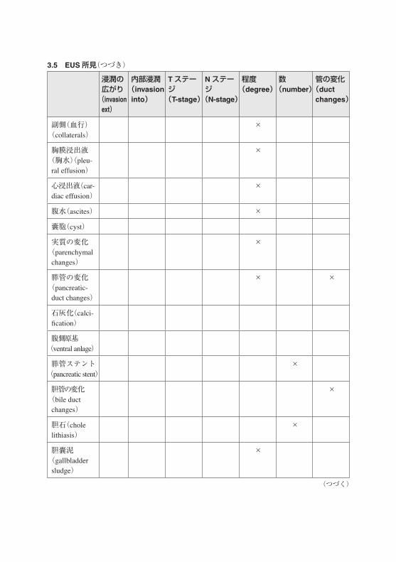
3.5 EUS所見(EUS findings)(table in 3 parts)
部位(location)
由来(origin)
層の由来(layer origin)
外境界(outer limit)
サイズ(size)
壁内病変(intramural lesion)
× × × ×
壁肥厚(wall thicken-ing)
× × ×
ポリープ(polyp) × × ×
腫瘤(mass) × × × × ×
リンパ節[lymph node(s)]
× ×
狭窄(stricture) ×
3.4 臓器別 ERCP所見(つづき)
(つづく)

部位(location)
由来(origin)
層の由来(layer origin)
外境界(outer limit)
サイズ(size)
壁外腫瘤(extramural mass)
× × ×
静脈瘤(varices) × ×
副側(血行)(collaterals)
×
胸膜浸出液(胸水)(pleural effusion)
×
心浸出液(cardiac effusion)
腹水(ascites)
囊胞(cyst) × ×
実質の変化(parenchymal changes)
×
膵管の変化(pancre-atic duct changes)
×
石灰化(calcification) × ×
腹側原基(ventral anlage)
膵管ステント(pancreatic stent)
×
胆管の変化(bile duct changes)
×
胆石(cholelithiasis) × ×
胆囊泥(gallbladder sludge)
胆管ステント(biliary stent)
×
膵管癒合不全(pancreas divisum)
3.5 EUS所見(つづき)
(つづく)

所見の属性(finding attributes)
由来(origin)
層の由来(layer origin)
外境界(outer limit)
関連臓器(relevant organs)
粘膜(mucosa) 粘膜(mucosa)
粘膜筋板(muscularis mu-cosae)
粘膜筋板(muscularis mu-cosae)
粘膜下層(submucosa)
粘膜下層(submucosa)
固有筋層(muscularis pro-pria)
固有筋層(muscularis pro-pria)
固有筋層を越え(beyond muscu-laris propria)
壁をまたいだ(transmural)
すべての層(all layers)
不確定な(indeterminate)
形態(shape/character)
辺縁(mar-gins)
超音波像(echo-features)
囊胞像(cyst features)
壁内病変(intramural lesion)
× × ×
壁肥厚(wall thickening) ×
ポリープ(polyp) × × ×
腫瘤(mass) × × ×
リンパ節[lymph node(s)]
× × ×
狭窄(stricture)
3.5 EUS所見(つづき)
(つづく)

形態(shape/character)
辺縁(mar-gins)
超音波像(echo-features)
囊胞像(cyst features)
壁外腫瘤(extramural mass)
× × ×
静脈瘤(varices) ×
副側(血行)(collaterals) ×
胸膜浸出液(胸水)(pleural effusion)
心浸出液(cardiac effusion)
腹水(ascites)
囊胞(cyst) × × ×
実質の変化(parenchymal changes)
× ×
膵管の変化(pancreatic duct changes)
×
石灰化(calcification) ×
腹側原基(ventral anlage)
膵管ステント(pancreatic stent)
胆管の変化(bile duct changes)
胆石(cholelithiasis)
胆囊泥(gallbladder sludge)
胆管ステント(biliary stent)
膵管癒合不全(pancreas divisum)
丸い(round) 明瞭な(well defined)
正常(normal) 無エコーの(anechoic)
3.5 EUS所見(つづき)
(つづく)

形態(shape/character)
辺縁(mar-gins)
超音波像(echo-features)
囊胞像(cyst features)
卵円形(oval) 不明瞭な(poorly defined)
無エコーの(anechoic)
均質な(homo-geneous)
三角の(triangular)
滑らかな(smooth)
低エコーの(hypoechoic)
不均質な(het-erogeneous)
半月上の(crescent-shaped)
不規則(irregular)
内エコーの(isoechoic)
胆泥あり(debris present)
蛇行(tortuous)
囲む(encasing)
高エコーの(hyperechoic)
隔壁のある(septated)
管状の(tubular)
台状圧排像(abutting)
高エコー病変(hyperechoic foci)
単房性(unilocular)
分葉した(lobulated)
境界エコーのない(loss of interface)
高エコー部位(hypenterface)
多房性(multilocular)
無茎性(sessile)
浸潤している(invading)
顆粒状(granular)
小囊胞性(microcystic)
有茎性(pedunculated)
管腔内発育(intraluminal growth)
均質な(homo-geneous)
大囊胞性(macrocystic)
潰瘍性の(ulcerated)
不均質異質(heterogeneous)
無囊胞(no of cysts)
不規則な(irregular)
囊胞成分(cystic components)
小囊胞サイズ(microcyst size)
周辺の(circum-ferential)
充実性の(solid) 壁肥厚(wall thickness)
拡大した(enlarged)
シャドウを引く(shadowing)
壁不整(wall irregularity)
萎縮性(atrophic)
遠位部強調(distal enhance-ment)
壁内小結節(mural nod-ules)
3.5 EUS所見(つづき)
(つづく)

形態(shape/character)
辺縁(mar-gins)
超音波像(echo-features)
囊胞像(cyst features)
びまん性(diffuse)
石灰化(calci-fication)
汎発性の(generalized)
管との交通(duct commu-nication)
限局性(localized)
関連した腫瘤(associated mass)
サイズ/数(size/number)
浸潤の広がり(invasion ext)
内部浸潤(invasion into)
Tステージ(T-stage)
Nステージ(N-stage)
程度(degree)
数(number)
管の変化(duct changes)
壁内病変(intramural lesion)
× ×
壁肥厚(wall thickening)
ポリープ(polyp)
腫瘤(mass) × × × ×
リンパ節[lymph node(s)]
×
狭窄(stricture)
× × ×
壁外腫瘤(extramural mass)
× × × ×
静脈瘤(varices) ×
3.5 EUS所見(つづき)
(つづく)
浸潤の広がり(invasion ext)
内部浸潤(invasion into)
Tステージ(T-stage)
Nステージ(N-stage)
程度(degree)
数(number)
管の変化(duct changes)
副側(血行)(collaterals)
×
胸膜浸出液(胸水)(pleu-ral effusion)
×
心浸出液(car-diac effusion)
×
腹水(ascites) ×
囊胞(cyst)
実質の変化(parenchymalchanges)
×
膵管の変化(pancreatic-duct changes)
× ×
石灰化(calci-fication)
腹側原基(ventral anlage)
膵管ステント(pancreatic stent)
×
胆管の変化(bile duct changes)
×
胆石(cholelithiasis)
×
胆囊泥(gallbladder sludge)
×
3.5 EUS所見(つづき)
(つづく)

浸潤の広がり(invasion ext)
内部浸潤(invasion into)
Tステージ(T-stage)
Nステージ(N-stage)
程度(degree)
数(number)
管の変化(duct changes)
胆管ステント(Biliary stent)
×
膵管癒合不全(Pancreas divisum)
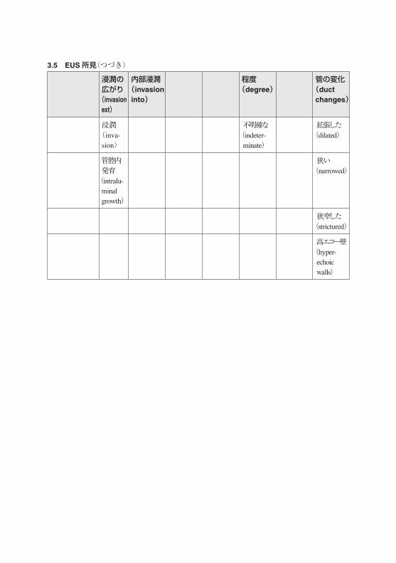
浸潤の広がり(invasion ext)
内部浸潤(invasion into)
程度(degree)
管の変化(duct changes)
口径不整(encase-ment)
関連臓器(relevant
organs)
なし(none) 正常(normal)
境界なし<1.5cm(inter-
face loss<1.5cm)
最小の(minimal)
規則的な(regular)
境界なし<1.5cm(inter-
face loss<1.5cm)
中等度の(moderate)
不規則な輪郭(ir-regular contour)
アバットメント(台上の圧排像)(abut-
ment)
広範な(extensive)
珠状の(beaded)
3.5 EUS所見(つづき)
(つづく)
浸潤の広がり(invasion ext)
内部浸潤(invasion into)
程度(degree)
管の変化(duct changes)
浸潤(inva-sion)
不明確な(indeter-
minate)
拡張した(dilated)
管腔内発育(intralu-
minal growth)
狭い(narrowed)
狭窄した(strictured)
高エコー壁(hyper-
echoic walls)
3.5 EUS所見(つづき)

3.6 EUS 病変-個別属性のリスト(EUS lesion-specific attributes lists)3.6.1 形態/性状(shape/character)
形態 /性状(shape/character)
円い(round)
卵円形の(oval)
三角形の(tri-angular)
線状の(linear)
三ヶ月状の(crescent-shaped)
蛇行した(tortuous)
管状の(tubular)
分葉状の(lobulated)
無茎の(sessile)
有茎性(pedun-culated)
壁内病変(intramural lesion) × × × × × ×
ポリープ(polyp) × × ×
腫瘤(mass) × × × × × ×
リンパ節[lymph node(s)] × × × × × ×
壁外腫瘤(extramural mass) × × × × × ×
静脈瘤(varices) × ×
副側(血行)(collaterals) × ×
囊胞(cyst) × × × ×
実質の変化(parenchymal changes)
膵管の変化(pancreatic duct changes)
石灰化(calcification)
(つづく)

3.6.1 形態/性状(つづき)
形態 /性状(shape/character)
潰瘍性の(ulcerated)
不規則な(irregular)
周辺部(circum-ferential)
拡大した(enlarged)
萎縮性(atrophic)
びまん性(diffuse)
汎発性の(general-ized)
限局性の(local-ized)
数(number)
壁内病変(intramural lesion) × × ×
ポリープ(polyp) × × ×
腫瘤(mass) × × × ×
リンパ節[lymph node(s)] × ×
壁外腫瘤(extramural mass) × ×
静脈瘤(varices)
副側(血行)(collaterals) × × ×
囊胞(cyst) × ×
実質の変化(parenchymal changes)
× × ×
膵管の変化(pancreatic duct changes)
× ×
石灰化(calcification) × ×
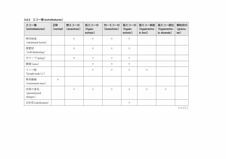
3.6.2 エコー像(echofeatures)
エコー像(echofeatures)
正常(normal)
無エコーの(anechoic)
低エコーの(hypo-echoic)
均一エコーの(isoechoic)
高エコーの(hyper-echoic)
高エコー病変(hyperecho-ic foci)
高エコー部位(hyperecho-ic strands)
顆粒状の(granu-lar)
壁内病変(intramural lesion)
× × × ×
壁肥厚(wall thickening)
× × × ×
ポリープ(polyp) × × × ×
腫瘤(mass) × × ×
リンパ節[lymph node(s)]
× × × ×
壁外腫瘤(extramural mass)
×
実質の変化(parenchymal
changes)
× × × × × ×
石灰化(calcification) ×
(つづく)

3.6.2 エコー像(つづき)
エコー像(echofeatures)
均質な(homogeneous)
異質な(heterogeneous)
囊胞成分(cystic-components)
充実した(solid)
シャドウを引く(shadowing)
末端強調(distal- enhancement)
壁内病変(intramural lesion)
× × × × × ×
壁肥厚(wall thickening)
× × × × × ×
ポリープ(polyp) × × × × × ×
腫瘤(mass) × × × × × ×
リンパ節[lymph node(s)]
× × × × × ×
壁外腫瘤(extramural mass)
実質の変化(parenchymal
changes)
× × × ×
石灰化(calcification) × × ×

3.7 臓器別 EUS所見(EUS findings per organ)
食道(esophagus)
胃(stomach)
十二指腸(duodenum)
膵(pan-creas)
胆道(biliary tract)
大腸(colorectal)
壁内病変(intramural lesion)
× × × × ×
壁肥厚(wall thickening)
× × × × ×
ポリープ(polyp)
× × × ×
腫瘤(mass) × × × × × ×
リンパ節[lymph node(s)]
× × × × × ×
狭窄(stricture) × × × ×
壁外腫瘤(ex-tramural mass)
× × × × ×
囊胞(cyst) × × × × × ×
静脈瘤(varices)
× ×
副側(血行)(collaterals)
× × ×
胸膜浸出液(胸水)(pleural
effusion)
×
心浸出液(car-diac effusion)
×
腹水(ascites) × × ×
肛門周囲瘻孔(perianal fistula)
×
実質の変化(parenchymal changes)
×
(つづく)

食道(esophagus)
胃(stomach)
十二指腸(duodenum)
膵(pan-creas)
胆道(biliary tract)
大腸(colorectal)
膵管の変化(pancreatic duct changes)
×
石灰化(calcification)
×
腹側原基(ventral anlage)
×
膵管ステント(pancreatic stent)
×
胆管の変化(bile duct changes)
×
胆石(choleli-thiasis)
×
総胆管瘤(cho-ledochocele)
×
胆囊泥(gall-bladder sludge)
×
胆管ステント(biliary stent)
×
他(other) × × × × × ×
4 内視鏡検査の理由(reasons for endoscopy)Indications are often used in place of reasons for endoscopy. An indication is often used to define
the reason for an endoscopy which complies with generally accepted standards of practice. There may be reasons for an endoscopy which are not indications. For example, a patient may want to undergo annual colonoscopy for colorectal cancer surveillance even though there is no prior history of polyps or family history of colon cancer.
Reasons for have been divided into symptoms and diseases. . For the symptoms, a qualifier for duration and/or degree may be relevant. For diseases, the following qualifiers may all be relevant
3.7 臓器別 EUS所見(つづき)

Suspected, established, follow-up of.., sampling of…, treatment of…and screening for…. Screening will cover normal risk, as well as high risk individuals without previous findings.
If a disease is implicated, a disease should be listed as the indication. If the software allows several reasons/indications, then diseases should be prioritized to symptoms.
4.1 上部消化管内視鏡(upper GI endoscopy)
範疇(category) 用語(term) 修飾語(qualifier)
症状(symptoms) 腹部不快感/腹痛(abdominal distress/pain) 期間(duration) 消化不良(dyspepsia) 程度(degree)胸やけ(heartburn)嚥下困難(dysphagia)嚥下痛(odynophagia)嘔吐(vomiting)吐血(hematemesis)下血(melena)悪心(nausea)体重減少(weight loss)貧血(anemia)下痢(diarrhea)食欲不振
疾患(diseases) アカラシア(achalasia) 疑い(suspected) 吻合部リーク(anastomic leak) 確かな(established)血管拡張症(angioectasia) の経過観察
(follow-up of)萎縮性胃炎(atrophic gastritis) の抽出(sampling of)
バレット食道(Barrett's esophagus) の治療(treatment of)腐蝕物嚥下(caustic ingestion) のスクリーニング
(screening for)セリアック病(celiac disease)憩室(diverticulum)十二指腸狭窄(duodenal stricture)十二指腸潰瘍(duodenal ulcer)食道狭窄(esophageal stricture)食道静脈瘤(esophageal varices)瘻孔(fistula)異物(foreign body)胃潰瘍(gastric ulcer)胃静脈瘤(gastric varices)胃食道逆流症(GERD)
(つづく)

範疇(category) 用語(term) 修飾語(qualifier)
リンパ腫(lymphoma)転移,原発不明(metastasis, unknown origin)その他の食道炎(other esophagitis)悪性貧血(pernicious anaemia)ポリープ(polyp)前癌病変(precancerous lesions)幽門狭窄(pyloric stenosis)逆流性食道炎(reflux esophagitis)表在(面)型腫瘍病変(superficial neoplastic lesion)腫瘍 /腫瘤(tumor/mass)
4.2 大腸内視鏡(colonoscopy)
範疇(category) 用語(term) 修飾語(qualifier)
症状(symptoms) 血便(hematochezia) 期間(duration)下血(melena) 程度(degree) 下痢(diarrhea)腹部不快感/腹痛(abdominal distress/pain)交替制便通異常(altered bowel habits)貧血(anemia)体重減少(weight loss)便秘(constipation)排便異常(defecation disorder)腹部腫瘤(abdominal mass)
疾患(diseases) 吻合部リーク(anastomic leak) 疑い(suspected) 血管拡張症(angioectasia) 確かな(established)大腸閉塞症(イレウス)(colonic obstruction) の経過観察
(follow-up of)大腸がん(colorectal cancer) の抽出(sampling of)クローン病(Crohn's disease) の治療(treatment of)憩室(diverticula) のスクリーニング
(screening for)内分泌腫瘤(endocrine mass)瘻孔(fistula)虚血性大腸炎(ischemic colitis)転移,原発不明(metastasis, unknown origin)
4.1 上部消化管内視鏡(つづき)
(つづく)

範疇(category) 用語(term) 修飾語(qualifier)
その他の大腸炎(other colitis)ポリープ(polyps)パウチ炎(pouchitis)偽膜性大腸炎(pseudomembranous colitis)表在(面)型腫瘍病変(superficial neoplastic lesion)潰瘍性大腸炎(ulcerative colitis)
4.3 小腸内視鏡(enteroscopy)
範疇(category) 用語(term) 修飾語(qualifier)
症状(symptoms) 腹部不快感/腹痛(abdominal distress/pain) 期間(duration) 嘔吐(vomiting) 程度(degree)下血(melena)貧血(anemia)下痢(diarrhea)
疾患(diseases) 疾患(diseases) 疑い(suspected) セリアック病(celiac disease) 確かな(established)クローン病(Crohn's disease) の経過観察
(follow-up of)遺伝性ポリポーシス(hereditary polyposis syndrome)
の抽出(sampling of)
リンパ腫(lymphoma) の治療(treatment of)NSAID腸病変(NSAID enteropathy) のスクリーニング
(screening for)ポリープ(polyps)狭窄(stricture)腫瘤(mass)
4.2 大腸内視鏡(つづき)

4.4 ERCP
範疇(category) 用語(term) 修飾語(qualifier)
症状(symptoms) 黄疸(jaundice) 期間(duration) 膵胆道部痛(pancreatobiliary pain) 程度(degree) 瘙痒(pruritus)腫瘍マーカー異常
疾患(diseases) 急性膵炎(acute pancreatitis) 疑い(suspected) 乳頭部腫瘤(ampullary mass) 確かな(established)吻合部狭窄(anastomotic stricture) の経過観察
(follow-up of)胆管損傷(bile duct injury) の抽出(sampling of)胆管リーク(bile duct leak) の治療(treatment of)胆管結石(bile duct stone) のスクリーニング
(screening for)胆管狭窄(bile duct stricture)カロリー病(Caroli's disease)総胆管囊腫(choledochal cyst)総胆管瘤(choledochocele)慢性膵炎(chronic pancreatitis)瘻孔(fistula)胆囊ポリープ(gallbladder polyp) 胆囊結石(gallbladder stone)胆囊腫瘤(gallbladder mass)肝胆道腫瘤(hepatobiliary mass)膵管内乳頭粘液性腫瘍(IPMN)ミリッツィ症候群(Mirizzi syndrome)膵管リーク(pancreatic duct leak)膵損傷(pancreatic injury)膵仮性囊胞(pancreatic pseudocyst)膵結石(pancreatic stone)膵腫瘤(pancreatic mass)乳頭機能不全(papillary dysfunction)寄生虫(parasites)肝実質疾患(parenchymal liver disease)原発性硬化性胆管炎(primary sclerosing cho-langitis)化膿性胆管炎(purulent or suppurative cholangitis)ステント機能不良(stent dysfunction)

4.5 EUS
範疇(category) 用語(term) 修飾語(qualifier)
症状(symptoms) 腹痛(abdominal pain) 期間(duration)下痢(diarrhea) 程度(degree)黄疸(jaundice)膵胆道部痛(pancreatobiliary pain)体重減少(weight loss)
疾患(diseases) 腹部腫瘤(abdominal mass) 疑い(suspected) 乳頭部腫瘤(ampullary mass) 確かな(established)胆管腫瘤(bile duct mass) の経過観察
(follow-up of) 胆管結石(bile duct stone) の抽出(sampling of)胆管狭窄(bile duct stricture) のステージング
(staging of)大腸壁内病変(colorectal intramural lesion) の治療(treatment of)大腸腫瘤(colorectal mass) のスクリーニング
(screening for)大腸狭窄(colorectal stricture)十二指腸壁内病変(duodenal intramural lesion)十二指腸腫瘤(duodenal mass)十二指腸狭窄(duodenal stricture)食道壁内病変(esophageal intramural lesion)食道腫瘤(esophageal mass)食道狭窄(esophageal stricture)胃壁内病変(gastric intramural lesion)胃腫瘤(gastric mass)肝腫瘤(liver mass)縦隔内リンパ節 /腫瘤(mediastinal lymph node/mass)膵囊胞(pancreatic cyst)膵腫瘤(pancreatic mass)膵仮性囊胞(pancreatic pseudocyst)膵石(pancreatic stone)膵炎-急性(pancreatitis acute)膵炎-慢性(pancreatitis chronic)直腸周囲腫瘤(perirectal mass)肺腫瘤(pulmonary mass)

用語(term)
迷入膵(aberrant pancreas)アカラシア(achalasia)バレット食道(Barrett's esophagus)良性狭窄(benign stricture)原因不明の出血(bleeding of unknown origin)セリアック病(celiac disease)クローン病(Crohn's disease)デュラフォア病変(Dieulafoy lesion)腺腫良性十二指腸腫瘍(duodenal benign tumor)十二指腸球部変形(duodenal bulb deform-ity)十二指腸がん(duodenal cancer)十二指腸憩室(duodenal diverticulum)十二指腸瘻(duodenal fistula)十二指腸ポリープ(duodenal polyp) 過去の十二指腸手術後所見(duodenal postoperative appearance)十二指腸粘膜下腫瘍(duodenal submucosal tumor)表在(面)型十二指腸腫瘍(duodenal super-ficial neoplasm) 十二指腸潰瘍(duodenal ulcer)出血を伴う十二指腸潰瘍(duodenal ulcer with bleeding)びらん性十二指腸病変(duodenopathy erosive)出血性十二指腸病変(duodenopathy hemor-rhagic)充血性十二指腸病変(duodenopathy hyper-emic)好酸球性食道炎(eosinophilic esophagitis)良性食道腫瘍(esophageal benign tumor)腐蝕剤による食道障害(esophageal caustic injury)食道がん(esophageal cancer)食道カンジダ症(esophageal candidiasis)食道憩室(esophageal diverticulum)
食道瘻孔(esophageal fistula)食道異物(esophageal foreign body) 食道ポリープ(esophageal polyp)過去の食道手術後所見(esophageal post-operative appearance)食道狭窄(esophageal stricture)表層性食道新生物(esophageal superficial neoplasm)食道粘膜下腫瘍(esophageal submucosal tumor)食道静脈瘤(esophageal varices)壁外性圧迫(extrinsic compression) 良性胃腫瘍(gastric benign tumor)胃がん(gastric cancer)MALTリンパ腫(MALToma)胃憩室(gastric diverticulum)胃瘻孔(gastric fistula)胃異物(gastric foreign body)腐蝕剤による胃障害(gastric caustic injury)胃リンパ腫(gastric lymphoma)胃ポリープ[gastric polyp(s)]過去の胃手術後所見(gastric postoperative appearance)胃内うっ滞(gastric retention) 胃粘膜下腫瘍(gastric submucosal tumor)表在(面)型胃新生物(gastric superficial neoplasm)胃潰瘍(gastric ulcer)出血を伴う胃潰瘍(gastric ulcer with bleed-ing)胃潰瘍-吻合部(gastric ulcer-anastomotic)胃静脈瘤(gastric varices)胃病変-びらん性(gastropathy-erosive)胃病変-出血性(gastropathy-hemorrhagic)胃病変-充血性(gastropathy-hyperemic)胃病変-肥厚性(gastropathy-hypertrophic)胃病変-NSAID起因性(gastropathy-NSAID-related)
5 内視鏡診断(endoscopic diagnosis)
5.1 上部消化管内視鏡(upper endoscopy)
(つづく)

用語(term)
胃病変-門脈圧亢進性(gastropathy-portal hypertensive)胃病変-あばた状(gastropathy-varioloform)裂孔ヘルニア(hiatus hernia)マロリー・ワイス裂傷(Mallory-Weiss tear)他の食道炎(other esophagitis)寄生虫(parasites)
硬化療法後所見(post sclerotherapy appear-ance)幽門狭窄(pyloric stenosis)逆流性食道炎(reflux esophagitis) シャッキィ輪(Schatzki ring)瘢痕(scar)粘膜下腫瘍(submucosal tumor)
用語(term)
血管拡張症(angiectasia)原因不明の出血(bleeding of unknown origin)大腸炎-虚血性(colitis-ischemic) 大腸炎-偽膜性(colitis-pseudomembranous)大腸腺腫大腸がん(colorectal cancer)表在(面)型大腸腫瘍(colorectal superficial neoplasm)コンジロマ(condylomata)クローン病-活動性(Crohn's disease-active)クローン病-劇症型(Crohn's disease-fulmi-nant)クローン病-寛解期(Crohn's disease-quies-cent)憩室炎(diverticulitis)多発性憩室症(diverticulosis)瘻孔(fistula)異物(foreign body)痔核(hemorrhoids)回腸炎(ileitis)
脂肪腫(lipoma)リンパ腫(lymphoma)黒皮症 /メラノーシス(melanosis)寄生虫(parasites)大腸囊胞状気腫症(pneumatosis coli)ポリープ(polyp)ポリポーシス症候群(polyposis syndrome)手術後の所見(postoperative appearance)直腸炎(proctitis)直腸潰瘍(rectal ulcer)孤在性潰瘍(solitary ulcer)狭窄-炎症性(stricture-inflammatory)狭窄-悪性(stricture-malignant)狭窄-術後(stricture-postoperative)粘膜下腫瘍(submucosal tumor)潰瘍性大腸炎-活動性(ulcerative colitis-ac-tive)潰瘍性大腸炎-劇症型(ulcerative colitis-ful-minant)潰瘍性大腸炎-寛解期(ulcerative colitis-quiescent)
5.1 上部消化管内視鏡(つづき)
5.2 大腸内視鏡(colonoscopy)

用語(term)
血管拡張症(angiectasia)セリアック病(celiac disease)クローン病(Crohn's disease)憩室(diverticulum)(pl. 1a)腸病変-びらん性(enteropathy-erosive)腸病変-出血性(enteropathy-hemorrhagic)腸病変-充血性(enteropathy-hyperemic)腸病変-NSAID起因性(enteropathy-NSAID-related)びらん(erosions)家族性腺腫性ポリポーシス(FAP)消化管間葉性細胞腫(GIST)GVHD(graft vs host disease)若年性ポリポーシス(juvenile polyposis)脂肪腫(lipoma)
リンパ管拡張症(lymphangioectasia)リンパ腫(lymphoma)NSAID腸病変(NSAID-enteropathy)寄生虫(parasites)プッツ・ジェーガースポリポーシス(Peutz-Jeghers polyposis)ポリープ[polyp(s)]放射性腸炎(radiation enteritis) 良性小腸腫瘍(small bowel benign tumor)悪性小腸腫瘍(small bowel malignant tumor)表在(面)小腸新生物(small bowel superfi-cial neoplasm)粘膜下腫瘍(submucosal tumor)潰瘍(ulcer)
5.3 小腸内視鏡(enteroscopy)
5.4 ERCP4
用語(term)
膵胆管合流異常(abnormal pancreatobiliary-junction)吻合部狭窄(anastomotic stricture) 胆道瘻 /リーク(biliary fistula/leak) 胆道損傷(biliary injury)胆道閉塞(biliary occlusion)胆道ステント閉塞(biliary stent occlusion)胆石 /胆道結石[biliary stone(s)]胆道狭窄(biliary stricture)カロリー病(Caroli's disease)総胆管囊胞(choledochal cyst)総胆管腔(choledochocele)胆囊管結石(cystic duct stones)胆囊結石[gallbladder stone(s)]胆囊腫瘍(gallbladder tumor)血性胆汁(hemobilia)膵管内乳頭粘液性腫瘍(IPMN)
総胆管癌(cholangiocarcinoma)肝実質疾患(liver parenchymal disease)ミリッツィ症候群(Mirizzi syndrome)輪状膵(pancreas annulare)膵管癒合不全(pancreas divisum)膵囊胞(pancreatic cyst)膵管瘻 /リーク(pancreatic duct fistula/leak)膵管損傷(pancreatic duct injury) 膵管狭窄(pancreatic duct stricture)膵管ステント閉塞(pancreatic stent occlu-sion)膵石(pancreatic stone)膵腫瘍(pancreatic tumor)膵炎-急性(pancreatitis-acute) 膵炎-慢性(pancreatitis-chronic) 自己免疫性膵炎(autoimmune-pancreatitis)乳頭狭窄(papillary stenosis)
4 Upper endoscopy diagnoses may be relevant for the luminal part of the ERCP procedure as well.
(つづく)

用語(term)
乳頭腫瘍(papillary tumor)原発性硬化性胆管炎(primary sclerosing cho-langitis)
サンプ症候群(sump syndrome)化膿性胆管炎(suppurative cholangitis)
用語(term)
大動脈瘤(aortic aneurysm)腹水(ascites)胆管拡張(bile duct dilation)胆石 /胆管結石(bile duct stone)胆管狭窄(bile duct stricture)胆管腫瘍(bile duct tumor)肝泥(biliary sludge)大腸がん(colorectal cancer) 大腸ポリープ(colorectal polyp) 大腸狭窄(colorectal stricture)大腸粘膜下腫瘍(colorectal submucosal mass)大腸潰瘍(colorectal ulcer)クローン病(Crohn's disease)十二指腸がん(duodenal cancer)十二指腸ポリープ(duodenal polyp)十二指腸狭窄(duodenal stricture)十二指腸粘膜下腫瘤(duodenal submucosal mass)十二指腸潰瘍(duodenal ulcer)内分泌腫瘍(endocrine tumor)食道がん(esophageal cancer)食道腫瘤(esophageal mass)食道ポリープ(esophageal polyp)食道狭窄(esophageal stricture) 食道粘膜下腫瘤(esophageal submucosal mass)食道潰瘍(esophageal ulcer) 胆囊泥(gallbladder sludge) 胆囊結石(gallbladder stone) 胃がん(gastric cancer)胃ポリープ(gastric polyp)胃粘膜下腫瘤(gastric submucosal mass)
胃潰瘍(gastric ulcer)消化管間葉細胞腫瘍 GIST(GIST tumor)膵管内乳頭粘液性腫瘍(IPMT)スキルス型胃がん(linitis plastica)脂肪腫(lipoma)リンパ腫(lymphoma)縦隔内リンパ節[mediastinal lymphnode(s)]縦隔腫瘤(mediastinal mass)メネトリエ 病(Menetrier's disease)腸間膜動脈瘤(mesenteric artery aneurysm)膵管癒合不全(pancreas divisum)膵がん(pancreatic cancer)膵管拡張(pancreatic duct dilation)膵管狭窄(pancreatic duct)stricture膵腫瘤(pancreatic mass)膵仮性囊胞(pancreatic pseudocyst)膵石(pancreatic stone)急性膵炎(pancreatitis-acute) 慢性膵炎(pancreatitis-chronic)乳頭腫瘤(papillary mass)心囊滲出液 /胸水(pericardial effusion/pleu-ral effusion)ポリープ(polyp)門脈血栓(portal vein thrombosis)放射線照射後変化(postradiation changes)偽動脈瘤(pseudoaneurysm)脾動脈瘤(splenic artery aneurysm)粘膜下腫瘍(submucosal tumor)TNM分類のステージ(TNM-stage)潰瘍性大腸炎(ulcerative colitis)静脈瘤(varices)壁肥厚(wall thickening)
5.5 EUS
5.4 ERCP(つづき)

6 手技(procedures)Procedures should be described, both in terms of technical aspects and whether the aim of the
procedure was achieved.Because some procedures are difficult to connect directly to a described finding, procedures are
suggested to be linked generally to the endoscopy per se, not to lesions. However, software should be able to connect the two as needed. Thus, sampling, as well as treatment of a lesion should be available linked to the description of the lesion itself. The choice of treatment modalities should be adapted to the individual lesions, to avoid listing of irrelevant procedures.
6.1 管腔での内視鏡手技(luminal procedures)
方法(method) 属性(attribute) 属性値(attribute values)
採取(sampling) 病変(lesion) link to relevant finding OR 正常部位[normal location(s)]
型(type) 生検(biopsy)不完全切除(incomplete removal)完全分割切除(complete piecemeal re-moval)一括切除(en bloc resection)ブラシング採取(brush sampling)液体採取(fluid sampling)
最終判断(final read-ing)
後での登録(late entry)
バルーン拡張(balloon dila-tion)
様式(type)5 記述する(specify)
口径(caliber) 記述する(specify)時間(time) 記述する(specify) 終了時の口径(end caliber)
記述する(specify)
ブジー拡張(bougie dilation) 最初の口径(start cal-iber)
記述する(specify)
終了時の口径(end caliber)
記述する(specify)
バンド結紮(band ligation) 数(number) 記述する(specify)結果(result) 成功(success)
一部成功(partial success)失敗(failure)
注入療法(injection therapy) 材料(material) 記述する(specify)量(volume) 記述する(specify)結果(result) 成功(success)
5 Type/tool details should be optional for most procedures.
(つづく)

方法(method) 属性(attribute) 属性値(attribute values)
一部成功(partial success)失敗(failure)
クリッピング(clipping) 方式(type) 記述する(specify)数(number) 記述する(specify)結果(result) 成功(success)
一部成功(partial success)失敗(failure)
凝固(coagulation) 方式(type) 記述する(specify)結果(result) 成功(success)
一部成功(partial success)失敗(failure)
APC 使用ガス(gas used) ml結果(result) 成功(success)
一部成功(partial success)失敗(failure)
ポリープ切除(polypectomy) 方法(method) スネアのみで(snare only)ループとスネアで(loop and snare)クリップとスネアで(clips and snare)
術前注入(pre-injec-tion)
はい /いいえ(yes/no)
術前注入液(pre-inj fluid)
術前注入液(pre-inj fluid)
術前注入液量(pre-inj volume)
ml
結果(result) 成功(success)一部成功(partial success)失敗(failure)
ホットバイオプシー(hot biopsy)
結果(result) 成功(success)一部成功(partial success) 失敗(failure)
異物除去(foreign body removal)
器具(tool) 記述する(specify)
結果(result) 成功(success)一部成功(partial success)失敗(failure)
内視鏡的粘膜切除術(EMR) 器具(tool) 記述する(specify)術前注入(局注)液の種類(pre-inj fluid)
記述する(specify)
術前注入(局注)液量(pre-inj volume)
ml
(つづく)
6.1 管腔での内視鏡手技(つづき)

方法(method) 属性(attribute) 属性値(attribute values)
結果(result) 全体切除(en bloc resection) 完全分割切除(complete piecemeal removal)部分切除(partial resection)失敗(failure)
組織のサイズ(specimen size)
mm
内視鏡的粘膜下層剝離術(ESD)
器具(tool) 記述する(specify)
術前注入液(pre-inj fluid)
記述する(specify)
術前注入液量(pre-inj volume)
ml
結果(result) 一括切除(en bloc resection)部分切除(partial resection)失敗(failure)
組織のサイズ(specimen size)
mm
ステント留置(stent place-ment)
型(type) 記述する(specify)
サイズ(size) 記述する(specify)術前拡張(pre-dilation) なし /mm(no/mm)結果(result) 成功(success)
一部成功(partial success)失敗(failure)
PEG造設(PEG placement) 方式(type) 記述する(specify)サイズ(size) 記述する(specify)結果(result) 成功(success)
失敗(failure)チューブ挿入(tube place-ment)
方式(type) 記述する(specify)
サイズ(size) 記述する(specify)部位(location) 記述する(specify)結果(result) 成功(success)
一部成功(partial success)失敗(failure)
経消化管壁ドレナージ(transmural drainage)
方法(method) 記述する(specify)
部位(location) 記述する(specify)
6.1 管腔での内視鏡手技(つづき)
(つづく)

方法(method) 属性(attribute) 属性値(attribute values)
病変(lesion) 記述する(specify)EUSによる(EUS guided)
はい /いいえ(yes/no)
材料(material) 記述する(specify)ステント留置(stents-placed)
記述する(specify)
結果(result) 成功(success)一部成功(partial success)失敗(failure)
焼灼術(ablation therapy) 方式(type) 記述する(specify)部位(location) 記述する(specify)機材の状況(settings) 記述する(specify)
6.2 ERCP手技(ERCP procedures)
方法(method) 属性(attribute) 属性値(attribute values)
カニュレーション(cannula-tion)
胆(biliary) 試みず(not attempted)
偶然に(incidental)表層性(superficial)深い(deep)失敗した(failed)
膵(pancreatic) 試みず(not attempted)偶然に(incidental)表層性(superficial)深部(deep)失敗した(failed)
副乳頭(minor papil-la)
試みず(not attempted)
表層性(superficial)深部(deep)
造影(opacification) 胆(biliary) 完全(complete)不完全,未遂(incomplete, missing)胆道造影失敗の原因(no biliary, due to)胆道非造影の理由(no biliary intended)
膵(pancreatic) 完全(complete)不完全,未遂(incomplete, missing)
6.1 管腔での内視鏡手技(つづき)
(つづく)

方法(method) 属性(attribute) 属性値(attribute values)
膵管造影失敗の原因(no biliary, due to)膵管非造影の理由(no biliary intended)
乳頭括約筋切開術(papillotomy)
形式(type) 方法/プレカット(access/precut)
治療のため(therapeutic)瘻孔切開術(fistulotomy)
器具(tool) ワイヤーナイフ(wire knife) ニードルナイフ(needle knife)
管(duct) 胆管(bile duct)膵管(pancreatic duct)両方(both ducts)
バルーン拡張(balloon dila-tation)
バルーンの直径(balloon caliber)
mm
拡張後の(end cali-ber)
mm
ブジー拡張(bougie dilata-tion)
ブジーの直径(bougie caliber)
フレンチ(French)
拡張後の直径(resultcaliber)
mm
採石(stone extraction) 方法(method) バルーン(balloon)バスケット(basket)
破砕(fragmentation) はい(yes)いいえ(no)
結果(result) 完全除去(complete clearance)部分除去(partial clearance)不明(questionable)
ステント設置(stent place-ment)
型(type) 記述する(specify)
数(number) 記述する(specify)サイズ /口径(size/caliber)
記述する(specify)
ステント除去(stent re-moval)
範疇(category) 経乳頭(transpapillary)
上方への逸脱(upwards dislocated)器具(tool) 記述する(specify)結果(result) 成功(success)
失敗(failure)止血(hemostasis) 方法(method) 注入(injection)
クリップ(clips)バルーン圧迫(balloon compression)アルゴンプラズマ凝固術(APC)
6.2 ERCP手技(つづき)
(つづく)
方法(method) 属性(attribute) 属性値(attribute values)
凝固(coagulation)結果(result) 成功(success)
部分的成功(partial success)失敗(failure)
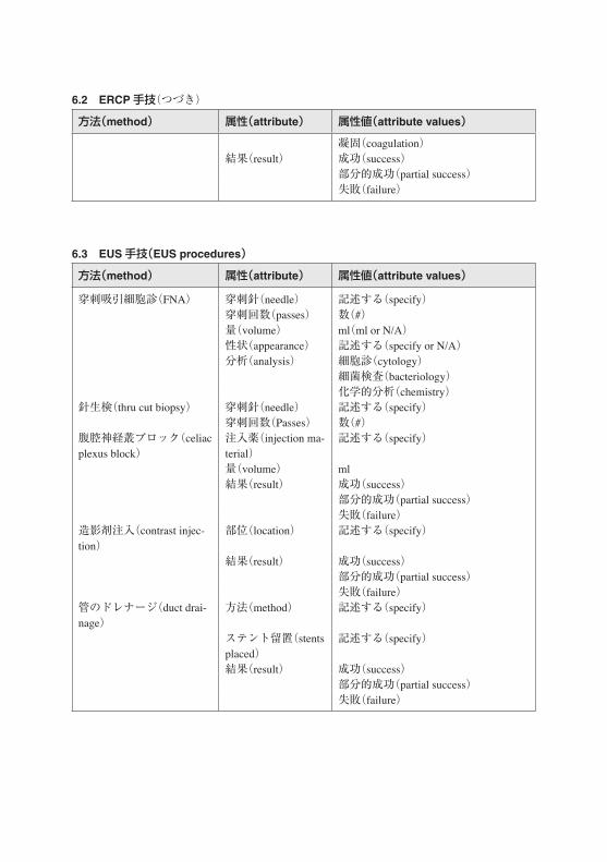
6.3 EUS手技(EUS procedures)
方法(method) 属性(attribute) 属性値(attribute values)
穿刺吸引細胞診(FNA) 穿刺針(needle) 記述する(specify)穿刺回数(passes) 数(#)量(volume) ml(ml or N/A)性状(appearance) 記述する(specify or N/A) 分析(analysis) 細胞診(cytology)
細菌検査(bacteriology)化学的分析(chemistry)
針生検(thru cut biopsy) 穿刺針(needle) 記述する(specify)穿刺回数(Passes) 数(#)
腹腔神経叢ブロック(celiac plexus block)
注入薬(injection ma-terial)
記述する(specify)
量(volume) ml結果(result) 成功(success)
部分的成功(partial success)失敗(failure)
造影剤注入(contrast injec-tion)
部位(location) 記述する(specify)
結果(result) 成功(success)部分的成功(partial success)失敗(failure)
管のドレナージ(duct drai-nage)
方法(method) 記述する(specify)
ステント留置(stents placed)
記述する(specify)
結果(result) 成功(success)部分的成功(partial success)失敗(failure)
6.2 ERCP手技(つづき)

7 偶発症(adverse events)Adverse events should be a part of the endoscopy report inasmuch they are noted at the time of the
procedure. However, it must be recognized that this will not constitute a complete complications registry without post-hoc registration of late complications.
Thus, an endoscopy database should acknowledge the need for additional late data input, for complications, as well as for e.g. sampling reports.
7.1 術中の偶発症(intraprocedure events)
方法(method) 属性(attribute) 属性値(attribute values)
管腔の穿孔(perforation of lumen)
局部(location) 主部位(main sites)
…との交通(communicating to)
後腹膜(retroperitoneum)
腹膜(peritoneum)縦隔(mediastinum)
形式(type) 内視鏡による(by endoscope)内視鏡的乳頭括約筋切開術関連(sphinc-terotomy-related)ポリープ切除術関連(polypectomy-related)拡張術関連(dilation-related)
出血(bleeding 局部(location) 主部位(main sites)心肺(cardiopulmonary) 様式(type) 無呼吸 /呼吸数減弱(apnoea/hypopnea)
気管支痙攣(bronchospasm)声門痙攣(laryngospasm)低血圧(hypotension)高血圧(hypertension)心停止(arrest)不整脈(dysrhythmia)吸引(aspiration)
行為(action) 補助呼吸(ventilation assist)救急コード(emergency code)下記のリスト(list below)
装置不良(equipment mal-function)
様式(type) 記述する(specify)
器具の埋伏(equipment im-paction)
様式(type) 記述する(specify)
アレルギー反応(allergic re-action)
薬剤(agent) 記述する(specify)
様式(type) 記述する(specify)行為(action) 補助呼吸(ventilation assist)
救急コード(emergency code)(つづく)

方法(method) 属性(attribute) 属性値(attribute values)
下記のリスト(list below)他の出来事(other events) 様式(type) 記述する(specify)
7.2 術後の偶発症(postprocedure events)
方法(method) 属性(attribute) 属性値(attribute values)
心肺障害(cardiopulmonary event) 様式(type) 記述する(specify)発症日(date of onset)
記述する(specify)
関連性(relation) おそらく(probable)可能性あり(possible)可能性少なし(unlikely)
腹痛(abdominal pain) 発症日(date of onset)
記述する(specify)
関連性(relation) おそらく(probable)可能性あり(possible)可能性少なし(unlikely)
感染(infection) 様式(type) 記述する(specify)発症日(date of onset)
記述する(specify)
関連性(relation) おそらく(probable)可能性あり(possible)可能性少なし(unlikely)
血栓塞栓症(thromboembolic event) 様式(type) 記述する(specify)発症日(date of onset)
記述する(specify)
関連性(relation) おそらく(probable)可能性あり(possible)可能性少なし(unlikely)
遅延性穿孔(late perforation) 様式(type) 記述する(specify)発症日(date of onset)
記述する(specify)
遅延性出血(late bleeding) 様式(type) 記述する(specify)発症日(date of onset)
記述する(specify)
膵炎(pancreatitis) 発症日(date of onset)
記述する(specify)
7.1 術中の偶発症(つづき)

7.3 行為とその結果(actions and outcomes)
行為(actions) 治療内視鏡施行(endoscopic intervention)手技の中断(aborted procedure)薬剤治療(medical intervention)入院(admission to hospital[days)]ICUへの搬入(admission to ICU)内視鏡再試行(repeat endoscopy)放射線治療(radiologic intervention)外科手術(surgery)
結果(outcomes) 回復(recovery)不具合の残存(permanent disability)死亡(術後の日数)[died(days after procedure)]
8 付録(appendices)
8.1 分類(classifications)
8.1.1 食道炎の Los Angels分類(LA classification of erosive esophagitis)
grade A 粘膜裂が 5mm未満(mucosal break< 5 mm in length)grade B 粘膜裂が 5mm以上(mucosal break> 5mm)grade C 粘膜裂が 2つ以上の襞に連続(mucosal break continuous between> 2 mucosal folds)grade D 粘膜裂が全周の 75%以上連続(mucosal break> 75% of esophageal circumference)
Reference: Lundell L, Dent J, Bennett J, et al. Endoscopic assessment of esophagitis: clinical and functional correlates and further validation of Los Angeles classification. Gut 1999 45:172-80
8.1.2 食道静脈瘤の大きさ分類(size classification of esophageal varices)
grade 1 小さくて屈曲蛇行せず,送気で平坦化(small and nontortuous esophageal varices, flattened with insufflations)
grade 2 屈曲しているが食道全体の 50%以下(tortuous esophageal varices but covering less than 50% of the radius of the distal esophagus)
grade 3 屈曲しており食道の 50%以上を占める(large and tortuous esophageal varices covering more than 50% of the radius of the distal esophagus)
Reference: Pungpapong S,Keaveny A, Raimondo M, Dickson R, Woodward T, Harnois D, Wallace: Accuracy and interobserver agreement of small-caliber vs. conventional esophagogastroduodenoscopy for evaluating esophageal varices. Endoscopy 2007 39 673-80

8.1.3 バレット上皮の食道伸展でのプラハ CM分類(Prague C & M classification of Barrett's
esophagus extension)Cは周囲,Mは極大で次の様に計算される[The C(Circumferential)and M(maximal)measurements are calculated, respectively, as]
C=内視鏡で観察された胃食道境界から全周性バレット上皮の口側辺縁までの距離(The difference in endoscope insertion distances between the positions recorded for the“GEJ”and the“proximal margin of the circumferential Barrett's epithelium.) M=内視鏡で観察された胃食道境界からバレット上皮が舌状に伸びた最長部分の口側辺縁までの距離(島状バレット上皮は含まず)(The difference in endoscope insertion distances between the positions recorded for the“GEJ”and the“proximal margin for the longest“tongue-like”segment of Barrett's epithelium(do not include Barretts islands in this assessment.)
Reference: Sharma P, Dent J, Armstrong D, Bergman JJ, Gossner L, Hoshihara Y, Jankowski JA, Junghard O, Lundell L, Tytgat GN, Vieth M. The development and validation of an endoscopic grading system for Barrett's esophagus: the Prague C & M criteria. Gastroenterology.2006 131:1392-9
8.1.4 腫瘍病変に関するパリ分類(Paris classification of neoplastic lesions)
type 0- 表層性隆起,平坦陥凹,ないし陥凹を示す腫瘍病変(superficial polypoid, flat/depressed, or excavated tumors)
type 1-ポリープ様癌,通常は広茎性(polypoid carcinomas, usually attached on a wide base) type 2- 境界鮮明で周堤を伴う潰瘍形成型の癌(ulcerated carcinomas with sharply demarcated and
raised margins) type 3- 境界不鮮明な浸潤を伴う潰瘍形成型の癌(ulcerated, infiltrating carcinomas without definite
limits)type 4-潰瘍を伴わない広範な浸潤癌(nonulcerated, diffusely infiltrating carcinomas)type 5-分類不能な進行癌(unclassifiable advanced carcinomas)
Reference: The Paris endoscopic classification of superficial neoplastic lesions: esophagus, stomach, and colon(no authors listed). Gastrointestinal Endosc 58 6, Supplement 1: S3-S43
8.1.5 表層型腫瘍病変に対する JGCA分類(JGCA classification of superficial neoplastic
lesions)
隆起型(protruding)- 有茎型(pedunculated) 0-Ⅰp- 無茎型(sessile) 0-Ⅰs表面型(non-protruding and nonexcavated)- 表面隆起型(slightly elevated) 0-Ⅱa- 表面平坦型(completely flat) 0-Ⅱb- 表面陥凹型(slightly depressed) 0-Ⅱc- 表面隆起+陥凹型(elevated and depressed types) 0-Ⅱc+IIa6
0-Ⅱa+IIc
6 The distinction betweenⅡa+Ⅱc andⅡc+Ⅱa is based on the relative importance of the two features. The same applies to the distinction betweenⅡc+ⅢandⅢ+Ⅱc.

陥凹型(excavated)- 陥凹型(ulcer) 0-Ⅲ- 隆起+陥凹型(excavated and depressed types) 0-Ⅱc+Ⅲ
0-Ⅲ+Ⅱc
Reference: Endoscopic classification review group. Update on the Paris endoscopic classification of superficial neoplastic lesions in the digestive tract. Endoscopy 2005 37: 570-8(日本語訳は消化器内視鏡用語集第 2版,1997によった)
8.1.6 潰瘍出血に対する Forrest分類(Forrest classification of ulcer bleeding)
ForrestⅠa 噴出性動脈出血(arterial, spurting hemorrhage)ForrestⅠb 毛細血管性出血(oozing hemorrhage)ForrestⅡa 露出血管あり(visible vessel g= vessel< 2 mm G= vessel> 2 mm )ForrestⅡb 凝血(adherent clot)ForrestⅡc ヘマチン付着(hematin-covered lesion)ForrestⅢ 新しい出血の痕跡なし(no signs of recent hemorrhage)
Reference Forrest JA, Finlayson ND, Shearman DJ. Endoscopy in gastrointestinal bleeding. Lancet 1974; 2 394-397(Heldwein W, Schreiner J, Pedrazolli J, et al: Is the Forrest classification a useful tool for planning endoscopic therapy of bleeding peptic ulcers? Endoscopy 21: 258-262, 1989に基づく OMED: Minimal Standard Terminology for Gastrointestinal Endoscopy MST 3.0より引用・改変)
8.1.7 痔核の Goligherグレーディング(Goligher grading of hemorrhoids)
grade 1 出血のある痔核(hemorrhoids with bleeding)grade 2 自然寛解のある出血・隆起を伴う痔核(hemorrhoids with bleeding and protrusion, with
spontaneous reduction)grade 3 用手環納の必要がある出血・隆起を伴う痔核(hemorrhoids with bleeding and protrusion
that require manual reduction)grade 4 修復不能な脱出を伴う痔核(hemorrhoids with prolapse that cannot be replaced)
Reference: T.R. Schrock, Hemorrhoids: nonoperative and interventional management. In: J. Barkin and C.A. O'Phelan, Editors, Advanced therapeutic endoscopy, Raven Press, New York(1991).
8.1.8 大腸粘膜表層バターンの工藤分類(ピットバターン)(Kudo classification of colon polyp
surface pattern)(p.103参照)

8.1.9 肝内胆管癌の Bismuth分類(Bismuth classification of cholangiocarcinoma)
type I: 肝外胆管に限局(extrahepatic involvement only)type II: 肝外胆管および肝門部のみ(extrahepatic and hilar involvement)type IIIa: 肝外胆管および肝門部から右側区域まで(extrahepatic, hilar and right-sided segmental
involvement)type IIIb: 肝外胆管および肝門部から左側区域まで(extrahepatic, hilar and left-sided segmental
involvement)type IV: 肝外胆管および両側肝区域に広がる(extrahepatic and bilateral segmental involvement)
Reference: Bismuth H, Castaing D, Traynor O. Resection or palliation: priority of surgery in the treatment of hilar cancer. World J Surg 1988 12: 39-47
8.1.10 食道胃領域の定義(definitions of the esophagogastric region)
The distal esophagus and the proximal part of the stomach, or gastric cardia, constitute the esophagogastric region, with specific anatomical landmarks. There are no conspicuous proximal and distal limits of the esophagogastric region. The landmarks are selected arbitrarily: 2 cm above and below the EGJ is a frequent standard.(食道下部とそれに接する胃の部分,すなわち噴門部は解剖学的に特別な目安となる食道胃領域を含んでいる.しかし,この食道胃領域の境界は明瞭ではない.食道胃領域は EGJより 2 cm以内とすることが習慣的に多く使用されている)The EGJ(esophagogastric junction)is the point where the tubular esophagus joins the stomach at the cardia, with an angle between the opened esophagus and the gastric greater curvature. Other markers of the EGJ include the proximal extent of the gastric folds and the distal extent of longitudinal palisade vessels.(ECJは管状の食道と,食道と胃の大彎の間で角度を形成する,噴門の接合部である.その他の目安となるものには胃の皺襞の上端および食道の長い柵状血管の下端がある)The SCJ(squamo-columnar junction, Z-line)represents the transition between esophageal squamous epithelium and the more reddish columnar epithelium. In the normal situation, this conspicuous landmark is located in the distal esophagus, just above the pinch of the diaphragm and the dilated lumen of the stomach(Diagram 1). In endoscopic vision, the normal esophagus is covered with a pale pink epithelium with an even surface the stomach is covered with a darker epithelium with crests and pits. SCJ(扁平上皮-円柱上皮接合部または Z-line)と呼ばれるものは,食道の扁平上皮とより赤い円柱上皮の接合部である.正常の状態では,この顕著な目標は食道の下端にあり横隔膜での狭細部上で胃の上縁に位置する.内視鏡所見では正常食道粘膜は蒼白なピンク色の粘膜で覆われ,胃粘膜はより暗い色で皺襞および小窩をもっている)In the presence of a hiatal hernia, the relative positions of the three endoscopic landmarks have been changed due to the intra-thoracic location of the proximal stomach. The SCJ and the anatomical junction of the esophagus with the stomach have moved to a position frankly proximal to the pinch of the diaphragm. However, the SCJ and the EGJ still coincide. The size of the hiatal hernia is measured from the EGJ to the impression of the hiatus of the diaphragm.(食道裂孔ヘルニアがあるときは,胃の上縁が胸腔内に移動することによって,これら 3つの内視鏡的ランドマークの相対的な位置が変化する.EGJとSCJともに横隔膜の狭細部の上方に移動する.しかし,EGJと SCJは一致している.食道裂孔ヘルニアの長さは EGJと横隔膜での裂孔圧痕部までの距離を測定することによって決める.)In the presence of columnar metaplasia of the esophagus, the SCJ is located proximally to the EGJ(the

anatomical junction of the esophagus and stomach). With the(histological)verification of intestinal metaplasia with goblet cells, this is called Barrett's esophagus. The extension of the metaplasia is described using the Prague C&M classification(see above).(食道の円柱上皮化生があるときは SCJが ECJを越えて上方に存在する.組織学的に杯細胞を伴う腸上皮化生を確かめられれば,この状態をバレット食道と呼ぶ.進展度は上記プラハ分類を参照)
Adapted from: Sharma P, Dent J, Armstrong D, Bergman JJ, Gossner L, Hoshihara Y, Jankowski JA, Junghard O, Lundell L, Tytgat GN, Vieth M. The development and validation of an endoscopic grading system for Barrett's esophagus: the Prague C & M criteria. Gastroenterology.2006 131:1392-9