mri protocol
TRANSCRIPT

MSK MRI PROTOCOLSMSK MRI PROTOCOLS

ContentsContents►► Upper ExtremityUpper Extremity PagePage►► ShoulderShoulder►► ElbowElbow►► WristWrist►► FingerFinger►► ThumbThumb►► Lower ExtremityLower Extremity►► Hip Hip ►► PelvisPelvis►► ThighThigh►► KneeKnee►► Lower Extremity/ShinLower Extremity/Shin►► AnkleAnkle►► FootFoot►► Special CasesSpecial Cases►► Soft Tissue MassSoft Tissue Mass►► Metal ProtocolMetal Protocol
UPPER EXTREMITYUPPER EXTREMITY

MR Shoulder Indications:MR Shoulder Indications:
►► Routine ShoulderRoutine ShoulderIndications:Indications:►►Rotator Cuff Pathology/EvaluationRotator Cuff Pathology/Evaluation
►► MR ArthrogramMR ArthrogramIndications:Indications:►►Labrum Pathology/Tear Labrum Pathology/Tear
►► Post Gadolinium ShoulderPost Gadolinium ShoulderIndications: Labral Pathology/Instability without ability Indications: Labral Pathology/Instability without ability to do a direct arthrogramto do a direct arthrogramContraindications: PostContraindications: Post--opop

ShoulderShoulder--RoutineRoutine
AxialAxialProton Proton FSEFSEFatSatFatSat
1010 512 x 256512 x 25622
4/0.54/0.5 30003000 2020 -- -- 88 1616
CorCorObliqueObliqueSTIRSTIR
1616--1818 256 x 192256 x 19233
4/0.54/0.5 >1500>1500 4040 120120 9090 88 1616
CorCorObliqueObliqueT1 SET1 SENonNonFatSatFatSat
1616--1818 256 x 256256 x 25611
3/0.53/0.5 400400--800800 400400--800800 -- -- 1616
SagSagObliqueObliqueT2 FSET2 FSENonNonFat SatFat Sat
1616 256x 256256x 25622
3/0.53/0.5 >2000>2000 110110 -- -- 88 1616
Seq. FOV Matrix/ Slice TR TE TI Flip ETL BWNex
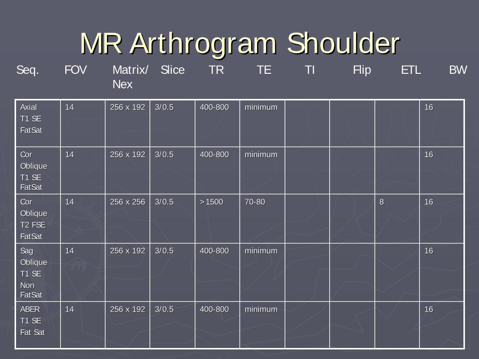
MR Arthrogram ShoulderMR Arthrogram Shoulder
AxialAxialT1 SET1 SEFatSatFatSat
1414 256 x 192256 x 192 3/0.53/0.5 400400--800800 minimumminimum 1616
CorCorObliqueObliqueT1 SE T1 SE FatSatFatSat
1414 256 x 192256 x 192 3/0.53/0.5 400400--800800 minimumminimum 1616
CorCorObliqueObliqueT2 FSET2 FSEFatSatFatSat
1414 256 x 256256 x 256 3/0.53/0.5 >1500>1500 7070--8080 88 1616
SagSagObliqueObliqueT1 SET1 SENon Non FatSatFatSat
1414 256 x 192256 x 192 3/0.53/0.5 400400--800800 minimumminimum 1616
ABERABERT1 SET1 SEFat SatFat Sat
1414 256 x 192256 x 192 3/0.53/0.5 400400--800800 minimumminimum 1616
Seq. FOV Matrix/ Slice TR TE TI Flip ETL BWNex

ShoulderShoulder--Post GadoliniumPost Gadolinium
Axial T1Axial T1FatSatFatSat
1414 256x192256x192 4/.54/.5 400400--800800 minimumminimum 1616
AxialAxialT2 FSET2 FSEFatSatFatSat
1414 256x 256256x 256 4/1mm4/1mm 20002000--60006000
6060--7070 88
CorCorObliqueObliqueT1 T1 FatSatFatSat
1414 256x192256x192 4/.54/.5 400400--800800 minimumminimum 1616
SagSagObliqueObliqueT1 T1 FatSatFatSat
1414 256x192256x192 4/.54/.5 400400--800800 minimumminimum 1616
CorCorObliqueObliqueT2 FSET2 FSEFatSatFatSat
1212 256x256256x256 4/14/1 20002000--60006000
6060--7070 88 1616
Seq. FOV Matrix/ Slice TR TE TI Flip ETL BWNex

ShoulderShoulder--Axial Imaging PlaneAxial Imaging Plane
Relevant Anatomy
HumeralHead
Bony Glenoid
Clavicle
Axial Imaging PlanePrescribe plane parallel to humeral shaft. Cover
from AC joint through proximal humeral diaphysis.
AC Joint(Not shown on MR)

ShoulderShoulder--Coronal Imaging PlaneCoronal Imaging Plane
Coronal Imaging PlanePrescribe coronal plane off of axial images parallel to
supraspinatus muscle
Relevant Anatomy
Supraspinatus

ShoulderShoulder--Sagittal Imaging PlaneSagittal Imaging Plane
Humeral Head
Bony GlenoidLabrum
CartilaginousLabrumAnt. and Post.
Relevant Anatomy Sagittal Imaging PlanePrescribe sagittal plane off axial images with
line parallel to bony glenoid. Image from bony glenoid through deltoid muscle.
Deltoid Muscle

MR Elbow Indications:MR Elbow Indications:
►► Routine ElbowRoutine ElbowIndications:Indications:►►Biceps/Triceps tearBiceps/Triceps tear►►Medial/Lateral Collateral Ligament TearsMedial/Lateral Collateral Ligament Tears►►Common Flexor/Common Extensor Tendon PathologyCommon Flexor/Common Extensor Tendon Pathology
►► MR Arthrogram ElbowMR Arthrogram ElbowIndications:Indications:►► Intra articular Body evaluationIntra articular Body evaluation►►Sometimes Medial/Lateral Collateral Ligament EvaluationSometimes Medial/Lateral Collateral Ligament Evaluation
►► Post Gadolinium ElbowPost Gadolinium ElbowIndications:Indications:►► IA bodyIA body

ElbowElbow--RoutineRoutine
AxialAxialPD FSEPD FSENon Non FatSatFatSat
1010 512 x 256 512 x 256 22
4/0.54/0.5 30003000 4040 88 1616
AxialAxialT2 FSET2 FSEFatSatFatSat
1010 256 x 256256 x 25622
4/0.54/0.5 >2000>2000 7070--8080 88 1616
CoronalCoronalT1 SET1 SENon Non FatSatFatSat
1616--1818 256 x 256256 x 25611
3/0.53/0.5 400400--800800 minimalminimal 1616
CoronalCoronalSTIRSTIR
1616--1818 256 x 192256 x 19233
3/0.53/0.5 > 1500> 1500 4040 120120 9090 88 1616
SagSagT2 FSET2 FSEFatSatFatSat
1616 256 x 256256 x 25622
3/0.53/0.5 >1500>1500 7070--8080 88 1616
Seq. FOV Matrix/ Slice TR TE TI Flip ETL BWNex

MR Arthrogram ElbowMR Arthrogram Elbow
AxialAxialT1 SET1 SENon Non FatSatFatSat
1212--1414 256 x 256256 x 256 4/14/1 400400--800800 minimumminimum 1616
AxialAxialT2T2FSEFSEFatSatFatSat
1212--1414 256 x 256256 x 256 4/14/1 >1500>1500 7070--8080 88 1616
CoronalCoronalT1 SET1 SEFatSatFatSat
1212--1414 256 x 256256 x 256 4/14/1 400400--800800 minimumminimum 1616
CoronalCoronalT2T2FSE FSE FatSatFatSat
1212--1414 256 x 256256 x 256 4/14/1 >1500>1500 7070--8080 88 1616
SagSagT2 SET2 SEFatSatFatSat
1212--1414 256 x 256256 x 256 4/14/1 >1500>1500 7070--8080 88 1616
SagSagT1 SET1 SEFatSatFatSat
1212--1414 256 x 256256 x 256 4/14/1 400400--800800 minimumminimum 1616
Seq. FOV Matrix/ Slice TR TE TI Flip ETL BWNex

ElbowElbow--Axial Imaging PlaneAxial Imaging Plane
Relevant AnatomyAxial Imaging Plane
*Prescribe plane perpendicular to coronalplane (©). Scan from humeral metaphysis
through radial tuberosity.
©©
*
*
Radial Tuberosity(bump on medial radius)
Lateral and MedialHumeral Condyles
UlnaRadius
Expected location radial tuberosity(Not shown)

ElbowElbow--Coronal Imaging PlaneCoronal Imaging Plane
Relevant AnatomyCoronal Imaging Plane
*Prescribe plane parallel to anterior humerus at condyles. Scan through entire elbow.
*
*
Lateral Humeral Condyle
Medial Humeral Condyle
Olecranon processof Ulna
Humerus

ElbowElbow--Sagittal Imaging PlaneSagittal Imaging Plane
Sagittal Imaging Plane*Prescribe plane perpendicular to coronal plane (©). Scan
through entire elbow.
©
©
*
*Lateral Humeral Condyle
Medial Humeral Condyle
Olecranon processof Ulna
Humerus
Relevant Anatomy
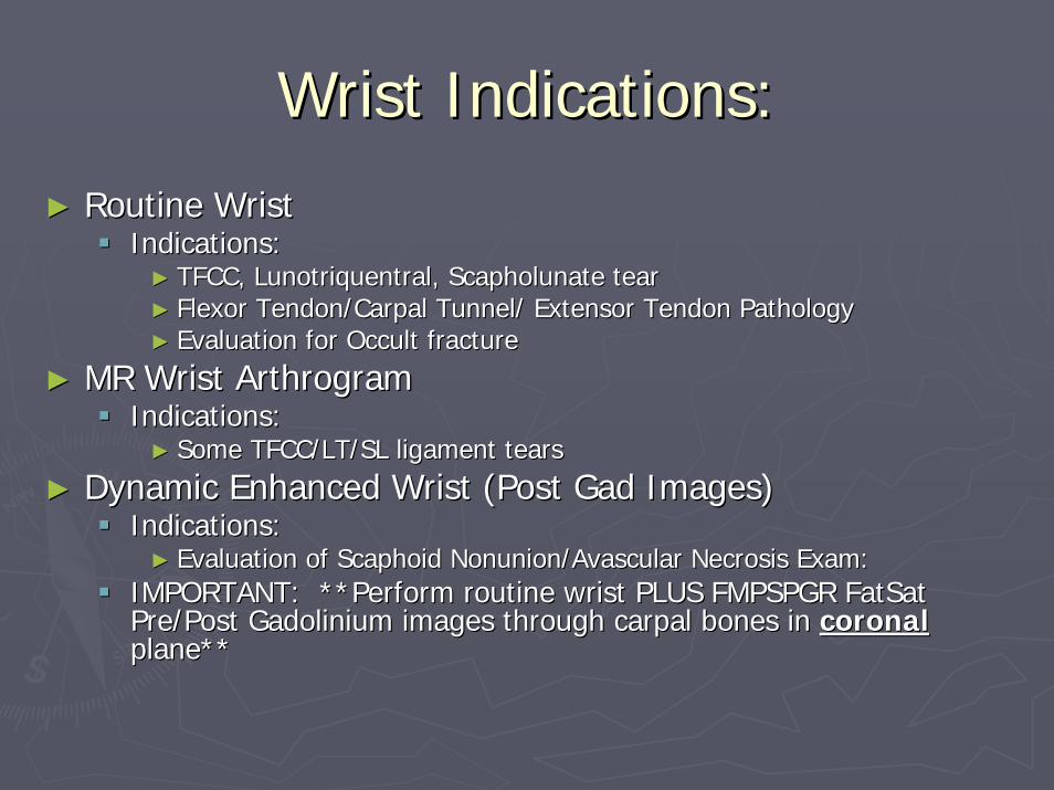
Wrist Indications:Wrist Indications:
►► Routine WristRoutine WristIndications:Indications:►► TFCC, TFCC, LunotriquentralLunotriquentral, , ScapholunateScapholunate teartear►► Flexor Tendon/Carpal Tunnel/ Extensor Tendon PathologyFlexor Tendon/Carpal Tunnel/ Extensor Tendon Pathology►► Evaluation for Occult fractureEvaluation for Occult fracture
►► MR Wrist ArthrogramMR Wrist ArthrogramIndications:Indications:►► Some TFCC/LT/SL ligament tearsSome TFCC/LT/SL ligament tears
►► Dynamic Enhanced Wrist (Post Gad Images)Dynamic Enhanced Wrist (Post Gad Images)Indications:Indications:►► Evaluation of Evaluation of ScaphoidScaphoid Nonunion/Avascular Necrosis Exam: Nonunion/Avascular Necrosis Exam:
IMPORTANT: **Perform routine wrist PLUS FMPSPGR IMPORTANT: **Perform routine wrist PLUS FMPSPGR FatSatFatSatPre/Post Gadolinium images through carpal bones in Pre/Post Gadolinium images through carpal bones in coronalcoronalplane**plane**

WristWrist--RoutineRoutine
CoronalCoronalT1 SE T1 SE Non Non FatSatFatSat
1212--1414 512 x 256512 x 25611
3/0.53/0.5 400400--800800 minimumminimum 1616
CoronalCoronalT2 FSET2 FSEFatSatFatSat
1212--1414 256 x 256256 x 25622
3/0.53/0.5 >1500>1500 7070--8080 88 1616
CoronalCoronalGradientGradient
1212--1414 256 x 192256 x 19222
1/01/0 6060 minimumminimum 3030--4040 88 1616
AxialAxialPD FSEPD FSENon Fat Non Fat SatSat
88--1010 256 x 256256 x 25622
3/0.53/0.5 30003000 4040 88 1616
SagSagSTIRSTIR
1212 256 x 192256 x 19233
3/0.53/0.5 >1500>1500 4040 120120 9090 88 1616
Seq. FOV Matrix/ Slice TR TE TI Flip ETL BWNex

MR Arthrogram Wrist (dir or MR Arthrogram Wrist (dir or indind))
CoronalCoronalT1 SET1 SEFatSatFatSat
88--1010 256 x 256256 x 256 3/0.53/0.5 400400--800800 1010--2020 1616
CoronalCoronalT2 FSET2 FSEFatSatFatSat
88--1010 256 x 256256 x 256 3/0.33/0.3 >1500>1500 7070--8080 88 1616
CoronalCoronal3D 3D GradientGradient
88--1010 256 x 192256 x 192 1/01/0 6060 minimumminimum 3030--4040 88 1616
AxialAxialT2 FSET2 FSEFatSatFatSat
88--1010 256 x 256256 x 256 3/0.53/0.5 >1500>1500 7070--8080 88 1616
AxialAxialT1 SET1 SENonNon--FatSatFatSat
1212 3/0.53/0.5 400400--800800 1010--2020 1616
SagSagT2 FSET2 FSEFatSatFatSat
1212 3/0.53/0.5 >1500>1500 7070--8080 88 1616
Seq. FOV Matrix/ Slice TR TE TI Flip ETL BWNex

WristWrist--Axial Imaging PlaneAxial Imaging Plane
Distalulna
lun
scaph
cap
triq
Distradius
trapmtrapz ham
Relevant AnatomyAxial Imaging Plane
Prescribe plane parallel to distal radius. Scanfrom proximal metacarpals through distal
radial/ulnar metaphysis.
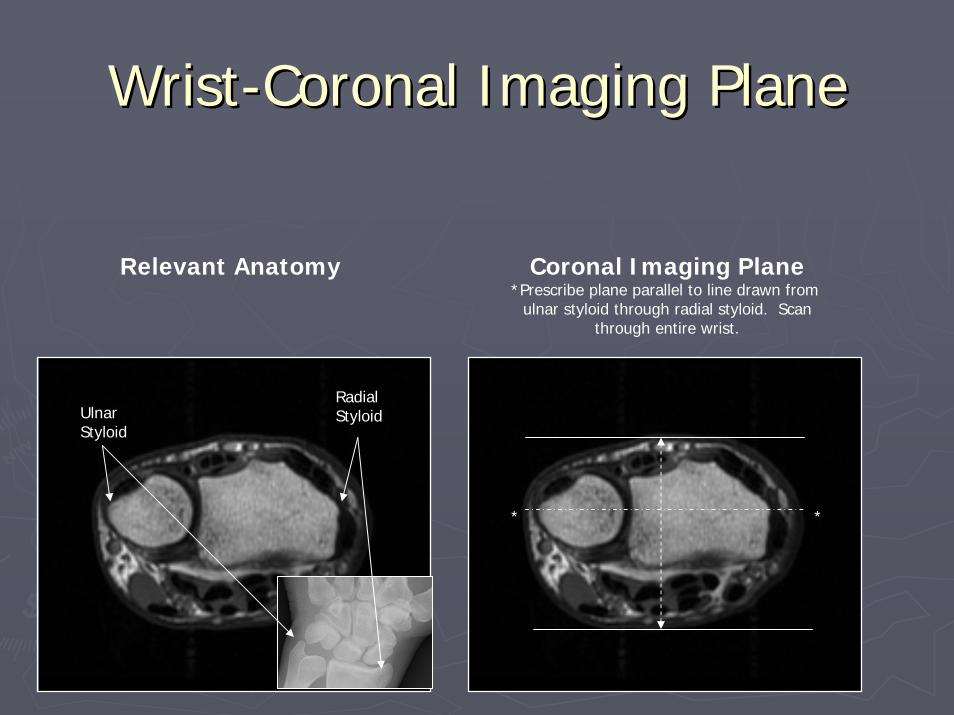
WristWrist--Coronal Imaging PlaneCoronal Imaging Plane
Relevant Anatomy Coronal Imaging Plane*Prescribe plane parallel to line drawn from
ulnar styloid through radial styloid. Scanthrough entire wrist.
* *
UlnarStyloid
RadialStyloid

WristWrist--Sagittal Imaging PlaneSagittal Imaging Plane
UlnarStyloid
RadialStyloid
Relevant AnatomySagittal Imaging Plane
*Prescribe plane perpendicular to coronal plane (©).Scan through entire wrist.
©
©
**
UlnarStyloid
RadialStyloid

Thumb Indications:Thumb Indications:
►►Routine ThumbRoutine ThumbIndications: Indications: ►►GamekeeperGamekeeper’’s thumb (s thumb (UlnarUlnar Collateral Ligament tear)Collateral Ligament tear)►►Flexor/Extensor Tendon TearFlexor/Extensor Tendon Tear►►R/O Occult FractureR/O Occult Fracture

ThumbThumb
CoronalCoronalT1 SET1 SENon Non FatSatFatSat
1010 256 x 192256 x 192 3/0.33/0.3 400400--800800 minimalminimal 1616
CoronalCoronalT2 FSET2 FSENon Non FatSatFatSat
1010 256 x 256256 x 256 3/0.33/0.3 20002000--60006000
9090--110110 1616 1616
SagSagSTIRSTIR
1010 256 x 192256 x 192 3/0.33/0.3 150150 1616 1616
AxialAxialT1 SET1 SENon Non FatSatFatSat
1010 256 x 192256 x 192 3/0.33/0.3 400400--800800 minimalminimal 1616
AxialAxialT2 FSET2 FSEFatSatFatSat
1010 256 x 256256 x 256 3/0.33/0.3 20002000--60006000
6060--7070 1616 1616
Seq. FOV Matrix/ Slice TR TE TI Flip ETL BWNex

ThumbThumb--Axial Imaging PlaneAxial Imaging Plane
Relevant AnatomyAxial Imaging Plane
Prescribe plane perpendicular to midshaft ofproximal 1st phalanx. Scan from CMC joint
through thumb.
CMC
Joint
MCP
Joint
IPJoint
Metacarpal
Proximal Phalanx

ThumbThumb--Coronal Imaging PlaneCoronal Imaging Plane
Relevant Anatomy Coronal Imaging Plane*Prescribe plane with line bisecting
sesamoid bones. Scan through entire thumb.
Sesamoids
Thumb
*

ThumbThumb--Sagittal Imaging PlaneSagittal Imaging Plane
Sesamoids
Thumb
Relevant Anatomy Sagittal Imaging Plane*Prescribe plane perpendicular to coronal
imaging plane (©). Scan through entire thumb.
©
*

Finger Indications:Finger Indications:
►►Routine FingerRoutine FingerIndications:Indications:►►Pulley rupture/Flexor or Extensor Tendon InjuryPulley rupture/Flexor or Extensor Tendon Injury
►►Post Gadolinium FingerPost Gadolinium FingerIndications:Indications:►►MassMass►►**Perform routine finger plus Axial and either **Perform routine finger plus Axial and either
Coronal or Sagittal (whichever plane mass best seen) Coronal or Sagittal (whichever plane mass best seen) pre/post gadolinium FMPSPGR imagespre/post gadolinium FMPSPGR images

FingerFinger--RoutineRoutine
SagSagT1 SET1 SENon Non FatSatFatSat
256 x 192256 x 192 3/0.33/0.3 400400--800800 minimumminimum 1616
SagSagT2 FSET2 FSEFatSatFatSat
256 x256256 x256 3/0.33/0.3 20002000--60006000
6060--7070 1616 1616
AxialAxialT1 SET1 SENon Non FatSatFatSat
256 x 192256 x 192 3/0.33/0.3 400400--800800 minimumminimum 1616
AxialAxialT2 FSET2 FSEFatSatFatSat
256 x256256 x256 3/0.33/0.3 20002000--60006000
6060--7070 1616 1616
CoronalCoronalSTIRSTIR
256 x256256 x256 3/0.33/0.3 >2000>2000 2020--4040 1616 1616
Seq. FOV Matrix/ Slice TR TE TI Flip ETL BWNex

FingerFinger--MassMass
AxialAxialT1 SET1 SENon Non FatSatFatSat
256 x 192256 x 192 3/0.33/0.3 400400--800800 minimumminimum 1616
Axial Axial T2 FSET2 FSEFatSatFatSat
256 x 256256 x 256 3/0.33/0.3 20002000--60006000
6060--7070 1616 1616
Sag/Sag/CorCorT2 FSET2 FSEFatSatFatSat
256 x 256256 x 256 3/0.33/0.3 20002000--60006000
6060--7070 1616 1616
AxialAxialFMPSPGRFMPSPGRFatSatFatSatPre/PostPre/Post
256 x ??256 x ?? 200 ?200 ? 4 ?4 ? 1616
Sag/Sag/CorCorFMPSPGRFMPSPGRFatSatFatSatPre/PostPre/Post
256 x ??256 x ?? 200 ?200 ? 4 ?4 ? 1616
Seq. FOV Matrix/ Slice TR TE TI Flip ETL BWNex

FingerFinger--Axial Imaging PlaneAxial Imaging Plane
DistalPhalanx
MidPhalanx
Metacarpal
Proximal Phalanx
Relevant Anatomy
Axial Imaging PlanePrescribe best fit line. Scan from
proximal metacarpal through entirefinger.

FingerFinger--Coronal Imaging PlaneCoronal Imaging Plane
Relevant Anatomy Coronal Imaging Plane*Prescribe plane parallel to anterior
metacarpal head. Scan through entirefinger.
*
Thumb
Extensor Tendon

FingerFinger--Sagittal Imaging PlaneSagittal Imaging Plane
Relevant AnatomySagittal Imaging Plane
*Prescribe plane perpendicular to coronal plane (©). Scan through entire
finger.
*
©
Thumb
Extensor Tendon

LOWER EXTREMITYLOWER EXTREMITY

Bony pelvisBony pelvis►► Indication: hip fracture, AVN, Mets, transient osteoporosis, Indication: hip fracture, AVN, Mets, transient osteoporosis,
bursitis, arthritis, tendonitis, hip pain over age 50bursitis, arthritis, tendonitis, hip pain over age 50►► If If metsmets/marrow, add in/out of phase coronal of full pelvis/marrow, add in/out of phase coronal of full pelvis►► PA Torso Coil is 1PA Torso Coil is 1stst choicechoice►► Sagittal may be skipped in ED hip fracture, elderly, multiSagittal may be skipped in ED hip fracture, elderly, multi--
traumatrauma
CoronalCoronalFSEFSE--STIRSTIR
3636--4040 256x192256x19222--33
4/14/1 >2000>2000 2020--4040 150150 88 1616
AxialAxialT2 FSET2 FSEFatSatFatSat
3636--4040 256 x 256256 x 25622
5/1.55/1.5 30003000 3030--4040 88 1616
CoronalCoronalT1 SET1 SENon Non FatSatFatSat
3636--4040 256 x 256256 x 25611
4/14/1 400400--800800 MinimumMinimum 1616
SagSagT2 FSET2 FSEFatSatFatSat(Hip to Hip)(Hip to Hip)
1212--1616 256 x 256256 x 25611--22
5/15/1 >2000>2000 4040--5050 88 1616
Seq. FOV Matrix/ Slice TR TE TI Flip ETL BWNex

Coronal (PELVIS) Coronal (PELVIS) T1 SET1 SENon Non FatSatFatSat
3636--4040 256 x 256256 x 25611
4/14/1 400400--800800 MinimumMinimum 1616
Coronal (PELVIS)Coronal (PELVIS)FSEFSE--STIRSTIR
3636--4040 256x192256x19222--33
4/14/1 >2000>2000 2020--4040 150150 88 1616
Axial (PELVIS)Axial (PELVIS)T2 FSET2 FSEFatSatFatSat
3636--4040 256 x 256256 x 25611--22
5/1.55/1.5 >2000>2000 4040--5050 88 1616
Ax oblique (HIP)Ax oblique (HIP)T1 T1 FatSatFatSat
1414--1616 384 x 256384 x 2561.51.5
4/.54/.5 400400--800800 MinimumMinimum 1616
Coronal (HIP)Coronal (HIP)T1 T1 FatSatFatSat
1414--1616 384 x 256384 x 2561.51.5
4/.54/.5 400400--800800 MinimumMinimum 1616
Sag (HIP)Sag (HIP)T1 T1 FatSatFatSat
1616 384 x 256384 x 2561.51.5
4/.54/.5 400400--800800 MinimumMinimum 1616
Seq. FOV Matrix/ Slice TR TE TI Flip ETL BWNex
Direct arthrogram hipDirect arthrogram hip►► Indication: hip labrum tear, FAI or dysplasia, hip pain under Indication: hip labrum tear, FAI or dysplasia, hip pain under
age 50age 50►► PA Torso coil is 1PA Torso coil is 1stst choicechoice►► Prescribe Prescribe obliquesobliques along femoral neck from coronal localizeralong femoral neck from coronal localizer

Coronal (PELVIS) Coronal (PELVIS) T1 SET1 SENon Non FatSatFatSat
3636--4040 256 x 256256 x 25611
4/14/1 400400--800800 MinimumMinimum 1616
Coronal (PELVIS)Coronal (PELVIS)FSEFSE--STIRSTIR
3636--4040 256x192256x19222--33
4/14/1 >2000>2000 2020--4040 150150 88 1616
Axial (PELVIS)Axial (PELVIS)T2 FSET2 FSEFatSatFatSat
3636--4040 256 x 256256 x 25611--22
5/1.55/1.5 >2000>2000 4040--5050 88 1616
Ax oblique (HIP)Ax oblique (HIP)T1 T1 FatSatFatSat
1414--1616 384 x 256384 x 2561.51.5
4/.54/.5 400400--800800 MinimumMinimum 1616
Coronal (HIP)Coronal (HIP)T1 T1 FatSatFatSat
1414--1616 384 x 256384 x 2561.51.5
4/.54/.5 400400--800800 MinimumMinimum 1616
Sag (HIP)Sag (HIP)T1 T1 FatSatFatSat
1616 384 x 256384 x 2561.51.5
4/.54/.5 400400--800800 MinimumMinimum 1616
Seq. FOV Matrix/ Slice TR TE TI Flip ETL BWNex
Indirect arthrogram hipIndirect arthrogram hip►► Indication: hip labrum tear, FAI or dysplasia, hip pain under Indication: hip labrum tear, FAI or dysplasia, hip pain under
age 50 when direct MR arthrography not availableage 50 when direct MR arthrography not available►► PA Torso coil is 1PA Torso coil is 1stst choicechoice►► Prescribe Prescribe obliquesobliques along femoral neck from coronal localizeralong femoral neck from coronal localizer

Coronal (PELVIS) Coronal (PELVIS) T1 SET1 SENon Non FatSatFatSat
3636--4040 256 x 256256 x 25611
4/14/1 400400--800800 MinimumMinimum 1616
Coronal (PELVIS)Coronal (PELVIS)FSEFSE--STIRSTIR
3636--4040 256x192256x19222--33
4/14/1 >2000>2000 2020--4040 150150 88 1616
Axial (PELVIS)Axial (PELVIS)T2 FSET2 FSEFatSatFatSat
3636--4040 256 x 256256 x 25611--22
5/1.55/1.5 >2000>2000 4040--5050 88 1616
Ax oblique (HIP)Ax oblique (HIP)PD FSE PD FSE FatSatFatSat
1414--1616 256 x 256256 x 25622
4/14/1 30003000 3030--4040 44 1616
Coronal (HIP)Coronal (HIP)T2 FSET2 FSEFatSatFatSat
1414--1616 256 x 256256 x 25622
4/14/1 >2000>2000 4040--5050 88 1616
Sag (HIP)Sag (HIP)T2 FSET2 FSEFatSatFatSat
1616 256 x 256256 x 25622
4/14/1 >2000>2000 4040--5050 88 1616
Seq. FOV Matrix/ Slice TR TE TI Flip ETL BWNex
Noncontrast hipNoncontrast hip►► Indication: hip labrum tear, FAI or dysplasia when direct MR arIndication: hip labrum tear, FAI or dysplasia when direct MR arthrogram is not throgram is not
possible, hip pain under age 50possible, hip pain under age 50►► PA Torso coil is 1PA Torso coil is 1stst choicechoice►► Prescribe Prescribe obliquesobliques along femoral neck from coronal localizeralong femoral neck from coronal localizer
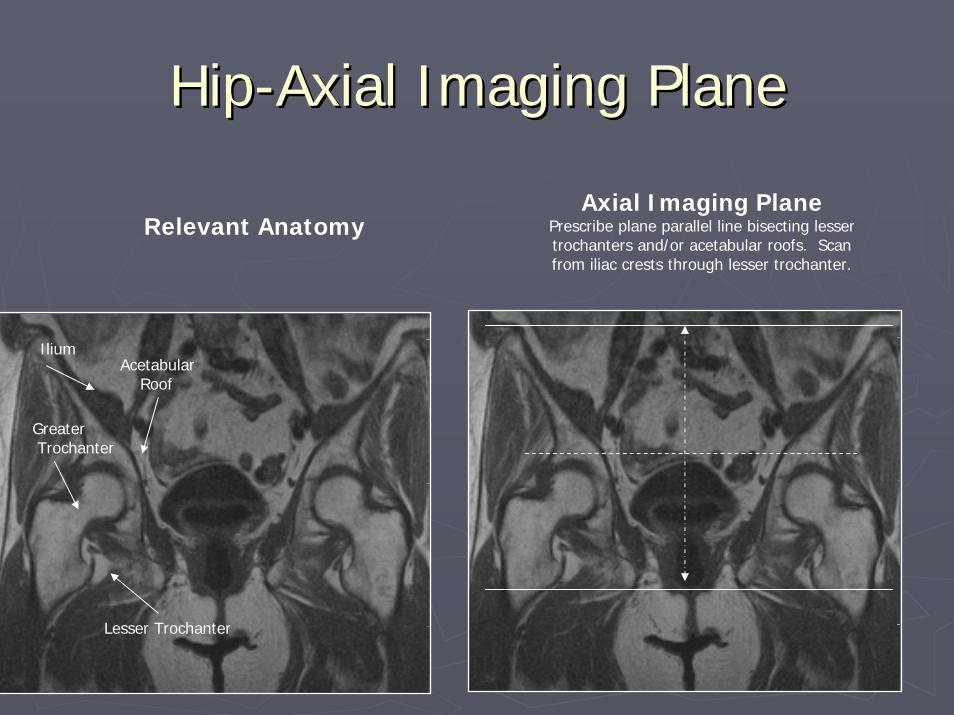
HipHip--Axial Imaging PlaneAxial Imaging Plane
Lesser Trochanter
GreaterTrochanter
IliumAcetabular
Roof
Relevant AnatomyAxial Imaging Plane
Prescribe plane parallel line bisecting lessertrochanters and/or acetabular roofs. Scanfrom iliac crests through lesser trochanter.

HipHip--Coronal Imaging PlaneCoronal Imaging Plane
Greater Trochanter
FemoralNeck
Superior Pubic Ramus
Relevant AnatomyCoronal Imaging Plane
*Prescribe plane parallel femoral heads. Scan from ischium through pubic
symphyses.
Ischium
*
FemoralHead
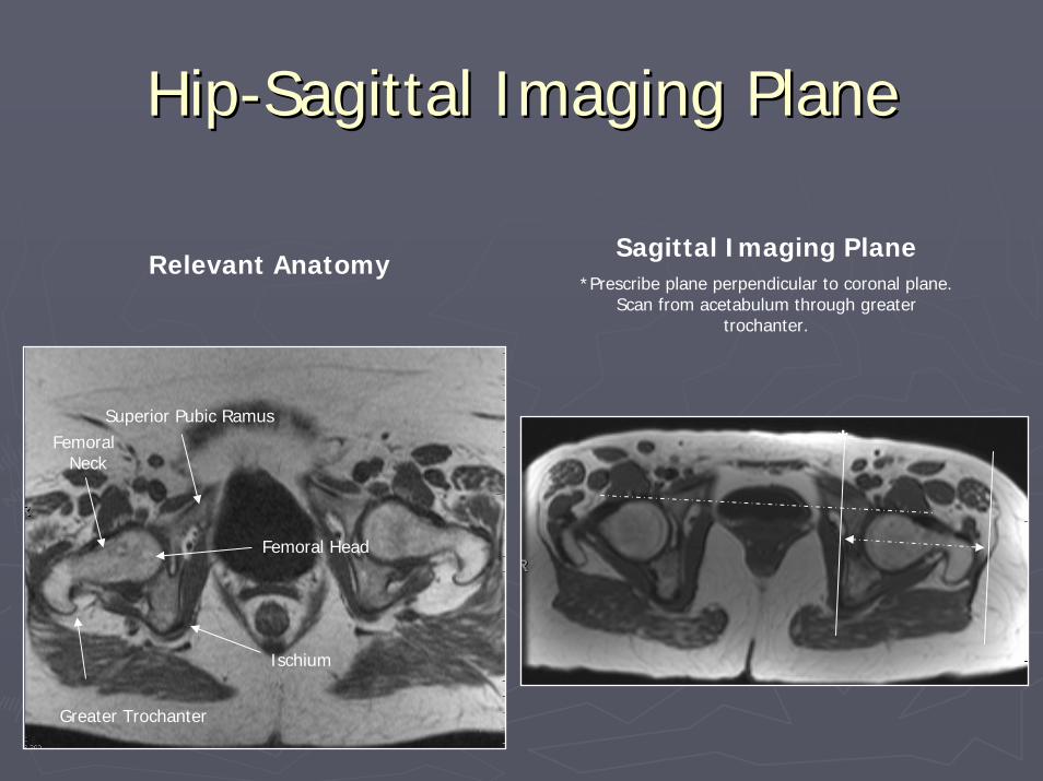
HipHip--Sagittal Imaging PlaneSagittal Imaging Plane
Greater Trochanter
FemoralNeck
Superior Pubic Ramus
Femoral Head
Ischium
Relevant AnatomySagittal Imaging Plane
*Prescribe plane perpendicular to coronal plane. Scan from acetabulum through greater
trochanter.
*

Axial Oblique Imaging PlaneAxial Oblique Imaging Plane(For (For FemoracetabularFemoracetabular Impingement Patients Only)Impingement Patients Only)
Axial Oblique PlanePrescribe plane parallel to femoralneck. Scan through entire femoral
neck.
Relevant Anatomy
FemoralNeck
FemoralHead
CoronalCoronalSTIRSTIR
2828--3636(Both hips)(Both hips)
256x192256x19222--33
4/14/1 >2000>2000 2020--4040 150150 88 1616
CoronalCoronalT1 SET1 SE
2828(Both hips)(Both hips)
256x256256x25611--22
4/14/1 400400--800800 minimumminimum 1616
Axial T2 FSEAxial T2 FSEFat SatFat Sat
2828(Both hips)(Both hips)
256x256256x25622--33
5/15/1 >2000>2000 5050--6060 88 1616
Axial Axial OblObl PD FSEPD FSENonfatsatNonfatsat
2020 256x192256x19211--22
4/.54/.5 3000 3000 (max)(max)
2525--3030 44 1616
Axial Axial OblObl T2 FSE T2 FSE Fat SatFat Sat
2020 256x192256x19222--33
4/.54/.5 >2000>2000 5050--6060 88 1616
Sag T2 FSESag T2 FSEFat SatFat Sat
2020--2222 256x192256x19222--33
4/.54/.5 >2000>2000 5050--6060 88 1616
PD FSE (optional)PD FSE (optional)NonfatsatNonfatsat
2020--2222 256x256256x25622--33
4/.54/.5 >2000>2000 2020--3030 88 1616
Seq. FOV Matrix/NEX Slice TR TE TI ETL BW
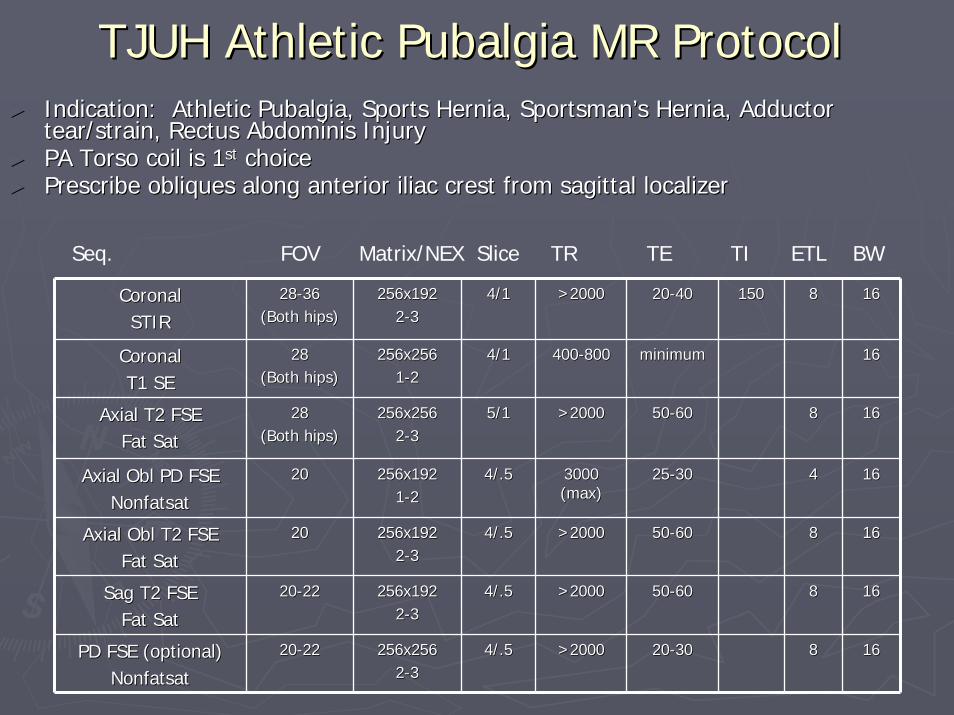
TJUH Athletic Pubalgia MR ProtocolTJUH Athletic Pubalgia MR Protocol►► Indication: Athletic Pubalgia, Sports Hernia, SportsmanIndication: Athletic Pubalgia, Sports Hernia, Sportsman’’s Hernia, Adductor s Hernia, Adductor
tear/strain, Rectus Abdominis Injurytear/strain, Rectus Abdominis Injury►► PA Torso coil is 1PA Torso coil is 1stst choicechoice►► Prescribe Prescribe obliquesobliques along anterior iliac crest from sagittal localizeralong anterior iliac crest from sagittal localizer

Axial Oblique Imaging PlaneAxial Oblique Imaging Plane(Adductor unfolding plane)(Adductor unfolding plane)
Relevant Anatomy
Axial Oblique Imaging Plane*Prescribe plane to line paralleling anteriorIliac crest*. Be sure to scan across pubic
symphysis at midline
*
*
Iliac c
rest
Hip joint

Axial Oblique Imaging PlaneAxial Oblique Imaging Plane(Adductor unfolding plane)(Adductor unfolding plane)

Bony PelvisBony Pelvis(Special Cases)(Special Cases)
*Follow Hip Planes**Follow Hip Planes*
►► Post Gadolinium PelvisPost Gadolinium PelvisIndications:Indications:►►OsteomylitisOsteomylitis
IMPORTANT:IMPORTANT:►►Same as Routine Hip Protocol but perform axial and coronal Same as Routine Hip Protocol but perform axial and coronal
images on both sides. images on both sides. ►► In addition, perform FMPSPGR fat saturated images pre/post in In addition, perform FMPSPGR fat saturated images pre/post in
both axial and coronal planes.both axial and coronal planes.
►► In/Out of Phase PelvisIn/Out of Phase PelvisIndications:Indications:►►Possible Bony MetastasesPossible Bony Metastases
IMPORTANT:IMPORTANT:►►Perform In/Out of Phase images in Coronal PlanePerform In/Out of Phase images in Coronal Plane

Bony Pelvis Bony Pelvis OsteoOsteo –– pre/post optionpre/post option
CoronalCoronalT1T1Non Non FatSatFatSat
3030--4545 256 x 192256 x 192 4/14/1 400400--800800 minimalminimal 1616
CoronalCoronalSTIRSTIR
3030--4545 256 x 192256 x 192 4/14/1 >2000>2000 2020--4040 88 1616
AxialAxialT1T1Non Non FatSatFatSat
3030--4545 256 x 192256 x 192 4/14/1 400400--800800 minimalminimal 1616
AxialAxialT2 FSET2 FSEFatSatFatSat
3030--4545 256 x 256256 x 256 4/14/1 20002000--60006000
6060--7070 88 1616
SagSagT2 FSET2 FSEFatSatFatSat
2020 256 x 256256 x 256 4/14/1 20002000--60006000
50?50?(60(60--70)70)
88 1616
Axial/Axial/CorCorFMPSPGRFMPSPGRFatSatFatSatPre/PostPre/Post
1212 256 x 192256 x 192 3/.53/.5 5050 55 3030--4040 88 1616
Seq. FOV Matrix/ Slice TR TE TI Flip ETL BWNex

Sacrum Sacrum –– Sacroiliac jointsSacroiliac joints
CoronalCoronalSTIRSTIR
3030--4545 256 x 192256 x 192 4/14/1 >2000>2000 2020--4040 88 1616
AxialAxialT2 FSET2 FSEFatSatFatSat
3030--4545 256 x 256256 x 256 4/14/1 >1500>1500 7070--8080 88 1616
CorCor ObliqueObliqueT1 SET1 SENon Non FatSatFatSat
1818--2222 256 x 256256 x 256 3/.53/.5 400400--800800 minimalminimal
CorCor ObliqueObliqueT2 FSET2 FSEFatSatFatSat
1818--2222 256 x 256256 x 256 3/.53/.5 >1500>1500 7070--8080 88 1616
SagSagT2 FSET2 FSEFatSatFatSat
1818--2222 256 x 256256 x 256 3/.53/.5 >1500>1500 7070--8080 88 1616
AxialAxialT1 SET1 SENon Non FatSatFatSat
1818--2222 256 x 256256 x 256 3/.53/.5 400400--800800 minimalminimal
Seq. FOV Matrix/ Slice TR TE TI Flip ETL BWNex
Indications:Indications:Possible Sacroileitis (Septic/Rheumatoid or Rheumatoid Variant Possible Sacroileitis (Septic/Rheumatoid or Rheumatoid Variant Arthritis)Arthritis)

Sacrum Sacrum –– Sacroiliac jointsSacroiliac joints►► Prescribing the coronal oblique plane: parallel the sacrum Prescribing the coronal oblique plane: parallel the sacrum
on a sagittal localizeron a sagittal localizer

Routine ThighRoutine Thigh(Follow Hip Imaging Planes)(Follow Hip Imaging Planes)
►►Routine ThighRoutine ThighIndications:Indications:►►PolymyositisPolymyositis/Diabetic /Diabetic MyonecrosisMyonecrosis

ThighThigh--RoutineRoutine
CoronalCoronalT1 SET1 SENon Non FatSatFatSat
4040 256 x 192256 x 192 4/14/1 400400--800800 minimalminimal 1616
CoronalCoronalT2 FSET2 FSEFatSatFatSat
4040 256 x 256256 x 256 4/14/1 >1500>1500 7070--8080 88 1616
SagSagSTIRSTIR
2626 256 x 192256 x 192 4/14/1 >2000>2000 2020--4040 150150 88 1616
Axial SEAxial SET1T1Non Non FatSatFatSat
2626 256 x 192256 x 192 4/14/1 400400--800800 minimalminimal 1616
AxialAxialT2 FSET2 FSEFatSatFatSat
2626 256 x 256256 x 256 4/14/1 >1500>1500 7070--8080 88 1616
Seq. FOV Matrix/ Slice TR TE TI Flip ETL BWNex

Knee IndicationsKnee Indications
►► Routine KneeRoutine KneeIndications:Indications:►►Meniscal Tear/Medial or Lateral Ligament Tear/ACL/PCLMeniscal Tear/Medial or Lateral Ligament Tear/ACL/PCL
►► Direct ArthrogramDirect ArthrogramIndications:Indications:►►Meniscal ReMeniscal Re--tear tear ►► Intra articular BodyIntra articular Body
►► Post Gadolinium KneePost Gadolinium KneeIndicationsIndications►►Meniscal ReMeniscal Re--teartear
IMPORTANT: Image 20 minutes post gadoliniumIMPORTANT: Image 20 minutes post gadolinium

KneeKnee--RoutineRoutine
SagSagPD FSEPD FSENonFatSatNonFatSat
1414--1616 512 x 256512 x 25622
4/0.54/0.5 30003000 1515--2020 88 1616
SagSagT2 FSET2 FSEFatSatFatSat
1414--1616 256 x 256256 x 25622
4/0.54/0.5 20002000 7070--8080 88 1616
CorCorT1 SET1 SENon Non FatSatFatSat
1616--1818 256 x 192256 x 19211
3/0.53/0.5 400400--800800 MinimalMinimal 1616
CoronalCoronalT2 FSET2 FSEFat SatFat Sat
1616--1818 256 x 256256 x 25622
3/0.53/0.5 >2000>2000 7070--8080 88 1616
AxialAxialT2 FSET2 FSEFatSatFatSat
1414--1616 256 x 256256 x 25622
3/0.53/0.5 >2000>2000 7070--8080 88 1616
Seq. FOV Matrix/ Slice TR TE TI Flip ETL BWNex

MR Arthrogram Knee (dir or MR Arthrogram Knee (dir or indind))
SagSagT1 SeT1 SeFatSatFatSat
1414--1616 256 x 192256 x 192 4/14/1 400400--800800 MinimalMinimal 1616
SagSagPD FSEPD FSENonFatSatNonFatSat
1414--1616 512 x 256512 x 25622
4/0.54/0.5 30003000 1515--2020 88 1616
AxialAxialT2 FSET2 FSEFatSatFatSat
1414--1616 256 x 256256 x 256 4/14/1 >1500>1500 7070--8080 88 1616
CoronalCoronalT2 FSET2 FSEFatSatFatSat
1616--1818 256 x 256256 x 25622
4/14/1 >1500>1500 7070--8080 88 1616
CoronalCoronalT1 SET1 SEFatSatFatSat
1616--1818 256 x 192256 x 19211
4/14/1 400400--800800 MinimalMinimal 1616
AxialAxialT1 SET1 SEFatSatFatSat
1414--1616 256 x 192256 x 192 4/14/1 400400--800800 MinimalMinimal 1616
Seq. FOV Matrix/ Slice TR TE TI Flip ETL BWNex

KneeKnee--Axial Imaging PlaneAxial Imaging Plane
PAT
Tibia
Femur
PatellarTendon Insertion
Relevant Anatomy Axial Imaging PlaneImage from distal quad tendon
through patellar tendon insertion
Distal Quadriceps tendon
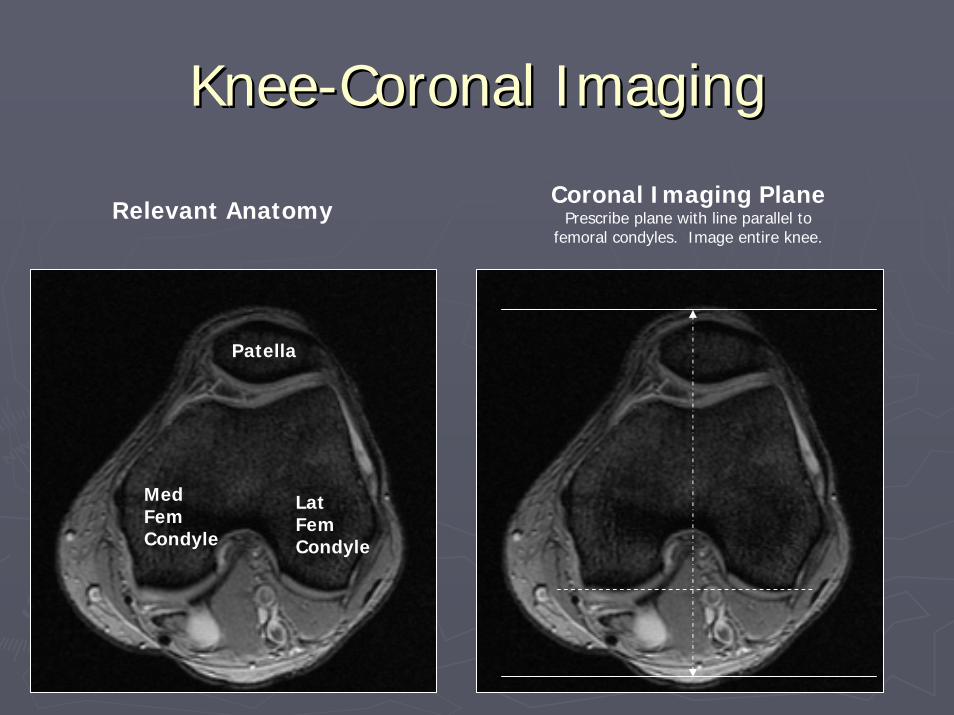
KneeKnee--Coronal ImagingCoronal Imaging
Relevant Anatomy
MedFemCondyle
LatFemCondyle
Patella
Coronal Imaging PlanePrescribe plane with line parallel to
femoral condyles. Image entire knee.

KneeKnee--Sagittal Imaging PlaneSagittal Imaging Plane
MedFemCondyle
LatFemCondyle
Patella
Relevant AnatomySagittal Imaging Plane
*Prescribe plane perpendicular to coronal plane (©).Scan from the medial to the lateral femoral condyle.
©©
*
*

Lower Extremity/ShinLower Extremity/Shin
►►Indication:Indication:Shin SplintsShin Splints
►►IMPORTANT:IMPORTANT:Acquire coronal and axial (STIR) sequences Acquire coronal and axial (STIR) sequences covering both shins, but sagittal and axial (t2 covering both shins, but sagittal and axial (t2 FatSatFatSat only of side in questiononly of side in questionPlace a marker on pain / tendernessPlace a marker on pain / tenderness

Lower Extremity/Shin AreaLower Extremity/Shin Area
CoronalCoronalT1 SET1 SENon Non FatSatFatSat
3535--4040 256 x 256256 x 256 4/14/1 400400--800800 minimalminimal 1616
CoronalCoronalSTIRSTIR
3535--4040 256 x 192256 x 192 4/14/1 >2000>2000 2020--4040 150150 88 1616
AxialAxialSTIRSTIR
3535--4040 256 x 192256 x 192 4/14/1 >2000>2000 2020--4040 150150 88 1616
AxialAxialT2 FSET2 FSEFatSatFatSat(through (through marker marker region)region)
1414--1616 256 x 256256 x 256 4/14/1 >1500>1500 7070--8080 88 1616
SagSagT2 FSET2 FSEFatSatFatSat
256 x 256256 x 256 4/14/1 >1500>1500 7070--8080 88 1616
Seq. FOV Matrix/ Slice TR TE TI Flip ETL BWNex

Ankle IndicationsAnkle Indications
►►Routine AnkleRoutine AnkleLigament Sprain/Tendon pathology/Tarsal Ligament Sprain/Tendon pathology/Tarsal Tunnel/Sinus Tarsi/Occult fracture, PTT, Plantar Tunnel/Sinus Tarsi/Occult fracture, PTT, Plantar fasciitisfasciitis
►►Ankle ArthrogramAnkle ArthrogramIndications:Indications:►►IntraIntra--articular Bodyarticular Body
►►Post Gadolinium AnklePost Gadolinium Ankle

AnkleAnkle--RoutineRoutine
Sag Sag T1 SET1 SENon Non FatSatFatSat
1616--1818 256 x 256256 x 25611
3/13/1 400400--800800 MinimalMinimal 1616
SagSagSTIRSTIR
1616--1818 256 x 192256 x 19233
3/13/1 >1500>1500 4040 120120 9090 88 1616
AxialAxialPD FSEPD FSENon Non FatSatFatSat
1414--1616 384 x 256384 x 25622
4/14/1 30003000 4040 88 1616
AxialAxialT2 FSET2 FSEFatSatFatSat
1414--1616 256 x 256256 x 25622
4/14/1 >2000>2000 7070--8080 88 1616
CoronalCoronalT2 FSET2 FSEFatSatFatSat
1414 256 x 256256 x 25633
3/13/1 >2000>2000 4040--5555 88 1616
Seq. FOV Matrix/ Slice TR TE TI Flip ETL BWNex

MR Arthrogram Ankle (dir or MR Arthrogram Ankle (dir or indind))
SagSagT1 SET1 SEFatSatFatSat
1616 256 x 192256 x 192 4/14/1 400400--800800 minimalminimal 1616
SagSagSTIRSTIR
1818 256 x 192256 x 192 4/14/1 >2000>2000 2020--4040 150150 88 1616
Axial Axial T1 SET1 SEFatSatFatSat
1414 256 x 256256 x 256 4/14/1 800800 minmin 1616
AxialAxialPD FSEPD FSENon Non FatSatFatSat
1414--1616 513 x 256513 x 25622
4/14/1 30003000 4040 88 1616
CoronalCoronalT1 SET1 SEFatSatFatSat
1414 256 x 192256 x 192 4/14/1 400400--800800 minimalminimal 1616
CoronalCoronalT2 FSET2 FSEFatSatFatSat
1414 256 x 256256 x 256 4/14/1 >1500>1500 7070--80 80 88 1616
Seq. FOV Matrix/ Slice TR TE TI Flip ETL BWNex

AnkleAnkle--Post GadoliniumPost Gadolinium
AxialAxialFMPSPGRFMPSPGRFatSatFatSatPre/PostPre/Post
1414 256 x XX256 x XX 200 ?200 ? 4 ?4 ? 1616
Sag Sag T1 SET1 SENon Non FatSatFatSat
1616 256 x 192256 x 192 4/14/1 400400--800800 minimalminimal 1616
AxialAxialT1 SET1 SEFatSatFatSat
1212 256 x 256256 x 256 4/14/1 ?800?800 MinimalMinimal 1616
AxialAxialPD FSEPD FSEFatSatFatSat
1414 256 x 256256 x 256 4/14/1 >2000>2000 4040--5050 44 1616
CorCorT1 SET1 SEFatSatFatSat
1414 256 x 256256 x 256 4/14/1 400400--800800 minimalminimal 1616
Seq. FOV Matrix/ Slice TR TE TI Flip ETL BWNex

AnkleAnkle--Axial Imaging PlaneAxial Imaging Plane
Tibia
Talus
Calcaneus
Relevant AnatomyAxial Imaging Plane
Prescribe plane parallel to axis of calcaneus.Scan ankle from distal tibia through subcutaneous
soft tissues (include plantar fascia).
Black band is plantar fascia

AnkleAnkle--Coronal Imaging PlaneCoronal Imaging Plane
Coronal Imaging PlanePrescribe plane perpendicular
to axial imaging plane. Scan ankle from calcaneus through metatarsal bases.
Calcaneus
Talus
Cuboid
METATARSALS
Relevant Anatomy

AnkleAnkle--Sagittal Imaging PlaneSagittal Imaging Plane
Talus
LatMal
Med
Mal
Ach
Sagittal Imaging PlanePrescribe plane with line parallel
to talus. Cover ankle from medial through lateralmalleolus.
Relevant Anatomy

FootFoot--IndicationsIndications
►► Routine FootRoutine FootIndicationsIndications►►Plantar Plate Injury, R/O fracture, Plantar Plate Injury, R/O fracture, LisfrancLisfranc injury, injury,
tarsal/metatarsal fracturetarsal/metatarsal fracture
►► Post Gadolinium FootPost Gadolinium FootIndications:Indications:►►MortonMorton’’s s NeuromaNeuroma►►OsteomylitisOsteomylitis
IMPORTANT: Perform routine foot plus coronal IMPORTANT: Perform routine foot plus coronal FMPSPGR fat saturated pre and post gad images and FMPSPGR fat saturated pre and post gad images and axial POST gad FMPSPGR fat saturated images.axial POST gad FMPSPGR fat saturated images.

FootFoot--RoutineRoutine
Coronal Coronal (short axis)(short axis)T1T1Non Non FatSatFatSat
1010 256 x 256256 x 25611
3/0.53/0.5 400400--800800 minimalminimal 1616
Coronal Coronal (short axis)(short axis)T2 FSET2 FSEFatSatFatSat
1010 256 x 256256 x 25622
3/0.53/0.5 >2000>2000 7070--8080 88 1616
SagSagSTIRSTIR
1212--1414 256 x 192256 x 19233
3/0.53/0.5 >1500>1500 4040 120120 9090 88 1616
SagSagT1T1Non Non FatSatFatSat
1212--1414 256 x 256256 x 25611
3/0.53/0.5 400400--800800 minimalminimal 1616
AxialAxial(long axis)(long axis)PD FSEPD FSENon Non FatSatFatSat
1212--1414 256 x 256256 x 25622
3/0.53/0.5 >2000>2000 3030 88 1616
AxialAxial(long axis)(long axis)T2 FSET2 FSEFatSatFatSat
1212--1414 256 x 256256 x 25622
3/0.53/0.5 >2000>2000 9090 88 1616
Seq. FOV Matrix/ Slice TR TE TI Flip ETL BWNex

FootFoot--Mass/OsteomyelitisMass/Osteomyelitis
CoronalCoronalT1 SET1 SENon Non FatSatFatSat
1010--12 12 256 x 256256 x 25611
3/0.53/0.5 400400--800800 minimalminimal 1616
CoronalCoronalT2 FSET2 FSEFatSatFatSat
1010--1212 256 x 192256 x 19222
3/0.53/0.5 >2000>2000 7070--8080 88 1616
SagSagSTIRSTIR
1212--1414 256 x 192256 x 19222
3/0.53/0.5 >1500>1500 4040 120120 9090 88 1616
SagSagT1T1Non Non FatSatFatSat
1212--1414 256 x 256256 x 25611
3/0.53/0.5 400400--800800 minimalminimal 1616
CoronalCoronalFMPSPGRFMPSPGRFatSatFatSatPre/PostPre/Post
1010--1212 256 x 192256 x 19222
3/.53/.5 6060 55 3030--4040 88 1616
AxialAxialFMPSPGRFMPSPGRFatSatFatSatPostPost
1010--1212 256 x 192256 x 19222
3/.53/.5 6060 55 3030--4040 88 1616
Seq. FOV Matrix/ Slice TR TE TI Flip ETL BWNex

FootFoot--Axial Imaging PlaneAxial Imaging Plane
Relevant Anatomy Axial Imaging PlanePrescribe plane parallel to 2rd or 3rd
metatarsal. Scan foot from navicular throughphalanges.
2nd meta
tarsal
NAV
Phalanges
CUN

FootFoot--Coronal Imaging PlaneCoronal Imaging Plane
Relevant Anatomy
Metatarsals
12
3
4
5
Coronal Plane*Prescribe plane parallel to 1st and 5th metatarsal
Shafts. Scan through entire foot.
*

FootFoot--Sagittal Imaging PlaneSagittal Imaging Plane
**
Metatarsals
12
3
4
5
Sagittal Plane*Prescribe plane perpendicular to coronal
Plane (©). Scan through entire foot.
Relevant Anatomy
©
©
*

Soft Tissue Mass ProtocolSoft Tissue Mass Protocol
►►Place Vitamin E tablet on skinPlace Vitamin E tablet on skin►►Perform routine imaging of the body part Perform routine imaging of the body part
imaged PLUS Pre and Post FMPSPGR fat imaged PLUS Pre and Post FMPSPGR fat saturated images in the axial plane and also saturated images in the axial plane and also in the sagittal OR coronal plane (whichever in the sagittal OR coronal plane (whichever plane mass best seen).plane mass best seen).
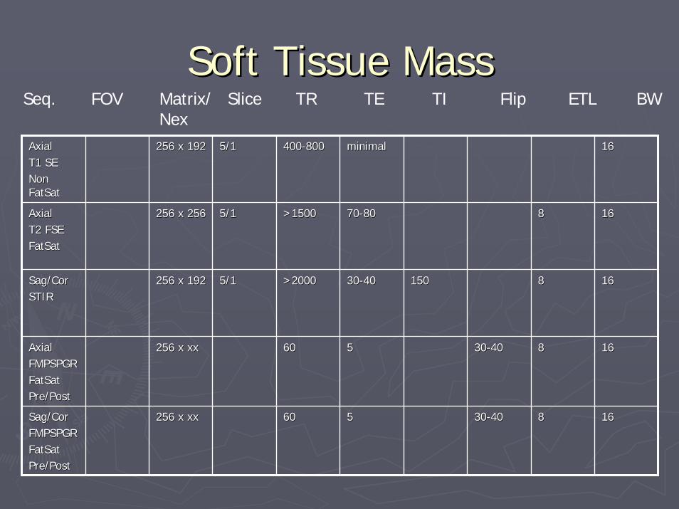
Soft Tissue MassSoft Tissue Mass
AxialAxialT1 SET1 SENon Non FatSatFatSat
256 x 192256 x 192 5/15/1 400400--800800 minimalminimal 1616
AxialAxialT2 FSET2 FSEFatSatFatSat
256 x 256256 x 256 5/15/1 >1500>1500 7070--8080 88 1616
Sag/Sag/CorCorSTIRSTIR
256 x 192256 x 192 5/15/1 >2000>2000 3030--4040 150150 88 1616
AxialAxialFMPSPGRFMPSPGRFatSatFatSatPre/PostPre/Post
256 x xx256 x xx 6060 55 3030--4040 88 1616
Sag/Sag/CorCorFMPSPGRFMPSPGRFatSatFatSatPre/PostPre/Post
256 x xx256 x xx 6060 55 3030--4040 88 1616
Seq. FOV Matrix/ Slice TR TE TI Flip ETL BWNex

Metal Protocol Metal Protocol –– general general recommendationsrecommendations
►►Assess severity of artifact on scout Assess severity of artifact on scout –– discuss discuss with MSK radiologistwith MSK radiologist
►►Maximize BW (bandwidth)Maximize BW (bandwidth)►►Lower TE (T2 and STIR)Lower TE (T2 and STIR)►►Remove fat suppression (call MSK Remove fat suppression (call MSK radrad first)first)►►Avoid GREAvoid GRE►►Use STIR instead of T2w fat satUse STIR instead of T2w fat sat