mri physics and artifacts.ppt - home - the american ... annual meeting/handouts/fritz asn sun… ·...
TRANSCRIPT
12/21/2012
1
American Society of Neuroimaging36th Annual Meeting
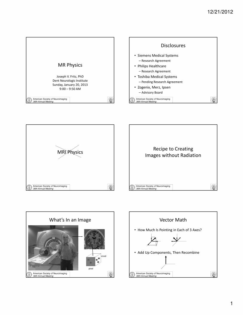
MR Physics
Joseph V. Fritz, PhD
Dent Neurologic Institute
Sunday, January 20, 2013
9:00 – 9:50 AM
American Society of Neuroimaging36th Annual Meeting
Disclosures
• Siemens Medical Systems
– Research Agreement
• Philips Healthcare
– Research Agreement
• Toshiba Medical Systems
– Pending Research Agreement
• Zogenix, Merz, Ipsen
– Advisory Board
American Society of Neuroimaging36th Annual Meeting
MRI Physics
American Society of Neuroimaging36th Annual Meeting
Recipe to Creating
Images without Radiation
American Society of Neuroimaging36th Annual Meeting
What’s In an Image
voxel
pixel
H+
H+
H+
H+
H+
American Society of Neuroimaging36th Annual Meeting
Vector Math
• How Much Is Pointing in Each of 3 Axes?
• Add Up Components, Then Recombine
12/21/2012
2
American Society of Neuroimaging36th Annual Meeting
N
S
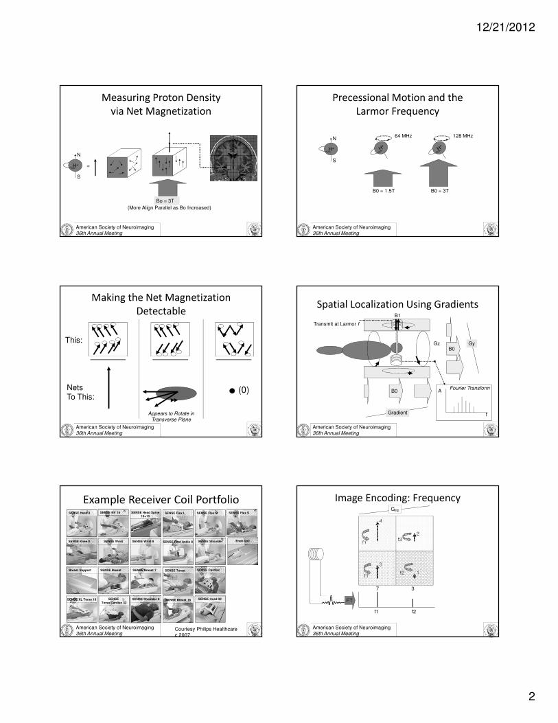
Measuring Proton Density
via Net Magnetization
H+
Bo = 1.5T
=
(More Align Parallel as Bo Increased)
Bo = 3T
American Society of Neuroimaging36th Annual Meeting
Precessional Motion and the
Larmor Frequency
B0 = 1.5T
N
S
H+
64 MHz
B0 = 3T
128 MHz
American Society of Neuroimaging36th Annual Meeting
Making the Net Magnetization
Detectable
NetsTo This:
(0)
This:
Appears to Rotate in Transverse Plane
American Society of Neuroimaging36th Annual Meeting
Spatial Localization Using Gradients
B0
Transmit at Larmor f
B1
Gradient
Gz
Receive Mix ofLarmor f’s
B0Gy
EchoA
f
Fourier Transform
American Society of Neuroimaging36th Annual Meeting
SENSE Head 8
SENSE Knee 8
SENSE Flex L SENSE Flex SSENSE Flex M
SENSE Shoulder
SENSE Head Spine16+15
SENSE Foot Ankle 8SENSE Wrist 8
Breast Support SENSE Breast
Endo coil
SENSE NV 16
SENSE Breast 7
SENSE
Torso/Cardiac 32SENSE XL Torso 16
SENSE Torso SENSE Cardiac
Example Receiver Coil Portfolio
SENSE Shoulder 8 SENSE Head 32SENSE Breast 16
SENSE Wrist
Courtesy Philips Healthcarec 2007
American Society of Neuroimaging36th Annual Meeting
Image Encoding: Frequency
4
3
2
1
f1 f2
7 3
FT
f1
f1
f2
f2
GFE
12/21/2012
3
American Society of Neuroimaging36th Annual Meeting
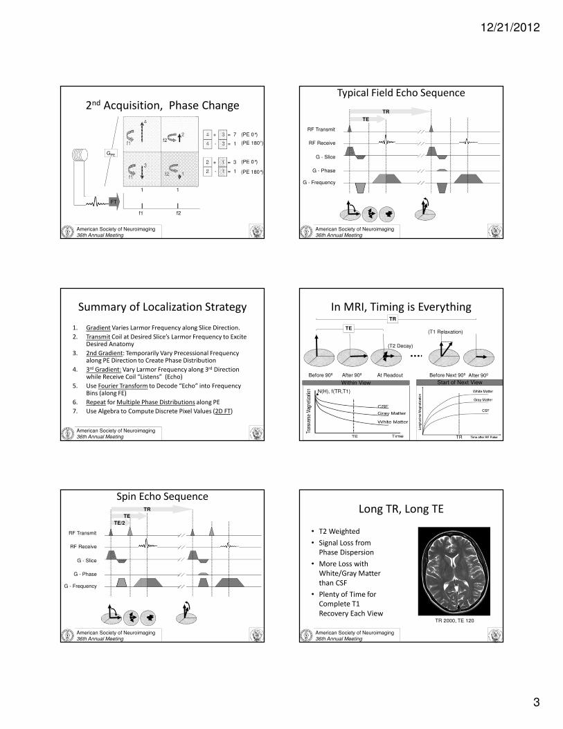
2nd Acquisition, Phase Change
4
3
2
1
f1 f2
1 1
FT
f1
f1
f2
f2
4 + 3 = 7
4 - 3 = 1
2 + 1 = 3
2 - 1 = 1
GPE
(PE 0°)
(PE 0°)
(PE 180°)
(PE 180°)
American Society of Neuroimaging36th Annual Meeting
TR
TE
Typical Field Echo Sequence
RF Transmit
RF Receive
G - Slice
G - Phase
G - Frequency
American Society of Neuroimaging36th Annual Meeting
Summary of Localization Strategy
1. Gradient Varies Larmor Frequency along Slice Direction.
2. Transmit Coil at Desired Slice’s Larmor Frequency to Excite Desired Anatomy
3. 2nd Gradient: Temporarily Vary Precessional Frequency along PE Direction to Create Phase Distribution
4. 3rd Gradient: Vary Larmor Frequency along 3rd Direction while Receive Coil “Listens” (Echo)
5. Use Fourier Transform to Decode “Echo” into Frequency Bins (along FE)
6. Repeat for Multiple Phase Distributions along PE
7. Use Algebra to Compute Discrete Pixel Values (2D FT)
In MRI, Timing is Everything
Before 90º After 90º Before Next 90º After 90º
(T1 Relaxation)
Within View
TR
At Readout
(T2 Decay)
TE
Start of Next View
TR
N(H), f(TR,T1)
American Society of Neuroimaging36th Annual Meeting
TR
TE
Spin Echo Sequence
RF Transmit
RF Receive
G - Slice
G - Phase
G - Frequency
TE/2
American Society of Neuroimaging36th Annual Meeting
Long TR, Long TE
• T2 Weighted
• Signal Loss from
Phase Dispersion
• More Loss with
White/Gray Matter
than CSF
• Plenty of Time for
Complete T1
Recovery Each ViewTR 2000, TE 120
12/21/2012
4
American Society of Neuroimaging36th Annual Meeting
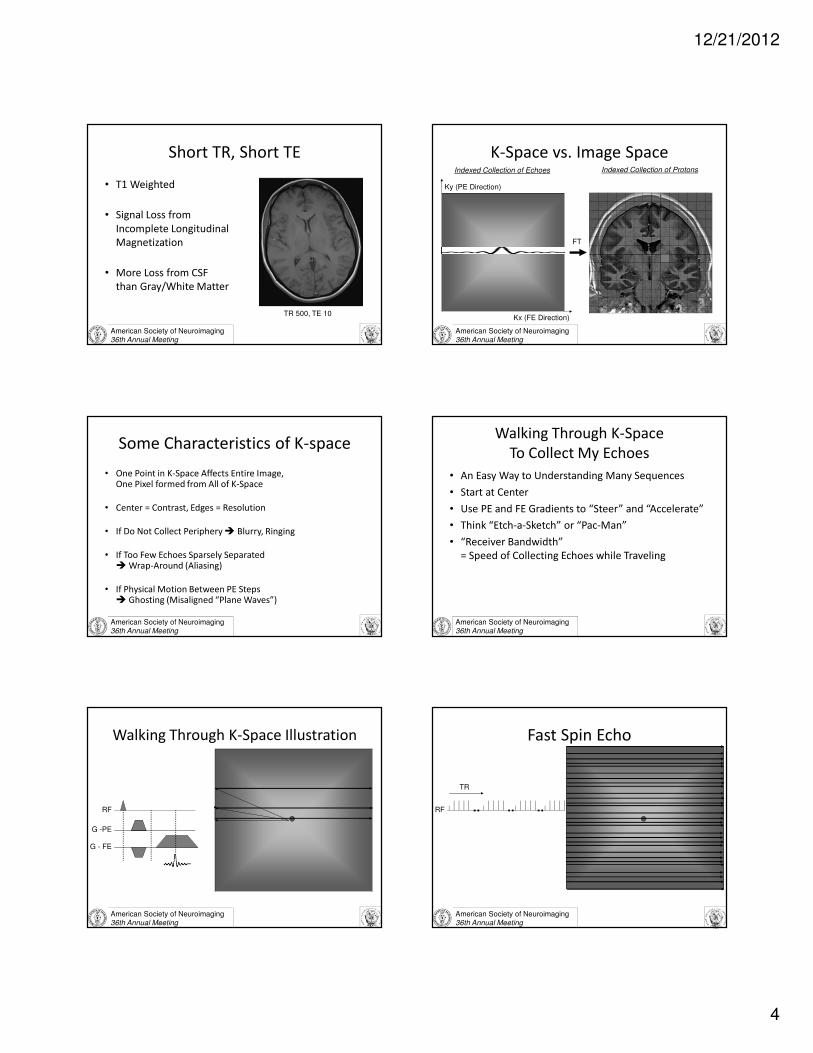
Short TR, Short TE
• T1 Weighted
• Signal Loss from
Incomplete Longitudinal
Magnetization
• More Loss from CSF
than Gray/White Matter
TR 500, TE 10
American Society of Neuroimaging36th Annual Meeting
K-Space vs. Image Space
FT
Indexed Collection of Echoes Indexed Collection of Protons
Ky (PE Direction)
Kx (FE Direction)
American Society of Neuroimaging36th Annual Meeting
Some Characteristics of K-space
• One Point in K-Space Affects Entire Image,One Pixel formed from All of K-Space
• Center = Contrast, Edges = Resolution
• If Do Not Collect Periphery � Blurry, Ringing
• If Too Few Echoes Sparsely Separated � Wrap-Around (Aliasing)
• If Physical Motion Between PE Steps � Ghosting (Misaligned “Plane Waves”)
American Society of Neuroimaging36th Annual Meeting
Walking Through K-Space
To Collect My Echoes
• An Easy Way to Understanding Many Sequences
• Start at Center
• Use PE and FE Gradients to “Steer” and “Accelerate”
• Think “Etch-a-Sketch” or “Pac-Man”
• “Receiver Bandwidth”
= Speed of Collecting Echoes while Traveling
American Society of Neuroimaging36th Annual Meeting
Walking Through K-Space Illustration
RF
G -PE
G - FE
American Society of Neuroimaging36th Annual Meeting
Fast Spin Echo
RF
TR
12/21/2012
5
American Society of Neuroimaging36th Annual Meeting
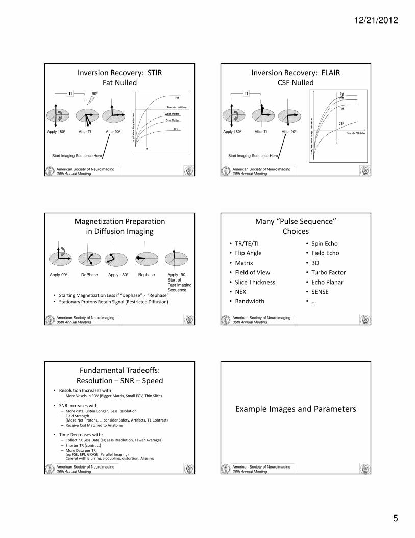
Inversion Recovery: STIR
Fat Nulled
After TI After 90º
TI
Start Imaging Sequence Here
Apply 180º
90º
American Society of Neuroimaging36th Annual Meeting
Inversion Recovery: FLAIR
CSF Nulled
Apply 180º After TI
TI
After 90º
Start Imaging Sequence Here
American Society of Neuroimaging36th Annual Meeting
Magnetization Preparation
in Diffusion Imaging
• Starting Magnetization Less if “Dephase” ≠ “Rephase”
• Stationary Protons Retain Signal (Restricted Diffusion)
Apply 90º DePhase Apply 180º Rephase Apply -90
Start ofFast Imaging
Sequence
American Society of Neuroimaging36th Annual Meeting
Many “Pulse Sequence”
Choices
• TR/TE/TI
• Flip Angle
• Matrix
• Field of View
• Slice Thickness
• NEX
• Bandwidth
• Spin Echo
• Field Echo
• 3D
• Turbo Factor
• Echo Planar
• SENSE
• …
American Society of Neuroimaging36th Annual Meeting
Fundamental Tradeoffs:
Resolution – SNR – Speed • Resolution Increases with
– More Voxels in FOV (Bigger Matrix, Small FOV, Thin Slice)
• SNR Increases with– More data, Listen Longer, Less Resolution
– Field Strength (More Net Protons, … consider Safety, Artifacts, T1 Contrast)
– Receive Coil Matched to Anatomy
• Time Decreases with:– Collecting Less Data (eg Less Resolution, Fewer Averages)
– Shorter TR (contrast)
– More Data per TR (eg FSE, EPI, GRASE, Parallel Imaging)Careful with Blurring, J-coupling, distortion, Aliasing
American Society of Neuroimaging36th Annual Meeting
Example Images and Parameters
12/21/2012
6
Name that Sequence…Name that Sequence
Name that Sequence Name That Sequence
Name That Sequence What is the Sequence on the Rt?
12/21/2012
7
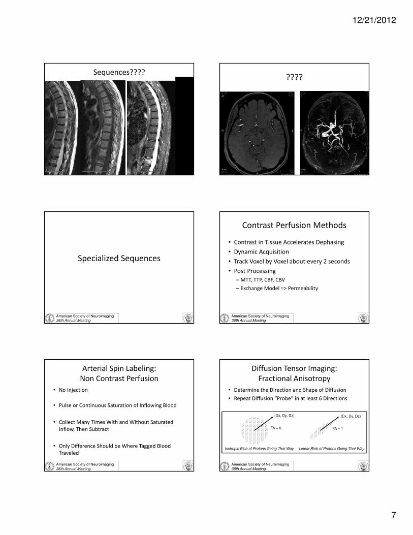
Sequences????????
American Society of Neuroimaging36th Annual Meeting
Specialized Sequences
American Society of Neuroimaging36th Annual Meeting
Contrast Perfusion Methods
• Contrast in Tissue Accelerates Dephasing
• Dynamic Acquisition
• Track Voxel by Voxel about every 2 seconds
• Post Processing
– MTT, TTP, CBF, CBV
– Exchange Model => Permeability
American Society of Neuroimaging36th Annual Meeting
Arterial Spin Labeling:
Non Contrast Perfusion
• No Injection
• Pulse or Continuous Saturation of Inflowing Blood
• Collect Many Times With and Without Saturated
Inflow, Then Subtract
• Only Difference Should be Where Tagged Blood
Traveled
American Society of Neuroimaging36th Annual Meeting
Diffusion Tensor Imaging:
Fractional Anisotropy
• Determine the Direction and Shape of Diffusion
• Repeat Diffusion “Probe” in at least 6 Directions
Isotropic Blob of Protons Going That Way Linear Blob of Protons Going That Way
FA = 0 FA ≈ 1
(Dx, Dy, Dz) (Dx, Dy, Dz)

12/21/2012
8
American Society of Neuroimaging36th Annual Meeting
Resulting Graphics of White Matter
• FA = Brightness
• Color = Direction
• Connect Similar Dots =
Representation of Tracts
that Linearly Restrict
Diffusion
American Society of Neuroimaging36th Annual Meeting
Functional Imaging: BOLD
• Blood Oxygenation Level Dependent Imaging
• Oxy vs. De-oxy Hg Changes Iron Environment
• Iron Affects Nearby Magnetic Field
• Variation in Larmor Frequencies within Voxel
Changes Net Magnetization
American Society of Neuroimaging36th Annual Meeting
Simple Paradigm to Tag Function
• Scan Entire Brain Every Couple Seconds
– FE-EPI = fast single shot, & susceptibility weighted
• Do Something that Affects Local O2 Uptake
• Continue Scanning While Interleaving Another
Task With Different O2 Affect
• Subtract Image Sets (after a lot of averaging)
• Threshold “Meaningful” Differences
• Overlay on a Registered Anatomic Image
American Society of Neuroimaging36th Annual Meeting
Surgical Guidance Using Combined
FiberTracts and fMRI
Courtesy: Philips &University of Leuven, Belgium
AVM on T2
Perfusion abnormality
Arcuate Fasciculus (DTI)
Broca & Wernicke (fMRI)
American Society of Neuroimaging36th Annual Meeting
Very Basic MR Spectroscopy
• eg Fat and Water 3.5 ppm Separation
• Larmor Frequency of Protons in
Water = 1.5T x 42.58 MHz/T = 64MHz
Fat = 3.5 ppm slower due to e- shielding
= about 220 Hz Lower
Fat
Water
64 MHz
.000220 MHz (3.5ppm)
American Society of Neuroimaging36th Annual Meeting
Water – Fat Shift
Chemical Shift due to Shielding => Different Larmor f ’s
• Tune “RF Slice Select” or “Sat Bands” to Excite or Destroy Fat or Water
• Phase Cycling: Fat & Water Precess In and Out of Phase (Another Fat Suppression Method)
• Different Frequency => Chem Shift Artifact Misplaces Fat (Incorrect Frequency Encoding)
12/21/2012
9
American Society of Neuroimaging36th Annual Meeting
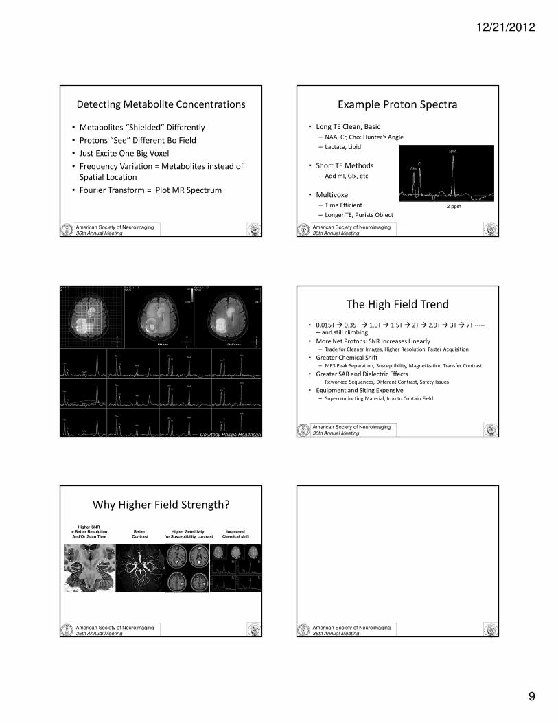
Detecting Metabolite Concentrations
• Metabolites “Shielded” Differently
• Protons “See” Different Bo Field
• Just Excite One Big Voxel
• Frequency Variation = Metabolites instead of
Spatial Location
• Fourier Transform = Plot MR Spectrum
American Society of Neuroimaging36th Annual Meeting
Example Proton Spectra
• Long TE Clean, Basic
– NAA, Cr, Cho: Hunter’s Angle
– Lactate, Lipid
• Short TE Methods
– Add mI, Glx, etc
• Multivoxel
– Time Efficient
– Longer TE, Purists Object
2 ppm
Hunter’s Angle
American Society of Neuroimaging36th Annual Meeting
Spectroscopic Imaging in Tumors
(2 minutes)
Courtesy Philips Healthcare
American Society of Neuroimaging36th Annual Meeting
The High Field Trend
• 0.015T � 0.35T � 1.0T � 1.5T � 2T � 2.9T � 3T � 7T ------- and still climbing
• More Net Protons: SNR Increases Linearly
– Trade for Cleaner Images, Higher Resolution, Faster Acquisition
• Greater Chemical Shift
– MRS Peak Separation, Susceptibility, Magnetization Transfer Contrast
• Greater SAR and Dielectric Effects
– Reworked Sequences, Different Contrast, Safety Issues
• Equipment and Siting Expensive
– Superconducting Material, Iron to Contain Field
American Society of Neuroimaging36th Annual Meeting
Why Higher Field Strength?
Higher SNR = Better ResolutionAnd/Or Scan Time
Higher Sensitivityfor Susceptibility contrast
Better Contrast
IncreasedChemical shift
American Society of Neuroimaging36th Annual Meeting
12/21/2012
10
American Society of Neuroimaging36th Annual Meeting
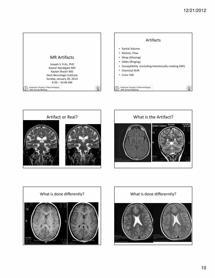
MR Artifacts
Joseph V. Fritz, PhD
Kaveer Nandigam MD
Kaylan Shastri MD
Dent Neurologic Institute
Sunday, January 20, 2013
9:50 – 10:40 AM
American Society of Neuroimaging36th Annual Meeting
Artifacts
• Partial Volume
• Motion, Flow
• Wrap (Aliasing)
• Gibbs (Ringing)
• Susceptibility (including Intentionally creating SWI)
• Chemical Shift
• Cross Talk
Artifact or Real? What is the Artifact?
What is done differently? What is done differerntly?

12/21/2012
11
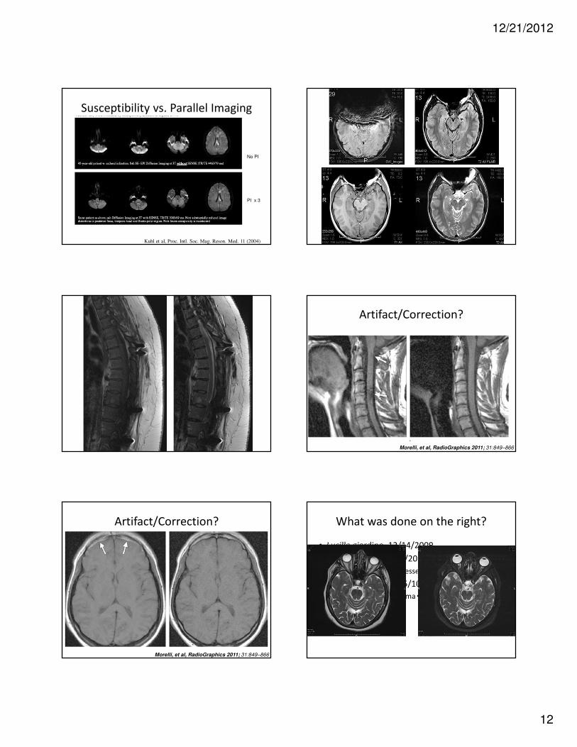
Artifact? Correction?
Morelli, et al, RadioGraphics 2011; 31:849–866
Artifact, Correction?
Artifact/Correction?
American Society of Neuroimaging36th Annual Meeting
Surface Coils and Parallel Imaging
• Small Surface Coil Improves “Filling Fraction”
– Better SNR
– Larger FOV with Array (eg. Spine)
– Depth of Penetration Limited
• PI Trick:
– Arrange Multiple Coils to Look at Same Anatomy
– Signal Drops Off Differently from Each Coil
– Another “Set of Equations” for Free
– Proportionally Fewer PEs Needed
• Sqrt (N) SNR Loss
– High Field Advantage: Convert Extra SNR to Speed
Parallel Imaging
If Reduce FOV by half…Half Scan time but Aliasing Unfolding
(Post processing)Coil #1
Coil #2
Full FOV
No wrap around
Half scan time
Sensitivity
Distribution
Multiple receiver coils
Coil #1
Coil #2
Courtesy Toshiba Medical Systems
12/21/2012
12
Susceptibility vs. Parallel Imaging
Kuhl et al, Proc. Intl. Soc. Mag. Reson. Med. 11 (2004)
No PI
PI x 3
Artifact/Correction?
Morelli, et al, RadioGraphics 2011; 31:849–866
Artifact/Correction?
Morelli, et al, RadioGraphics 2011; 31:849–866
What was done on the right?
• Lucille giardino, 12/14/2009
• Edward bajdas, 2/12/2010
– Find f/yu w fat suppressed orbits 2/24/10
• Amanda obrien, 6/25/10
– see f/u potential lipoma with fat sat
12/21/2012
13
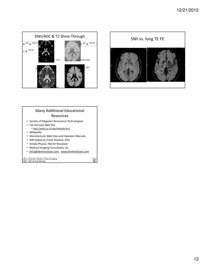
DWI/ADC & T2 Shine Through
b=0 (T2*) b=1000 (DWI)
ADC eADC
e -bD e -TE/T2*
= e -TE/T2*
e -bD e -TE/T2*
e -bDD
SWI vs. long TE FE
American Society of Neuroimaging36th Annual Meeting
Many Additional Educational
Resources
• Society of Magnetic Resonance Technologists
• Joe Hornack Web Site
– http://www.cis.rit.edu/htbooks/mri/
• Wikipedia
• Manufacturer Web Sites and Operator Manuals
• MRI Safety by Frank Shellock, PhD
• Simply Physics, Moriel Nessaiver
• Medical Imaging Consultants, Inc
• [email protected], www.DentInstitute.com