mri of the wrist and hand - national institutes of...
TRANSCRIPT

1
MR Imaging of the Wrist and
Hand
William B. Morrison MDThomas Jefferson University Hospital
MRI of the Wrist
• Occult fracture• Ganglion Cyst• Tumor• Ligament tear• Avascular necrosis• Arthritis• Tendon Pathology• Nerve Impingement• Infection
Occult fracture
-Not visible on initial radiographs-follow-up xray, CT
-MRI: -MRI very sensitive for dx
-Use T2fs / STIR to detect-Use T1 to DDx fx vs. bone bruise
-Determine extent of injury-Osseous, soft tissue
-can dx alternate cause of pain
-Capitate fracture
-Distal radial fracture -Occult scaphoidFractureNBA player

2
Ganglion Cyst
- Common at wrist, esp. dorsal- May simulate mass, or may
be occult source of pain if small or deep
- Joint >> tendon sheath- MRI:
-Lobulated-Fluid signal-Rim-enhancement-May indicate underlying ligament tear
Ganglia: Common Locations• Dorsal
– Deep to tendons– Adjacent to lunate/capitate joint– Weak area of capsule– Extends around dorsal intercarpal ligament
• Volar– Radial aspect off radioscaphoid joint– Adjacent to radial artery – may be confused for
vessel / aneurysm• Other areas
– Into carpal tunnel– Off tendon sheaths
Ganglion Cyst from JointExtending Around Tendons
Fluid signal
Rim enhancement
Dorsal intercarpalligament
The “Angry Ganglion”
Volar RadioscaphoidGanglion
Extensor Tendon Ganglion
Tumor- MRI may help DDx:
-Malignant / benign lesion vs. ‘pseudomass’- Most soft tissue ‘masses’ are benign lesions with characteristic MRI features
-Lipomas-Ganglion cysts-Hemangiomas / vascular malformations-Giant cell tumor of tendon sheath
- Osseous lesions-Radiographs important for DDx-MRI: solid vs. cystic (esp w contrast)

3
-Lipoma
Fat signalNo internal complexity
-Giant cell tumor of tendon sheath (GCTTS)
Location: tendon sheathSignal: low T1, T2
-Nerve lesionFibrolipomatoushamartoma
Location: neuralSignal: high T1, fascicular pattern
-Glomus tumor
Location: distal digitSignal: ‘light bulb’ on T2, Gd
Malignant lesion-Synovial sarcoma
Solid, complex mass
“Pseudomass”-Accessory muscle
Characteristic locationse.g., palmaris longusSignal: same as muscle

4
-Aneurysmalbone cyst
Fluid-fluidlevels
Ligament tear
-Intrinsic ligaments-Scapholunate-Lunatotriquetral-Triangular fibrocartilage complex
-central (radial aspect)-peripheral (ulnar side)
-MR arthrography-Increases accuracy for dx of tear
Triangular Fibrocartilage“Complex” (TFCC)
Anatomy
• Triangular fibrocartilage• Dorsal and volar radioulnar ligaments• Ulnar-triquetral ligament• Meniscal homologue• ECU sheath
TFCCANATOMY
• TFC• Dorsal and
volar RU lig• UT ligament• Meniscal
homologue• ECU sheath
Central: Attaches to cartilage of Radius
Peripheral: twoattachments-Look at slice with styloid
Attaches to bone centrallyUnlike theTFC
TFCC - perforation
Perforations may not be clinically significant
Central TFCC Tear

5
-Peripheral TFCC tear Peripheral TFCC Tear / LT Tear
ECU Subluxation / Peripheral TFCC Tear
ECU Tenosynovitis / Peripheral TFCC Tear
Ulno-lunate Abutment
-Positive ulnarvariance
-Cystic changein lunate
-TFCC tear
Indirect Arthrogram – tear of central TFC with ulnar-lunate abutment

6
Scapholunate and Lunatotriquetral Ligaments DORSAL AND VOLAR BANDS
These bands are more mechanically important than central membrane
Scapholunate Ligament Tear
Direct MR arthrogram –scapholunate tear
Scapholunate tearPalmarflexion of scaphoid
Dorsal tilt of lunate(DISI deformity)
SL or LT tear can cause carpal malalignment
Scapholunate advanced collapse(SLAC wrist)
DISI deformity
Proximal migration of capitateCarpal osteoarthritis
Radiographic progressionOf SLAC
Early-radioscaphoidjoint narrowing
Intermediate
Late

7
SLAC wrist SLAC secondary to rheumatoid arthritis
Extensive synovitisMarrow edema
Inflammatory arthropathiescan cause intrinsic ligament tears
Lunatotriquetral Ligament TearLunatotriquetralligament tear
Lunate may tiltin palmar directionalong with scaphoid(VISI)
Avascular necrosis
-Lunate (negative ulnar variance)-Scaphoid (fracture)Progression: density, fracture, collapse, OA
-Keinbock’s disease
Replacementof fat signal c/w AVN

8
-Scaphoid fracture with AVNof the proximal pole
-Scaphoid nonunion-Humpback deformity-Acts like an SL lig tear-Radiocarpal OA… and AVN lunate
“SNAC” WristScaphoid Nonunion Advanced Collapse
Arthritis
-Osteoarthritis-Subchondral cysts cartilage loss, spurs-Distribution depends on etiology-Trauma, instability, predisposing factors
-Inflammatory arthropathies-Classic: rheumatoid arthritis
-Carpus, MCPs-Diffuse involvement-Synovitis, erosions
-Scapholunate Advanced Collapse (SLAC)
-Type 2 lunate with secondary OA
Lunate articulates with hamate
Rheumatoid ArthritisMarked synovial proliferation

9
Rheumatoid ArthritisMRI can monitor activity, response to Tx
Erosions
Rheumatoid Arthritis
Tenosynovitis in multiplesheaths suggests anInflammatory arthropathy
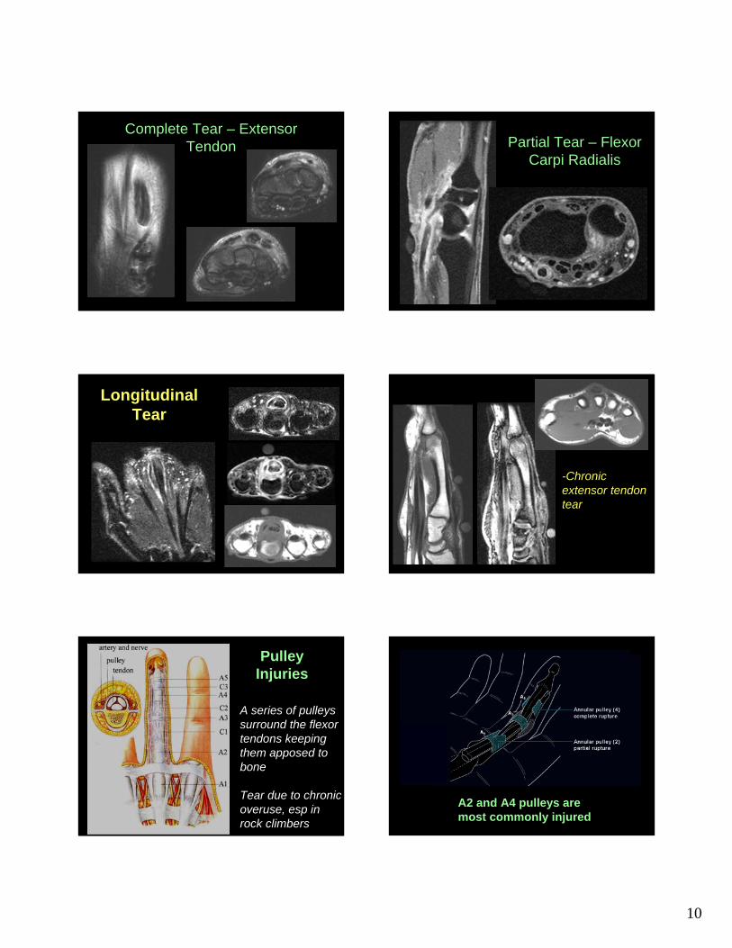
Tendon Pathology
-Tenosynovitis-Tendon tear-Pulley lesions
Extensor Tendons
COMPARTMENT 1Extensor Pollicis BrevisAbductor Pollicis Longus
COMPARTMENT 2Extensor carpi radialis brevisExtensor carpi radialis longus
COMPARTMENT 3Extensor pollicis longus
COMPARTMENT 4Extensor digitorumExtensor indicis
COMPARTMENT 5Extensor digiti minimi
COMPARTMENT 6Extensor carpi ulnaris
DeQuervain’sTenosynovitis1st EXTENSOR COMPARTMENT
Intersection syndrome
Inflammation at distal forearm at crossing point of first and secondextensor compartments
10
Complete Tear – Extensor Tendon Partial Tear – Flexor
Carpi Radialis
Longitudinal Tear
-Chronic extensor tendontear
Pulley Injuries
A series of pulleys surround the flexor tendons keeping them apposed to bone
Tear due to chronic overuse, esp in rock climbers
A2 and A4 pulleys are most commonly injured

11
Flexor tendonpulley injury
Pulley Lesionpre / post stress
Stenosing Tenosynovitis
Tendon ‘catches’ in sheath esp at pulleys
Thumb: UlnarCollateral
Ligament Injury
Line coronals up with sesamoids
Small FOV
Ulnar collateral ligament tear
Dorsal Hood Injury
Disrupted ‘sagittal band’allows ulnar subluxation of extensor tendon at MCP with flexion
Common in boxers
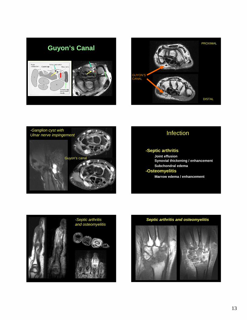
Nerve Impingement
Median nerve-Carpal tunnel syndromeUlnar nerve-Guyon’s canal

12
Carpal Tunnel
– Pisiform / hamatemedially
– Carpal bones dorsal
– Flexor retinaculum volar
– Median nerve deep to retinaculum
– Flexor tendons– Flexor carpi radialis:
outside the carpal tunnel
Carpal tunnel syndrome
-Flexortenosynovitis
-Separation of tendons by synovial tissue
-Mass effect frommuscle in carpal tunnel
CTS: Flexor RetinaculumBowing
CTS: Proximal Enlargement and Fasciculation
proximal distal
Fasciculation:Looks like dots inside
-Volar ganglion cystin carpal tunnel
13
Guyon’s Canal
GUYON’SCANAL
PROXIMAL
DISTAL
-Ganglion cyst withUlnar nerve impingement
Guyon’s canal
Infection
-Septic arthritis Joint effusionSynovial thickening / enhancementSubchondral edema
-OsteomyelitisMarrow edema / enhancement
-Septic arthritisand osteomyelitis
Septic arthritis and osteomyelitis

14
MRI ProtocolRoutine MRI wrist:-Tendon pathology-Carpal tunnel syndrome-Ganglion cyst-Acute trauma-Osteoarthritis-AVNMRI wrist with IV contrast:-Mass-Infection-Inflammatory arthropathyMR arthrogram:-Ligament tear
THANK YOU