mri of epidural cavernous malformations of the spine: … · 2016-12-26 · mon in posterior...
TRANSCRIPT
Journ al of the Korean Radiological Society, 1994 : 30(3) : 411-415
MRI of Epidural Cavernous Malformations of the Spine: Correlation with Surgical and
Histopathologic Findings1
Pyeongho Yoon. M.D .• Dongik Kim. M.D. , Choonsik Yoon. M.D. ,
Taesub Chung , M.D. , ‘JunghoSuh, M.D.
Purpose: The purpose of this study is to describe Magnetic Resonance( MR) findings oftwo epidural cavernous malformations ofthespine .
Materials and Methods: MR imaging was performed in 2 patients(29-year-old man and 54-year-old woman). Sagittal T1- , T2-weighted images and Gadolinium (Gd)-enhanced axial and sagittal images were acquired. Two patients had surgery and MR findings were compared with surgical and histopathological findings.
Results: MR imaging showed high- and low-signal intensity components of these lesions that were characteristic of an epidural cavernous malformation in one case. The other case showed a high signal intensity on T2- and strong enhancement on Gd-enhanced T1-weighted images. We think that the former may be due to mixed subacute and chronic hemorrhage and the latter may be due to blood within the endothelium-lined sinusoids without hemorrhage.
Conclusion : These findings were well correlated with the surgical and histo-pathological findingsof cavernous malformation.
Index Words: Spine , cavernous malformation Spine, epidural space Spine, MRI
Cavernous malformations are uncommon vascular malformations of the central nervous system that may be generally found in cerebral hemisphere(1). In the spine , epidural lesions are uncommon in contrast to intramedullary and extramedullary intradural lesions (2). The majority of these epidural lesions are secondary to extension of vertebral hemangiomas, and purely epidural lesions are rare(3 , 4). We report two cases of epidural cavernous malformation of the spine and present the MR findings correlated with the surgical and histopathological findings.
ness of both legs with absent knee jerk. He showed hypesthesia and hypoalgesia below T6 level. A myelogram showed decreased anteroposterior diameter at T3 - T5 level by a large fusiform mass with a typical epidural configuration
MRI of the thoracic spine demonstrated a posterior epidural mass at T3-T5 level. The mass showed small areas of mixed low - and high - signal intensity on T1 ' weighted images. These hypointednsities were more marked on T2 ' weighted images and these hypointense areas showed no enhancement (Fig. 1) These findings were consistent with hemosiderin depOSltS CASE REPORTS
Case1 A 29-year-이 d man was presented to the this hos
pital complaining of weakness of the lower limbs with acute onset. Neurological examination revealed weak-
1 Department ofD iagnostic Radiology Yonsei Un iversity Col lege 01 Medicine Received 8eptember4, 1993; Acepted October 22, 1993 Address repr int requests to : Pyeongho Yoon , M.D., Departmentol Diagnostic Radiology Yonsei University College 01 Medicine, 146.92, Dogok-Dong , Kangnam.Gu , 8eoul , Korea. Tel. (82-2)3450 -3622
• 411
At surgery , this was a dark reddish hematoma intermingled with fat tissue and enlarged abnormal vessels at T3-T5Ievel(Fig. 1). Histopathologic examination of the specimen showed organizing hemorrhage and thrombi with areas of irregular vascular spaces(Fig. 1).
Case2 A 54 -year -old woman had progressive low back
pain radiating to lower extremities since a month ago.

Journal of the Korean Radiological Society, 1994 ; 30( 3) : 411 -41 5
She had a history of a fall - down trauma 23 years ago and low back pain developed from thattime
Neurological examination on admission revealed sensory impairment in L2 dermatome and mild paresis of left knee without pathologic reflexes. She had no difficulty in micturition and defecation.
MRI showed a smooth , somewhat elongated , posterior epidural mass located at L2 -L3level. The mass had a homogeneous, intermediate signal intensity similar to that of the spinal cord on T1 -weighted sagittal images. Sagittal T2-weighted images showed a homogeneous high - signal intensity similar to that of CSF. Sagittal and axial T1 - weighted images obtained
a b
'.
d e
after administration of contrast material demonstrated homogeneous enhancement of the mass and displaced dural sac to the right side by the mass. The lesion extended into the left intervertebral foramen with widening at L3-L4 level. At L2-L3 level , MR imaging showed ruptured disc material that compressed the anterior aspect of dural sac. The dural sac at L2 -L3level was severely compressed anteriorly and posteriorly by the ruptured disc material and the epidural mass(Fig 2).
At surgery , a dark reddish mass that was located between the dura and ligamentum flavum at L2 -L3level and extended into the left intervertebral foramen at
c
Fig. 1. Case 1. A 29-year-old man with an epidural cavernous mallormation in the thoracic spine T1-weighted sagittal image(a) shows small areas 01 mixed low- and high-signal intensities. These hypointense areas are more marked on T2-weighted image(b) Alter administration 01 contrast material (c) these hypointense areas show no enhancement. At surgery(d) , there was a dark reddish hematoma intermingled with lat tissue and enlarged abnormal vessels Histopathologic examination(e) 01 the specimen shows organizing hemorrhage and thrombi with areas 01 irregular vascular space
- 412 -

Pyeong Ho Yoon , e t a/ : MRI of Epidural Cavernous Malformations of the Spine
a b c
d e Fig. 2. Case 2. A 54-year-old woman with epidural cavernous mallormation in the lumbar spine. smooth , somewhat elongated, posterior epidural mass at L2-L3level has a homogeneous, intermediate signal intensity on T1-weighted image(a) and ruptured disc material compresses the anterior aspect 01 the dural sac. T2-weighted image(b) shows a homogeneous high-signal intensity mass Sagittal T1-weighted image(c) obtained after administration 01 contrast material demonstrates homogeneous enhancement 01 the mass. This mass extends into the left intervertebral loramen with widening at L3-L4 level(d). Histoparhologic examination(e) 01 the postoperative specimen shows variable-sized endothelium-lined sinusoids consistent with cavernous mallormation
L3-L4 level was excised and the ruptured disc material at L2-L3 level was also excised(Fig. 2) Histopathologic analysis of the postoperative specimen showed variable -sized endothelium - lined sinusoids consistent with cavernous malformation(Fig. 2).
DISCUSSION
Of the epidural tumors in the adult pop비 ation , the proportion of tumors primarily involving the vertebrae is as high as 50 percent(5). The vast majority of these tumors will be metastasizes to bone and epidural
space , and a still smaller proportion will be benign or malignant tumors arising from the osseous or notochord structures of the spinal column(5) . Purely epidural lesions are rare , and they contain vascular malformations, Iymphoma, lipoma, meningioma, neurofibroma, metastatic lesions,etc.
Cavernous malformations are uncommon vascular malformations of the central nervous system that may be generally found in cerebral hemisphere(1) . In the spine, these lesions usually occur in the vertebral body and epidural lesions are rare(3 , 4). Of cavernous malformations that involve the epidural space , lesions
413 -

Journal of the Korean Radiological Society, 1994; 30(3 ) ‘ 411 - 415
in the vertebral bodies with extension into the epidural space are much more common than are purely epidural(4 , 6). Purely epidural spinal cavernous malformations account for less than 4% of spinal epidural masses(4). Grossly , the usual cavernous malformation is a red -blue , soft , spongy mass with 1 to 2 cm in diameter. Histologically, the mass is sharply defined , but not encaps비 ated , and made up of large,
cavernous , vascular spaces , partly or completely filled with blood separated by a scanty connective tissue stroma. Intravascular thrombosis or rupture of channels may modify the histologic appearance(7). It is reported that an epidural cavernous malformation grossly showed a firm , red - purple mass and variable sized endothelium-lined sinusoids within a fibrous stroma microscopically(4, 8). Recent articles have described the MR appearance of epidural cavernous malformation(8 , 9) , but the MR appearance of this lesion has not been fully documented.
In our case 1, T1 - weighted MR images showed a mass with mixed areas of high signal and low signal intensity at the T3 - T5 level. The hypointense areas were more marked on T2 -weighted images. Selective reduction of T2 can occur with certain paramagnetic materials , such as hemosiderin(10, 11) , and hemosiderin appears dark when compared with the surrounding tissue. Small areas of hyperintensity on T1 -and T2 -weighted images suggest subacute and chronic hematomas. These hematomas are known to have a relatively short T1 relaxation time, resulting in areas of increased signal intensity , which may be due to methemoglobin(12, 13). This correlates well with the pathologic findings of thrombi and old liquefied blood identified at microscopic examination. The presence of mixed subacute and chronic hemorrhage, suggested by mixed high - and low-signal intensity components of this lesion , are characteristic of cavernous malformation(14)
In case 2, T1 - weighted MR images showed a well circumscribed mass with intermediate signal intensity similar to that of the spinal cord. T2 -weighted images demonstrated a high -signal intensity mass with slightly higher intensity than that of CSF. T1 - weighted images after the administration of contrast material showed a strong enhancement. These findings might be due to the blood within the endothelium - lined sinusoids. Gd -DTPA enhanced axial images revealed the extension ofthe mass into the left interv
were most consistent with a neurofibroma. Dumbbell shaped cavernous malformations have been reported before(4, 9, 15). Padovani et al. (4) gave evidence of bony erosion and enlargement ofthe intervertebral foramen in dumbbell-shaped cavernous malformation . Thus , we must now include epidural cavernous malformation in the differential diagnosis of a dumbbell shaped lesion because ofthe similar MR findings
Of the purely epidural cavernous malformations that have been reported , most of the lesions are more common in posterior epidural space than anterior epidural space(4, 8, 9). In our two cases , all are in posterior epidural space. Thus , we think that vascular anatomy of the epidural space may be related to the higher incidence of epidural cavernous malformation in posterior epidural space and the reason for this should be investigated. In summary , cavernous malformations are uncommon vascular malformations in the epidural space that show different stages of hemorrhage, calcification , and thrombi . The first case showed the characteristic findings , mixed subacute and chronic hemorrhage, of an epidural cavernous malformation. But the second case showed the MR findings similar to those of neoplastic condition. In the present cases , the MR findings are well correlated with the surgical and histopathologic findings.
REFERENCES
1. Russel DS, Rubinstein LJ. Pathology of the nervous sytem . 4th ed. 8altimore : Williams & Wilkins, 1977 ; 129-134
2. Rubinstein LJ. Tumors olthe central nervous system.ln : Atlas 01 tumor pathology. second series lascicle 6. Washington , D.C ,.
Armed Forces Institute of Pathology, 1972 ; 245-246 3. Ri chardson RR , Cerullo LJ. Spinal epidural cavernous
hemangioma. Surg Neuro/1979; 12: 266-268 4. Padovani R, Tognetti F, Proietti D, Pozatti E, Servadei F
Extrathecal cavernous hemangioma. Surg Neurol 1982 ; 18 463-465
5. Zimmerman RA , 8ilaniuk L T. Imaging 01 tumors 01 the spinal canal and cord. Radiol Clin North Am 1988 ; 26 : 965-1 007
6. Guthkelch AN. Hemangiomas involving the spinal epidural space. J Neurol Neurosurg Psychiatry 1948 ; 11 : 199-21 0
7. Ramsi SC, Vinay K, Stanley LR. Pathologic basis of disease. 4th ed. Philadelphia: W. 8. SaundersCompany, 1989 ; 588
8. Golwyn DH , Cardenas CA, Murtagh FR , 8alis GA , Klein JB. MRI 01 a cervical extradural cavernous hemangioma. Neuroradi
ology 1992 ; 34 : 68-69 9. Feider HK , Yuille DL. An epidural cavernous hemangioma 01 the
spine. AJNR 1991 ; 12: 243-244 10. New PFJ,Ojemann RG , Davis KR , et al. MR and CT 01 occult
vascular mallormations olthe brain. AJNR 1986 ; 7 :771-779 11 . Gomori JM , Grossman RI , Goldberg RI , Zimmerman RA‘
8ilaniuk L T. Intracranial hematomas : imaging by high-lield MR Radiology 1985 ; 157: 87-93
12. Lee 8CP , Herzberg L, Zimmerman RD, Deck MDF. MR imaging 01 cerebralvascular mallormations. AJNR 1985 ; 6 : 863-870
13. Lemme-Plaghos L, Kucharczyk W, 8rant-Zawadzki M, et al. MR
- 414 -

Pyeong Ho Yoon , et al: MRI of Epidural Cavernous Malformations of the Spine
imaging olangiographically occult vascular mallormations. 166: 839-841
AJNR1986 ; 7: 217-222 15. Marioka T, Nakagaki H, Hasuo K. Oumbbell-shaped spinal epi-
14. Fontaine S, Melanson 0 , Cosgrove R, Betrand G. Cavernous dural cavernous angioma. Surg Neuro/1986 ; 25: 142-144
hemangiomas 01 the spinal cord ‘ MR imaging. Radio/ogy 1988;
대 한 방 사 선 의 학회 지 1994; 30(3): 411-415
척추의 경막부위에서 발생한 해면상 혈관기형의 자기공명영상소견: 수술및 병리학적 소견과의 비교
윤평호·김동익·윤춘식·최태섭·서정호
연세대학교 의과대학 진 단방사선과학교실
척추의 경막부위에서 발생하는 해면상 혈관기형은 매우 드문 질환으로서 저자들은 자기공명영상상 고신호강도와 저신호
강도가 섞여 있어 전형적인 해면상 혈관기형의 소견을 보이는 한 예와 T2 강조영상 및 초영증강후의 Tl 강조영상에서 강한
조영증강을 보인 또 다른 한 예를 보고한다. 전자는 아급성기와 만성기의 출혈이 섞여 있는 것으로 생각되며 후자는 출혈없
이 상피내세포로둘러싸여 있는동양혈관(sinusoid)내에 혈액이 차 있는것으로생각되었으며 이러한소견은수술과초직병
리학적 소견과일치하였다.
415-
연푼별 전공의 연수교육 주제
년 도 기 별 끼 2s. 제
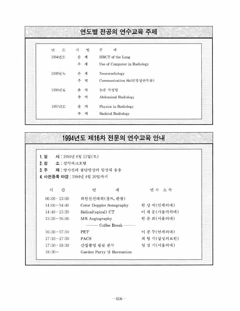
1994년도 춘 계 HRCT of the Lung
추 계 Use of Computer in Radiology
1 995년도 춘 계 Neuroradiology
추 계 Comrnunication Skill( 영 상판독술)
1996년도 춘 계 논문작성법
추 계 Abdominal Radiology
1997년도 춘 계 Physics in Radiology
추 계 Skeletal Radiology
1994년도 제16차 전문의 연수교육 안내
1. 일 시 : 1994년 6월 11일(토)
2. 장 소 : 설악파크호텔
3. 주 저1 : 방사선과 첨단영상의 임상적 응용
4. 사전등록 마감 : 1994년 4월 30일까지
시 간
06 :00-13 :00
14 :00 -14:40
14 :40 - 15: 20
15: 20 -16:00
16 :30-17: 10
17 : 10 - 17 : 50
17 :50- 18 :30
18 :30
연 저l 연사 소속
회원친선대회(골프, 관광) v“
-
h
-
때 熾
n
T
’
B
c
C
i
e
s
h
f
앙 떼 째 뻐
빼 빼 뺑 4
D
U
m -
r
3
A
-
뼈 마 R
-
C
H
M
한상석(인제의대)
이 재 문(가톨릭의대)
한문 희(서울의대)
PET
PACS
간접촬영 필름 판독
이 종 두(연세의대)
최 형 식(삼성의료원)
임 정 기(서울의대)
Garden Party 및 Recreation
- 416 -