mrcp in the evaluation of choledocholithiasis prior to ...978-3-642-60461-4/1.pdf · appendix 1...
TRANSCRIPT
APPENDIX 1
MRCP in the Evaluation of Choledocholithiasis Prior to Laparoscopic Cholecystectomy
Introduction
Since 1987, when Mouret performed the first laparoscopic cholecystectomy, there has been a remarkable change in the surgical treatment of gallstones [1]. In most centers laparoscopic cholecystectomy has become the procedure of choice for symptomatic cholelithiasis [2]. The laparoscopic approach is more acceptable due to lower morbidity, reduced postoperative pain, lower cost, shorter hospital stay, and earlier return to work. Because of the limited contraindications (e.g., severe acute peritonitis, severe portal hypertension, carcinoma of the gallbladder, coagulopathies, third trimester of pregnancy) about 95% of patients with gallstones are treated by laparoscopic cholecystectomy [3].
The new therapeutic approach has brought new diagnostic needs related to the evaluation of the associated choledocholithiasis, which has been demonstrated in 3%-33% of patients, according to various authors [4]. Several studies have examined the problem related to choledocholithiasis, but there is no consensus regarding diagnosis or therapy. From the therapeutic point of view, preoperative diagnosis of CBD stones requires either a conversion to an open operation or attempted laparoscopic extraction or leaving the calculi in situ with either a subsequent endoscopic sphincterotomy or conservative management [5]. Currently most authors prefer a preoperative endoscopic procedure, with a laparoscopic exploration of the CBD restricted to selected patients [6].
Preoperative Evaluation
At the preoperative stage various techniques have been proposed for the evaluation of choledocholithiasis: abdominal ultrasound, CT, intravenous cholangiography, and ERCP. A different approach to evaluating the possible presence of stones is to perform an intraoperative study by cholangiography and/or ultrasound. Abdominal ultrasound has an overall sensitivity of 55% in detecting CBD stones, but if the diameter of the CBD is less than 6 mm (in up to 86% of patients) stones cannot be detected. CT has been widely evaluated in the past as a noninvasive screening method in diagnosing CBD stones, with sensitivity reported between 83% and 90% [8, 9], but more recent studies [10] have found lower sensitivity (75%). The major limitation of

132 APPENDIX 1 MRCP in the Evaluation of Choledocholithiasis
preoperative intravenous cholangiography is the lack of opacification of the biliary system (30%-40% of cases). In a prospective study intravenous cholangiography was useful in detecting choledocholithiasis in only 1.5% of cases [11]. Moreover, there are risks related to adverse reaction of contrast agents, high costs, and poor anatomic definition of the intra- and extrahepatic bile ducts [12].
The use of ERCP prior to a laparoscopic cholecystectomy is controversial. Good results have been obtained by combining ERCP and endoscopic sphincterotomy in the diagnosis and management of suspected CBD stones. However, patient selection is very important because 10% of patients who undergo this procedure develop some type of complication, with a mortality of 0.37%-1.0% [13]. Even if ERCP cannot be considered as a screening procedure in preoperative evaluation of patients with suspected CBD stones, it can be performed if a high clinical suspicion for the presence of stones is evident.
Several authors use a series of clinical, laboratory, and radiological indicators to determine the presence of stones [14, 151. A direct correlation be~ tween the number of positive criteria and the presence of stones has been established. However, in the better series 4%-10% of patients with CBD stones are not detected due to the lack of symptoms.
Intraoperative Evaluation
The second approach is to perform an intraoperative study by intraoperative cholangiography and ultrasonography. Intraoperative cholangiography performed during a laparoscopic procedure is inconvenient, time-consuming, and potentiaily harmful because a bile duct injury can occur [16]. Intraoperative cholangiography therefore does not appear to have a role unless the demonstration of stones in the duct leads to immediate exploration of the CBD, which is a technically difficult procedure. Laparoscopic ultrasonography has some limitations due to technical problems and can be considered as an ancillary technique to intraoperative cholangiography.
MRCP for Detecting CBD Stones Before Laparoscopic Cholecystectomy
Recent studies have reported a high reliability of MRCP in detecting stones in the CBD. A larger series showed a sensitivity of 88% and a specificity of 98%, a positive predictive value of ~I %, a negative predictive value of 97%, and an overall diagnostic accuracy of 96%.
We conducted a study among 40 patients undergoing laparoscopic chlecystectomy (14 men, 26 women; age 28-72 years, mean 54). The patients were studied with MRCP to evaluate the presence of CBD stones. Inclusion criteria, other than the known gallbladder stones, were one or more of the following: previous episodes of jaundice, increased alkaline phosphatase, and dilation of bile ducts at ultrasound. MRCP was performed 2-4 days prior to surgery. In all patients intraoperative cholangiography was performed during the laparoscopic procedure for diagnostic confirmation and for a therapeutic
a
References 133
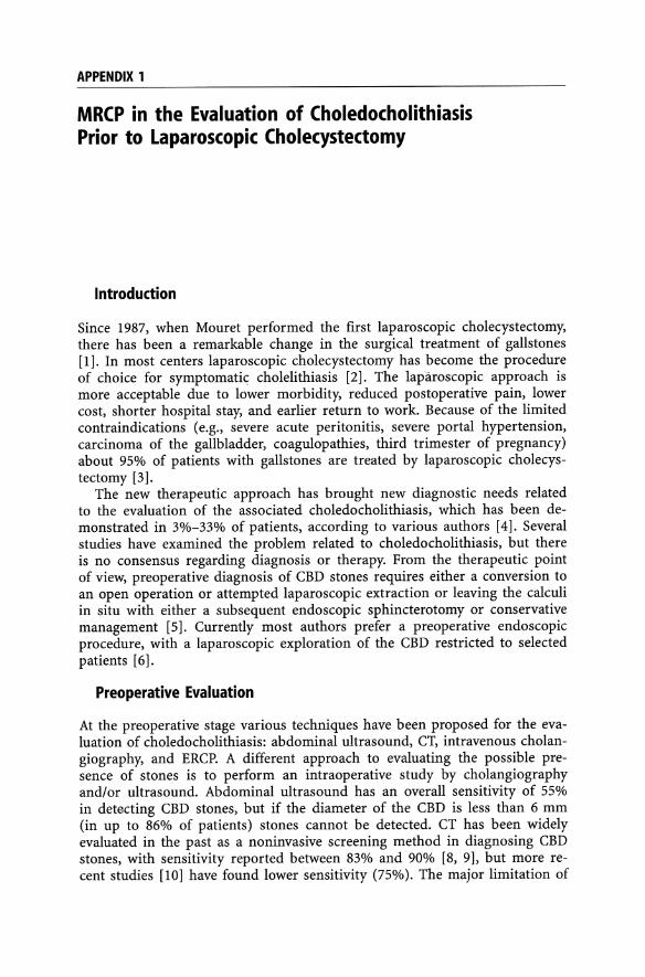
Fig. la,b. Patient with known gallbladder stones. a On MRCP on MIP-reconstructed image an inhomogeneous signal is appreciable at the distal tract of the CBD. b On the source image the stone is clearly evident
approach, when necessary. Of the 40 patients 23 (57.5%) had CBD stones, as confirmed at intraoperative procedure. MRCP correctly evaluated all of these 23 cases (Fig. 1).
Role of MRCP
Despite its high accuracy in detecting CBD stones MRCP cannot be considered as a screening technique to be performed in all patients prior to surgery due to the high costs of the procedure. It should be confined to patients with positive predictive factors. The accuracy of the predictive factors is less good. Enzymes values each have an accuracy of 17%-22%; jaundice is present in a minority of patients, and ultrasound is able to detect CBD stones in only 23%-80%. In a large surgical series of 950 patients, 100 presented stones at intraoperative cholangiography, but the predictive signs were positive in only 66%.
References
1. Mouret (1991) From the first laparoscopic cholecystectomy to the frontiers of laparosc6'pic surgery: the future prospectives. Dig Surg 8:124-125
2. Franceschi D, Brandt C, Margolin D, Szopa B, Ponsky J, Priebe P, Stellato T, Eckhauser ML (1993) The management of common bile duct stones in patients undergoing laparoscopic cholecystectomy. Am Surg 59:525-532
3. National Institute of Health Consensus Development Conference Statement on Gallstones and Laparoscopic Cholecystectomy (1993). Am J Surg 165:390-398
b
134 APPENDIX 1 MRCP in the Evaluation of Choledocholithiasis
4. Arnold DJ (1970) Cholecystectomy in Ohio: results of a survey in Ohio hospital by the Gallbladder Survey Committee, Ohio Chapter, American College of Surgeons. Am J Surg 119:714-717
5. Patel JC, McInnes GC, Bagley JS, Needham G, Krukowski ZH (1993) The role of intravenous cholangiography in pre-operative assessment for laparoscopic cholecystectomy. Br J Radiol 66:1125-1127
6. Berci G (1991) Cholangiography and choledochoscopy during laparoscopic cholecystectomy, its place and value. Dig Surg 8:92-96
7. Gross BH, Harter LP, Gore RM et al (1983) Ultrsonic evaluation of common bile duct stones. Prospective comparison with endoscopic retrograde cholangiopancreatography. Radiology 146:471
8. Baron RL, Stanley RJ, Lee JKT (1982) Prospective comparison of the evaluation of biliary obstruction using computed tomography and ultrasonography. Radiology 145:91
9. Jeffrey RB, Federle MP, Laing FC, Wall S, Rego J, Moss AA (1983) Computed tomography in choledocolithiasis. AJR 140:1179
10. Baron RL (1987) Common bile ducts stones. Reassessment of criteria for CT diagnosis. Radiology 162:419
11. Patel, McInnes G C, Bagley JS, Needham G, Krukowski ZH (1993) The role of intravenous cholangiography in pre-operative assessment for laparoscopic cholecystectomy. Departments of Diagnostic Radiology and Surgery, Aberdeeen Royal Infirmary, Foresterhill, Aberdeen
12. Shehadi WH, Toniolo G (1980) Adverse reaction to _.contrast media. Diagn Radiol 137:299-302
13. Cotton PB, Lehman G, Vennes J, Geenen JE, Russell RCG et al (1991) Endoscopic sphincterotomy complications and their management: an attempt at consensus. Gastrointest Endosc 37:383-393
14. Reis R, Deutsch AA, Nudelman I, Kott I (1984) Statistical value of various clinical parameters in predicting the presence of choledochal stones. Surg Gynecol Obstet 159:273-276
15. Hauer-Jensen M, Karesen R, Nygaard K, Solheim K, Amlie E et al (1985) Predictive ability of choledocholithiasis indicators: a prospective evaluation. Ann Surg 202:64-68
16. Grogono JL, Woods WGA (1986) Selective use of operative cholangiography. World J Surg 10:1009-1013
APPENDIX 2
MR Cholangiography in the Postoperative Evaluation of Patients Undergoing Laparoscopic Cholecystectomy
Introduction
Gallbladder stones is one of the most widespread diseases worldwide. In the United States alone 500000 cholecystectomies are performed every year [1]. Laparoscopic cholecystectomy is the current accepted surgical procedure for the treatment of gallbladdt:r stones; this innovative technique was first proposed in 1987 by Mouret. The laparoscopic procedures performed for cholecystectomy are relatively standardized, and all surgical centers in the world have accepted them [2-4].
In addition to perioperative complications, recurrent symptoms may arise after laparoscopic cholecystectomy and may be due to a number of causes, including residual stones in the main bile duct and iatrogenic lesions of the bile ducts. These are the most feared complications, and their incidence varies with the experience of the surgeon. Values ranging between 0.6 and 0.16 are reported in literature [5-8].
Diagnostic evaluation in symptomatic patients following laparoscopic cholecystectomy requires a complete overview of the status of the biliary tree. ERCP is usually performed for this. However, symptoms are often mild (recurrent colic), no treatment is generally required, and the use of a relatively invasive procedure as ERCP is not welcomed either by the patient or the surgeon.
MR cholangiography can also play a diagnostic role in patients undergoing previous laparoscopic cholecystectomy; titanium clips present no problems. We evaluated the MRCP findings in a series of patients with symptoms after laparoscopic cholecystectomy, in which MRCP was performed as primary diagnostic modality.
Personal Series
We used MRCP to examine 34 patients (mean age 56 years) with recurrent symptoms after laparoscopic cholecystectomy. Surgery had been performed between 10 days and 2 years previously by various groups of surgeons (our hospital is a referring center for biliary disease). Symptoms included dyspepsia, nausea and vomiting, pain and recurrent colic, and recurrent episodes of jaundice.

136 APPENDIX 2 MR Cholangiography in the Postoperative Evaluation of Patients
ERCP was performed in 14 patients following MRCP and transhepatic biliary drainage in 8. In 17 cases an interventional procedure was performed (9 by endoscopic approach and 8 by percutaneous transhepatic approach). In 10 cases the biliary tree was evaluated only with MRCP. These cases, presenting with mild symptoms, were followed up clinically and no further diagnostic or interventional procedure was performed. In these cases the biliary tree was normal.
Recurrent bile duct stones were found in six patients, three of which also presented a significant dilatation of the bile ducts. The stones ranged in diameter between 3 and 8 mm and were located in the distal bile duct in five and at the hepatic ducts confluence in one. Stones were evident in the MIPreconstructed images in four cases, while only the evaluation of the single slices allowed detection of the stones in the remaining cases. In all these patients ERCP was performed following MRCP and removal of stones was per-
Fig. 1. MRCP image shows iatrogenic stricture of the CBD located more than 2 em from the carrefour
Fig. 2. MRCP image shows a wide dilation of the CBD with a distal smooth narrowing at ampulla

a
Personal Series
Fig. 3. MRCP of papillitis with moderately dilated biliary tract that shrinks distally to the flute beak
137
Fig. 4. a Smooth narrowing of the distal tract of the CBD, clear on MIP-reconstructed image. b At the confluence of the cystic duct an area of relative stenosis is evident on the source image
formed by this approach. In four cases sphincterotomy had to be performed after endoscopic.
In 12 cases dilatation of the bile ducts was present, with size ranging from 12 to 15 mm. No stones was evident in this group of patients and the dilatation was considered caused by papillary inflamation. In 2 cases axial slices showed a thickening of the papilla, suggestive for this diagnosis. Due to the presence of recurrent symptoms, sphyncterotomy through ERCP was performed in 4 this patients, while 5 patients with mild symptoms were fol-
b

138 APPENDIX 2 MR Cholangiography in the Postoperative Evaluation of Patients
Fig. 5. A significant stenosis at the level of the cystic duct confluence is evident on MRCP MIP-reconstructed (a) and source image (b)
lowed up clinically. In three cases a peculiar finding was discovered. At the confluence of the cystic duct an area of relative stenosis was evident, better evaluated on the single slices of the acquisition. A clinical follow-up was undertaken in these three patients, due to the mildness of the symptoms.
Twelve patients had significant stenosis at the level of the cystic duct confluence. In seven there was residual lumen at the level of the stenosis in the analysis of single slices, and this finding was confirmed at subsequent ERCP (n=4) or percutaneous transhepatic biliary drainage (PTBD) (n=3). In five cases there was a complete absence of signal in the area of the lesion, without bile signal. In three of these cases PTBD performed afterwards showed the presence of residual lumen, with overstimation by MRCP. In two cases PTBD confirmed the diagnosis of complete obstruction.
In one case the fibrosis had extended to completely separate the right, and left duct system and percutaneous treatment with double approach was performed.
a
b

Discussion 139
Discussion
Laparoscopic cholecystectomy is currently the accepted procedure for treatment of gallbladder stones. Despite being a minimally invasive surgical procedure, complications may occur. These are generally related to the development of fibrosis at the level of the confluence of the cystic duct. Dilatation of the stenosis and placement of temporary stents by an endoscopic or percutaneous approach is the indicated treatment for these benign iatrogenic stenosis. Imaging of the bile ducts and evaluation of the stenosis is currently performed as the initial part of the therapeutic procedure.
Complications following surgery are not the only cause of symptoms in patients undergoing laparoscopic cholecystectomy. Symptoms may be due to the presence of recurrent stones or to inflammatory changes at the level of the papilla or may have no morphological background and result only from functional alteration in bile flow. Therefore a valid noninvasive imaging procedure is needed for screening patients to undergo invasive procedures and for determining the appropriate approach, whether percutaneous or endoscopic. Our experience shows that MRCP can be considered a valid procedure for the noninvasive evaluation of this group of patients.
In our experience ten patients presented with normal findings. Mild symptoms following removal of the gallbladder were well known even before the application of the new surgical procedures [9]. These symptoms often have no morphological cause. The bile ducts present normal size, there is no evidence of morphological alteration at the level of the papilla, and no residual stones are found. In this group of patients only follow-up must be performed. The value of MRCP is particularly evident in these patients since only a relatively invasive procedure could provide clues for diagnosis, considering the poor performance of intravenous cholangiography and also the fact that manufacturers have removed cholagiographic contrast medium from the market in many European countries due to the high cost of production and restricted indications for the procedure. Among the causes of the symptoms are functional disturbances to bile flow. If symptoms persist in these patients, a functional study must be perfomed. Manometric evaluation of the bile ducts has been proposed and is currently performed in selected instances [10]. Depending on the results of the manometric studies, sphyncterotomy may be indicated. Although in our series sphyncterotomy was never performed among such patients, we believe that MRCP allows restriction of the indications to sphyncterotomy only to patients really in need for this procedure.
CBD stones are present in 5%-10% of patients with gallbladder stones [11]. Preoperative identification of main bile duts stones can be achieved in 50%-70% of patients by using ultrasound [12]). Intravenous cholangiography is 'ho longer perfomed, and therefore many patients undergo gallbladder removal without a correct preoperative diagnosis of CBD stones. Opinions in the literature differ as to the best procedure for treating these patients. Some surgeons routinely perform intraoperative cholangiography followed by removal of the CBD stones by a trans cystic or transcholedochal approach [13].

140 APPENDIX 2 MR Cholangiography in the Postoperative Evaluation of Patients
Other surgeons prefer not to perform intraoperative cholangiography and perform endoscopic removal of CBD stones following surgery in cases with positive symptoms. In our series we evaluated various groups of patients undergoing gallbladder surgery. No CBD stones were found in those in whom intraoperative cholangiography had been performed.
The stones were evident on MIP images in only four cases, while in the remaining cases the stones could be shown in the single slices. A careful evaluation of each slice acquired on MRCP must be performed in identifying CBD stones, as shown in previous series. MRCP in these patients allowed a noninvasive evaluation to screen for patients to undergo endoscopic removal of stones. It is relevant that the symptoms of patients with stones did not differ from those of patients without significant morphological findings and mild symptoms (nausea, vomiting, residual colic, and general discomfort) or from those with bile duct stenosis and severe symptoms (jaundice and colics). MRCP is therefore a valid screening procedure also in this group of patients.
Dilatation of the bile ducts is a common finding following cholecystect~ omy. Without the presence of significant symptoms, dilatation of the bile ducts is considered part of the postoperative findings. In 12 of our patients there was dilatation of bile ducts of 12-15 mm. A morphological cause of the dilatation may exist, related to papillary phlogosis, or a functional cause, related to bile duct flow alteration. In either case MRCP is able to define the anatomy of the bile ducts and to exclude other causes of symptoms, such as stones and iatrogenic stenosis. In two cases of our series we also detected a thickening of the papilla due to inflammatory changes. The decision to perform therapeutic sphyncterotomy is made upon the importance of the symptoms. In our cases with bile duct dilatation sphyncterotomy was performed following MRCP in only four of nine patients. Again, the advantage of MRCP in screening for these patients and the importance of the symptoms in defining the cases to undergo ERCP and sphyncterotomy is evident in our series.
There was a very peculiar finding in three patients of our series, each of whom had mild symptoms following surgery. On MRCP no evidence of morphological alteration was evident in the MIP-reconstructed images, and there was only minimal dilatation of the bile ducts. However, the evaluation of single slices showed an area of relative reduction in size on the marginal slices (anterior in two cases and posterior in one case). These stenosis were considered clinically insignificant due to the minimal symptoms present in these patients. To our knowledge, this is, the first time that minimal stenosis following cholecystectomy has been shown morphologically. In our cases, due to mild symptoms, ERCP could not be performed for lack of consent by the patients. We presume that these patients, since they reported symptoms related to heavy meals, suffer symptoms only in cases of stress for the bile system, with increase in relative flow. It will be interesting to follow-up these patients over the years to determine whether they develop significant symptoms.
Stenosis of the bile ducts following cholecystectomy is one of the most feared complications of biliary surgery. Its incidence has increased due to the
References 141
use of laparoscopy, and the role of surgeon experience is evident [13, 14]. In the presence of iatrogenic stenosis symptoms may vary from increasing jaundice, to cholestasis without jaundice (increased phosphatases alkaline), to mild symptoms or recurrent colics. A noninvasive procedure to evaluate the biliary tree has the goal of screening for patients with stenosis to undergo interventional procedure and to provide an anatomic evaluation of the biliary tree and of the extension of the stenosis to define the most correct approach for treatment, by either transhepatic or endoscopic approach.
In 12 cases we correctly demostrated iatrogenic stenosis following laparoscopic cholecystectomy. The stenosis was present at the level of the confluence of the cystic duct in 11 patients, while in one there was a large fibrotic reaction, with complete extension to the confluence of the hepatic ducts and complete separation of the right and left bile duct systems.
In conclusion, MRCP has been shown to provide valid results in evaluating patients presenting symptoms following laparoscoic cholecystectomy. The various symptoms presented by these patients are not correlated with morphological findings. It is well known that dyspeptic symptoms are more often related to nonbiliary digestive pathology (e.g., gastritis, colitis). Therefore MRCP is the ideal noninvasive procedure for differentiating the cases not to undergo any intervention from those requiring intervention either for endoscopic removal of stones or sphyncterotomy or for dilatation of iatrogenic stenosis. Mapping the anatomy of the biliary tree is also helpful for defining the best approach in treating iatrogenic stenosis, either by transhepatic or endoscopic approach. The noninvasiveness of MRCP is important both to patients and to surgeons, considering the medicolegal implication that complications following this type of surgery may have.
References
1. National Center for Health Statistic (1987) Summary: national hospital discharge survey. In: Advance data from vital and health statistics, no 159. National Center for Statistics, Hyattsville (DHHS publication no 88-1250)
2. NHI Consensus Conference (1993) Gallstones and laparoscopic cholecystectomy. JAMA 269(8):1018
3. Cuschieri A, Dubois F, Mouiel J, Mouret P, Becker H, Buess G, Trede M, Troidl H (1991) The European experience with laparoscopic cholecystectomy. Am J Surg 161:385
4. Gedacz TR (1993) US experience with laparoscopic cholecystectomy. Am J Surg 165:450
5. Ponsky JL (1991) Complications of laparoscopic cholecystectomy. Am J Surg 161:393 6. Harvey RB Hartman W (1993) Complications after laparoscopic cholecystectomy. Am J
Surg 165:533 7. Wright TB, Bertino RB, Bishop AF et al (1993) Complications of laparoscopic cholecys
tectomy and their interventional radiologic management. Radiographics 13:119-128 8. Hunter J (1991) Avoidance of bile duct injury during laparoscopic cholecistectomy. Am
J Surg 162:71 9. Speranza V (1991) Surgical treatment of extrahepatic stones. Changing concepts in bili
ary stones. Management Problems Gen Surg 8:283 10. De~Masi E, Corazziari E, Mobih et al (1984) Manometric study of the sphinter of Oddi
in patients with and without common bile duct stones. Gut 25:275 11. Hermann R (1991) The spectrum of biliary stone disease. Am J Surg 161:171-173 12. Pasanen P, Partanen K, Pikkarainen P, Alhava E, Pirinen A, Janatuinen E (1992) Ultra
sonography, CT and ERCP in the diagnosis of choledochal stones. Acta Radiologica 33:53-56

142 APPENDIX 2 MR Cholangiography in the Postoperative Evaluation of Patients
12. Leroche E, Paganini A, Lomanto A et al (1996) Laparoscopic treatment of galldbladder and common bile duct stones: World J Surg (in press)
13. Hunter JG (1991) Avoidance of bile duct injury during laparoscopic cholecystectomy. Am J Surg 162:71-76
14. Ress AM, Sarr MG, Nagormy DM et al (1993) Spectrum and managment of mayor complications of laparoscopic cholecystectomy. Am J Surg 165:655-669
APPENDIX 3
Does MRCP Improve the Diagnosis of Pancreatic Pathologies?
Introduction
Diagnostic evaluation of pancreatic diseases is based primarily on ultrasound and CT morphological findings. Such diseases as chronic pancreatitis, stones, pseudocysts, and in some cases pancreatic neoplsmscan be characterized only in association with study of the pancreatic parenchyma and evaluation of the pancreatic duct. No contrast agent is excreted by the pancreas; therefore opacification of the pancreatic duct was achieved until recently only by direct contrast agent injection with ERCP. However, cannulation of the pancreatic duct is not possible in all cases, and some risks are related to this invasive procedure.
MRI provides morphological images of the pancreas by means of T1- and T2-weighted images. Its accuracy in staging pancreatic neoplasms is reported to be similar to that of CT [1]. Scattered reports have appeared on the use of MRI in the evaluation of chronic pancreatitis. However, reduced spatial resolution and the lack of evidence of calcifications has until now limited the potential diagnostic role of MRI in this field.
MRCP has recently been proposed for imaging both biliary and pancreatic ducts. Various pulse sequences can be employed with the same aim: to obtain a very high signal of the stationary fluids, canceling at the same time the signal of surrounding solid tissues. Both heavily T2-weighted turbo spin echo sequences and steady-state free precession sequences can be employed. Depending on the technique employed, breath-hold or non-breath-hold examinations can be carried out. The latter must be associated with respiratory compensation techniques.
Soto [2] and Takehara [3] have confirmed the value of MRCP in visualizing pancreatic duct abnormalities. Each' of these series used high-field systems. Most equipment in Europe is currently of medium-field systems. Newer systems with high gradient strength (over 15 mT/m) provide the possibility of acquiring images with turbo spin echo techniques, using long echo times and long echo train length.
We optimized a non-breath hold 3D turbo spin echo MRCP sequence for visualizating the pancreatic duct. The sequence was used as a first diagnostic procedure in association with morphological Tl- and T2-weighted spin echo sequences in patients with pancreatic pathology. The aim of the study was to compare the diagnostic value of MRCP to that of MRI in pancreatic pathol-

144 APPENDIX 3 Does MRCP Improve the Diagnosis of Pancreatic Pathologies?
ogy to determine the types of cases in which this technique could replace CT and ERCP.
Personal Series
In the period from January 1995 to January 1996 a total of 63 consecutive patients (41 male, 22female; ranging in age from 20 to 82 years; mean age 57.4 years) underwent MRI for evaluation of pancreatic pathology. All had been referred by the gastroenterological or surgical department to the radiological department for evaluation of suspected pancreatic pathology, based on clinical and ultrasound findings. The ultrasound data were not known when MRI was performed. The final diagnoses, obtained at surgery in 21 cases, at ERCP/PTC in 15 cases, at CT in 11 cases, and at follow-up in 16 cases, were: pancreatic carcinoma, 39; pancreatitis, 19; duodenal carcinoma, 2; and ampullary tumor, 3.
In this work only fiogistic and neoplastic pancreatic diseases have been considered, thus excluding the three cases of ampullary tumors and the two cases of duodenal carcinoma.
ERCP and PTC procedures were performed in 15 of 58 patients (13 ERCP and 2 PTC), with interventional indication in nine and for further diagnostic evaluation in six. In three the pancreatic duct could not be filled by ERCP because of anatomic or techincal difficulties. CT was perfomed in 11 cases following MRI (third-generation equipment, CT Pace, GE Medical, Milwaukee, WI, USA). Contiguous 8-mm- thick slices were acquired before and after contrast agent injection. An automatic injector was employed (Medrad, etc.) to perform a biphasic injection: a bolus of 50 ml at 3 mlls, followed by an infusion of 100 ml at 1 mlls. Nonionic contrast agent was employed in every case at 300 mgI/ml concentration (Iomeprol, Bracco, Milan, Italy).
Image Analysis
MR images were analyzed prospectively by two of the authors (P.P., A.L.). The cases were evaluated immediately after performing MRI. Both MIP reconstruction and source images of the MRCP sequence were available. In addition to defining the morphological findings, they were asked also to consult with the enoscopist, and together they decided whether to perform ERCP following MRCP. Image quality, parenchymal alteration (masses, atrophy, gross calcification), visualization of the pancreatic duct, size of the duct, presence of stenosis, and intraluminal defects were evaluated.
Results
Image Quality
MRI images were of optimal quality in all of the cases. MRCP provided images of optimal quality in 43 (68%) and good in 20 (32%). Poor quality of the images was not related to significant motion artifacts in any patients,

Personal Series 145
even uncooperative ones. Similarly, poor quality of the images did not impair the diagnostic value of the procedure in any patients.
Parenchymal Alterations
Focal Pancreatic Masses. Focal pancreatic masses were present in 52 patients (82.5): 39 cases of adenocarcinomas, eight of focal chronic pancreatitis, three ampullary tumors, and two duodenal carcinomas. The lesions were in all cases clearer in Tl-weighted images, while intrinsic contrast was limited in T2-weighted images. Lesion size averaged 3.7 cm (range 1.5-6 cm).
In 43 cases the MRI diagnosis was pancreatic cancer, and in four focal chronic pancreatitis, based on the presence of gross calcifications, detected as very low signal intensity lesions on Tl- and T2-weighted images. No cases of focal chronic pancreatitis (IS) were misdiagnosed as cancer. However, three cases at surgery and one at clinical follow-up (patient refused surgery) proved to be areas of focal chronic pancreatitis. In all these cases, there was a small hypointense lesion i~ the head of the pancreas, wIthout infiltration of the peripancreatic fat, with morphological features similar to those of panc;:reatic cancer. The resulting sensitivity was 100%, specificity 78.9%, positive predictive value 90.6, negative predictive value 100%, and diagnostic accuray 93.1 %.
Diffuse Areas of Signal Intensity Changes. Diffuse parenchymal alterations were evident in 12/19 cases (63.1%). There were diffuse patchy areas of hypointensity, evident on Tl-weighted images. In T2-weighted images the signal intensity was generally inhomogeneus, but the definition of areas of parenchymal alteration was not as evident as in Tl-weighted images. The finding was considered compatible with chronic pancreatitis, and depending on the clinical findings no further diagnostic modalities were performed. In two cases ERCP was performed to further define the pathology, with the aim of performing interventional procedures.
Parenchymal Atrophy. A total of 16/19 patients presented a marked reduction in pancreatic parenchyma. The atrophy was diffuse to all the body in twelve cases and was related to diffuse chronic pancreatitis. In these cases the reduction in parenchyma corresponded to low signal intensity of the residual parenchyma, a finding consistent with chronic pancreatitis. In four cases the atrophy was limited to the tail region due to occlusion of the pancreatic duct related to tumor in one case, focal chronic pain in one, and pseudocysts in two. No further diagnostic procedure was performed in this group of patients.
Pseudacysts. Pseudocysts were present in weven patients (11 %). In five there were multiple cysts. The diameter ranged between 0.8 cm and 12 cm (mean 5.3 cm)
In four cases there was clear contact with the pancreatic duct, also evident in the source images. This finding was considered suggestive for communica-

146 APPENDIX 3 Does MRCP Improve the Diagnosis of Pancreatic Pathologies?
tion with the duct. In five patients ERCP was performed that discovered communication with the duct, not evident in MRCP in two other patients.
Bile Dud Involvement
Visualization of the Common Bile Duct. The bile ducts were normal in 26 patients. Bile ducts were dilated in 32 cases, with marked dilatation in 14 (duct size over 15 mm). In normally sized bile ducts, full evidence of the CBD was obtained in 10 cases, with only partial visualization in six.
Pancreatic Duct Involvement
Visualization of the Pancreatic Duct. The pancreatic duct was fully visualized in 51 patients (81 %) with concomitant visualization of the accessory duct in four cases (6.3%). In seven of the patients (14%), the Wirsung duct was normal. In the remaining 44 cases, the Wirsung duct was dilated: 2-4 mm in 21 cases (33%) and more than 4 mm in 23 (36.5%). In 13 cases (21%), secondary ducts were visualized. A beaded appearance was present in six cases (36%) of chronic pancreatitis, while a homogeneous dilation was evident in 24 cases (41.4%).
Stenosis. Duct strictures were present in 23 cases. In 16 cases the stricture was single and located in the head of the pancreas, due to a focal mass in the head of the pancreas. In two cases there was a stricture in the body-tail of the pancreas, and in five there were multiple strictures, alternating with dilatation of the duct, with a beaded appearance. ERCP confirmed the stenotic involvement of the duct in the cases in which it was performed. MRCP slightly overestimated the stenosis in two of five cases of chronic pancreatitis with single or multiple stenosis.
Filling Defects. In six cases there were flliing defects due to stones, single in two and multiple in four. The stones were evident in the MIP images in one of six patients, while the source images the stones could be determined in the remaining patients only after careful evaluation of. ERCP was performed in all cases for interventional purpose to confirm the diagnosis. A further case with panceatic pseuodcyst showed the presence of a impacted stone in the distal part of the stone, which as evident on MRCP source images only after careful retrospective evalution.
MRCP Improvement
In eight cases (20.5%) with pancreatic carcinoma and in 12 cases (63%) with chronic pancreatitis, MRCP findings improved the diagnostic value of conventional MRI.
References 147
Pseudocysts
In ten patients MRCP showed saccular dilatation adjacent to the pancreatic duct, due to pseudocysts; these were 0.5-10 em in size. In two there was clear contact with the pancreatic duct, also evident in the source images. This finding was suggested communication with the duct. In six patients ERCP was performed, and in two it revealed discovered communication with the duct not evident in MRCP. In four cases no ERCP was performed, and the patients underwent immediate surgical drainage of the pseudocyst.
Discussion
Our series shows that MRI supported by MRCP sequences is highly accurate in determining pancreatic pathology. MRCP improved the diagnostic yield of MRI in all cases with biliary or pancreatic ducts involvement. This was important in neoplastic cases, but the improvement was evident particularly in patients with chronic pancreatitis. In this group of patients MRCP allowed complete and detailed anatomic evaluation of the pancreatic duct involvement, showing patterns similar to those on ERCP. The overlap of findings obtained with the two procedures was such that ERCP was considered to provide no further diagnostic information, and its use was limited mostly to interventional procedures. The only advantage of ERCP was in determining the communication of pseudocysts with the pancreatic duct, a finding not provided by MRCP.
At our insititution we began using MRCP over 1 year ago. In this period the confidence of the referring physicians and of the radiologists has grown to such an extent that MRCP is now the first diagnostic procedure to be performed in evaluating patients with suspected pancreatic pathology (with the exception of acute pancreatitis). The results obtained are positive, and the use of other diagnostic procedures following MRI is limited.
From a cost-effective point of view, this saves in expenditure. We replace two procedures commonly usually performed in this field (CT and ERCP) by single procedure (MRI with MRCP). CT should be used only in rare cases, principally when calcifications are suspected on MRI and need to be verified by CT. ERCP will retain an important role only for interventional procedures. However, many patients undergo immediate surgical intervention without the need for preoperative endoscopic intervention. It is our feeling that in many cases endoscopists place biliary stents even in patients who are potential surgical candidates. The use of MRI to determine the indications for treatment (surgical or endoscopic) will limit the number of stents to be placed.
References
1. Megibow AJ, Zhou XH, Rotterdam H, Francis IR, Zerhouni EA, Balfe DM, Weinreb JC, Aisen A, Kuhlman J, Heiken JP et al (1995) Pancreatic adenocarcinoma: CT versus MR imaging in the evaluation of resectability-report of the Radiology Diagnostic Oncology Group. Radiology 195:327-332
148 APPENDIX 3 Does MRCP Improve the Diagnosis of Pancreatic Pathologies?
2. Soto JA, Barish MA, Yucel EK, Clarke P, Siegenberg D, Chuttani R, Ferrucci JT (1995) Pancreatic duct: MR cholangiopancreatography with a three dimensional fast spin echo technique. Radiology 196: 459-464
3. Takehara Y, Ichijo K, Tooyama N, Kodaira N et al (1994) Breath-hold MR cholangiopancreatography with a long echo train fast spin echo sequence and a surface coil in chronic pancreatitis. Radiology 192:73-78

Subject Index
Accuracy 70, 71, 133, 145 Acute pancreatitis 104, 106, 107 Adenocarcinoma 79, 145 Artifact reduction 11
Benign biliary stenosis 66 Bile 39 Biliary ducts 80, 141 Biliary enteric anastomosis 112, 113 Biliary involvement 94-96, 146 Biliaryobstruction 41, 44 Biliary strictures 124, 125 Biliary tree 32,37,39,41,50,66, 113 Bilirubinemia 41 Blood flow artifacts 11 Breath-hold techniques 15, 16, 143
CA 19-9 79 Chemsat 10 Cholangiocarcinoma 88 Cholangitis 91, 113, 120, 121, 124 Choledocholithiasis 33, 39, 50, 91, 112,
114, 131, 132 Chronic pancreatitis 107, 108, 145-147 CNR 15 Common bile duct 24-26, 34, 42, 50, 51,
53, 56, 57, 60, 66, 72, 80, 88, 98, 112, 131-13~ 139, 140, 146
Congenital dilatation of biliary tract 32 CT 1, 34, 37, 40, 47, 51, 73, 80, 87, 88, 96,
107, 110, 113, 125, 129, 131, 143, 144, 147
Cystic duct 28, 138, 139
3-D 13, 14, 16, 17, 19,46,52, 143 3-D rare techniques 16, 17 Duodenum 42
ERCP 1, 2, 32, 35, 37, 40, 42, 45, 47, 48, 51, 53, 56, 59, 67, 68, 71, 80, 81, 92, 96, 98,100,102,104,107-109, 113, 114, 118, 121, 125, 129-132, 135-140, 143-147
ETL 1, 8, 16
Fast spin echo 7, 14 Fat suppression 10, 15, 82 Fibrotic papillary stenosis 67 Filling defects 53, 146
Focal pancreatic masses 145 FOV 1,2, 16, 17 Functional papillary stenosis 68
Gallbladder carcinoma 91, 92 Gallbladder 28, 79, 88 Gallbladder stones ,59, 135 Gallbladder's anomalies 29, 31 Gd-DTPA 88 Glucagon 68 Gradient echo sequences 3, 14
Hepatic artery 27 Hepatic ducts 26, 88, 121 Hepatobiliary scintigraphy 40, 66, 68
Iatrogenic biliary stenosis 72, 74, 75, 91, 139
Intraoperative evaluation 132 Intravenous cholangiography 1 Intravenous cholangiography 50 Invasive methods 35 Inversion recovery 10
Jaundice 39-41, 70, 79, 91, 92, 94, 113, 118, 120, 133
Klatskin's tumor 88 k space 2, 11
Laparoscopic cholecystectomy 131, 132, 135, 139
Laparoscopic ultrasonography 132 Liver transplanted patients 124
Main bile duct 24 Matrix 1, 17 Metastases 87, 94 MIP 16, 18, 27, 35, 42, 44-46, 52, 53, 60,
114, 136, 140, 144, 146 Mirizzi's syndrome 112 Modified gradient echo 4 MR pancreatography 98, 100 MRCP resolution 56
NEX 1 Non breath-hold techniques 17, 143 Non-invasive methods 34
150
Oddi's sphincter 66-68, 124
Pancreatic carcinoma 79, 80, 83, 87 Pancreatic duct 24, 80, 98, 100, 102, 107-
109, 145, 147 Pancreatitis 113 Papillitis 66, 77 Partial volume reformating 21 Portal vein 27 Pseudocysts 145, 147 PTBD 138 PTC 1, 2, 35, 73, 113, 114, 118, 121, 144
Rapid GRE 7 Respiratory artifacts 11 Respiratory triggering 11, 15 Retrograde pancreatography 107
SNR 15, 17,21 Spin echo 4, 143
Spoiled gre sequences 5 Staging 87, 109
Subject Index
Stenosis 122, 127, 138-140, 146 Stones 52, 53, 57, 59, 77, 80, 88, 92, 108,
113, 114, 122, 130-133, 135, 136, 139, 140
TE 16, 17,81 Three-dimensional imaging 12 TR 2,7, 8, 17, 81 Turbo spin echo 7, 8
Ultrasound 1, 39, 50, 59, 73, 107, 113, 125, 130, 131
Vascular involvement 87 Vater's papilla 66-68, 70, 98, 139 Volume rendering technique 19
Wirsung 42,67, 100