mr findings in spinal hemangioblastoma: correlation with
TRANSCRIPT

206
AJNR Am J Neuroradiol 22:206–217, January 2001
MR Findings in Spinal Hemangioblastoma:Correlation with Symptoms and with Angiographic and
Surgical Findings
Bao-Cheng Chu, Satoshi Terae, Kazutoshi Hida, Matakazu Furukawa, Satoru Abe, and Kazuo Miyasaka
BACKGROUND AND PURPOSE: To our knowledge, a detailed analysis of MR findings inspinal hemangioblastoma has not been conducted to date. Our purpose was to elucidate theMR features of this disease with special attention to tumor size, correlation with MR findingsand clinical symptoms, and any differences between patients with and without von Hippel-Lindau disease (VHLD).
METHODS: MR images in five patients with VHLD and seven patients without VHLD werereviewed retrospectively for spinal hemangioblastoma by two neuroradiologists. The MR find-ings were correlated with clinical symptoms and with angiographic and surgical findings.
RESULTS: The MR features depended on the size of the spinal hemangioblastoma. Small(10 mm or less) hemangioblastomas were mostly isointense on T1-weighted images, hyperin-tense on T2-weighted images, and showed homogeneous enhancement. Larger hemangioblas-tomas tended to be hypointense or mixed hypo- and isointense on T1-weighted images, hetero-geneous on T2-weighted images, and tended to show heterogeneous enhancement. Smallhemangioblastomas were located at the surface of the spinal cord, most frequently along itsposterior aspect. These were subpial in location at surgery and showed well-demarcated, intenseenhancement. Symptomatic small hemangioblastomas had relatively large associated syringes,whereas asymptomatic ones did not. A hemangioblastoma larger than 24 mm was invariablyaccompanied by vascular flow voids. There was no difference in the MR findings between thetwo patient groups except for the multiplicity and higher percentage of small tumors in patientswith VHLD.
CONCLUSION: Knowledge of these MR features helps to differentiate spinal hemangioblas-toma from other diseases that show enhancing nodules.
Imaging findings in spinal hemangioblastoma havebeen reported for studies performed with myelog-raphy, CT, and angiography (1–4). Although MRimaging has become the radiologic study of choicefor spinal tumors, published reports on the MRfindings in spinal hemangioblastoma have beenbased on relatively few cases (3–13). In this study,we evaluated the MR findings in spinal hemangio-blastoma in 12 patients to determine the character-istic MR features and to compare these with clin-ical symptoms and with angiographic and surgicalfindings. We also compared the MR findings in spi-
Received February 14, 2000; accepted after revision June 26.From the Departments of Radiology (B-C.C., S.T., M.F.,
K.M.) and Neurosurgery (K.H.), Hokkaido University Schoolof Medicine; and the Department of Radiology, Sapporo Aza-bu Neurosurgical Hospital (S.A.), Sapporo, Japan.
Address reprint requests to Satoshi Terae, MD, Departmentof Radiology, Hokkaido University, School of Medicine, Sap-poro, 060-8638, Japan.
q American Society of Neuroradiology
nal hemangioblastoma in patients with von Hippel-Lindau disease (VHLD) against those in patientswith sporadic hemangioblastomas.
MethodsTwelve consecutive patients with spinal hemangioblastoma
were treated between November 1989 and March 2000. In fivepatients (two men and three women, aged 16–49 years), thehemangioblastomas were associated with VHLD. Of these fivepatients, three were confirmed to have spinal hemangioblas-toma at surgery. In the other two patients, diagnosis of spinalhemangioblastoma was based on the exclusion of other dis-eases, such as metastasis and sarcoidosis, by laboratory tests,general imaging studies, and follow-up MR examinations. Inseven patients (five men and two women, aged 30–59 years)hemangioblastoma was not associated with VHLD, and in allseven of these, hemangioblastoma was proved at surgery.
The primary symptoms in nine of the 12 patients includedpain, proprioceptive sensation disturbance of position and vi-bration, motor weakness, and bowel and urinary dysfunction(Table). Two patients with VHLD had no subjective symptoms;however, neurologic examination revealed sensory disturbancein one of them. In one patient without VHLD, a hemangio-

AJNR: 22, January 2001 SPINAL HEMANGIOBLASTOMA 207
Summary of clinical manifestations
Case No. Sex/Age (y) Duration Neurologic Symptoms Other Disease Processes
1 M/16 5 Bowel and urinary incontinence, hypalgesia and hypesthesia(below C2), tetraparesis
VHLD, CH
2 F/42 · · · No subjective symptoms (hypalgesia and decreased positionsense in bilateral L/E)
VHLD, RCC
3 F/24 4 mo Pain in L foot VHLD, RCC4 F/44 · · · Asymptomatic VHLD, RCC, CH, RH5 M/49 30 y Pain, weakness, and decreased vibration sense in bilateral L/E VHLD, RCC6 M/30 5 y Paraparesis, hypalgesia, and hypesthesia (below T8); de-
creased vibration and position sense in bilateral L/E; bow-el and urinary incontinence
· · ·
7 F/49 6 y Pain in L shoulder, dysesthesia in face, weakness of L U/E;decreased vibration and position sensation in L U/E
· · ·
8 F/59 10 y Tetraparesis and hypalgesia in bilateral U/E and L/E; de-creased vibration sense in bilateral L/E
· · ·
9 M/52 1 y Brown-Sequard syndrome · · ·10 M/41 2 y Weakness and numbness in R hand; bowel and urinary
incontinence· · ·
11 M/44 7 mo Decreased deep sense in L L/E; bowel and urinaryincontinence
· · ·
12 M/44 · · · Asymptomatic (pain in R leg due to herniated disk atL5–S1)
· · ·
Note.—L/E indicates lower extremities; U/E, upper extremities; VHLD, von Hippel Lindau disease; CH, cerebellar hemangioblastoma; RCC,renal carcinoma; RH, retinal hemangioblastoma.
blastoma was found incidentally at MR examination performedfor lumbar disk herniation. This patient had no symptoms re-lated to the tumor.
MR examinations were performed on 1.5-T units using aspinal coil. Spin-echo (SE) or fast spin-echo (FSE) or turbospin-echo (TSE) T1-weighted imaging was performed in thesagittal plane in 11 patients and in the axial plane in eight.T2-weighted imaging was performed in the sagittal plane in10 patients and in the axial plane in seven. Contrast materialwas injected intravenously at a dose of 0.1 mmol/kg bodyweight to obtain contrast-enhanced T1-weighted images inboth sagittal and axial planes in 11 patients, and in the sagittalplane only in the remaining patient. TR/TE was 500–800/12–20 for the SE T1-weighted images and 2200/90 for the SE T2-weighted images. TR/TEeff was 600–700/10–12 for the TSEor FSE T1-weighted images, 4100–6220/80–112 for the TSEor FSE T2-weighted images. Slice thickness was 3 to 5 mmwith an interslice gap of 0.3 to 0.5 mm for the sagittal plane,and 5 mm with variable interslice gaps for the axial plane.
Images were reviewed by two neuroradiologists who wereblinded to the angiographic and surgical findings. The MRimages were then evaluated for the following criteria: location,shape, size, signal intensity, enhancement pattern, marginal def-inition of the tumor, vascular flow voids, superficial enhance-ment of the spinal cord, syrinx or cyst, and edema. Tumorlocation was classified into four groups: intramedullary, intra-and extramedullary, intradural extramedullary, and extradural.In cases of intramedullary tumors, the location was furtherdivided into superficial or deep groups. In cases of superficiallylocated tumors, the location was further divided into posterior,lateral, or anterior aspects. The location was defined as intra-and extramedullary if an extramedullary component was ob-served in continuity with an intramedullary component. Themaximal diameter of a tumor was measured by using calipers.Signal intensity was characterized as hypo-, hyper-, or isoin-tense relative to signal intensity of normal-appearing spinalcord. The enhancement of a tumor was recorded as homoge-neous or heterogeneous with respect to the texture, and as in-tense, moderate, or mild with respect to the intensity. Intenseenhancement was defined as that which was equal to or higherthan the signal intensity of subcutaneous fat or of epidural
venous plexus on contrast-enhanced T1-weighted images. Tu-mors were classified as well demarcated or poorly demarcateddepending on the relationship between enhancing tumor andadjacent tissue. Generally, it is difficult to distinguish cyst fromsyrinx even on histopathologic specimens; therefore, in thisstudy, we defined a syrinx as a cystic cavity larger than onevertebral segment in length and containing fluid. A peritumoralcyst was defined as a cystic cavity adjacent to the tumor andconfined within one vertebral segment. It was mandatory thatneither cystic cavity showed enhancement of the wall of thecavity. Edema was represented as a pencil-shaped intramed-ullary lesion with blurry margins that was hypointense onT1-weighted images, hyperintense on T2-weighted images,and showed no contrast enhancement.
MR findings were correlated with clinical symptoms andwith angiographic findings, which were available in six pa-tients. The feeders, tumor stain, and draining veins were iden-tified by reviewing the angiograms. The MR findings were alsocompared with the surgical findings, which were available in10 tumors in nine patients, with respect to tumor location,feeding vessels and draining veins, and presence of syrinx andedema.
ResultsA total of 32 tumors were found in the 12 pa-
tients. Among the five patients with VHLD, fourhad multiple tumors and one had a solitary tumor(total, 25 tumors). The seven patients withoutVHLD all had a solitary tumor.
Tumor Location
Ten (31%) tumors were located in the cervicalregion, two in cervicothoracic region, 16 (50%) inthe thoracic region, one in the thoracolumbar re-gion, two in the lumbar region, and one in the sa-cral region.

AJNR: 22, January 2001208 CHU
Of the 32 tumors, 24 were intramedullary in lo-cation. Two were intra- and extramedullary, andthese were 60 and 17 mm in size, respectively (Fig1). One tumor, which was 15 mm in size, was in-tradural and extramedullary. One tumor, 25 mm insize, was located at the left extradural S1 nerve(Fig 2), and three small tumors were located alongthe dorsal nerve root to the cord surface (Fig 3).The location of one tumor could not be determinedbecause transverse slices were not obtained. Of the24 intramedullary tumors, 21 (88%) were locatedsuperficially, most frequently at the posterior aspectof the spinal cord (n 5 16) (Fig 4) and occasionallyat the anterior aspect (n 5 4) (Fig 5) or the pos-terolateral aspect (n 5 1). As for the small intra-medullary tumors (10 mm or less in size), 18 (95%)of 19 were superficial in location (Figs 4–7). Therewas no significant difference between patients withand without VHLD as to whether the tumor waslocated intramedullary or completely extramedul-lary (Fisher’s exact test; P 5 .69).
Tumor Shape and SizeOf the 25 tumors in the patients with VHLD, 23
were small (10 mm or less), and nodular or ovoidin shape. They were located intramedullary oralong the dorsal nerve root (Figs 3–6 and 8). Oneintramedullary tumor was sausagelike and mea-sured 60 mm along its major axis (Fig 6). The oneremaining extradural S1 nerve tumor was roundand 25 mm in size (Fig 2).
Of the seven tumors in the patients withoutVHLD, two were nodular and less than 10 mm insize and located superficially at the spinal cord (Fig7). Three were ovoid and 15 to 17 mm in size. Onewas 40 mm, and the remaining one was 60 mmalong the major axis (Fig 1). The percentage ofoccurrence of small tumors (10 mm or less) wassignificantly higher in the patients with VHLD thanin those without VHLD (Fisher’s exact test; P 5.002).
Signal Intensity of the TumorT1- and T2-weighted images were available for
22 and 21 tumors, respectively. Signal intensitywas described for the tumor parenchyma, exclud-ing vascular flow voids. On T1-weighted images,16 tumors were isointense with surrounding tis-sue, five were of low signal intensity, and oneshowed mixed low intensity and isointensity. OnT2-weighted images, 14 tumors showed high sig-nal intensity, three were isointense, and four wereof mixed high intensity and isointensity or highand low intensity. There was no difference in sig-nal intensity pattern between patients with andwithout VHLD. However, signal intensity corre-sponded to the size of the tumor: among smalltumors (10 mm or less), all but one (14 of 15)were isointense on T1-weighted images and allbut two (13 of 15) were of high signal intensity
on T2-weighted images (Fig 7); medium-sized tu-mors (11–20 mm) tended to be hypointense onT1-weighted images (three of four), and large tu-mors (.20 mm) were heterogeneous (hyper- andisointense) on T2-weighted images (three ofthree) (Figs 1 and 2).
Enhancement Pattern and MarginalDefinition of the Tumors
All tumors showed intense enhancement andwere well demarcated from adjacent tissues on con-trast-enhanced MR images. Twenty-three tumorsshowed homogeneous enhancement and nineshowed heterogeneous enhancement. The hetero-geneous enhancement was caused by vascular flowvoids in seven of the nine tumors. The enhance-ment pattern also corresponded to tumor size. Ofthe 23 small tumors, 22 showed homogeneous en-hancement. Of the nine medium-to-large tumors,eight showed heterogeneous enhancement (Figs 1,2, 6, and 8).
Vascular Flow VoidsVascular flow voids were found in seven tumors,
ranging in size from 15 to 60 mm (four were in-tramedullary, the remaining three were either intra-medullary with an extramedullary component, in-tradural extramedullary, or extradural) (Figs 1, 2,and 6). Vascular flow voids were not detected intumors that were smaller than 15 mm. There werefive large tumors, ranging in size from 25 to 60 mm.These large tumors invariably had vascular flowvoids in or around the tumor. As for the medium-sized tumors, two (50%) of four were accompaniedby vascular flow voids.
Superficial enhancement of the spinal cord wasfound in four patients, in three of whom the en-hancement was associated with vascular flow voids(Figs 1, 6, and 8).
Syrinx or Cyst and EdemaNo peritumoral cysts were found in this study.
A syrinx was found in seven (64%) of the 11 pa-tients who had a tumor in an intramedullary loca-tion or in an intra- and extramedullary location.
Of the five patients with VHLD, three had a syr-inx. The syrinx extended over almost the wholespinal cord in two patients, and over two vertebralsegments in one (Figs 3 and 6). It was difficult todetermine which tumor was responsible for the syr-inx formation, because these patients had multipleintramedullary tumors. The size of the largest tu-mor was 60 mm in one patient (Fig 6) and 15 mmin another. In the remaining patient with a smallsyrinx, the tumor adjacent to the syrinx was 2 mmin size and was located at the surface of the spinalcord. Edema was observed in four patients. In twoof these, the edema was adjacent to the syrinx (cas-es 2 and 5) (Fig 3); in the other two, the edema
AJNR: 22, January 2001 SPINAL HEMANGIOBLASTOMA 209
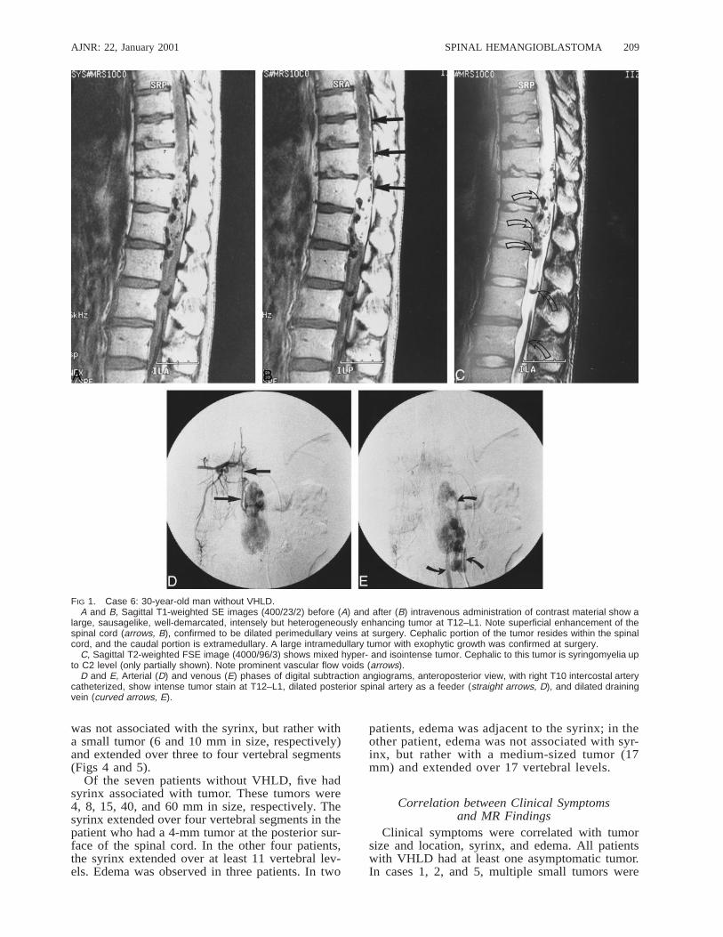
FIG 1. Case 6: 30-year-old man without VHLD.A and B, Sagittal T1-weighted SE images (400/23/2) before (A) and after (B) intravenous administration of contrast material show a
large, sausagelike, well-demarcated, intensely but heterogeneously enhancing tumor at T12–L1. Note superficial enhancement of thespinal cord (arrows, B), confirmed to be dilated perimedullary veins at surgery. Cephalic portion of the tumor resides within the spinalcord, and the caudal portion is extramedullary. A large intramedullary tumor with exophytic growth was confirmed at surgery.
C, Sagittal T2-weighted FSE image (4000/96/3) shows mixed hyper- and isointense tumor. Cephalic to this tumor is syringomyelia upto C2 level (only partially shown). Note prominent vascular flow voids (arrows).
D and E, Arterial (D) and venous (E) phases of digital subtraction angiograms, anteroposterior view, with right T10 intercostal arterycatheterized, show intense tumor stain at T12–L1, dilated posterior spinal artery as a feeder (straight arrows, D), and dilated drainingvein (curved arrows, E).
was not associated with the syrinx, but rather witha small tumor (6 and 10 mm in size, respectively)and extended over three to four vertebral segments(Figs 4 and 5).
Of the seven patients without VHLD, five hadsyrinx associated with tumor. These tumors were4, 8, 15, 40, and 60 mm in size, respectively. Thesyrinx extended over four vertebral segments in thepatient who had a 4-mm tumor at the posterior sur-face of the spinal cord. In the other four patients,the syrinx extended over at least 11 vertebral lev-els. Edema was observed in three patients. In two
patients, edema was adjacent to the syrinx; in theother patient, edema was not associated with syr-inx, but rather with a medium-sized tumor (17mm) and extended over 17 vertebral levels.
Correlation between Clinical Symptomsand MR Findings
Clinical symptoms were correlated with tumorsize and location, syrinx, and edema. All patientswith VHLD had at least one asymptomatic tumor.In cases 1, 2, and 5, multiple small tumors were

AJNR: 22, January 2001210 CHU
FIG 2. Case 3: 24-year-old woman with VHLD.A, Sagittal T1-weighted FSE image (700/10/6) shows a large, round, hypointense tumor in the left vertebral foramen between S1 and
S2. Note vascular flow voids within the tumor.B, Sagittal T2-weighted FSE image (4100/121/5) shows mixed iso- and hyperintense tumor.C, Sagittal T2-weighted FSE image (4100/121/5), medial to slice in B, shows serpentine vascular flow voids (arrows).D, Contrast-enhanced axial T1-weighted FSE image (740/8.6/4) shows intensely but heterogeneously enhancing tumor that was
confirmed to arise from left S1 nerve root at surgery.E and F, Arterial (E) and venous (F) phases of digital subtraction angiograms, right anterior oblique 308 view, with left internal iliac
artery catheterized, show intense tumor stain, dilated lateral sacral arteries that supply the tumor (curved arrows, E), and early venousfilling (straight arrows, F), indicating the arteriovenous shunt in the tumor. This vein corresponds with the flow voids seen in C.
observed. In these patients, clinical symptoms wereattributable mainly to the syrinx. In two of them,edema was associated with the syrinx and it waspossible, although not definite, that the edema wasalso a cause of the presenting symptoms. In case1, the large size of the tumor (60 mm) was alsopartly responsible for the symptoms. On the otherhand, in cases 3 and 4, a small intramedullary tu-mor (10 and 6 mm, respectively) with peritumoral
edema at the thoracic spinal cord did not producesymptoms (in case 3, it was the S1 nerve tumorthat caused symptoms).
Among the patients without VHLD, one wasasymptomatic. This patient had an intradural extra-medullary medium-sized tumor (15 mm) but hadno syrinx or edema. The tumor was found inciden-tally on an MR examination performed for lumbardisk herniation. In two patients, clinical symptoms

AJNR: 22, January 2001 SPINAL HEMANGIOBLASTOMA 211
FIG 3. Case 2: 42-year-old woman with VHLD without subjective symptoms.A and B, Contrast-enhanced sagittal T1-weighted FSE images (600/12/6) show nodular, intensely and homogeneously enhancing
tumors at the posterior aspects of the spinal cord at levels C5, C6, T8–T9, and T9–T10.C, Sagittal T2-weighted TSE image (4500/112/3) shows a syrinx at T7–T8 and adjacent edema at T9–T10. This image is a composite
of the upper and lower halves of the MR images of the spine.D, Contrast-enhanced axial T1-weighted TSE image (1000/12/2) shows the tumor surrounding a posterior nerve root at C6, as is also
the case with other tumors at C5 and C7–T1 (not shown).
were attributable primarily to the syrinx, with in-tramedullary tumors of 4 and 15 mm, respectively.In one patient with Brown-Sequard syndrome, thesymptoms were attributable primarily to the tu-mor (17 mm in size) and not to the associatededema. In the remaining three patients, with tu-mors of 8, 40, and 60 mm, respectively, symptomswere attributable both to the tumor itself and to thesyrinx.
For both groups of patients, in cases of smalltumors (10 mm or less), clinical symptoms werecaused by the syrinx. A small tumor without as-sociated syrinx did not cause spinal cord symp-toms. Among the patients who had medium-to-large tumors, clinical symptoms were attributableto the tumor itself and/or to the syrinx. Peritumoraledema without syrinx did not contribute to the pa-tients’ symptoms.
Correlation between MR andAngiographic Findings
Contrast enhancement of the tumors on MR im-ages was compatible with tumor stain on angio-grams in one patient with VHLD and in five pa-tients without VHLD. In the patient with VHLD,vascular flow voids on MR images corresponded tothe feeding artery and draining vein on angiogramsin one tumor arising from the S1 nerve root (Fig2). Among the five patients without VHLD, twohad vascular flow voids on MR images, corre-sponding to the feeding arteries and/or drainingveins on angiograms (cases 6 and 11; tumor size:
60 and 40 mm, respectively) (Fig 1). In three patients(cases 7, 9, and 10; tumor size: 8, 17, and 15 mm,respectively), no vascular flow voids were noted onMR images, but abnormal vessels were shown onangiograms. The feeding arteries originated fromthe anterior spinal artery in three tumors and fromthe posterior spinal artery in four. The drainingveins emptied into the anterior spinal vein in twotumors, into the posterior spinal vein in two, andinto the radiculomedullary vein in one.
Correlation between MR and Surgical FindingsIn the patients with VHLD, one tumor with a
superficial location on MR images was confirmedto be subpial in location at surgery. In two tumorsseen as deeply located within the spinal cord onMR images, one was confirmed to be deeply lo-cated within the cord at surgery (Fig 6) and theother was found to be subpial in location at sur-gery. In the latter patient, the spinal cord waspushed ventrally by the tumor (15 mm in size),becoming very thin, and the enhancing tumor waslocated centrally in the dural sac on MR images(Fig 8). One tumor, located within the left S1 nerveon MR images (Fig 2), was confirmed to arise fromthe S1 nerve at surgery, and was totally excisedalong with the nerve. In the patients withoutVHLD, four tumors in superficial locations on MRimages were confirmed to be subpial at surgery.Two tumors, both intra- and extramedullary in lo-cation, and one tumor, intradural-extramedullary inlocation on MR images, were proved to have been

AJNR: 22, January 2001212 CHU
FIG 4. Case 3: 24-year-old woman with VHLD.A, Sagittal T1-weighted FSE image (700/10/6) shows cord enlargement and decreased signal intensity at T3–T6.B, Contrast-enhanced sagittal T1-weighted FSE image (700/10/6) shows a small, ovoid, well-demarcated, intensely and homoge-
neously enhancing tumor at T4–T5.C, Sagittal T2-weighted FSE image (4220/120/5) shows isointense tumor (arrow) and hyperintense pencil-shaped lesion from T3
to T6.D, Contrast-enhanced axial T1-weighted FSE image (540/9/3) shows the tumor is superficially located at the posterior aspect of the
spinal cord.
FIG 5. Case 4: 44-year-old woman with VHLD who had no spinal cord symptoms.A, Contrast-enhanced sagittal T1-weighted SE image (800/14/2) shows a small, ovoid, well-demarcated, intensely enhancing intra-
medullary tumor at the anterior aspect of the spinal cord at T10.B, Sagittal T2-weighted TSE image (4500/112/3) shows edema (arrow).C, Contrast-enhanced axial T1-weighted SE image (660/15/1) shows the tumor is superficially located at the anterior aspect of the
spinal cord.
correctly located at surgery (cases 6, 9, and 12)(Fig 1). The extramedullary tumor arose from aposterior nerve root. The superficial enhancementof the spinal cord corresponded to perimedullarydraining veins at surgery. The peritumoral cystic
cavity was identified by confirming the efflux offluid after incision of the wall of the cavity.
Intraoperative sonography was used in some ofthe patients, but the sonographic findings were notincluded in this study.

AJNR: 22, January 2001 SPINAL HEMANGIOBLASTOMA 213
FIG 6. Case 1: 16-year-old boy withVHLD.
A, Contrast-enhanced sagittal T1-weight-ed TSE image (700/12/2) shows a large,well-demarcated, intensely and heteroge-neously enhancing intramedullary tumor atC7–T2. Note vascular flow voids in andaround the tumor (curved arrows) and su-perficial enhancement of the spinal cord(straight arrows), proved to be dilated per-imedullary veins at surgery. Syrinx extendsfrom medulla oblongata to T12. Also notemultiple small intensely and homoge-neously enhancing tumors, most of whichare located superficially at the posterioraspect of the spinal cord.
B, Contrast-enhanced axial T1-weightedSE image (660/15/1) at C7–T1 shows en-largement of the spinal canal and the en-hancing tumor. The tumor occupies entiredural sac and the spinal cord is barely dis-cernible. The tumor was interpreted asdeeply located within the spinal cord onMR image, and was confirmed at surgery.
MR findings of syrinx and edema were compat-ible with the surgical findings in all but one tumor(case 6). On MR images, this tumor had edema atC2–C3, which was beyond the surgical field. Path-ologically, all hemangioblastomas were denselyvascular tumors consisting of thin-walled, closelypacked blood vessels interspersed with large stro-mal cells.
DiscussionHemangioblastomas of the spinal cord are rare
benign tumors representing 1.6% to 6.4% of spinaltumors (1, 7, 14). According to a review of 85 his-tologically documented case reports by Browne etal (1), about two thirds of spinal hemangioblasto-mas are sporadic and the remainder are associatedwith VHLD. The lesions are single in 79% of cas-es. In patients with VHLD, hemangioblastomas areoften multiple. Sixty percent of spinal hemangio-blastomas are intramedullary, 11% are intra- andextramedullary, 21% are intradural extramedullary,and 8% are extradural (1). The thoracic spinal cordis most frequently involved (51%), followed by thecervical spinal cord (38%). Forty-three percent ofall spinal hemangioblastomas have associated syr-inx, and, when only cases of intramedullary spinalhemangioblastoma are considered, 67% have as-sociated syrinx. No MR findings are included inthis review.
To our knowledge, no detailed analysis of MRfindings in spinal hemangioblastoma has been con-ducted to date. Previously published MR imagingfindings in spinal hemangioblastoma have beenbased primarily on case reports (3–13). Amongthese articles, those published before 1989 did notinclude contrast-enhanced MR studies (4–6, 12).Although Xu et al (14) reported 13 cases of intra-medullary hemangioblastoma of the spinal cord,the focus was more on surgical treatment. In a pic-torial essay by Baker et al (13), the MR findings
were based on a literature review and on some oftheir own cases; however, their material was notexplicitly described. In the present study, we ana-lyzed the MR findings in spinal hemangioblastomawith special attention to the relationship betweentumor size and other MR findings, including tumorlocation, signal intensity, vascular flow voids, as-sociated syrinx, and edema. We compared the MRfindings in hemangioblastoma in patients withVHLD with those in sporadic hemangioblastoma toascertain any differences. We also correlated theMR findings with clinical symptoms and with an-giographic and surgical findings.
We found several characteristic MR findings inspinal hemangioblastoma, among which was thefinding of well-demarcated, intense enhancement.This property was evident in all tumors in thisstudy (Figs 1–8) and reflects the associated abnor-mal, densely vascular tumor parenchyma, whichconsisted of thin-walled, closely packed blood ves-sels interspersed with large stromal cells.
The second characteristic MR finding was thesuperficial location of the intramedullary tumors(21/24) (Figs 4–7), most often at the posterior as-pect of the spinal cord (16/21) (Figs 3, 4, and 6),reflecting the surgically proved subpial location ofthe intramedullary tumor. The third characteristicfinding was the relatively large size of the syrinxas compared with the size of the intramedullarytumor (Figs 3 and 7). The frequency of syrinx for-mation was high (64%) in cases of intramedullarytumors. Although syrinx is not specific to spinalhemangioblastomas, as it may be associated withother spinal tumors, such as ependymoma and as-trocytoma (13), a small superficially located tumorwith a large syrinx is considered characteristic(Figs 3 and 7). Once the tumor was totally re-moved, the syrinx disappeared or shrunk withoutthe need for additional drainage or for a shuntprocedure.

AJNR: 22, January 2001214 CHU
FIG 7. Case 7: 49-year-old woman without VHLD.A and B, Noncontrast (A) and contrast-enhanced (B) sagittal T1-weighted SE images (600/15/2) show a small, homogeneously and
intensely enhancing nodular tumor at C5–C6. The tumor is isointense on noncontrast T1-weighted image. Note extensive syrinx frommedulla oblongata to T3 level.
C, Contrast-enhanced axial T1-weighted SE image (600/15/2) shows the tumor is well demarcated, superficially located at the anterioraspect of the spinal cord.
FIG 8. Case 5: 49-year-old man withVHLD.
A, Contrast-enhanced sagittal T1-weighted TSE image (700/12/3) shows amedium-sized, ovoid, heterogeneously en-hancing tumor at T9. Note several small,homogeneously enhancing nodules ce-phalic to this tumor. Also note flow voidswithin the tumor and superficial enhance-ment of the spinal cord.
B, Contrast-enhanced axial T1-weightedSE image (660/15/1) shows the tumor atT9 occupies entire dural sac. The tumorwas thought to be located deeply withinthe spinal cord, but turned out to be sub-pial in location at surgery.
The pathogenesis of syringeal formation in spi-nal hemangioblastoma remains unknown (15, 16).One hypothesis is that a syrinx is formed by a soft-ening of the spinal cord resulting from damage tothe cord or to the intramedullary vessels caused bythe tumor. However, this mechanism is not likely
in our cases, because a large syrinx was associatedwith even a tiny tumor that could hardly cause ex-tensive damage to the spinal cord or intramedullaryvessels. Samii et al (16) suggested that total or sub-total obstruction of CSF flow by an intramedullarytumor plays a major role in the development of a

AJNR: 22, January 2001 SPINAL HEMANGIOBLASTOMA 215
syrinx. This mechanism was not the cause of thesyringomyelia in our cases either, because most ofthe tumors were small and did not cause CSF ob-struction. Transudation from the tumor vessels andsecretion by tumor cells are generally believed tobe the major causes of syringeal development inpatients with intramedullary tumors (17, 18), andwe speculate that this mechanism is most likely thecause of syringomyelia in spinal hemangioblasto-ma, because the tiny tumor would behave as a mu-ral nodule of a cyst or syrinx, as frequently seen incerebellar hemangioblastoma.
One may speculate that peritumoral edema leadsto syringeal development; however, no such evi-dence has been reported to date. In our study, twopatients who had edema were followed up by MRimaging, and in neither case did the edema changein extent or convert into a syrinx over a 2- or 3-yearperiod, respectively (Figs 4 and 5). It is more likelythat edema results from local venous congestioncaused by the hypervascular tumor, which has ar-teriovenous shunts (4). Another hypothesis includesthe production of an edema-promoting factor by theneoplasm. It is possible that hemangioblastomas se-crete agents that induce increased capillary per-meability (4).
The fourth characteristic MR finding observed inour study was the presence of vascular flow voidsin or around medium-sized to large tumors (Figs 1,6, and 8). This reflects distended feeding arteries ordraining veins that were confirmed by angiographyand/or surgery. We also found that tumor size wasrelated to the tumor’s shape, apparent location, sig-nal intensity, homogeneity of enhancement, and thepresence of vascular flow voids. When the tumorwas small (10 mm or less), its shape was invariablynodular or ovoid, and its location was at the surfaceof the spinal cord or along a nerve root (Figs 3–7).When the tumor was larger than 10 mm, it wasfound either at the surface of or deep in the spinalcord, or it had an intra- and extramedullary location(exophytic growth), or was completely extramedul-lary in location (Fig 1). (It may be difficult to esti-mate the true location of a tumor that is larger than10 mm, because the spinal cord may be compressedby the tumor and be barely recognizable on routineMR images.) Small tumors were primarily isoin-tense on T1-weighted images and of high intensityon T2-weighted images (Fig 7), whereas larger tu-mors tended to be hypointense or mixed hypo- andisointense on T1-weighted images and of heteroge-neous intensity on T2-weighted images (Figs 1 and2). Most of the small tumors showed homogeneousenhancement, whereas medium-sized to large tu-mors tended to show heterogeneous enhancement(Figs 1, 2, 6, and 8).
In the present study, vascular flow voids wereobserved in or around the tumor in seven (58%) ofthe patients. We found that the vascular flow voidswere absent when the tumor was less than 15 mmin size, whereas they were invariably present whenthe tumor size was 25 mm or greater. Therefore, a
tumor is not likely to be a hemangioblastoma if itis 25 mm or larger and is not associated with vas-cular flow voids on MR images.
It has been reported that the diagnostic featuresof hemangioblastoma include the presence of en-larged arterial feeding vessels, a densely stainingtumor nodule, and rapid shunt into a distended ve-nous structure at angiography (1). In our study, MRimaging failed to detect dilated vessels in two me-dium-sized tumors (cases 9 and 10; 17 and 15 mm,respectively) and in one small tumor (case 7) thatharbored abnormal vessels on angiograms. Theseresults indicate that MR imaging underestimatesthe presence of dilated vessels. MR angiography isexpected to have higher sensitivity in the detectionof spinal vascular disease than conventional MRimaging. It has been reported that both phase-con-trast MR angiography and 3D contrast-enhancedMR angiography have high accuracy in the char-acterization of spinal vascular disease and can de-pict the arterial pedicles, although the latter tech-nique has shown better results (19, 20). Thus, MRangiography may be a good supplementary tech-nique for characterizing spinal tumors, especiallywhen vascular flow voids are indistinct on conven-tional MR studies. Although MR angiography mayhave sufficient spatial resolution for the detectionof abnormal vascular structures, its time resolutionis not sufficient to distinguish arterial feeders fromdraining veins. Moreover, acquisition time typicallyis longer than 20 seconds to obtain sufficient spatialresolution. Because MR angiography usually de-picts arteries and venous structures simultaneously,it may be difficult to differentiate between feedingarteries and draining veins. In planning surgicaltreatment of a large hypervascular spinal tumor, itis essential to clarify the 3D relationship amongtumor parenchyma, feeding arteries, and drainingveins, because the main draining vein should notbe interrupted before all feeding arteries have beencompletely devascularized by coagulation. If themain draining vein is coagulated before devascu-larization is complete, the change in hydrodynam-ics may increase the difficulty of the surgical pro-cedure and damage the spinal cord (14). Piecemealremoval of the tumor should be avoided, becauseit may increase the possibility of tumor bleeding.Therefore, we believe that angiography is indicatedfor treatment planning when a large spinal tumorassociated with vascular flow voids is found on MRimages.
Angiography was performed in six of our pa-tients (cases 3, 5, 6, 9, 10, and 11) to determinethe location of feeding arteries and draining veinsin order to plan a treatment strategy. In cases 3 and6, the feeding arteries were embolized by usingpolyvinyl alcohol before surgery, and these tumorswere removed safely by surgery. The ideal surgicalprocedure for spinal hemangioblastoma is to co-agulate all feeding arteries before interrupting themain draining vein, and to remove the tumor enbloc. In some cases, distended draining veins reside

AJNR: 22, January 2001216 CHU
in front of the tumor and feeding arteries so thatthey hamper the surgical approach to feeding ar-teries. In such cases, we think that preoperative em-bolization of the feeding arteries is useful to reducearterial flow and subsequently to shrink the drain-ing veins and lower the risk of tumor bleeding.Chang et al (21) have shown that stereotactic ra-diosurgery provides a high likelihood of local con-trol of hemangioblastoma and can be an alternativeto multiple surgical procedures for patients withVHLD.
Although we do not consider that angiography isindicated for a small spinal tumor to confirm thediagnosis of hemangioblastoma, we performed an-giography in one patient with a small tumor (case7), because we initially had difficulty in reaching adiagnosis on MR images owing to a lack of expe-rience early on. We did not perform angiographyin case 8, because the tumor was small and locatedsuperficially at the posterior aspect of the spinalcord, and thus we expected the tumor to be excisedeasily and safely. Two patients (cases 1 and 5) de-clined to undergo angiography. We did not performangiography in cases 2 and 4, because neither pa-tient had subjective symptoms and the tumor wassmall; both were followed up with MR examina-tions.
Superficial enhancement of the spinal cord wasconcomitant with flow voids and was confirmed tobe dilated perimedullary veins at surgery (Figs 1,6, and 8). This finding may also be characteristicfor spinal hemangioblastoma, although other spinalcord diseases with pial or subpial extension, suchas high-grade astrocytoma, metastatic tumor, sar-coidosis, and tuberculosis, may have similar MRappearances.
In the present study, spinal hemangioblastomaswere associated with VHLD in five (42%) of the12 patients. We found that spinal hemangioblasto-mas in patients with VHLD were different fromsporadic spinal hemangioblastomas only in theirmultiplicity and the size of the tumor. Multiplehemangioblastomas were found only in patientswith VHLD. The percentage of small hemangio-blastomas was significantly higher in patients withVHLD than in those without VHLD. This was be-cause the asymptomatic hemangioblastomas werefound incidentally on MR examinations performedfor other symptomatic tumors, or they were foundon the screening examination for CNS tumors inpatients with VHLD. There was no difference inthe other MR features between the two groups.
No differences were found in symptoms be-tween the patients with and without VHLD. Themain symptoms included pain, proprioceptive sen-sation disturbance of position and vibration, motorweakness, and bowel and urinary dysfunction.These manifestations are consistent with previousfindings (1).
In the patients with VHLD, most small tumors(10 mm or less) were considered asymptomatic.Solitary spinal hemangioblastoma was found in one
patient with VHLD and in seven patients withoutVHLD. Two of these patients had asymptomatictumors, 6 and 15 mm in size, respectively. It isnoteworthy that spinal hemangioblastoma wasfound incidentally even in a patient without VHLD.This relatively high rate of occurrence of asymp-tomatic hemangioblastoma is attributable to thehigh detectability of spinal tumors by MR imaging.We thus suggest that in patients with VHLD theentire neuraxis should be screened by MR imagingfor potential association of hemangioblastoma,even when neurologic symptoms are absent. Con-trast-enhanced T1-weighted imaging is most appro-priate for this purpose, because it is the most sen-sitive in detecting small hemangioblastomas.
In cases of small tumors, clinical symptoms werebelieved to be caused by the associated syrinx andnot by the tumor itself. As for medium-sized tolarge tumors, the clinical symptoms were attribut-able to the syrinx or to the tumor itself. Peritumoraledema without syrinx did not contribute to the pa-tients’ symptoms. We speculate that the small tu-mors without associated syrinx did not damage thespinal cord, because their location was completelysubpial. When a tumor becomes larger, it will causesymptoms by virtue of its mass effect on the spinalcord. The fact that a small subpial hemangioblas-toma without an associated syrinx is asymptomaticmay be important in differentiating other diseases,which may produce small, enhancing foci (with orwithout spinal cord edema), such as metastatic tu-mors, sarcoidosis, tuberculosis, neurosyphilis, fun-gal infection, and multiple sclerosis, because thesediseases may be symptomatic even when their en-hancing lesions are small.
In general, the differential diagnosis of spinal tu-mors is based primarily on the location of the tumoron imaging studies. On MR images, an intramedul-lary tumor is most likely a hemangioblastoma whenit is small, well demarcated, intensely enhanced, lo-cated at the surface of the spinal cord, and associatedwith a relatively large syrinx. Peritumoral edema isnot always present. When the patient is asymptom-atic, syrinx may be absent. As for an intramedullarytumor larger than 10 mm, it is most likely a hem-angioblastoma when an associated syrinx and vas-cular flow voids are present in or around the tumor.Spinal hemangioblastoma can also be found either inan intradural extramedullary or an extradural location(1, 2, 8, 14). It can arise from filum terminale, intra-dural nerve roots, extradural nerves, or it can existindependent of these structures. The distinguishingMR feature of a hemangioblastoma in these locationsis the presence of vascular flow voids. However,when vascular flow voids are present on MR images,spinal hemangioblastomas should be differentiatedfrom arteriovenous malformations (AVMs) and otherhypervascular tumors, including paraganglioma andmetastatic tumors (especially from renal cell carci-nomas), that frequently occur in patients with VHLD(5, 8, 14, 22). A well-defined enhancing mass shouldserve to distinguish spinal tumors from AVMs (14).

AJNR: 22, January 2001 SPINAL HEMANGIOBLASTOMA 217
A thorough search for a primary tumor, such as arenal carcinoma, is necessary, especially in patientswith VHLD. Spinal paragangliomas are usuallyfound in the intradural extramedullary compartment(22) and are difficult to differentiate from extramed-ullary hemangioblastoma.
ConclusionMR features of spinal hemangioblastoma depend
on the size of the tumor. Small (10 mm or less) hem-angioblastomas are mostly isointense on T1-weightedimages and hyperintense on T2-weighted images andshow homogeneous enhancement, whereas largerones tend to be hypointense or mixed hypo- andisointense on T1-weighted images and heteroge-neous on T2-weighted images, and tend to showheterogeneous enhancement. Small hemangioblas-tomas are located at the surface of the spinal cord,most frequently at the posterior aspect, and showwell-demarcated, intense enhancement. A heman-gioblastoma larger than 24 mm is accompanied byvascular flow voids on MR images. Symptomaticsmall hemangioblastomas have a relatively largeassociated syrinx. Asymptomatic hemangioblasto-mas do not have an associated syrinx, althoughthey may have peritumoral edema. Spinal heman-gioblastomas in patients with VHLD tend to bemultiple. Although the percentage of small hem-angioblastomas is significantly higher in patientswith VHLD than in those without VHLD, there isno difference in the other MR findings between thetwo groups. Because asymptomatic hemangioblas-tomas are frequently found in patients with VHLD,the entire neuraxis should be screened by contrast-enhanced MR imaging. Knowledge of these MRfeatures helps to differentiate spinal hemangioblas-tomas from other spinal tumors and other diseasesthat produce enhancing nodules. Angiography is in-dicated for treatment planning when a large spinaltumor associated with vascular flow voids is foundon MR images.
References1. Browne TR, Adams RD, Roberson GH. Hemangioblastoma of
the spinal cord: review and report of five cases. Arch Neurol1976;33:435–441
2. Wisoff HS, Suzuki Y, Llena JF, Fine DIM. Extramedullary hem-angioblastoma of the spinal cord. J Neurosurg 1978;48:461–464
3. Corr P, Dicker T, Wright M. Exophytic intramedullary heman-gioblastoma presenting as an extramedullary mass on myelog-raphy. AJNR Am J Neuroradiol 1995;16:883–884
4. Solomon RA, Stein BM. Unusual spinal cord enlargement re-lated to intramedullary hemangioblastoma. J Neurosurg 1988;68:550–553
5. Sato Y, Waziri M, Smith W, et al. Hippel-Lindau disease: MRimaging. Radiology 1988;166:241–246
6. Kaffenberger DA, Shah CP, Murtagh FR, et al. MR imaging ofspinal cord hemangioblastoma associated with syringomyelia.J Comput Assist Tomogr 1988;12;495–498
7. Murota T, Symon L. Surgical management of hemangioblasto-ma of the spinal cord: a report of 18 cases. Neurosurgery 1989;25:699–708
8. Arbelaez A, Castillo M, Armao D. Hemangioblastoma of thefilum terminale: MR imaging. AJR Am J Roentgenol 1999;173:857–858
9. Silbergeld J, Cohen WA, Maravilla KR, et al. Supratentorial andspinal cord hemangioblastomas: gadolinium enhanced MR ap-pearance with pathologic correlation. J Comput Assist Tomogr1989;13:1048–1051
10. Friedman DP, Flanders AE, Tartaglino LM. Vascular neoplasmsand malformations, ischemia, and hemorrhage affecting thespinal cord: MR imaging findings. AJR Am J Roentgenol 1994;162:685–692
11. Yu JS, Short MP, Schumacher J, et al. Intramedullary hemor-rhage in spinal cord hemangioblastoma. J Neurosurg 1994;81:937–940
12. Neumann HPH, Eggert HR, Weigel K, et al. Hemangioblastomasof the central nervous system: a 10-year study with specialreference to von Hippel-Lindau syndrome. J Neurosurg 1989;70:24–30
13. Baker KB, Moran CJ, Wippold FJ II, et al. MR imaging of spinalhemangioblastoma. AJR Am J Roentgenol 2000;174:377–382
14. Xu QW, Bao WM, Mao RL, Yang GY. Magnetic resonance im-aging and microsurgical treatment of intramedullary heman-gioblastoma of the spinal cord. Neurosurgery 1994;35:671–676
15. Enomoto H, Shibata T, Ito A, et al. Multiple hemangioblastomasaccompanied by syringomyelia in the cerebellum and the spi-nal cord. Surg Neurol 1984;22:197–203
16. Samii M, Klekamp J. Surgical results of 100 intramedullarytumors in relation to accompanying syringomyelia. Neurosur-gery 1994;35:865–873
17. Gardner WJ. Hydrodynamic mechanism of syringomyelia: itsrelationship to myelocele. J Neurol Neurosurg Psychiatry 1965;28:247–259
18. Kiwitt JCW, Lanksch WR, Fritsch H, et al. Magnetic resonancetomography of solid spinal cord tumors with extensive sec-ondary syringomyelia. Adv Neurosurg 1988;16:211–215
19. Mascalchi M, Quilici N, Ferrito G, et al. Identification of thefeeding arteries of spinal vascular lesions via phase-contrastMR angiography with three-dimensional acquisition andphase display. AJNR Am J Neuroradiol 1997;18:351–358
20. Binkert CA, Kollias SS, Valavanis A. Spinal cord vascular dis-ease: characterization with fast three-dimensional contrast-en-hanced MR angiography. AJNR Am J Neuroradiol 1999;20:1785–1793
21. Chang SD, Meisel JA, Hancock SL, et al. Treatment of hem-angioblastomas in von Hippel-Lindau disease with linear ac-celerator-based radiosurgery. Neurosurgery 1998;43:28–35
22. Sundgren P, Annertz M, Englund E, et al. Paragangliomas of thespinal canal. Neuroradiology 1999;41:788–794